Anais da Academia Brasileira de Ciências (2018) 90(3): 3081-3097 (Annals of the Brazilian Academy of Sciences)
Printed version ISSN 0001-3765 / Online version ISSN 1678-2690 http://dx.doi.org/10.1590/0001-3765201820180364
www.scielo.br/aabc | www.fb.com/aabcjournal
Glucocorticoid susceptibility and
in vivo
ABCB1 activity differ in murine B cell subsets
KELLI M. DA COSTA1,2
,RAPHAEL C. VALENTE1,4
, JOYLE M.C. DA SILVA3
, LUCIANA S. DE PAIVA3 and VIVIAN M. RUMJANEK1
1
Instituto de Bioquímica Médica Leopoldo de Meis, Universidade Federal do Rio de Janeiro, Av. Carlos Chagas Filho 373, Sala H2-03, Ilha do Fundão, 21941-902 Rio de Janeiro, RJ, Brazil 2
Instituto de Biofísica Carlos Chagas Filho, Universidade Federal do Rio de Janeiro, Av. Carlos Chagas Filho 373, Sala C1-42, 21941-902 Ilha do Fundão, Rio de Janeiro, RJ, Brazil 3
Instituto de Biologia, Departamento de Imunobiologia, Universidade Federal Fluminense, Outeiro de São João Batista, s/n, Campus do Valonguinho, Prédio Núcleo de Animais de Laboratório,
2º andar, Laboratório de Imunorregulacão, 24020-141 Niterói, RJ, Brazil 4
Faculdade de Ciências Médicas, Departamento de Microbiologia, Imunologia e Parasitologia, Universidade do Estado do Rio de Janeiro, Av. Prof. Manuel de Abreu 444, 3º andar, Vila Isabel, 20550-170 Rio de Janeiro, RJ, Brazil
Manuscript received on April 16, 2018; accepted for publication on June 22, 2018
ABSTRACT
Glucocorticoids are produced and released by the adrenal gland and become elevated in response to stress. Although glucocorticoids are well known for their immunosuppressive effects, less is known about their effects on B cells. ABCB1 is an efflux pump expressed in both cancer and normal cells, modulating the gradient of various metabolites, including hydrocortisone. Our goal was to evaluate the effect of this glucocorticoid on murine B cell differentiation and whether sensitivity to hydrocortisone could be related to ABCB1 activity in vivo. C57BL/6 mice received one or three consecutive i.p. injections of hydrocortisone (70, 140 and 200 mg/kg/day). ABCB1 activity was evaluated via the rhodamine-123 transport and inhibited by cyclosporin A in hydrocortisone-treated and control mice. Cells from bone marrow, spleen and blood were counted, incubated with antibodies and analyzed by flow cytometry. A single hydrocortisone injection did not alter the number of bone marrow subsets. Conversely, three daily injections were able to reduce the cell number of most bone marrow subsets, excepting c-kit-sca-1+ and mature B cells. This treatment reduced marginal zone, follicular and transitional B cells, though splenic subsets were more resistant than bone marrow B cells. Recirculating follicular B cells in the blood were resistant to hydrocortisone. With the exception of follicular B cells, all subpopulations exhibited ABCB1 activity. However, hydrocortisone treatment did not affect ABCB1 activity in most subsets analyzed. Results suggest that hydrocortisone is able to regulate B cell lymphopoiesis although ABCB1 activity is not related to the susceptibility to that glucocorticoid in B cell subsets.
Key words: ABC transporters, B cells, immunoregulation, steroid.
Correspondence to: Vivian Mary Rumjanek E-mail: [email protected]
INTRODUCTION
Glucocorticoids are a class of steroid hormones secreted by the adrenal gland cortex (zona fasciculata) as end-products of stress-induced activation of the hypothalamic-pituitary-adrenal axis (Garrod 1958). Systemically, glucocorticoids act counteracting insulin effects by inducing hepatic gluconeogenesis, glycogenolysis, lipolysis and also catabolic reactions within skeletal muscle, thus protecting the organism from hypoglycemia (Habib et al. 2001).
Endogenous and synthetic glucocorticoids are analogous molecules that exert their effects predominantly through the glucocorticoid receptor (GR). Only one gene is known to encode GR, which belongs to the superfamily of ligand-activated nuclear transcription factors (Barnes 2006). This gene encodes two isoforms: GRα, widely expressed in most cells as part of a complex containing chaperones located in the cytoplasm, mediating most of the known effects of glucocorticoids; GRβ, a dominant inhibitor of GRα-induced activation of specific genes (Gross and Cidlowski 2008).
G e n o m i c s i g n a l i n g p a t h w a y o f glucocorticoids requires the presence of a ligand to initiate dimerization and translocation of GRα to the nucleus where it interacts directly with DNA sequences or with transcription factors to regulate gene transcription (Barnes 2006). Non-genomic signaling pathway appears to be mediated by glucocorticoid interactions either with cell membranes, with cytoplasmic or membrane-bound GR (Stahn and Buttgereit 2008). It is unclear whether the membrane receptor is a GRα isoform. However, its signaling pathway is dependent on G protein and/or MAPKs as well as on cytoplasmic GRα presence (Nahar et al. 2015).
Specifically in the immune system, endogenous (as cortisol/hydrocortisone and corticosterone) and synthetic (as dexamethasone) glucocorticoids are well known both for their immunosuppressive
and anti-inflammatory properties, since they are able to induce apoptosis in lymphocytes (Garvy et al. 1993b, Wyllie 1980, Compton and Cidlowski 1986). Apoptosis appears to play a crucial role during the different steps leading to maturation of B and T cells from their precursor cells. In T cell lineage, the susceptibility to dexamethasone-induced apoptosis seems to be regulated during development, being CD4+CD8+ cells more susceptible (Berki et al. 2002). Such facet is related both to the relevant thymic atrophy observed after treatment with hydrocortisone (Rodrigues-Mascarenhas et al. 2006) and regulation of thymic selection by dexamethasone (Ashwell et al. 1996, Berki et al. 2002). Glucocorticoids as hydrocortisone, corticosterone, dexamethasone or methylprednisolone can influence multiple processes like antibody isotype switching (Jabara et al. 1991, 2001) and both synthesis (Tokuyama and Tokuyama 1994) and suppression of B cell antigenic response (Garvy and Fraker 1991, Voetberg et al. 1994). In addition, in vitro and in vivo treatments with those glucocorticoids led to a stage-dependent depletion of B cells in the bone marrow (Garvy et al. 1993a, b), where immature B cells exhibited a significant sensitivity to glucocorticoid-induced apoptosis, while mature B cells were resistant (Lu and Osmond 2000). In vitro, dexamethasone actively induce apoptosis of spleen B cells (Miyake et al. 1994), a feature considerably reduced with the use of the glucocorticoid receptor antagonist RU486 (Andreau et al. 1998).
ABCB1 EFFLUX AND GLUCOCORTICOID EFFECT IN B CELLS 3083
Louis, MO, USA); treated group - 70, 140 or 200 mg/kg/day hydrocortisone (Sigma-Aldrich, Saint Louis, MO, USA) diluted in RPMI 1640 medium. All groups were treated for three consecutive days at the same time in the morning, respecting a 24-hour interval among injections. After 24 h, mice were euthanized and bone marrow (n=9 for Figures 1, 2 and 3 and n=12 for Figure 6) or spleen (n=12 for Figure 4 and n=9 for Figure 7) were removed to obtain cell suspensions. Blood cells were obtained from the retro-orbital plexus (n=20 for Figure 5). 3-4 independent experiments were performed with 2-5 mice/group.
In vivo ABCB1 EFFLUX ACTIVITY
In vivo ABCB1-related activity was investigated by means of the use of the fluorescent dye Rhodamine 123 (Rho; Sigma-Aldrich, Saint Louis, MO, USA; channel FL1) in association or not with activity inhibitor Cycloporin A (CsA; Novartis Biosciences S.A., São Paulo, SP, Brazil) in an efflux assay. 84 mice from treated and control groups (42 mice/ group) were separated in subgroups: Rho, injected only with the dye; and Rho + CsA, injected with the dye and inhibitor. Subgroups received a single i.p. injection of 100 mg/kg CsA or diluent only (200
µL of RPMI 1640 medium), followed 1 h later by a single i.p. injection of 2.5 mg/kg Rho (n=12 for Figure 6 and n=9 for Figure 7). An additional mouse from the same litter was selected per experiment, injected only with diluent and used as the negative control. After 1 h, mice were euthanized and bone marrows and spleens were used to obtain cell suspensions (Leite et al. 2006). Four independent experiments were performed with 2-3 mice/group.
IMMUNOPHENOTYPES OF CELLS BY FLOW CYTOMETRY
Fresh cell suspensions from bone marrow, spleen and heparinized blood were stained with fluorochrome-conjugated monoclonal antibodies (mAbs) and monitored by flow cytometry. The 1996, Flens et al. 1996) and also in cells of the
immune system, more precisely in some progenitors from bone marrow and lymphocyte subsets (Leslie et al. 2005, Kyle-Cezar et al. 2007a). In T cells, the expression of this protein seems to be finely regulated during development (Kyle-Cezar et al. 2007b). CD4+CD8- and CD4-CD8+ thymocytes present ABCB1 activity and are more resistant to glucocorticoids, unlike CD4+CD8+ thymocytes (Leite et al. 2006). Gruol and Bourgeois (1994) demonstrated that ABCB1 expression in a murine thymoma line is a mechanism of escape from dexamethasone-induced apoptosis, since this pump reduced the cytoplasmic levels of this glucocorticoid. Thus, regulation of cytoplasmic glucocorticoid levels reduces the amount of activated GR and consequently, sensitivity to dexamethasone-induced apoptosis.
In the present work, we assessed the effects of the glucocorticoid hydrocortisone on B cell lymphopoiesis, as their effects on B cells have not been fully elucidated. Furthermore, we investigated whether ABCB1 activity is related to differences in susceptibility to hydrocortisone in mature B cell subsets and their precursors.
MATERIALS AND METHODS
ANIMALS
215 female mice C57BL/6, 2 to 3-month-old, were obtained from Fluminense Federal University. Animals were housed in a controlled environment with a 12 h/12 h light/dark cycle, receiving water and food ad libitum. All protocols herein were approved by the Ethics Committee on Animals Use of Federal University of Rio de Janeiro under protocol number IBQM048.
following mAbs specific for mouse surface antigens were used: anti-mouse IgM FITC (clone: R6-60.2), CD45R/B220 PE (RA3-6B2), CD117/c-kit PercP/Cy5.5 (2B8), sca-1/Ly6A/E Alexa Fluor 647 (D7), CD19 PercP/Cy 5.5 (6D5), CD21 APC (7G6) and CD23 PE (B3B4) were purchased from BD Biosciences (San Jose, CA, USA), eBioscience (San Diego, CA, USA) or Biolegend (San Diego, CA, USA). 106 cells were incubated with 5% fetal bovine serum (FBS, Cultilab, Campinas, SP, Brazil) diluted in PBS for 20 min to avoid nonspecific binding. Then, cells were washed with PBS and incubated with mAbs diluted in PBS for 20 min on ice, in accordance with the manufacturer instructions. Again, cells were washed with PBS and suspended in PBS supplemented with 5% FBS and kept on ice until the acquisition time in the flow cytometer. Data were acquired on a FACSCalibur device (BD Biosciences, San Jose, CA, USA) and analyzed using Summit v4.3 software (Dako cytomation, Fort Collins, CO, USA).
FLOW CYTOMETRY
B cells and progenitors were gated on basis of cell size and granularity from either bone marrow, spleen or peripheral blood and subsets were identified as follows: lymphoid progenitors (sca-1+c-kit-; sca-1+c-kit+ and sca-1-c-kit+), Pro/PreB-PreBI (c-kit+B220+IgM-), PreBII (c-kit-B220lowIgM-), immature B (B220lowIgM+), mature B (B220highIgM+), transitional B (CD19+CD23-CD21-), follicular B (CD19+CD23+CD21+), marginal zone B (CD19+CD23-CD21+) and peripheral blood B (CD19+). To analyze ABCB1-related transport activity, a fluorescence intensity histogram was made for each subpopulation, except in PreBII and B immature subpopulations. As IgM-FITC and Rho use the same detection channel, they were not used at the same time. For this reason, it was not possible to separate PreBII and immature B cells according to the expression of IgM. Therefore, these cells were named PreBII
and B immature and identified by the expression of B220low.
In the presence of Rho, three subpopulations were highlighted. Rhodamine negative or low cells (Rho neg and Rho low) exhibited either negative or low accumulation of the dye, therefore displaying background or low fluorescence levels, respectively. Rhodamine high cells (Rho high) showed increased incorporation of the dye and exhibit high fluorescence intensity.
STATISTICAL ANALYSIS
Treated and control groups data were analyzed by Kruskal-Wallis ANOVA or analysis of variance ANOVA one-way test, according to the D’Agostino-Pearson normality test. Rho profile data were analyzed by Mann-Whitney test or the unpaired t-test, according with normality. Data were analyzed by GraphPad Prism v5.0 software (La Jolla, CA, USA) and considered statistically significant when (*) p < 0.05, (**) p < 0.01 and (***) p < 0.001.
RESULTS
DEPLETION OF LYMPHOCYTES AND PROGENITORS IN BONE MARROW OF HYDROCORTISONE-TREATED MICE
ABCB1 EFFLUX AND GLUCOCORTICOID EFFECT IN B CELLS 3085
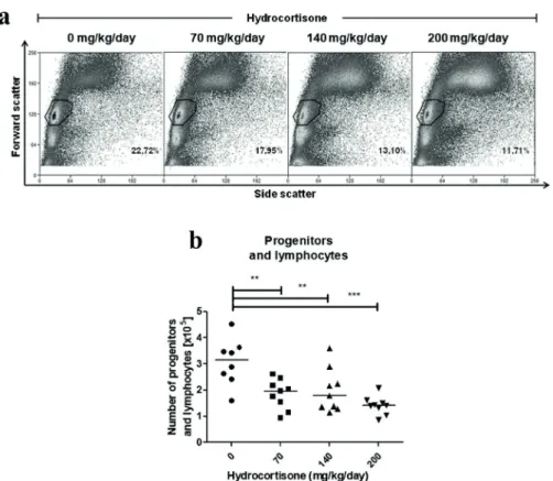
105 (140 mg/kg/day) and 1.42 x 10⁵ cells (200 mg/ kg/day) (Figure 1b). However, when a single i.p. injection of hydrocortisone was administered, there was no change in either percentage or number of progenitors or lymphocytes in any of the previously cited concentrations (data not shown).
DIFFERENTIAL SENSITIVITY OF BONE MARROW B CELLS TO HYDROCORTISONE TREATMENT
Some authors have suggested that glucocorticoids may regulate T cell development, because subpopulations have different degrees of susceptibility to steroids (Vacchio et al. 1994). Likewise, development of B cells also appears to be altered by exposure to glucocorticoids, although
the extent of glucocorticoid actions concerning this
matter is still unknown. Progenitors had a drastic
reduction in number of cells in
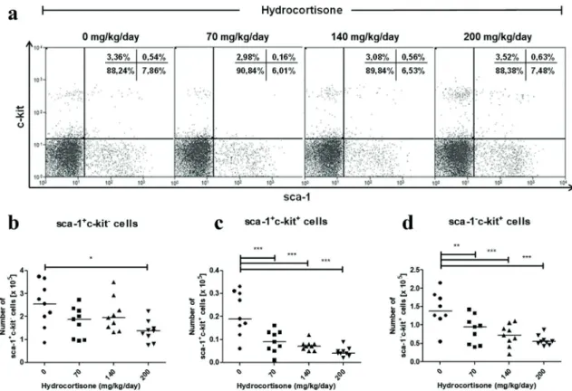
hydrocortisone-treated mice, excluding sca-1+c-kit- cells (Figure
2). The subset sca-1+c-kit+ was more sensitive to
hydrocortisone, with a depletion of 51% at 70 mg/
kg/day hydrocortisone (Figure 2c), while subset
sca-1-c-kit+ had a 37% decrease on average (Figure
2c). The subpopulation sca-1+c-kit-, which is the most undifferentiated profile in the development pathway, showed significant sensitivity only in the highest concentration of hydrocortisone employed
(200 mg/kg/day) (Figure 2b). Regarding the
percentage of cells, there was a mild increase in
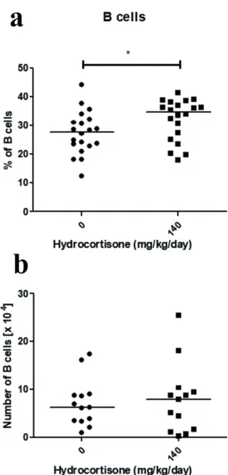
Figure 1 - Effect of in vivo treatment with hydrocortisone on bone marrow cells. Mice
were injected with diluent or hydrocortisone (70, 140 or 200 mg/kg/day) for 3 consecutive
days. In the next day, bone marrow cells were counted and analyzed by flow cytometry. (a) Percentage and (b) total number of progenitors and lymphocytes. Line represents the median,
(**) represents values of p<0.01 and (***) p<0.001, according to the statistical test. Results are
sca-1+c-kit- progenitors with treatment, which was statistically significant at 140 mg/kg/day.
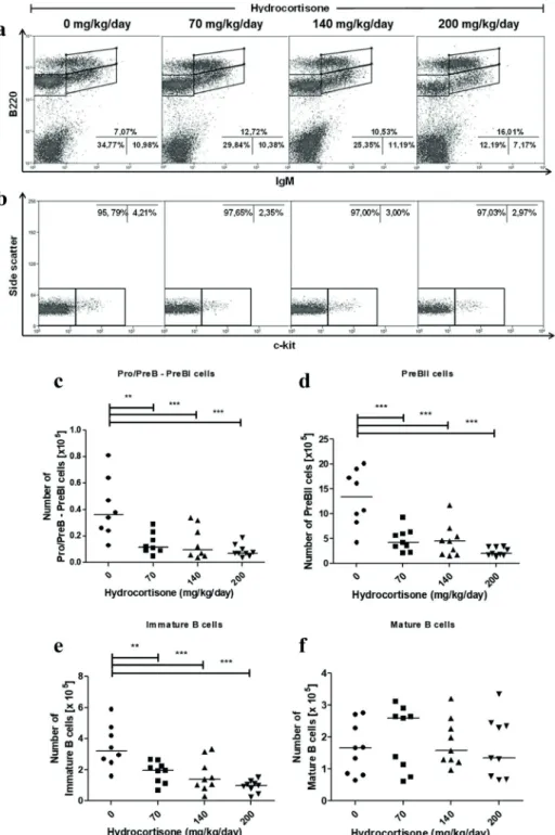
Most immature subsets of B cells showed a reduction in the percentage of cells when exposed to hydrocortisone, particularly at concentration of 200 mg/kg/day (Pro/PreB - PreBII and immature B cells). Consequently, mature B cells showed an increased percentage of cells (Figure 3a). The separation of the subpopulations Pro/PreB - PreBII by means of evaluation of c-kit expression demonstrated no difference in percentage of Pro/ PreB - Pre BI (c-kit+) and PreBII cells (c-kit-) after hydrocortisone treatment, thus suggesting that these two subpopulations reduced proportionally (Figure 3b). When evaluating the number of cells, subpopulations B220low (Pro/PreB - PreBI, PreBII and immature B cells) showed a dose-dependent
reduction after hydrocortisone exposure (Figure 3c-e). At concentration of 70 mg/kg/day, subsets Pro/PreB - Pre BI and PreBII showed a decrease of 56% in number of cells, while immature B cells decreased by 43%. Nonetheless, mature B cells in bone marrow proved to be very resistant to treatment, even at 200 mg/kg/day of hydrocortisone (Figure 3f). Mice treated with a single i.p. injection (70, 140 or 200 mg/kg of hydrocortisone) showed no change either in percentage or in number of cells in any of bone marrow subpopulations analyzed (data not shown).
DIFFERENTIAL SENSITIVITY OF PERIPHERAL B CELLS TO HYDROCORTISONE TREATMENT
Immature B cells that are weakly or non-self-reactive can mature, through the transition stage,
Figure 2 - Effect of in vivo treatment with hydrocortisone on B cell progenitors. Mice were injected with diluent
or hydrocortisone (70, 140 or 200 mg/kg/day) for 3 consecutive days. In the next day, bone marrow cells were counted
and stained with monoclonal antibodies for sca-1 and c-kit molecules for analysis by flow cytometry. (a) Percentage of hematopoietic progenitor subsets, (b) total number of sca-1+c-kit-, (c) sca-1+c-kit+ and (d) sca-1-c-kit+ cells. Line represents
the median, (*) represents values of p<0.05, (**) p<0.01 and (***) p<0.001, according to the statistical test. Results are
ABCB1 EFFLUX AND GLUCOCORTICOID EFFECT IN B CELLS 3087
Figure 3 - Effect of in vivo treatment with hydrocortisone on subsets of bone marrow B cells. Mice were injected with diluent or hydrocortisone (70, 140 or 200 mg/kg/day) for 3 consecutive days. In the next day, bone marrow cells were counted and stained with monoclonal antibodies for c-kit, B220
and IgM molecules for analysis by flow cytometry. (a) Percentage of Pro/PreB-PreBII (B220+IgM-), immature B (B220lowIgM+) and mature B cells (B220highIgM+), (b) percentage of Pro/PreB-PreBI (c-kit+) and PreBII (c-kit-), derived from Pro/PreB-PreBII subset, (c) total number of Pro/PreB-PreBI, (d) PreBII, (e) immature B and (f) mature B cells . Line represents the median, (**) represents
values of p<0.01 and (***) p<0.001, according to the statistical test. Results are representative of 3
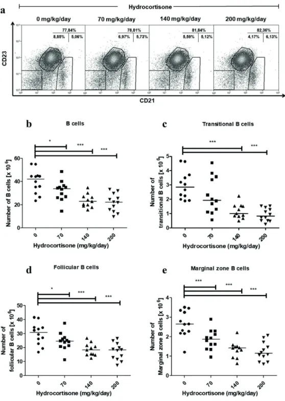
giving rise to follicular B and marginal zone B cells in the spleen (Pillai and Cariappa 2009). The percentage of marginal zone B cells did not change in control nor in hydrocortisone-treated mice. The percentage of transitional B cells was reduced by hydrocortisone treatment while the percentage of follicular B cells was increased (Figure 4a). However, the absolute number of splenic B cells decreased from 42.03 x 10⁶ in the control group to
33.83 x 10⁶, 22.91 x 10⁶ and 22.28 x 10⁶ (median) after treatment with 70, 140 and 200 mg/kg/day of hydrocortisone respectively (Figure 4b). The number of subsets decreased in a dose-dependent manner. Transitional B cells were more sensitive, showing 65% decrease in cell count at 140 mg/ kg/day of hydrocortisone (Figure 4c), followed by marginal zone B cells (Figure 4e) and follicular B cells (Figure 4d), which were reduced by 46% and 40%, respectively.
The majority of bone marrow mature B cells are recirculating follicular B cells, which migrate back into the bone marrow after maturation in the spleen. Since follicular B cells show susceptibility to hydrocortisone treatment, while bone marrow mature B cells are resistant, the concentration of 140 mg/kg/day of hydrocortisone was used to evaluate if sensitivity of peripheral blood B cells would be similar to that seen for bone marrow mature B cells or follicular B cells. There was an increase from 27.8% to 34.7% in the median of the percentage of peripheral blood B cells in hydrocortisone-treated mice (Figure 5a). However, absolute number of cells remained unchanged (Figure 5b) demonstrating that peripheral blood B cells are resistant to exposure to hydrocortisone, as well as bone marrow mature B cells.
IN VIVO MODULATION OF ABCB1 EFFLUX ACTIVITY IN B CELLS
ABCB1 efflux activity in B cells was assessed in vivo, mimicking a physiological process. For this, dye and inhibitor were both administered i.p. in
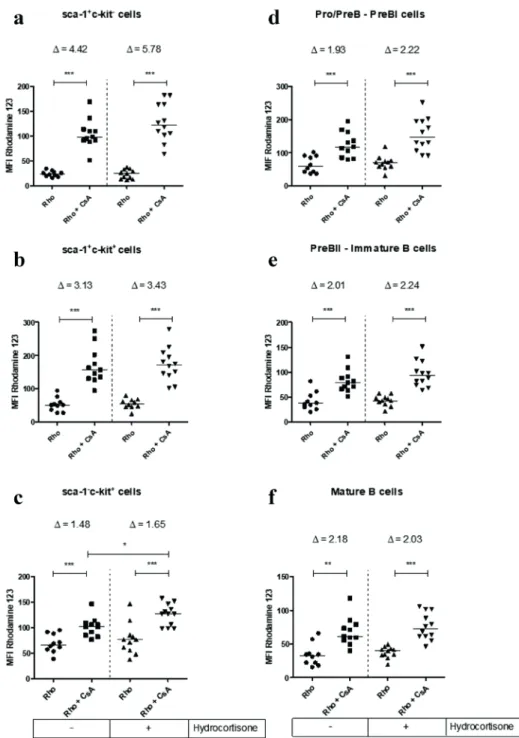
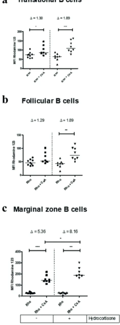
mice, as described in materials and methods. Rho is a lipophilic fluorescent dye that passively crosses cell membranes and can be transported through ABCB1 protein. In the presence of an inhibitor like CsA, cells can no longer extrude Rho and start to accumulate it, increasing median fluorescence intensity (MFI). The transport reversal index (Δ) is a ratio of MFI of the inhibited transport activity and MFI of transport activity (Δ = MFI Rho+CsA/ MFI Rho) (da Costa et al. 2018). Thus, a higher Δ indicates a greater pump efflux activity. Progenitor subsets of bone marrow cells showed ABCB1 transport activity, as can be seen by increased amount of MFI in CsA-injected mice (Figure 6a-c). The most undifferentiated subset sca-1+c-kit- presented an elevated transport activity, with Δ of 4.42 ± 0.61 (mean ± standard deviation), followed by sca-1+c-kit+ subset, with Δ of 3.13 ± 0.50 and sca-1-c-kit+ cells, with Δ of 1.48 ± 0.25. Subpopulations committed to B cell lymphopoiesis also exhibited ABCB1-related activity, with very similar Δ values, being 1.93 ± 0.44 for Pro/PreB - PreBI; 2.01 ± 0.54 for PreBII - immature B and 2.18 ± 0.56 for mature B cells (Figure 6d-f). The figure 7 indicates that splenic B cells showed distinct ABCB1 efflux activity levels. Transitional B cells showed little activity ABCB1 with Δ value of 1.30 ± 0,27. Follicular B cells showed no detectable ABCB1 activity, because there was no significant change in the MFI in the absence and presence of the inhibitor. Surprisingly, marginal zone B cells had an enormous ABCB1-related activity with Δ of 5.36 ± 1.17, the highest seen among B cell subpopulations (Figure 7a-c).
HYDROCORTISONE SENSITIVITY IS NOT RELATED TO ABCB1 EFFLUX ACTIVITY IN B CELLS
ABCB1 EFFLUX AND GLUCOCORTICOID EFFECT IN B CELLS 3089
Figure 4 - Effect of in vivo treatment with hydrocortisone on subsets of spleen B cells. Mice were
injected with diluent or hydrocortisone (70, 140 or 200 mg/kg/day) for 3 consecutive days. In the next day, spleen cells were counted and stained with monoclonal antibodies for CD19, CD21 and CD23 molecules for
analysis by flow cytometry. (a) Percentage of spleen B cells subsets, (b) total number of B cells (CD19+), (c) transitional B (CD19+CD21-CD23-), (d) follicular B (CD19+CD21lowCD23+) and (e) marginal zone B cells
(CD19+CD21highCD23-). Line represents the median, (*) represents values of p<0.05 and (***) p<0.001,
not increased considerably, with Δ value ranging from 1.48 ± 0.25 to 1.65 ± 0.39 after treatment. In spleen, treatment had varied effects: transitional B cells, displayed a Δ value ranging from 1.30 ± 0.27 to 1.89 ± 0.59 after treatment (when mean values were considered). However, there was no statistical significant change in relation to ABCB1 activity. Follicular B cells that showed no activity related to ABCB1 in untreated mice had detectable efflux activity with Δ of 1.89 ± 0.63. Marginal zone B cells which had the highest Δ values among B cells had an increased ABCB1 activity, with a Δ value ranging from 5.36 ± 1.17 to 8,16 ± 2.08 (Figure 7).
DISCUSSION
For over 40 years, Levine and Claman have shown that B cells from mouse bone marrow and spleen react differently to treatment with cortisone (Levine and Claman 1970). However, a detailed study of how these subsets are affected from the stem cells to the peripheral mature B cells had not yet been made. Mice treated with a single i.p. injection of hydrocortisone (70, 140 or 200 mg/kg) did not change either number or percentage of bone marrow B cell subsets (data not shown). These data differ from the results obtained with thymocytes, suggesting that B cells are more resistant to hydrocortisone. When thymocytes were studied, Rodrigues-Mascarenhas et al. (2006) showed that after 24 h of a single i.p. injection of 140 mg/kg of hydrocortisone a reduction in thymus weight and number of thymocytes could be observed. When mice were i.p. injected with hydrocortisone for three consecutive days, there was a depletion of the percentage and number of progenitors and lymphocytes, demonstrating that these cells are greatly affected when the treatment is prolonged. It is noteworthy that there was an increased percentage of monocyte and granulocyte subsets, as seen by the increased number of events above the highlighted region (Figure 1a). These results are in agreement
Figure 5 - Effect of in vivo treatment with hydrocortisone
on blood B cells. Mice were injected with diluent or hydrocortisone (140 mg/kg/day) for 3 consecutive days. In the next day, blood cells were counted and stained with monoclonal antibody for CD19 molecule for analysis by
flow cytometry. (a) Percentage and (b) the total number of B
cells (CD19+). Line represents the median of the data and (*) represents values of p<0.05, according to the statistical test.
Results are representative of 3 independent experiments with 3-5 mice/group (n=13-20).
ABCB1 EFFLUX AND GLUCOCORTICOID EFFECT IN B CELLS 3091
Figure 6 - Effect of in vivo treatment with hydrocortisone on ABCB1 efflux activity of subsets
of bone marrow cells. Mice were injected with diluent or hydrocortisone (70 mg/kg/day) for 3
consecutive days. In the next day, mice were injected with cyclosporin A (CsA, 100 mg/kg) and, one hour later, with Rhodamine 123 (Rho, 2.5 mg/kg), as described in materials and methods. Bone marrow cells were counted and stained with monoclonal antibodies for c-kit, sca-1 and
B220 molecules for analysis by flow cytometry. (a) Median fluorescence intensity for Rho on
sca-1+c-kit-, (b) sca-1+c-kit+, (c) sca-1-c-kit+, (d) Pro/PreB - PreBI, (e) PreBII - immature B and (f) mature B cells. Line represents the median, values represent mean of transport reversal index
(Δ=MFI Rho+CsA/MFI Rho), (*) represents values of p < 0.05, (**) p < 0.01 and (***) p < 0.001,
with Trottier et al. (2008), Laakko and Fraker (2002) who showed that corticosterone induce expansion of monocyte and granulocyte compartments over lymphocyte compartment. When evaluating the effect of hydrocortisone treatment for three consecutive days, it was possible to observe that the more undifferentiated progenitor cells, sca-1+c-kit-, were resistant to treatment while sca-1+c-kit+ and sca-1-c-kit+ cells were sensitive. These results confirm the data from Igarashi et al. (2005) that showed that all progenitors of lymphoid lineage were depleted after treatment with dexamethasone implant, however early lymphoid progenitors, which express both sca-1 and c-kit, were highly susceptible in treated mice and human cells. Cells committed to B cells, Pro/PreB - PreBI and PreBII cells were depleted in hydrocortisone-treated mice; while immature B cells were less and mature B cells were resistant. Garvy et al. (1993a) showed that chronic increase of plasma corticosterone in mice reduced the number of B cells in the S phase in the bone marrow. Such cells could be the B220low subpopulations (Pro/PreB - PreBI and PreBII), which are the proliferating cells in the developmental pathway of B cells (Karasuyama et al. 1994). In agreement with these data, Lill-Lill-Elghanian et al. (2002) demonstrated that cells with mature phenotype CD19+IgD+ were more resistant to dexamethasone than others subsets. They also observed that endogenous glucocorticoids were slightly less potent than the synthetic. Although the effects of dexamethasone on bone marrow have already been addressed previously by other authors (Miyake et al. 1994), their effects on splenic B cell maturation have not been fully elucidated. In hydrocortisone-treated mice, the change in the distribution of spleen cells resulted in a reduction in the number of transitional B cell, more sensitive, being followed in number by marginal zone B cells. Follicular B cells are more resistant compared to the other two splenic subpopulations. However, unlike B cell subsets from bone marrow, there
Figure 7 - Effect of in vivo treatment with hydrocortisone
on ABCB1 efflux activity of subsets of spleen B cells. Mice
were injected with diluent or hydrocortisone (140 mg/kg/day) for 3 consecutive days. In the next day, mice were injected with cyclosporin A (CsA, 100 mg/kg) and, one hour later, with rhodamine 123 (Rho, 2.5 mg/kg), as described in materials and methods. Spleen cells were counted and stained with monoclonal antibodies for CD19, CD21 and CD23 molecules for analysis by flow cytometry. (a) Median fluorescence intensity for rhodamine 123 on transitional B, (b) follicular B, (c) marginal zone B cells. Line represents the median, ∆ values
represent mean of transport reversal index (Δ=MFI Rho+CsA/ MFI Rho), (*) represents values of p < 0.05, (**) p < 0.01 and (***) p < 0.001, according to the statistical test. Results are
ABCB1 EFFLUX AND GLUCOCORTICOID EFFECT IN B CELLS 3093
were significant differences in the number of cells between treatments with 70 and 140 mg/kg/day and 70 and 200 mg/kg/day. The behavior of peripheral blood B cells was very similar to that of mature B cells from bone marrow: they were resistant. These results demonstrate that glucocorticoids such as hydrocortisone are also able to regulate the distribution and numbers of B cells from spleen and bone marrow, as already seen for differentiation of thymocytes (Vacchio et al. 1994). Moreover, our data support the view that the differentiation of B cells is inversely related to susceptibility to hydrocortisone. To B cells subsets, susceptibility to hydrocortisone is gradually decreased until the level of mature B cell.
Kyle-Cezar et al. (2007b), Drach et al. (1992) demonstrated ABCB1 activity in subpopulations of bone marrow and peripheral blood B cells, respectively. However, the in vivo activity of this glycoprotein along the differentiation pathway in a model that mimics the physiological had not been used so far. Our results demonstrate that the subsets expressing c-kit and/or sca-1 exhibit activity related to ABCB1. ABCB1 activity was highest in the more undifferentiated subset sca1+c-kit- decreasing along
the developmental pathway of B cells in the bone marrow. Surprisingly, splenic B cells also showed activity related to ABCB1, except for follicular B cells. Transitional B cells showed little ABCB1 activity, while marginal zone B cells had the largest activity among the subsets analyzed.
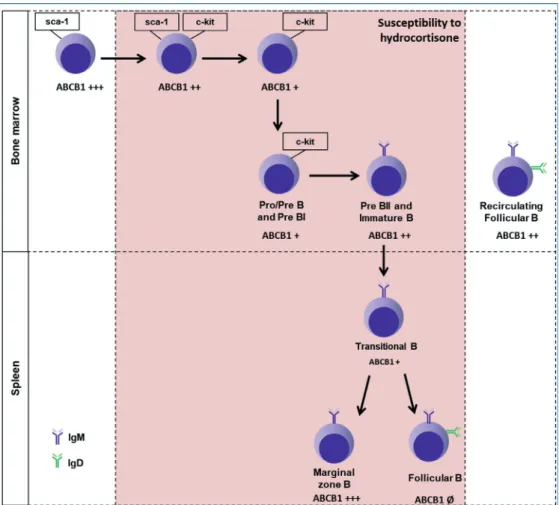
It can be seen that the activity of ABCB1 varies in different subsets, suggesting that this glycoprotein is finely regulated during the development of B cells (Table I and figure 8). Knowing that hydrocortisone is transported by ABCB1 (Silva et al. 2015) and is capable of inducing apoptosis in B cells (Garvy et al. 1993a, b), the role of ABCB1 in hydrocortisone sensitivity was evaluated. Since the glucocorticoid-induced genomic effects depend on the cytoplasmic GR, the entry and accumulation of the hormone is required for its binding to the
receptor. Therefore, any mechanism that influences the intracellular accumulation of hydrocortisone could affect the magnitude of the response and favor the resistant phenotype. B cell subsets presenting pump activity would survive and be present in greater numbers after treatment with hydrocortisone. In most subpopulations, there was an increase in the MFI to the mice of Rho + CsA group injected with hydrocortisone. However, it may be noted that the MFI of mice of the Rho group injected with hydrocortisone also underwent a slight increase or remained unchanged compared to untreated animals. Thus, the Δ values remained the same or have undergone little change compared to untreated animals in statistical terms, with the exception of sca1-ckit+, B follicular and marginal zone B subsets. Nevertheless, this increase is not enough to explain the sensitivity to glucocorticoids for two reasons: there was no increase in ABCB1 activity in other subsets analyzed, including in mature B cells that are resistant to glucocorticoid treatment. In addition, the subset that showed the highest index of ABCB1 activity was not the most resistant to the drug.
Figure 8 - Effect of in vivo consecutive treatment with hydrocortisone on ABCB1 activity of B cell subsets. The illustration shows B cell subsets sensitive and resistant to treatment with 3 consecutive i.p. injections (70, 140 and 200 mg/kg/day) hydrocortisone in C57BL/6 mice and reversion index of the ABCB1 transport (Δ) evaluated by rhodamine 123 efflux assay in the presence of the cyclosporin A inhibitor. Symbols as (Ø) represents no ABCB1 activity; (+), Δ < 2.0; (++), 2.0 ≤ Δ < 4.0; (+++), Δ ≤ 4.0.
cell in healthy donors (Bartholome et al. 2004) and its effects are associated to glucocorticoid-induced non-genomic signaling (Stahn and Buttgereit 2008).
The effect of glucocorticoids in T cell subpopulations suggest that an amount of factors may influence susceptibility such as the expression of GR and proteins of the Bcl-2 family. Thus, it may be that the susceptibility to glucocorticoid is multifactorial in B cells, as described by some authors in the context of inducing apoptosis, but still unrelated to the physiological process of differentiation and maturation of B cells as observed for T cells. In our study it was found that
B cells have different levels of ABCB1 transporter activity and that this activity is finely regulated during the differentiation process. However, ABCB1 activity could not be directly related to the resistant phenotype to hydrocortisone. In this context, more data should be gathered in order for a better understanding of the role of ABCB1 on B cells subsets, mainly on the ones from the marginal zone.
ABBREVIATIONS
ABCB1 EFFLUX AND GLUCOCORTICOID EFFECT IN B CELLS 3095
ABCG2 - ATP binding cassette subfamily G member 2 (BCRP)
CsA - Cyclosporin A
GR - Glucococorticoid receptor Rho - Rhodamine 123
ACKNOWLEDGMENTS
K.M.C. designed research and performed the experiments, analyzed, interpreted the results and drafted the manuscript. R.C.V. helped in carrying out the experiments evaluating ABCB1 activity, contributed to the interpretation of the results and revised the manuscript. J.M.C.S. helped in carrying out the bone marrow experiments. L.S.P. and V.M.R. designed research and critically revised the manuscript. All authors read and approved the final version of the manuscript. This work was
supported by grants from Conselho Nacional de Desenvolvimento Científico e Tecnológico (CNPq) and Fundação Carlos Chagas Filho de Amparo à Pesquisa do Estado do Rio de Janeiro (FAPERJ). K.M.C was a recipient from CNPq fellowship. The authors declare no conflict of interest.
REFERENCES
ANDREAU K, LEMAIRE C, SOUVANNAVONG V AND ADAM A. 1998. Induction of apoptosis by dexamethasone in the B cell lineage. Immunopharmacology 40: 67-76. ASHWELL JD, KING LB AND VACCHIO MS. 1996.
Cross-talk between the T cell antigen receptor and the glucocorticoid receptor regulates thymocyte development. Stem Cells 14: 490-500.
BARNES PJ. 2006. Corticosteroids: the drugs to beat. Eur J Pharmacol 533: 2-14.
BARTHOLOME B ET AL. 2004. Membrane glucocorticoid receptors (mGCR) are expressed in normal human peripheral blood mononuclear cells and up-regulated TABLE I
Effect of in vivo treatment with hydrocortisone on ABCB1 transport activity of B cell subsets.
[hydrocortisone] 0 mg/kg/day 70 mg/kg/day
Bone marrow
sca-1+ c-kit- +++ =
sca-1+ c-kit+ +++ =
c-kit+ sca-1- + ↑
Pro/PreB - PreBI ++ =
PreBII - Immature B ++ =
Mature B ++ =
[hydrocortisone] 0 mg/kg/day 140 mg/kg/day
Spleen
Transitional B + =
Follicular B Ø ↑
Marginal zone B +++ ↑
Symbols in table are representative of transport reversal index (Δ). (Ø), no ABCB1 activity; (+), Δ < 2.0; (++) 2.0 ≤ Δ < 4.0; (+++) and (+++) Δ ≤ 4.0. In mice treat with hydrocortisone (70 or 140 mg/kg/day), (=) represents no change in ABCB1 activity and (↑),
after in vitro stimulation and in patients with rheumatoid arthritis. FASEB J 18: 70-80.
BELLAMY WT. 1996. P-glycoproteins and multidrug resistance. Annu Rev Pharmacol Toxicol 36: 161-183. BERKI T, PALINKAS L, BOLDIZSAR F AND NEMETH
P. 2002. Glucocorticoid (GC) sensitivity and GC receptor expression differ in thymocyte subpopulations. Int Immunol 14: 463-469.
BOURGEOIS S, GRUOL DJ, NEWBY RF AND RAJAH FM. 1993. Expression of an mdr gene is associated with a new form of resistance to dexamethasone-induced apoptosis. Mol Endocrinol 7: 840-851.
COMPTON MM AND CIDLOWSKI JA. 1986. Rapid in vivo
effects of glucocorticoids on the integrity of rat lymphocyte genomic deoxyribonucleic acid. Endocrinology 118: 38-45.
DA COSTA KM, VALENTE RC, SALUSTIANO EJ, GENTILE LB, FREIRE-DE-LIMA L, MENDONCA-PREVIATO L AND MENDONCA-PREVIATO JO. 2018. Functional Characterization of ABCC Proteins from Trypanosoma cruzi and Their Involvement with Thiol Transport. Front Microbiol 9: 205.
DRACH D, ZHAO S, DRACH J, MAHADEVIA R, GATTRINGER C, HUBER H AND ANDREEFF M. 1992. Subpopulations of normal peripheral blood and bone marrow cells express a functional multidrug resistant phenotype. Blood 80: 2729-2734.
FLENS MJ, ZAMAN GJ, VAN DER VALK P, IZQUIERDO MA, SCHROEIJERS AB, SCHEFFER GL, VAN DER GROEP P, DE HAAS M, MEIJER CJ AND SCHEPER RJ. 1996. Tissue distribution of the multidrug resistance protein. Am J Pathol 148: 1237-1247.
GARROD O. 1958. The pharmacology of cortisone, cortisol (hydrocortisone) and their new analogues. Postgrad Med J 34: 300-304.
GARVY BA AND FRAKER PJ. 1991. Suppression of the antigenic response of murine bone marrow B cells by physiological concentrations of glucocorticoids. Immunology 74: 519-523.
GARVY BA, KING LE, TELFORD WG, MORFORD LA AND FRAKER PJ. 1993a. Chronic elevation of plasma corticosterone causes reductions in the number of cycling cells of the B lineage in murine bone marrow and induces apoptosis. Immunology 80: 587-592.
GARVY BA, TELFORD WG, KING LE AND FRAKER PJ. 1993b. Glucocorticoids and irradiation-induced apoptosis in normal murine bone marrow B-lineage lymphocytes as determined by flow cytometry. Immunology 79: 270-277. GROSS KL AND CIDLOWSKI JA. 2008. Tissue-specific
glucocorticoid action: a family affair. Trends Endocrinol Metab 19: 331-339.
GRUOL DJ AND BOURGEOIS S. 1994. Expression of the mdr1 P-glycoprotein gene: a mechanism of escape from
glucocorticoid-induced apoptosis. Biochem Cell Biol 72: 561-571.
HABIB KE, GOLD PW AND CHROUSOS GP. 2001. Neuroendocrinology of stress. Endocrinol Metab Clin North Am 30: 695-728.
IGARASHI H, MEDINA KL, YOKOTA T, ROSSI MI, SAKAGUCHI N, COMP PC AND KINCADE PW. 2005. Early lymphoid progenitors in mouse and man are highly sensitive to glucocorticoids. Int Immunol 17: 501-511. JABARA HH, AHERN DJ, VERCELLI D AND GEHA
RS. 1991. Hydrocortisone and IL-4 induce IgE isotype switching in human B cells. J Immunol 147: 1557-1560. JABARA HH, BRODEUR SR AND GEHA RS. 2001.
Glucocorticoids upregulate CD40 ligand expression and induce CD40L-dependent immunoglobulin isotype switching. J Clin Invest 107: 371-378.
KARASUYAMA H, ROLINK A, SHINKAI Y, YOUNG F, ALT FW AND MELCHERS F. 1994. The expression of Vpre-B/lambda 5 surrogate light chain in early bone marrow precursor B cells of normal and B cell-deficient mutant mice. Cell 77: 133-143.
KYLE-CEZAR F, ECHEVARRIA-LIMA J, DOS SANTOS GOLDENBERG RC AND RUMJANEK VM. 2007a. Expression of c-kit and Sca-1 and their relationship with multidrug resistance protein 1 in mouse bone marrow mononuclear cells. Immunology 121: 122-128.
KYLE-CEZAR F, ECHEVARRIA-LIMA J AND RUMJANEK VM. 2007b. Independent regulation of ABCB1 and ABCC activities in thymocytes and bone marrow mononuclear cells during aging. Scand J Immunol 66: 238-248. LAAKKO T AND FRAKER P. 2002. Rapid changes in the
lymphopoietic and granulopoietic compartments of the marrow caused by stress levels of corticosterone. Immunology 105: 111-119.
LEITE DF, ECHEVARRIA-LIMA J, SALGADO LT, CAPELLA MA, CALIXTO JB AND RUMJANEK VM. 2006. In vivo and in vitro modulation of MDR molecules in murine thymocytes. Int Immunopharmacol 6: 204-215. LESLIE EM, DEELEY RG AND COLE SP. 2005. Multidrug
resistance proteins: role of P-glycoprotein, MRP1, MRP2, and BCRP (ABCG2) in tissue defense. Toxicol Appl Pharmacol 204: 216-237.
LEVINE MA AND CLAMAN HN. 1970. Bone marrow and spleen: dissociation of immunologic properties by cortisone. Science 167: 1515-1517.
LILL-ELGHANIAN D, SCHWARTZ K, KING L AND FRAKER P. 2002. Glucocorticoid-induced apoptosis in early B cells from human bone marrow. Exp Biol Med (Maywood) 227: 763-770.
ABCB1 EFFLUX AND GLUCOCORTICOID EFFECT IN B CELLS 3097
MIYAKE K, YAMASHITA Y, HITOSHI Y, TAKATSU K AND KIMOTO M. 1994. Murine B cell proliferation and protection from apoptosis with an antibody against a 105-kD molecule: unresponsiveness of X-linked immunodeficient B cells. J Exp Med 180: 1217-1224. NAHAR J, HAAM J, CHEN C, JIANG Z, GLATZER NR,
MUGLIA LJ, DOHANICH GP, HERMAN JP AND TASKER JG. 2015. Rapid Nongenomic Glucocorticoid Actions in Male Mouse Hypothalamic Neuroendocrine Cells Are Dependent on the Nuclear Glucocorticoid Receptor. Endocrinology 156: 2831-2842.
PILLAI S AND CARIAPPA A. 2009. The follicular versus marginal zone B lymphocyte cell fate decision. Nat Rev Immunol 9: 767-777.
RODRIGUES-MASCARENHAS S, DOS SANTOS NF AND RUMJANEK VM. 2006. Synergistic effect between ouabain and glucocorticoids for the induction of thymic atrophy. Biosci Rep 26: 159-169.
SARKADI B, HOMOLYA L, SZAKACS G AND VARADI A. 2006. Human multidrug resistance ABCB and ABCG transporters: participation in a chemoimmunity defense system. Physiol Rev 86: 1179-1236.
SILVA R, VILAS-BOAS V, CARMO H, DINIS-OLIVEIRA RJ, CARVALHO F, DE LOURDES BASTOS M AND REMIAO F. 2015. Modulation of P-glycoprotein efflux pump: induction and activation as a therapeutic strategy. Pharmacol Ther 149: 1-123.
STAHN C AND BUTTGEREIT F. 2008. Genomic and nongenomic effects of glucocorticoids. Nat Clin Pract Rheumatol 4: 525-533.
SUZUKI M, SUZUKI H, SUGIMOTO Y AND SUGIYAMA Y. 2003. ABCG2 transports sulfated conjugates of steroids and xenobiotics. J Biol Chem 278: 22644-22649.
TOKUYAMA Y AND TOKUYAMA H. 1994. Retinoic acid and steroid hormones regulate IgA production by LPS-stimulated murine spleen cells. Immunopharmacology 28: 145-151.
TROTTIER MD, NEWSTED MM, KING LE AND FRAKER PJ. 2008. Natural glucocorticoids induce expansion of all developmental stages of murine bone marrow granulocytes without inhibiting function. Proc Natl Acad Sci U S A 105: 2028-2033.
VACCHIO MS, PAPADOPOULOS V AND ASHWELL JD. 1994. Steroid production in the thymus: implications for thymocyte selection. J Exp Med 179: 1835-1846.
VOETBERG BJ, GARVY BA, MAYER HK, KING LE AND FRAKER PJ. 1994. Apoptosis accompanies a change in the phenotypic distribution and functional capacity of murine bone marrow B-cells chronically exposed to prednisolone. Clin Immunol Immunopathol 71: 190-198.