Iranian Journal of Basic Medical Sciences
ijbms.mums.ac.ir
Low-frequency vibration treatment of bone marrow stromal cells
induces bone repair in vivo
Shengwei He
1, Wenzhi Zhao
1*, Lu Zhang
1, Lidong Mi
1, Guangyu Du
1, Chuanxiu Sun
1, Xuegang Sun
1 1 Department of Orthopedics, Second Affiliated Hospital, Dalian Medical University, Dalian, Liaoning Province, 116031, P. R. ChinaA R T I C L E I N F O A B S T R A C T
Article type: Original article
Objective(s): To study the effect of low-frequency vibration on bone marrow stromal cell differentiation and potential bone repair in vivo.
Materials and Methods: Forty New Zealand rabbits were randomly divided into five groups with eight rabbits in each group. For each group, bone defects were generated in the left humerus of four rabbits, and in the right humerus of the other four rabbits. To test differentiation, bones were isolated and demineralized, supplemented with bone marrow stromal cells, and implanted into humerus bone defects. Varying frequencies of vibration (0, 12.5, 25, 50, and 100 Hz) were applied to each group for 30 min each day for four weeks. When the bone defects integrated, they were then removed for histological examination. mRNA transcript levels of runt-related transcription factor 2, osteoprotegerin, receptor activator of nuclear factor -B ligan, and pre-collagen type 1 were measured.
Results: Humeri implanted with bone marrow stromal cells displayed elevated callus levels and wider, more prevalent, and denser trabeculae following treatment at 25 and 50 Hz. The mRNA levels of runt-related transcription factor 2, osteoprotegerin, receptor activator of nuclear factor -B ligand, and pre-collagen type 1 were also markedly higher following 25 and 50 Hz treatment.
Conclusion:Low frequency (25–50 Hz) vibration in vivo can promote bone marrow stromal cell differentiation and repair bone injury.
Article history: Received: Feb 15, 2015 Accepted: Jun 18, 2015
Keywords: Bone injury
Bone marrow stromal cells Pre-Col1a
RUNX2 Vibration stress
►
Please cite this article as:He Sh, Zhao W, Zhang L, Mi L, Du G, Sun Ch, Sun X, Low-frequency vibration treatment of bone marrow stromal cells induces bone repair in vivo. Iran J Basic Med Sci 2017; 20:23-28; http://dx.doi.org/10.22038/ijbms.2017.8088
Introduction
Bone nonunion and bone defects are the most important problems in bone fracture healing. To address these issues, autogenous and allogenous bone grafts are commonly used (1). However, limitations of these two methods, including limited bone resources and the immune response, make the development of alternative clinical treatments imperative (2).
Bone marrow stromal cells (BMSCs) are adult
stem cells that differentiate into mature bone cells (3, 4), a process that relies heavily on factors
within the surrounding microenvironment (5, 6). Several cytokines and growth factors, including osteoprotegerin (OPG), receptor activator of nuclear factor -B ligand (RANKL), runt-related transcription factor 2 (RUNX2), and pre-collagen type 1 (pre-Col1a) have critical roles in differentiating bone cells (7-9) and are standard indicators for normal BMSC
differentiation (9). In the clinical treatment of bone fractures, increased stress at the fracture site
improves healing (10). Studies have also linked increased cytokine production, as a result of specific
signal transduction pathways, with BMSC
differentiation, and in turn, bone fracture healing (11-13). Several studies have suggested that low-frequency vibration can effectively stimulate bone cell growth, increase the trabecular number and width, and prevent the loss of cortical bone strength (11, 14-17). Low-frequency signals propagate through the OPG pathway, stimulating the differentiation of bone precursor cells into osteoblasts, thereby affecting bone reconstruction (12). However, the exact frequency best suited to stimulation of osteogenesis remains controversial. Further, a recent study showed that 30 Hz vibration could induce BMSC differentiation in vitro (18). In this study, we used rabbits as an in vivo bone defect small animal model system to test varying frequencies and their effect on BMSC differentiation and bone fracture healing.
*Corresponding author:Wenzhi Zhao. Department of Orthopedics, Second Affiliated Hospital, Dalian Medical University, Dalian, Liaoning Province,
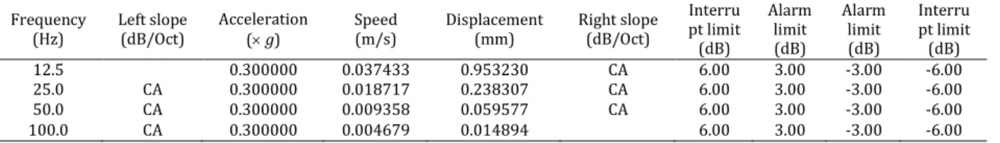
Table 1. Experimental set up for each frequency
Frequency (Hz)
Left slope (dB/Oct)
Acceleration ( g)
Speed (m/s)
Displacement (mm)
Right slope (dB/Oct)
Interru pt limit (dB)
Alarm limit (dB)
Alarm limit (dB)
Interru pt limit (dB)
12.5 0.300000 0.037433 0.953230 CA 6.00 3.00 -3.00 -6.00
25.0 CA 0.300000 0.018717 0.238307 CA 6.00 3.00 -3.00 -6.00
50.0 CA 0.300000 0.009358 0.059577 CA 6.00 3.00 -3.00 -6.00
100.0 CA 0.300000 0.004679 0.014894 6.00 3.00 -3.00 -6.00
CA: constant acceleration
Materials and Methods
Animal care and maintenance
Forty male New Zealand rabbits, weighing 2.5– 3.5 kg were caged and maintained at the Dalian Medical University Experimental Animal Centre. They were given standard rabbit food and drinking water, at a controlled room temperature of 22±1C. All experiments were conducted in accordance with the general guidelines of the Association for Assessment and Accreditation of Laboratory Animal Care. The Animal Care and Use Committee of Dalian Medical University, Dalian, Liaoning province, China reviewed and approved all animal-related procedures.
Methods of surgery and vibration
Rabbits were randomly and equally divided into five groups and assigned different frequencies of vibration: 0 Hz, 12.5 Hz, 25 Hz, 50 Hz, and 100 Hz. The surgery was carried out as previously described (9-12). Rabbits were anesthetized with 1 ml/kg of 3% sodium pentobarbital. Under sterile conditions, an anterolateral incision was made to expose the upper humerus. With a homemade ring saw, a circular 3-mm bilateral bone defect was induced at the proximal humerus. BMSCs (0.3 ml at a concentration of 5.0 × 10⁷/ml) with demineralized bones as a carrier vehicle were implanted into the opposite humerus. In each group, four rabbits were subject to right humerus bone defect and four to left humerus bone defect. All rabbits were separately caged after surgery and treated with their respective frequency of vibration using an ST-06 electronic vibration system made by Dalian University of Technology, Dalian, Liaoning province, China. The setup for each frequency is shown in Table 1.
Isolation and culture of BMSCs
Following anesthetization, a 16# bone needle was used to drill downward into the upper inside of femur. Between 4 and 5 ml of bone marrow was collected and mixed with D-Hank’s solution, and layered with two-fold rabbit bone marrow mesenchymal stem cell separation solution (TBD Inc., Tianjin, China). After centrifugation at 1500 rpm for 15 min, cells localized to the separation interface were collected in D-Hank’s solution, washed twice and suspended in LG-DMEM medium (Hyclone of
Thermo Fisher Scientific, Waltham, MA, USA) containing 15% fetal bovine serum (Seasons Green Inc., Hangzhou, China) and cultured at 37C in a 5% CO2 incubator. Cells were allowed to settle for 2 days, gently agitated to suspend the red blood cells every 24 hr, and one-half of the media replaced every 3–4 days. Between 2 and 3 weeks later, BMSCs were passaged at a 1:2 ratio and cells from passages 3 and 4 were frozen in liquid nitrogen for future experiments. To confirm the identity of these cells, CD44+CD105+CD45-CD34- BMSCs were first sorted and then evaluated under a microscope to verify the presence of morphologic characteristics such as adherent growth, fusiform morphology and colony formation.
Preparation of demineralized bones
Demineralized bones from the tibia and femur samples of sacrificed rabbits were prepared as previously described (19). Muscle and soft tissues were removed and the bones were washed completely with saline and dried in air. Bones were dissected with a 3 mm homemade ring saw, cleaned with distilled water, and immersed in a 1:1
chloroform to methanol mixture at room
temperature for 1 hr. The bones were then rinsed in distilled water, immersed in 0.6 mol/l hydrochloric acid at 2 C for 24 hr, 2 M CaCl2 at 5 C for 1 hr, 0.5 M
EDTA at 2 C for 1 hr, and 8 mol/L LiCl at 2 C for 1 hr. The bones were then rinsed and incubated in sterile distilled water at 55 C for 1 hr, and stored at -20 C until further use .
Gross and histologic study
callus formation stage, showing the increase of the range and density of the bone callus and the increase of the new trabecular bone callus in a regular arrangement. Grade IV represents the callus morphallaxis stage showing the transformation of bone structure in accordance with the principle of mechanics and the absorption of excess callus. For microscopy studies, the injured bone was fixed in 100 g/l paraformaldehyde, decalcified in 10% nitric acid, and embedded in paraffin. Continuous 5-m transverse sections were prepared and the growth of bone cells, calli, and trabeculae were observed under a light microscope (BX45; Olympus Corp., Shinjuku, Tokyo, Japan) at 100 magnification .
Electron microscopy
After soft tissues were removed, the bone defects site was placed on the stage of a scanning electron microscope (Quanta 200; FEI, Hillsboro, OR, USA) and observed at 250 magnification.
Alkaline phosphatase (ALP) staining
ALP staining was carried out according to a previously described protocol (20). Cells were fixed on a slide with 9:1 methanol/formaldehyde mixture
for 30 sec at 4C. After three washes with PBS, fresh incubation solution containing 1.2% sodium
pentobarbital, 0.384% -glycerophosphate, 12 mM magnesium sulfate, and 0.36% calcium nitrate was added to cells for 2 hr at 37C. The samples were then washed with water containing several drops of 2% calcium nitrate, incubated in 2% cobalt nitrate for 5 min, incubated in diluted (1:80) ammonium sulfate for 10 sec, and counterstained in 2% sand yellow solution for 10 sec.
Quantitative real-time fluorescence PCR
To analyze the expression levels of RUNX2, OPG,
RANKL, and pre-Col1a, total RNA was extracted from homogenized humeri with RNAisoTM Plus according to the manufacturer’s instructions (Takara, Dalian, China). Primers, dNTPs, and RNA were mixed and then incubated at 65 C for 5 min. 5PrimeScript buffer, reverse transcriptase, and RNase inhibitor were then added to initiate reverse transcription at 30 C for 10 min, 42 C for 20 min, and 95 C for 5 min. PrimeScriptTM A RT-PCR Kit was used for cDNA synthesis and an RT Master Mix was used for
real-time PCR (both from PrimeScript, TaKaRa, Dalian, China). The reaction consisted of 30 cycles at 94 C for 30 sec, 55 C for 30 sec, and 72 C for 90 sec. Glyceraldehyde-3-phosphatedehydrogenase (GAPDH) was used as an internal control when measuring the expression levels of RUNX2, OPG, RANKL, and pre-Col1a. The relative expression level was normalized to the expression level of each gene in the contralateral humerus. The primers used are listed below.
RUNX2: 5-CCACCTCTGACTTCTGCCTCT-3 and 5 -GGGATGAAATGCTTGGGAAC-3 ;
OPG: 5-ACTACACAGACACTTGGCACACC-3 and 5 -CTTCCTCGCATTCACACACAC-3 ;
RANKL: 5’-CAGCTATGATGGAAGGTTCGTG-3’ and 5’
-AACCCGATGGGATGTTGG-3’;
pre-Col1a: 5-TCTGCGACATGGACACTGG-3 and 5 -CTTCTCCTTGGGGTTCTTGCT-3 ;
GAPDH: 5-CCACTTTGTGAAGCTCATTTCCT-3 and 5 -TCGTCCTCCTCTGGTGCTC-3.
Statistical analyses
All statistical analyses were performed using SPSS 13.0 software (SPSS Inc., Chicago, IL, USA). Data are expressed as mean±standard error. A2 test was used for comparisons among multiple groups. P<0.05 was considered significant.
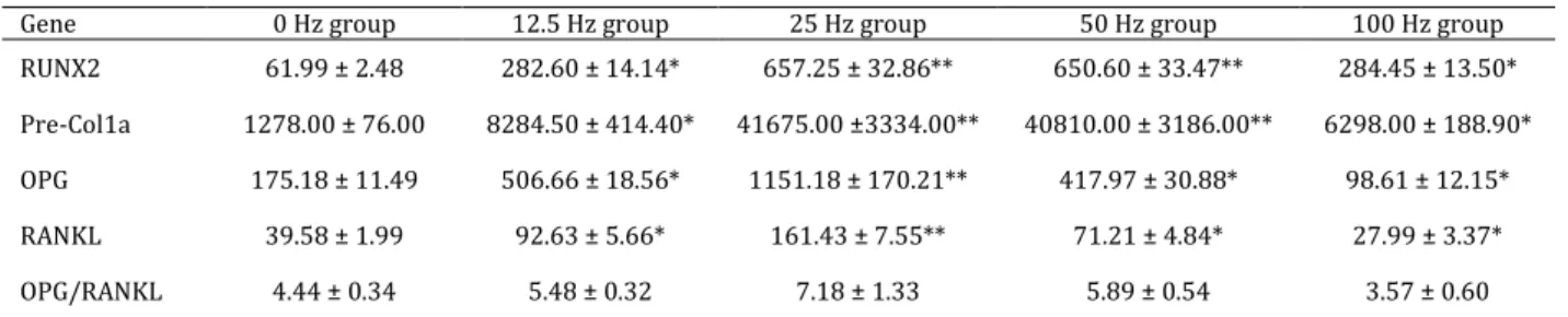
Table 2. mRNA levels of bone healing associated transcripts
Gene 0 Hz group 12.5 Hz group 25 Hz group 50 Hz group 100 Hz group
RUNX2 61.99 ± 2.48 282.60 ± 14.14* 657.25 ± 32.86** 650.60 ± 33.47** 284.45 ± 13.50*
Pre-Col1a 1278.00 ± 76.00 8284.50 ± 414.40* 41675.00 ±3334.00** 40810.00 ± 3186.00** 6298.00 ± 188.90*
OPG 175.18 ± 11.49 506.66 ± 18.56* 1151.18 ± 170.21** 417.97 ± 30.88* 98.61 ± 12.15*
RANKL 39.58 ± 1.99 92.63 ± 5.66* 161.43 ± 7.55** 71.21 ± 4.84* 27.99 ± 3.37*
OPG/RANKL 4.44 ± 0.34 5.48 ± 0.32 7.18 ± 1.33 5.89 ± 0.54 3.57 ± 0.60
OPG: osteoprotegerin; Pre-Col1a: pre-collagen type 1; RANKL: receptor activator of nuclear factor -B ligand; RUNX2: runt-related transcription factor 2
*P<0.05; **P<0.01 vs 0 Hz
Results
Low-frequency vibration treatment promotes bone fracture healing in experimental animals
To assess the effect of low-frequency vibration treatment, we first examined the gross morphology of humeri bone defects. At 4 weeks post-surgery, the bone defects were repaired effectively in the groups subjected to vibrations at frequencies of 12.5 Hz, 25 Hz, and 50 Hz (Figure 1). Specifically, bones from the 25 and 50 Hz treatment groups displayed increased callus formation, suggesting these two frequencies promote the best bone fracture healing and recovery .
Low-frequency vibration treatment promotes bone fracture healing by inducing ultrastructural changes
Following examination at the gross level, we sought to assess the effect of low-frequency vibration treatment on bone defects at the internal tissue, or microscopic, level. To do this, bone defects from each group were sectioned and observed using scanning electron microscopy. In the experimental groups treated with frequencies of 25 and 50 Hz, the implanted demineralized bones were tightly connected with the injured area, surrounded by an increased number of irregular new trabecular bones (Figure 2). Further, mesenchymal cells and osteoblasts were observed both in the
connective area and accumulated on the implanted demineralized bones (Figure 2). This phenotype was not observed in the group without low-frequency vibration treatment (0 Hz group) and was only
observed to a lesser degree in the 12.5 Hz and 100 Hz groups (Figure 2).
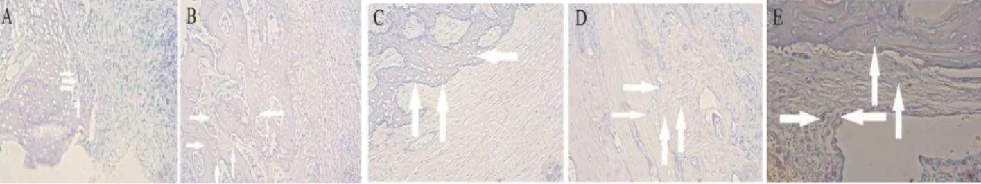
Low-frequency vibration treatment promotes stem cell activity of implanted BMSCs
To examine whether the better recovery of the bone defect is related to the activity of implanted BMSCs, ALP staining was carried out to identify pluripotent stem cells within bone sections. The degree of ALP staining gradually increased from a few pluripotent stem cells in the 0 Hz group to some positives in the 12.5 Hz and 100 Hz groups, to a substantial amount in the 25 Hz and 50 Hz groups (Figure 3). In addition, osteoblast staining was also much stronger in samples from the 25 Hz and 50 Hz groups, when compared with the 12.5 Hz and 100 Hz groups (Figure 3).
Low-frequency vibration treatment upregulates genes associated with BMSC differentiation
To examine the potential effect of the upregula- tion of osteogenesis-specific mRNA transcripts, we quantitated the mRNA levels of RUNX2, OPG, RANKL, and pre-Col1a in bone defects. The mRNA levels of RUNX2, OPG, RANKL, and pre-Col1a in the 12.5 Hz, 25 Hz, 50 Hz, and 100 Hz groups were substantially higher compared to the 0 Hz group (Table 2).
Discussion
Low-frequency vibration (10–100 Hz, intensity < 10 µε) can significantly promote bone anabolic effects without causing fractures (14-17). In ovariectomized
Figure 3. Low-frequency vibration treatment in vivo promotes stem cell activity of implanted bone marrow stromal cells. Representative images of alkaline phosphatase-stained sections from the 0 Hz group (A), 12.5 Hz group (B), 25 Hz group (C), 50 Hz group (D), and 100 Hz group (E). Magnification is 100×. Arrows indicate positively stained cells
rats, a small animal model for osteoporosis, low-frequency vibration increased osteogenesis-related gene expression and callus formation, stimulated new bone formation, prevented bone loss, and reduced bone strength decline (17). Furthermore, Xian Li et al (21) showed a 0.16 mJ/mm2, 0–3000 pulse shock wave in vitro could stimulate the expression of transforming growth factor-β; pre-Col1a, and osteocalcin promote the formation of calcium nodule, and increase the expression of oncogenic Ras in rats. These findings indicate that 500 shock pulses can effectively stimulate osteogenesis. In a similar study involving shock waves in a rodent model, Takahashi et al (22) found that in mice after 21 days of shock stimulus, femur bone mineral content and bone mineral density were increased by 8.46% and 5.80%,
respectively. Additionally, a significant increase in the osteogenesis-related genes, pre-Col1a,
osteocalcin, and osteopontin, was observed within the first four days of injury. After seven days, there was a growing intramembranous bone trabecular bone formation, and osteocalcin and pre-Col1a could be detected in the newborn inner trabecular bone of mature osteoblasts. Osteopontin was expressed in the immature bone cells, osteoblasts, and osteoclasts. Our results are consistent with these studies, showing the positive effects of low-frequency vibration stress on the increased expression of bone fracture-healing genes and the healing of the bone fractures.
How low-frequency vibration in New Zealand rabbits stimulates the differentiation of BMSCs remains unclear. However, several signaling mechanisms, such as OPG-RANKL and mitogen-activated protein kinase pathways, may play a role (13, 23). Cell surface molecules, including integrins and p38, have also been implicated (25, 26). However, further studies are needed to clarify the exact mechanisms involved.
Low-frequency vibration to stimulate bone formation has several advantages, including being non-invasive and easy to operate. Therefore, it could potentially be a great benefit to bone fracture healing, bone nonunion, osteoporosis, and
prevention of age-related bone loss.
Conclusion
This study demonstrates that low-frequency vibrations, most ideally at 50 Hz, promote the differentiation of BMSCs into osteoblasts, and in turn, promote efficient bone healing after injury. Several phenotypes were observed following treatment at 50 Hz, including faster absorption of callus hematoma, higher proliferation activity of osteoblasts, a more orderly distribution and prevalence of collagen fibers, and a substantially higher amount of callus. On a cellular level, we observed the upregulation of genes specifically associated with BMSC differentiation following treatment at 50 Hz. In summary, 50 Hz is the optimal frequency to stimulate BMSCs for bone injury repair in vivo.
Acknowledgment
This study was supported by a grant from the National Natural Science Foundation of China (Grant NO.30970708).
Conflict of Interest
The authors declare no conflict of interest.
References
1. Roberts TT, Rosenbaum AJ. Bone grafts, bone
substitutes and orthobiologics: the bridge between basic science and clinical advancements in fracture healing. Organogenesis 2012; 8:114-124.
2.Krebsbach PH, Kuznetsov SA, Bianco P, Robey PG.
Bone marrow stromal cells: characterization and clinical application. Crit Rev Oral Biol Med 1999; 10:165-181.
3.Mauney JR, Blumberg J, Pirun M, Volloch V,
Vunjak-Novakovic G, Kaplan DL. Osteogenic differentiation of human bone marrow stromal cells on partially
demineralized bone scaffolds in vitro. Tissue Eng
2004; 10:81-92.
4. Gimble JM, Wanker F, Wang CS, Bass H, Wu X, Kelly
K, et al. Regulation of bone marrow stromal cell
differentiation by cytokines whose receptors share the gp130 protein. J Cell Biochem 1994; 54:122-133.
5. Sun X, Gan Y, Tang T, Zhang X, Dai K. In vitro
proliferation and differentiation of human
mesenchymal stem cells cultured in autologous plasma derived from bone marrow. Tissue Eng Part A 2008; 14:391-400.
6. Komori T, Yagi H, Nomura S, Yamaguchi A, Sasaki
results in a complete lack of bone formation owing to maturational arrest of osteoblasts. Cell 1997; 89:755-764.
7. Otto F, Thornell AP, Crompton T, Denzel A, Gilmour
KC, Rosewell IR, et al. Cbfa1, a candidate gene for
cleidocranial dysplasia syndrome, is essential for osteoblast differentiation and bone development. Cell 1997; 89:765-771.
8. Huang W, Yang S, Shao J, Li YP. Signaling and transcriptional regulation in osteoblast commitment and differentiation. Front Biosci 2007; 12:3068-3092.
9. Frost HM. Wolff's Law and bone's structural
adaptations to mechanical usage: an overview for clinicians. Angle Orthod 1994; 64:175-188.
10. Leung KS, Shi HF, Cheung WH, Qin L, Ng WK, Tam
KF, et al. Low-magnitude high-frequency vibration
accelerates callus formation, mineralization, and fracture healing in rats. J Orthop Res 2009; 27:458-465.
11. Judex S, Gupta S, Rubin C. Regulation of mechanical signals in bone. Orthod Craniofac Res 2009; 12: 94-104.
12. Glossop JR, Cartmell SH. Effect of fluid flow-induced shear stress on human mesenchymal stem cells: differential gene expression of IL1B and MAP3K8 in MAPK signaling. Gene Expr Patterns 2009; 9:381-388.
13. Zen Fan, Xin Song and H. H et al. Impact of
vibration on bone structure and function after injury. Chinese Journal of Clinical Rehabilitation. 2006;45: 168-169.
14. Rubin C, Turner AS, Mallinckrodt C, Jerome C, McLeod K, Bain S. Mechanical strain, induced noninvasively in the high-frequency domain, is anabolic to cancellous bone, but not cortical bone. Bone 2002; 30: 445-452.
15. Omar H, Shen G, Jones AS, Zoellner H, Petocz P, Darendeliler MA. Effect of low magnitude and high frequency mechanical stimuli on defects healing in cranial bones. J Oral Maxillofac Surg 2008; 66:1104-1111.
16. Lau E, Al-Dujaili S, Guenther A, Liu D, Wang L, You L. Effect of low-magnitude, high-frequency vibration on osteocytes in the regulation of osteoclasts. Bone 2010; 46: 1508-1515.
17. Tezval M, Biblis M, Sehmisch S, Schmelz U, Kolios
L, Rack T, et al. Improvement of femoral bone quality after low-magnitude, high-frequency mechanical stimulation in the ovariectomized rat as an osteopenia model. Calcif Tissue Int 2011; 88:33-40. 18. Pre D, Ceccarelli G, Visai L, Benedetti L, Imbriani M, Cusella De Angelis MG, et al. High-frequency vibration treatment of human bone marrow stromal cells increases differentiation toward bone tissue. Bone Marrow Res 2013; 2013:803450.
19. Sadat Hashemi Z, Forouzandeh Moghadam M, Soleimani M. Comparison of the Ex Vivo Expansion of UCB-Derived CD34+ in 3D DBM/MBA Scaffolds with USSC as a Feeder Layer. Iran J Basic Med Sci 2013; 16:1075-1087.
20. Hashemibeni B, Esfandiari E, Sadeghi F, Heidary F, Roshankhah S, Mardani M, et al. An animal model
study for bone repair with encapsulated
differentiated osteoblasts from adipose-derived stem cells in alginate. Iran J Basic Med Sci 2014; 17:854-859.
21. Xian Li, Wang J, ZHOU Chang ren, et al Effects of
Chitooligosaccharide on the Expression of Cyclin D1 and RUNX2 in UCB-MSC Cells. J Materials Sci Eng 2010; 28:649-652.
22. Takahashi K, Yamazaki M, Saisu T, Nakajima A,
Shimizu S, Mitsuhashi S, et al. Gene expression for
extracellular matrix proteins in shockwave-induced osteogenesis in rats. Calcif Tissue Int 2004; 74:187-193.
23. Xiao G, Jiang D, Gopalakrishnan R, Franceschi RT. Fibroblast growth factor 2 induction of the osteocalcin gene requires MAPK activity and phosphorylation of the osteoblast transcription factor, Cbfa1/Runx2. J Biol Chem 2002; 277:36181-36187.
24. Kakita A, Suzuki A, Ono Y, Miura Y, Itoh M, Oiso Y. Possible involvement of p38 MAP kinase in prostaglandin E1-induced ALP activity in osteoblast-like cells. Prostaglandins Leukot Essent Fatty Acids 2004; 70:469-474.