ISSN 0101-2061 (Print)
ISSN 1678-457X (Online) Food Science and Technology
OI:
D http://dx.doi.org/10.1590/1678-457X.37816
1 Introduction
The total coliform group is used to indicate the sanitary conditions of food and is composed of the genera Escherichia,
Enterobacter, Klebsiella and Citrobacter, which belong to the family Enterobacteriaceae. Faecal coliforms, also denominated thermotolerant coliforms, are bacteria belonging to the group of the total coliforms that have the ability to continue to ferment lactose with gas production at a temperature of 45 °C. The main representative of this group is the genus Escherichia (Evangelista, 2001). Faecal coliform or E. coli counts are used to determine inadequate hygiene and faecal contamination. High scores of these microorganisms may be related to significant levels of enteric pathogens, such as Salmonella spp. and E. coli (Jay, 2000).
Enterohaemorrhagic E. coli, verotoxin-producing E. coli
(VTEC) or Shigatoxin-producing E. coli (STEC) has been associated with severe outbreaks and has been widely recognised as an important pathogen since the 1980s (Davis & Brogan, 1995; Duffy et al., 2006). VTEC can be isolated from the faeces of many animals, including ruminants (cattle, sheep and buffalo) and non-ruminant animals (horses, dogs and pigs) (Rivas et al., 2006).
The O157 is the most frequently isolated VTEC serogroup, however, several studies showed that non-O157 serogroups have been increasingly isolated (Nielsen et al., 2006; Stigi et al., 2012; Vally et al., 2012), particularly VTEC
serogroups O26, O45, O103, O111, O121, and O145 have been referred to as the non O157 VTEC “top six” pathogenic serogroups (Brooks et al., 2005).
Several foods have been incriminated in VTEC outbreaks, such as raw milk, fermented meat products, cheeses, unpasteurised juices, fruits and vegetables (Gyles, 2007; Karmali et al., 2010). However, increased risk is associated with the consumption of beef (Karmali et al., 2010; Nataro & Kaper, 1998). This serotype is one of the major bacteria that can contaminate meat and may potentially be transferred from the intestine or skin during the slaughtering process (Duffy et al., 2006). Furthermore, these bacteria can survive for hours or even days on hands, cloths or equipment, causing cross-contamination due to improper hygiene practices (Chen et al., 2001; Kusumaningrum et al., 2003).
Importer markets of Brazilian meat products are demanding with regard to the quality and microbiological safety of products, generally requiring the analysis of spoilage and the presence of pathogenic microorganisms. Through the Rapid Alert System for Food and Feed (RASFF) of the European Union, when a food product is identified as posing a risk to human health, measures are established to seize or reject it. From May 2015 to May 2016, nine shipments of Brazilian beef were retained and the presence of Shigatoxin-producing E. coli was notified (RASFF, 2016).
Survey of verotoxin-producing
Escherichia coli
and faecal coliforms in beef carcasses
destined for export at slaughterhouses in Brazil
Daniele BIER1,2, Márcio Roberto SILVA3, Carlos Alberto do Nascimento RAMOS1,
Giuliani D’Amico MORININGO2, Tâmiris Aparecida dos Santos SILVA2, Alaiza Corrêa de LIMA2,
Jenyfer Valesca Monteiro CHULLI2, Flábio Ribeiro de ARAÚJO4*
Received 12 Jan., 2017 Accepted 18 Sept., 2017
1 Faculdade de Medicina Veterinária e Zootecnia, Universidade Federal de Mato Grosso do Sul – UFMS, Campo Grande, MS, Brazil 2 Universidade Católica Dom Bosco – UCDB, Campo Grande, MS, Brazil
3 Empresa Brasileira de Pesquisa Agropecuária – Embrapa Gado de Leite, Juiz de Fora, MG, Brazil 4 Empresa Brasileira de Pesquisa Agropecuária – Embrapa Gado de Corte, Campo Grande, MS, Brazil *Corresponding author: flabio.araujo@embrapa.br
Abstract
This study investigated the presence of generic and verotoxin-producing E. coli as well as enumerated faecal coliforms in 30 beef carcasses in different parts of the slaughter process (after skinning, washing and cooling) at each of three slaughterhouses of the state of Mato Grosso do Sul, Brazil. Among the total number of carcasses examined (n = 90), 39 (43.3%) had generic E. coli. Among the 270 samples analysed, 25 (9.3%) were positive after skinning, 14 (5.2%) were positive after washing and nine (3.3%) were positive after cooling. The majority of isolates of E. coli was collected from samples after skinning, which is considered a critical point of the microbial contamination of carcasses. However, the highest concentration of faecal coliforms was found after the washing step. The cooling step proved to be important to reducing the amount of hygiene-indicator microorganisms. The E. coli isolates had no stx1 or stx2 genes associated with virulence.
Keywords: meat; enterobacteria; beef industry.
The analysis of microorganisms, such as faecal coliforms, as indicators of food quality can provide useful information for predicting the possibility of contamination by pathogens. In cattle slaughterhouses, this analysis is of considerable value to the determination of sanitary conditions and the effectiveness of contamination prevention actions. Moreover, the identification of potentially pathogenic microorganisms, such as verotoxin-producing E. coli, in beef carcasses during slaughter operations can contribute significantly to the implementation of quality monitoring programs and preventive measures, with a consequent reduction in risks to consumer health.
The aim of the present study was to investigate the presence of generic and verotoxin-producing E. coli in beef carcasses at three different parts of the slaughter process (after skinning, washing and cooling) in slaughterhouses of the state of Mato Grosso do Sul, Brazil, that export meat. A further aim was to enumerate faecal coliforms in the samples.
2 Materials and methods
Samples from beef carcasses were collected at three slaughterhouses registered with the Brazilian Federal Inspection Service in the state of Mato Grosso do Sul, Brazil. At each slaughterhouse, samples were taken from five carcasses per week for six consecutive weeks, as established by the European Union for microbiological tests on carcasses (Commission Regulation - EC, 2007).
Samples were taken from each animal using a non-destructive method at three different steps of the slaughter line: after skinning, after washing and after cooling. The detection of verotoxin-producing Escherichia coli was performed using the methodology of the 2001 Compendium of Methods for the Microbiological Examination of Foods (Meng et al., 2001). Dehydrated, sterilised sponges (Speci-Sponge - Nasco, Fort Atkinson, Wisconsin, USA) measuring 11.5 × 23.0 cm and individually packed in sterile plastic bags (Whirl Pak - Nasco,
Fort Atkinson, Wisconsin, USA) were used for the collection of the samples. The sponges were hydrated with 10 ml of 1% buffered peptone water (1% BPW) (HiMedia Laboratories, Mumbai, India) and rubbed onto the chest (100 cm2), flank (100 cm2) and rump close the occlusion of the rectum (200 cm2) of the carcass using a 10 × 10 cm sterile stainless steel mould (total surface sampled: 400 cm2). The sponges were transferred to the plastic bag (Nasco, Fort Atkinson, Wisconsin, USA) and transported to the laboratory under refrigeration.
Two hundred ml of 1% BPW were added to each plastic bag containing the sponges. The mixture was homogenised in a stomacher (Lab-blender 400BA 6021, Seward Laboratory, London, England) for 60 sec and placed in Erlenmeyer flasks. An aliquot of 25 ml of the homogenised sample was added to 225 ml of EC broth (HiMedia Laboratories, Mumbai, India), followed by incubation at 37 ± 1 °C for 18 ± 2 h. Then, 10 μl of the broth was streaked in Petri dishes containing sorbitol-MacConkey agar (SMAC) and sorbitol-MacConkey agar with cefixime-tellurite supplement (SMAC-CT) (HiMedia Laboratories, Mumbai, India). After incubation of the dishes at 35 ± 1 °C for 18 to 24 hours, five to ten colonies positive and negative for sorbitol were selected,
transferred to a TSAye medium (Merck KGaA, Darmstadt, Germany) and incubated at 35 ± 1 °C for 18 h to 24 h. The suspected E. coli
colonies were isolated on nutrient agar (HiMedia Laboratories, Mumbai, India), incubated at 35 ± 1 °C for 18 to 24 hours and subjected to further biochemical tests: indole, urea, motility, H2S production, methyl red, Voges-Proskauer, fermentation of carbohydrates (Triple Sugar Iron) and citrate All culture Media of biochemical tests were provided by HiMedia Laboratories, Mumbai, India.
The quantification of faecal coliforms in the samples was determined using the Most Probable Number (MPN) technique based on Kornacki & Johnson (2001). After the incubation period, 1 ml of the sample homogenised in BPW was transferred from each homogenate into tubes containing 10 ml of BPW (10-1 dilution) and submitted to decimal serial dilutions to 10-3 in BPW. The number of positive tubes for each dilution tested was used to determine the MPN of coliforms, based on Bacteriological Analytical Manual (2010). The value in the table was equivalent to MPN per ml of the sample. As each sample comprised 200 ml of BPW in which the material collected by the sponge over an area of 400 cm2 of carcass was suspended, each millilitre of sample was equivalent to an area of 2 cm2 (Silva, 2010).
Samples of bacteria suspended in tryptone soya broth (TSB) (Oxoid, Basingstoke, U.K) were used for DNA purification using a commercial kit (DNeasy Blood & Tissue Kit, QIAGEN, Valencia, CA, USA), following the manufacturer’s instructions. The isolates biochemically confirmed as E. coli were assayed by polymerase chain reaction (PCR) targeting the stx1 and stx2 genes (Paton & Paton, 1998) in a volume of 50 µl containing 0.2 mM of each dNTP, 0.5 µM of each primer, 5 µl of 10× PCR buffer (Invitrogen Corp., Carlsbad, CA, USA), 1.5 mM of MgCl2, 2.5 U of
Taq DNA Polymerase (Invitrogen Corp., Carlsbad, CA, USA) and 2 µl of DNA. The PCR cycling conditions used with the primers (Table 1) were as follows: an initial denaturation at 95 °C for 5 min followed by 35 cycles, each consisting of 45 s at 95 °C, 45 s at the annealing temperature (56 °C), and 30 s at 72 °C. After a final elongation step at 72 °C for 5 min, the amplicons were separated by 1.5% agarose gel electrophoresis in Tris-acetate-EDTA buffer – pH 8.2 (TAE 1×) with GelRedTM (Biotium, Hayward, CA, USA). The image was then photodocumented using the L-PIX Image EX (Loccus Biotechnology, Loccus Brazil, Cotia, SP, Brazil).
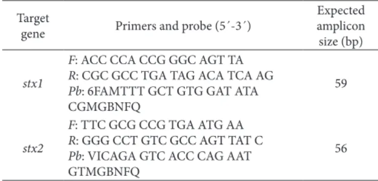
For real-time PCR (qPCR), primers and DNA probes for the TaqMan MGB system were designed with the Primer Express program (Applied Biosystems, Foster City, CA, USA) targeting
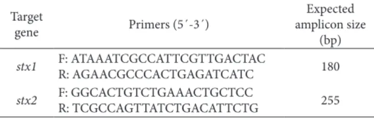
Table 1. Primers for detection of stx1 and stx2 genes, based on Paton
& Paton (1998).
Target
gene Primers (5´-3´)
Expected amplicon size
(bp)
stx1 F: ATAAATCGCCATTCGTTGACTAC
R: AGAACGCCCACTGAGATCATC 180
stx2 F: GGCACTGTCTGAAACTGCTCC
stx1 and stx2 (Table 2) and DNA detection was performed in the StepOne Plus system (Applied Biosystems, Foster City, CA, USA) in a final volume of 25 µl containing 12.5 µl of TaqMan
Universal PCR Master Mix (Applied Biosystems, Foster City, CA, USA), 7.5 µM of each primer, 1.5 µM of probe and 2 µl of the extracted DNA. The qPCR cycling conditions used were: an initial denaturation at 95 °C for 10 min followed by 40 cycles, each consisting of 25 s at 95 °C, and 15 s at 60 °C.
Strains of the reference microorganisms collection from FIOCRUZ-INCQS (Rio de Janeiro, RJ, Brazil) were used as negative and positive controls for all techniques: Escherichia coli INCQS 00033 (ATCC 25922), Escherichia coli INCQS 00171 (CDC EDL-933), Salmonella enterica subsp. enterica
serovar Enteritidis INCQS 00258 (ATCC 13076) and Salmonella enterica subsp. enterica serovar Typhimurium INCQS 00150 (ATCC 14028).
The rates of positive samples in each evaluation period (after skinning, after washing and after cooling) were compared using the OpenEpi program (Dean et al., 2013). The mid p-value determined using Fisher’s Exact test for matched pairs was used to test the strength of associations between paired evaluation periods and the presence/absence of E. coli.
3 Results and discussion
Eleven of the thirty beef carcasses (36.7%) were positive for generic E. coli at slaughterhouse I, 21/30 (70%) were positive at slaughterhouse II and 7/30 (23.3%) were positive at slaughterhouse III (Table 3). Thus, 39 (43.3%) of the 90 carcasses analysed at the three slaughterhouses exhibited E. coli.
Evaluating the presence of generic E. coli in beef carcasses at four slaughterhouses in Australia, Sumner et al. (2003) found an 18.8% positivity rate. However, the sampling area
was 200 cm2 per carcass, whereas the sampling area in the present investigation was 400 cm2, which may have enabled the greater recovery of bacteria, thereby determining a higher percentage of contaminated carcasses. Similarly, data from the Food Safety and Inspection Service of the US Department of Agriculture (USDA-FSIS) demonstrated the presence of bacteria in 16.6% of establishments under federal inspection (Eblen et al., 2005). The USDA-FSIS believes that generic (or non-pathogenic) E. coli is the best microbiological indicator of faecal contamination of beef carcasses, since the main contamination pathway by pathogenic bacteria, such as E. coli, is through the faeces (Department of Agriculture & Food Safety and Inspection Service, 1996). The level of contamination by E. coli in beef carcasses may vary greatly due to faecal contamination as well as other factors, such as skinning and gutting the animal or inadequate hygiene practices (Rigobelo et al., 2006). Table 3 shows infection rates per collection point at each slaughterhouse analysed.
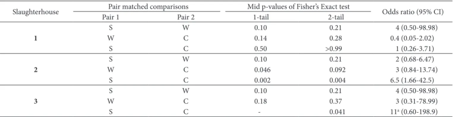
At slaughterhouse 1, no significant differences were detected in relation negative/positive carcasses between any two points analysed. At slaughterhouse 2, the chances of the negative results were threefold higher between washing and cooling and 6.5-fold times higher between skinning and cooling (p < 0.05). Thus, a significant trend in negative contamination results was found from skinning to cooling and this trend was more pronounced between washing and cooling than skinning and washing. At slaughterhouse 3, a significant chance of negativity was detected only between skinning and cooling, which was 11-fold higher than the chance for positive results (p < 0.05). A trend toward negativity was found between skinning and cooling at slaughterhouses 2 and 3, but was not at slaughterhouse 1 (Table 4).
The decrease in the contamination in slaughterhouses II and III was expected, since skinning the animal is considered a critical stage due to the possibility of the contamination of the carcass surface by microorganisms present on the skin, hair and hooves (Lambert et al., 1991). Moreover, the main goal of washing and cooling is to reduce microbial contamination (Ordóñez, 2005). However, the increase in contamination from washing to cooling found at slaughterhouse 1 may have been due to the cross-contamination of carcasses through the workers’ utensils, clothes or hands (Roça, 2004; Pardi et al., 2006), since low temperatures reduce the number, but do not cause the complete destruction of microorganisms (Ordóñez, 2005).
Among the 270 samples evaluated at the three slaughterhouses, 145 (53.7%) were negative (< 3 MPN/ml) for the presence of faecal coliforms and 125 (46.3%) were positive, ranging from 4 to > 1100 MPN/ml. The main representative of the group of faecal coliforms is E. coli. However, some strains of Klebsiella
Table 2. Primers and DNA probes for TaqMan MGB system targeting
Stx1 and Stx2.
Target
gene Primers and probe (5´-3´)
Expected amplicon size (bp)
stx1
F: ACC CCA CCG GGC AGT TA
R: CGC GCC TGA TAG ACA TCA AG
Pb: 6FAMTTT GCT GTG GAT ATA CGMGBNFQ
59
stx2
F: TTC GCG CCG TGA ATG AA
R: GGG CCT GTC GCC AGT TAT C
Pb: VICAGA GTC ACC CAG AAT GTMGBNFQ
56
Table 3. Beef carcass samples contaminated with Escherichia coli found after skinning, washing and cooling at three slaughterhouses in Mato
Grosso do Sul, Brazil.
Slaughterhouse Number of
carcasses Positive carcasses After skinning (%) After washing (%) After cooling (%)
1 30 11 (36.7%) 5 (16.6%) 2 (6.6%) 5 (16.6%)
2 30 21 (70%) 14 (46.6%) 9 (30%) 3 (10%)
3 30 7 (23.3%) 6 (20%) 3 (10%) 1 (3.3%)
and Enterobacter exhibit thermotolerance. Table 5 shows the number of positive samples in the multiple tubes of 270 samples from beef carcasses analysed for the presence of coliforms at 45 °C and E. coli. One hundred twenty-five samples (46.3%) had faecal coliforms and E. coli was isolated in 48 samples (17.8%). Resolution 12/2001 of the Brazilian National Health Surveillance Agency (ANVISA), which approved the Technical Regulations on Microbiological Standards for Food, does not establish the coliform limit for in natura chilled or frozen meat from cattle (Brazil, 2001). However, according to Florentino et al. (1997), the mere presence of faecal coliforms is considered an indicator of faecal contamination and can indicate the presence of pathogenic bacteria, posing a risk to consumers. Sofos et al. (1999) found that an increase in the coliform count serves as an indicator of the presence of pathogens and increases the odds of isolating
Salmonella. Thus, the presence of generic E. coli implies that other faecal microorganisms, including verotoxin-producing
E. coli, may be present (Jay, 2000).
Among the 125 samples positive for faecal coliforms, 11 had counts equal to or exceeding 1100 MPN/ml, nine of which were isolated from the same slaughterhouse. The high count of thermotolerant coliforms in the samples suggests that the sanitary conditions at this slaughterhouse are not satisfactory, suggesting improper handling during slaughter operations. At slaughterhouses 2 and 3, only one sample at each establishment exhibited contamination levels greater than 1100 MPN/ml. This shows that better hygiene conditions are maintained and that good manufacturing practices and quality programs are properly implemented at these establishments.
Among the 11 samples with a faecal coliform count equal to or greater than 1100 MPN/ml, nine samples were collected after washing. Washing with chlorinated water is performed at the end of the slaughter process to remove foreign materials, such as bone splinters, and reduce the microflora that may be adhered to the surface (Roça & Serrano, 1994). However, the washing step can actually spread bacterial contamination between distinct areas (Prasai et al., 1995; Yalçin et al., 2001). Moreover, the steps after skinning and prior to washing (gutting and sawing the carcass) are also important sources of contamination (Matos et al., 2013). Poor water quality is another problem that can occur during the washing of carcasses, as water can carry contamination within the processing plant, with the possible presence of spoilage microorganisms and even pathogenic microorganisms (Amaral et al., 2007).
The 48 samples isolated from carcasses and biochemically confirmed as E. coli were tested by PCR for stx1 and stx2, which are associated with virulence, and none of the isolates exhibited the stx genes, which encode the Shiga-like toxins. Most cases and outbreaks caused by verotoxin-producing E. coli have been attributed to the consumption of beef and pork (Stephan & Schumacher, 2001; Irino et al., 2005). The two enterotoxin Shiga (stx1 and stx2), which are produced by this strain, are responsible for clinical manifestations in patients (Jay, 2005) and are essential factors of virulence in the pathogenesis of the disease.
In the present study, no E. coli isolates showed fragments specific for the stx gene. This same has been reported in previous studies in which there was no isolation of verotoxin-producing E. coli in the beef carcasses and meat products analysed (Madden et al., 2001; Silva et al., 2001; Matos et al., 2013). In contrast, several studies report the presence of enterohaemorrhagic E. coli in meat products. In a study conducted over the span of one year in England, the authors isolated E. coli O157 in 1.4% of carcasses (Chapman et al., 2001). In Switzerland, Fantelli & Stephan (2001) isolated strains of E. coli that had the Shiga-toxin producer genes (stx1 and stx2) in 2.3% of minced meat samples.
Meyer-Broseta et al. (2001) performed a critical analysis of the results of 26 epidemiological studies on the prevalence of contamination of cattle by strains of verotoxin-producing
E. coli and argue that the absence of verotoxin-producing E. coli
(VTEC) in slaughtered cattle carcasses and the low prevalence
Table 5. Number of positive samples of bovine carcasses for presence
of faecal coliforms at 45 °C and Escherichia coli at slaughterhouses under Federal Inspection Service in municipalities of Mato Grosso do Sul, Brazil.
Slaughterhouse
Number of samples
Analysed Positive for faecal coliforms Positive for
E. coli
1 90 50 12
2 90 54 26
3 90 21 10
Table 4. Paired comparisons of beef samples contaminated with Escherichia coli at three points (after skinning, washing and cooling) in three
slaughterhouses in Mato Grosso do Sul, Brazil.
Slaughterhouse Pair matched comparisons Mid p-values of Fisher’s Exact test Odds ratio (95% CI)
Pair 1 Pair 2 1-tail 2-tail
1
S W 0.10 0.21 4 (0.50-98.98)
W C 0.14 0.28 0.4 (0.05-2.02)
S C 0.50 >0.99 1 (0.26-3.71)
2
S W 0.10 0.21 2 (0.68-6.47)
W C 0.046 0.092 3 (0.84-13.74)
S C 0.002 0.004 6.5 (1.66-42.5)
3
S W 0.10 0.21 4 (0.50-98.98)
W C 0.18 0.37 3 (0.31-78.99)
S C - 0.041 11a (0.60-198.9)
rates reported in numerous studies (Chapman et al., 2001; Fantelli & Stephan, 2001; Madden et al., 2001; Silva et al., 2001; Matos et al., 2013) are likely underestimated due to the inadequate sampling methods.
Most of the studies related to VTEC are restricted to O157 serogroups (Chapman et al., 2001; Matos et al., 2013; Vallières et al., 2013), because although E. coli O157: H7 is still considered the main STEC serogroup and this may be a bias that can explain the low prevalences of VTEC in the literature. Non-O157 are also recognized as important pathogens involved with outbreaks of foodborne disease (European Food Safety Authority, 2009; Mathusa et al., 2010; Schaffzin et al., 2012). In the work of Fantelli & Stephan, 2001, seven shigatoxin producing
E. coli were isolated from 400 samples (1.75%) of minced meat, and of the five distinct serotypes identified, none of the isolates belonged to O157: H7 serogroup. However, in the present study, the Stx 1 and Stx 2 genes were investigated in all isolates with biochemical characteristics of E. coli, which could be O157 or non-O157 (Gould et al., 2009).
Another reason for the low prevalence of VTEC may be the difficulty of recovering these isolates, which has already been reported by several authors (Chui et al., 2010, 2011; Pulz et al., 2003). Another hypothesis is a low pathogen’s inoculum size, hindering the growth on an agar plate (Vallières et al., 2013). Also, many bacterial species develop a viable but non-culturable (VBNC) state, characterized by a loss of culturability, therefore impairing their detection by conventional plate count techniques (Li et al., 2014; Pienaar et al., 2016; Ding et al., 2017). Nevertheless, Stx1 production was detected VBNC cells, pointing out the potential health risk of VBNC E. coli O157:H7 (Liu et al., 2010).
A number of factors may be affect the culturability of E. coli, including light, radiation, temperature, lack of nutrients and chemicals (chlorination) (Liu et al., 2010; Zhang et al., 2015), conditions that may occur in a slaughterhouse.
4 Conclusion
The highest rate of E. coli isolates was found in the samples collected after skinning, which is considered a critical point for the microbial contamination of carcasses. However, higher concentrations of thermotolerant coliforms were found after the washing stage. The cooling step proved to be important to reducing the amount of hygiene-indicator microorganisms. The E. coli isolates had no stx1 or stx2 genes associated with virulence.
Acknowledgements
Fundação de Apoio ao Desenvolvimento do Ensino, Ciência e Tecnologia do Estado de Mato Grosso do Sul – Fundect (grant 23/200.479/2014), Universidade Católica Dom Bosco, and Embrapa Gado de Corte (grants 03.14.00.047.00.00 and 03.13.10.008.00.00).
References
Amaral, L. A., Rossi, O. D. Jr., Nader, A. Fo., Ferreira, F. L. A., & Hagi, D. D. (2007). Água utilizada em estabelecimentos que comercializam
produtos cárneos, na cidade de Jaboticabal/SP, como via de contaminação dos alimentos. Revista Brasileira de Ciência Veterinária, 14(1), 3-6. http://dx.doi.org/10.4322/rbcv.2014.220.
Bacteriological Analytical Manual – BAM. (2010). Appendix 2: most probable number from serial dilutions. Retrieved from http://www.fda. gov/food/foodscienceresearch/laboratorymethods/ucm109656.htm Brasil, Agência Nacional de Vigilância Sanitária – ANVISA. (2001,
January 10). Resolução RDC n° 12. Aprova o Regulamento Técnico
sobre padrões microbiológicos para alimentos. Diário Oficial [da] República Federativa do Brasil.
Brooks, J. T., Sowers, E. G., Wells, J. G., Greene, K. D., Griffin, P. M., Hoekstra, R. M., & Strockbine, A. (2005). Non-O157 Shiga toxin-producing Escherichia coli infections in the United States, 1983_2002. The Journal of Infectious Diseases, 192(8), 1422-1429. PMid:16170761. http://dx.doi.org/10.1086/466536.
Chapman, P. A., Cerdán Malo, A. T., Ellin, M., Ashton, R., & Harkin, M. A. (2001). Escherichia coli O157 in cattle and sheep at slaughter, on beef and lamb carcasses and in raw beef and lamb products in South Yorkshire, UK. International Journal of Food Microbiology, 64(1-2), 139-150. PMid:11252496. http://dx.doi.org/10.1016/S0168-1605(00)00453-0.
Chen, Y., Jackson, K. M., Chea, F. P., & Schaffner, D. W. (2001). Quantification and variability analysis of bacterial cross-contamination rates in common food service tasks. Journal of Food Protection, 64(1), 72-80. PMid:11198444. http://dx.doi.org/10.4315/0362-028X-64.1.72. Chui, L., Couturier, M. R., Chiu, T., Wang, G., Olson, A. B., McDonald, R. R., Antonishyn, N. A., Horsman, G., & Gilmour, M. W. (2010). Comparison of Shiga toxin-producing Escherichia coli detection methods using clinical stool samples. The Journal of Molecular Diagnostics, 12(4), 469-475. PMid:20466837. http://dx.doi.org/10.2353/ jmoldx.2010.090221.
Chui, L., Lee, M.-C., Malejczyk, K., Lim, L., Fok, D., & Kwong, P. (2011). Prevalence of Shiga toxin-producing Escherichia coli (STEC) as detected by enzyme-linked immunoassays and real-time PCR during the summer months in northern Alberta, Canada. Journal of Clinical Microbiology, 49(12), 4307-4310. PMid:21940470. http:// dx.doi.org/10.1128/JCM.05211-11.
Davis, B. S., & Brogan, R. T. (1995). A widespread community outbreak of E. coli O157 infection in Scotland. Public Health, 109(5), 381-388. PMid:7480604. http://dx.doi.org/10.1016/S0033-3506(95)80011-5. Dean, A. G., Sullivan, K. M., & Soe, M. M. (2013). OpenEpi: Open Source Epidemiologic Statistics for Public Health. Updated 2013/04/06. Retrieved from www.openepi.com
Department of Agriculture – USDA, & Food Safety and Inspection Service – FSIS. (1996). Pathogen reduction; HAZARD Analysis and Critical Control Point (HACCP) Systems. Federal Register, 61(144), 38805-38989.
Ding, T., Suo, Y., Xiang, Q., Zhao, X., Chen, S., Ye, X., & Liu, D. (2017). Significance of Viable but Nonculturable Escherichia coli: induction, detection, and control. Journal of Microbiology and Biotechnology, 27(3), 417-428. PMid:27974738. http://dx.doi.org/10.4014/jmb.1609.09063. Duffy, G., Cummins, E., Nally, P., O’Brien, S., & Butler, F. (2006). A review of quantitative microbial risk assessment in the management of Escherichia coli O157:H7 on beef. Meat Science, 74(1), 76-88. PMid:22062718. http://dx.doi.org/10.1016/j.meatsci.2006.04.011. Eblen, D. R., Levine, P., Rose, B. E., Saini, P., Mageau, R., & Hill, W. E.
European Commission – EC. (2007). Commission regulation (EC) N° 1441/2007 of 5 december 2007 amending regulation (EC) No 2073/2005 on microbiological criteria for foodstuffs. Oficial Journal of the European Union, L322, 12-29.
European Food Safety Authority – EFSA. (2009). Technical specifications for the monitoring and reporting of verotoxigenic Escherichia coli (VTEC) on animals and food (VTEC surveys on animals and food) on request of EFSA. EFSA Journal, 7(11), 1366. http://dx.doi. org/10.2903/j.efsa.2009.1366.
Evangelista, J. (2001). Tecnologia de alimentos (2. ed.). São Paulo: Editora Atheneu.
Fantelli, K., & Stephan, R. (2001). Prevalence and characteristics of Shigatoxin-producing Escherichia coli and Listeria monocytogenes strains isolated from minced meat in Switzerland. International Journal of Food Microbiology, 70(1-2), 63-69. PMid:11759763. http:// dx.doi.org/10.1016/S0168-1605(01)00515-3.
Florentino, E. R., Leite, A. F. Jr, Sá, S. N., Araújo, M. S. O., & Martins, R. S. (1997). Avaliação da qualidade microbiológica da carne moída comercializada em Campina Grande - PB. Higiene Alimentar, 11(47), 38-41.
Gould, L. H., Bopp, C., Strockbine, N., Atkinson, R., Baselski, V., Body, B., Carey, R., Crandall, C., Hurd, S., Kaplan, R., Neill, M., Shea, S., Somsel, P., Tobin-D’Angelo, M., Griffin, P. M., & Gerner-Smidt, P. (2009, October 16). Recommendations for Diagnosis of Shiga
Toxin--Producing Escherichia coli Infections by Clinical Laboratories. MMWR Recommendations and Reports, 58(RR12), 1-14. Retrieved from http://www.cdc.gov/mmwr/preview/mmwrhtml/rr5812a1.htm Gyles, C. L. (2007). Shiga toxin-producing Escherichia coli: an overview. Journal of Animal Science, 85(13, Suppl.), E45-E62. PMid:17085726. http://dx.doi.org/10.2527/jas.2006-508.
Irino, K., Kato, M. A. M. F., Vaz, T. M. I., Ramos, I. I., Souza, M. A. C., Cruz, A. S., Gomes, T. A. T., Vieira, M. A. M., & Guth, B. E. C. (2005). Serotypes and virulence markers of Shiga toxin-producing Escherichia coli (STEC) isolated from dairy cattle in São Paulo state, Brazil. Veterinary Microbiology, 105(1), 29-36. PMid:15607081. http://dx.doi.org/10.1016/j.vetmic.2004.08.007.
Jay, J. M. (2000). Indicators of food microbiological quality and safety. In J. M. Jay, Modern food microbiology (6th ed., pp. 387-407). Maryland: Aspen Publication.
Jay, J. M. (2005). Microbiologia de alimentos. (E. C. Tondo et al., trad., 6. ed.). Porto Alegre: Artmed.
Karmali, M. A., Gannon, V., & Sargeant, J. M. (2010). Verocytotoxin-producing Escherichia coli (VTEC). Veterinary Microbiology, 140(3-4), 360-370. PMid:19410388. http://dx.doi.org/10.1016/j. vetmic.2009.04.011.
Kornacki, J. L., & Johnson, J. L. (2001). Enterobacteriaceae, coliforms and Escherichia coli as quality and safety indicators. In F. P. Downes & K. Ito (Eds.), Compendium of methods for the microbiological examination of foods (4th ed., chap. 8, pp. 69-80). Washington:
American Public Health Association.
Kusumaningrum, H. D., Riboldi, G., Hazeleger, W. C., & Beumer, R. R. (2003). Survival of foodborne pathogens on stainless steel surfaces and cross-contamination to foods. International Journal of Food Microbiology, 85(3), 227-236. PMid:12878381. http://dx.doi. org/10.1016/S0168-1605(02)00540-8.
Lambert, A. D., Smith, J. P., & Dodds, K. L. (1991). Shelf life extension and microbiological safety of fresh meat. A review. Food Microbiology, 8(4), 267-297. http://dx.doi.org/10.1016/S0740-0020(05)80002-4. Li, L., Mendis, N., Trigui, H., Oliver, J. D., & Faucher, S. P. (2014). The
importance of the viable but non-culturable state in human bacterial
pathogens. Frontiers in Microbiology. Microbial Physiology and Metabolism, 5(258), 1-20.
Liu, Y., Wang, C., Tyrrell, G., & Li, X. (2010). Production of Shiga-like toxins in viable but nonculturable Escherichia coli O157:H7. Water Research, 44(3), 711-718. PMid:19879621. http://dx.doi.org/10.1016/j. watres.2009.10.005.
Madden, R. H., Espie, W. E., Moran, L., Mcbride, J., & Scates, P. (2001). Occurrence of Escherichia coli O157:H7, Listeria monocytogenes, Salmonella and Campylobacter spp. on beef carcasses in Northern Ireland. Meat Science, 58(4), 343-346. PMid:22062423. http://dx.doi. org/10.1016/S0309-1740(00)00153-4.
Mathusa, E. C., Chen, Y., Enache, E., & Hontz, L. (2010). Non-O157 Shiga toxin-producing Escherichia coli in foods. Journal of Food Protection, 73(9), 1721-1736. PMid:20828483. http://dx.doi. org/10.4315/0362-028X-73.9.1721.
Matos, A. V. R., Nunes, L. B. S., Vianna, C., Spina, T. L. B., Zuim, C. V., Possebon, F. S., Xavier, D. M., Ferraz, M. C., & Pinto, J. P. A. N. (2013). Listeria monocytogenes, E. coli O157, Salmonella spp. e microrganismos indicadores em carcaças bovinas para exportação. Arquivo Brasileiro de Medicina Veterinária e Zootecnia, 65(4), 981-988. http://dx.doi.org/10.1590/S0102-09352013000400007. Meng, J. H., Feng, P., & Doyle, M. P. (2001). Pathogenic Escherichia
coli. In F. P. Downes & K. Ito (Eds.), Compendium of methods for the microbiological examination of foods (4th ed., chap. 35, pp. 331-342).
Washington: American Public Health Association.
Meyer-Broseta, S., Bastian, S. N., Arné, P. D., Cerf, O., & Sanaa, M. (2001). Review of epidemiological surveys on the prevalence of contamination of healthy cattle with Escherichia coli serogroup O157:H7. International Journal of Hygiene and Environmental Health, 203(4), 347-361. PMid:11434215. http://dx.doi.org/10.1078/1438-4639-4410041.
Nataro, J. P., & Kaper, J. B. (1998). Diarrheagenic Escherichia coli.Clinical Microbiology Reviews, 11(1), 142-201. PMid:9457432.
Nielsen, E. M., Scheutz, F., & Torpdahl, M. (2006). Continuous surveillance of Shiga toxin-producing Escherichia coli infections by pulsed-field gel electrophoresis shows that most infections are sporadic. Foodborne Pathogens and Disease, 3(1), 81-87. PMid:16602983. http://dx.doi. org/10.1089/fpd.2006.3.81.
Ordóñez, J. A. (2005). Tecnologia de alimentos: componentes dos alimentos e processos. Porto Alegre: Artmed.
Pardi, M. C., Santos, I. F., Souza, E. R., & Pardi, H. S. (2006). Aspectos higiênico-sanitários da carne. In M. C. Pardi, I. F. Santos, E. R. Souza, & H. S. Pardi (Eds.), Ciência, higiene e tecnologia da carne (cap. 4, 2. ed., vol. 1, pp. 271-490). Goiânia: UFG.
Paton, J. C., & Paton, A. W. (1998). Pathogenesis and diagnosis of Shiga toxin-producing Escherichia coli infections. Clinical Microbiology Reviews, 11(3), 450-479. PMid:9665978.
Pienaar, J. A., Singh, A., & Barnard, T. G. (2016). The viable but nonculturable state in pathogenic Escherichia coli: a general review. African Journal of Laboratory Medicine, 5(1), a368. PMid:28879110. http://dx.doi.org/10.4102/ajlm.v5i1.368.
Prasai, R. K., Phebus, R. K., Garcia Zepeda, C. M., Kastner, C. L., Boyle, A. E., & Fung, D. Y. C. (1995). Effectiveness of trimming and/or washing on microbiological quality of beef carcasses. Journal of Food Protection, 58(10), 1114-1117. http://dx.doi.org/10.4315/0362-028X-58.10.1114.
stool specimens. Journal of Clinical Microbiology, 41(10), 4671-4675. PMid:14532201. http://dx.doi.org/10.1128/JCM.41.10.4671-4675.2003. Rapid Alert System for Food and Feed – RASFF. (2016). Notifications by product category and notifying country. Brussels: European Commission. Retrieved from https://webgate.ec.europa.eu/rasff-window/portal/?event=searchResultList
Rigobelo, E. C., Stella, A. E., Ávila, F. A., Macedo, C., & Marin, J. M. (2006). Characterization of Escherichia coli isolated from carcasses of beef cattle during their processing at an abattoir in Brazil. International Journal of Food Microbiology, 110(2), 194-198. PMid:16720056. http://dx.doi.org/10.1016/j.ijfoodmicro.2006.03.013.
Rivas, M., Miliwebsky, E., Chinen, I., Roldán, C. D., Balbi, L., García, B., Fiorilli, G., Sosa-Estani, S., Kincaid, J., Rangel, J., & Griffin, P. M., Case-Control Study Group. (2006). Characterization and epidemiologic subtyping of Shiga toxin-producing Escherichia coli strains isolated from hemolytic uremic syndrome and diarrhea cases in Argentina. Foodborne Pathogens and Disease, 3(1), 88-96. PMid:16602984. http://dx.doi.org/10.1089/fpd.2006.3.88. Roça, R. O. (2004). Microbiologia da carne. Botucatu: Laboratório de
Tecnologia dos Produtos de Origem Animal. Retrieved from http:// dgta.fca.unesp.br/docentes/roca/carnes/Roca106.pdf
Roça, R. O., & Serrano, A. M. (1994). Operações de abate de bovinos. Higiene Alimentar, 8(34), 14-20.
Schaffzin, J. K., Coronado, F., Dumas, N. B., Root, T. P., Halse, T. A., Schoonmaker-Bopp, D. J., Lurie, M. M., Nicholas, D., Gerzonich, B., Johnson, G. S., Wallace, B. J., & Musser, K. A. (2012). Public health approach to detection of non-O157 Shiga toxin-producing Escherichia coli: summary of two outbreaks and laboratory procedures. Epidemiology and Infection, 140(2), 283-289. PMid:21554779. http:// dx.doi.org/10.1017/S0950268811000719.
Silva, N. (2010). Manual de métodos de análise microbiológica de alimentos e água. 4. ed. São Paulo. Livraria Varela.
Silva, N., Silveira, N. F. A., Contreras, C., Beraquet, N. J., Yokoya, F., Nascimento, C. A., Oliveira, V. M., & Tse, C. L. (2001). Ocorrência de Escherichia coli O157: H7 em produtos cárneos e sensibilidade dos métodos de detecção. Ciência e Tecnologia de Alimentos, 21(2), 223-227. http://dx.doi.org/10.1590/S0101-20612001000200018.
Sofos, J. N., Kochevar, S. L., Reagan, J. O., & Smith, G. C. (1999). Incidence of Salmonella on beef carcasses relating to the U.S Meat and Poultry Inspection Regulations. Journal of Food Protection, 62(5), 467-473. PMid:10340666. http://dx.doi.org/10.4315/0362-028X-62.5.467. Stephan, R., & Schumacher, S. (2001). Resistance patterns of non-O157
Shiga toxinproducing Escherichia coli (STEC) strains isolated from animals, food and asymptomatic human carriers in Switzerland. Letters in Applied Microbiology, 32(2), 114-117. PMid:11169054. http://dx.doi.org/10.1046/j.1472-765x.2001.00867.x.
Stigi, K. A., Macdonald, J. K., Tellez-Marfin, A. A., & Lofy, K. H. (2012). Laboratory practices and incidence of non-O157 Shiga toxin-producing Escherichia coli infections. Emerging Infectious Diseases, 18(3), 477-479. PMid:22377034. http://dx.doi.org/10.3201/ eid1803.111358.
Sumner, J., Petrenas, E., Dean, P., Dowsett, P., West, G., Wiering, R., & Raven, G. (2003). Microbial contamination on beef and sheep carcases in South Australia. International Journal of Food Microbiology, 81(3), 255-260. PMid:12485752. http://dx.doi.org/10.1016/S0168-1605(02)00220-9.
Vallières, E., Saint-Jean, M., & Rallu, F. (2013). Comparison of three different methods for detection of Shiga Toxin-Producing Escherichia coli in a Tertiary Pediatric Care Center. Journal of Clinical Microbiology, 51(2), 481-486. PMid:23175264. http://dx.doi. org/10.1128/JCM.02219-12.
Vally, H., Hall, G., Dyda, A., Raupach, J., Knope, K., Combs, B., & Desmarchelier, P. (2012). Epidemiology of Shiga toxin producing Escherichia coli in Australia, 2000-2010. BMC Public Health, 12(63), 1-12. PMid:22264221.
Yalçin, S., Nizamlioglu, M., & Gürbüz, U. (2001). Fecal coliform contamination of beef carcasses during the slaughtering process. Journal of Food Safety, 21(4), 225-231. http://dx.doi.org/10.1111/j.1745-4565.2001. tb00321.x.