Occurrence of
Salmonella
spp. and generic
Escherichia coli
on beef carcasses
sampled at a brazilian slaughterhouse
Fabiana Fernanda Pacheco da Silva, Mariana Bandeira Horvath,
Juliana Guedes Silveira, Luiza Pieta, Eduardo Cesar Tondo
Laboratório de Microbiologia e Controle de Alimentos, Instituto de Ciência e Tecnologia de Alimento, Universidade Federal do Rio Grande do Sul, Porto Alegre, RS, Brazil.
Submitted: April 2, 2012; Approved: April 1, 2013.
Abstract
A total of 120 beef carcasses were analyzed during processing at a slaughterhouse in southern Brazil. The carcasses were sampled by swab at three different steps of the slaughter line and then they were tested forSalmonellaandE. coli. TheSalmonellaisolates were also examined for antimicrobial sus-ceptibility.Salmonellaprevalence distribution was modeled and the probability of contamination was simulated using @Risk program and 10,000 interactions. Results demonstrated that 4 beef car-casses (3.3%) were positive forSalmonellaonly in the first point. The six isolates ofSalmonellawere classified:S. Newport (n = 3),S. Saintpaul (n = 2) andS. Anatum (n = 1). NoSalmonellastrains ex-hibited resistance to any of the antimicrobials tested. As expected, the most contaminated point with E. coliwas the first point (hide), presenting counts from 0.31 to 5.07 log cfu/100 cm2. Much smaller E. colicounts were observed in the other points. Results indicated low levels ofSalmonellaandE. colion the beef carcasses analyzed and also low probability of contamination of the carcasses by Sal-monella, suggesting adequate microbiological quality.
Key words:Salmonella, E.coli, beef carcasses, slaughter line, antimicrobial susceptibility.
Introduction
According to the United States Department of Agri-culture (USDA), Brazil is the leading exporter of beef in the world. In 2011, Brazil exported 1.8 million tons of beef and the main markets were Russia, Iran, Hong Kong, Egypt and Venezuela (USDA, 2011). Due to its importance in the in-ternational market, the quality and food safety of Brazilian beef must meet well-established international require-ments. In order to evaluate microbiological quality of meat, the quantification ofE. coliand the presence ofSalmonella have frequently been used because these microorganisms are considered good indicators of quality and food safety worldwide. The Food Safety and Inspection Service (FSIS) in the United States of America has established the criteria of a maximum 2.7% ofSalmonellafor cow and bull car-casses tested per set and a maximum of 102 cfu/cm2ofE. colibiotype I. The results of the quantitative analysis of car-casses at a specified frequency forEscherichia colibiotype
I have been used to guide the maintenance of sanitary con-ditions during slaughter (USDA, 1996).
Salmonellaspp. has been identified as the most im-portant contaminant of food and the leading bacterial agent responsible for foodborne outbreaks in several countries (Majowiczet al., 2010). In the last years, global surveil-lance data indicated that the number of salmonellosis has increased mainly associated with the consumption of raw or undercooked eggs, poultry, meat or dairy products, dem-onstrating the importance of controlling this pathogen in food production (Braden, 2006; Kimuraet al., 2004; Zhao et al., 2001). In the European Union, meat products were the second most common food group contributing to hu-man salmonellosis in 2005 (Norrung and Buncic, 2008).
In Brazil, a remarkable increase in the incidence of foodborne salmonellosis has been reported (Brasil, 2009). According to the Surveillance Service of Brazil, among the 6,349 foodborne outbreaks registered in this country from
Send correspondence to F.F.P. Silva. Laboratório de Microbiologia e Controle de Alimentos, Instituto de Ciência e Tecnologia de Alimento, Universidade Federal do Rio Grande do Sul, Avenida Bento Gonçalves 9500, Prédio 43212, Campus do Vale, Agronomia, Caixa Postal 15090, 91501-970 Porto Alegre, RS, Brazil. E-mail: fabi.p@terra.com.br.
1999 to 2009,Salmonellawas the most implicated bacterial agent, and was responsible for almost 20.7% of the reported outbreaks (Brasil, 2009). Corroborating these data, in Rio Grande do Sul (RS), southern Brazil,Salmonellawas iden-tified as the main causative agent of foodborne diseases, re-sponsible for 35.7% (116 outbreaks) of the total of 323 outbreaks investigated during 1997 to 1999, and the meat products were the third most common food involved (Cos-talunga and Tondo, 2002).
The increased use of antimicrobial agents in animal production and human medicine as a mean of preventing and treating diseases is a significant factor in the emergence of antibiotic-resistantSalmonella. Therefore, resistant Sal-monellawhich develops as a result of antibiotic use in ani-mal production can be transferred to humans through the food chain, and it is a problem to be considered. Contami-nation of food with antibiotic-resistant bacteria can be a major threat to public health, causing more difficulties in the treatment of infectious diseases (Arslan and Eyi, 2010).
It is well know that the microbiological quality of meat depends on the control measures implemented in the slaughter lines, and in slaughterhouses working 24 h a day the contamination events can occur very often. In order to correct identify contamination procedures and establish control measures, beef carcasses may be evaluated during different steps of the slaughter.
The aim of this study was to determine the occurrence and antimicrobial susceptibility ofSalmonellaspp. and the contamination withE. colion beef carcasses during differ-ent steps of slaughter in a slaughterhouse in southern Brazil.
Materail and Methods
Slaughterhouse and animals
Samples were taken at a slaughterhouse located in Rio Grande do Sul, regulated by the Brazilian Federal In-spection Service. The abattoir slaughtered approximately 200 animals daily and the slaughter was done mainly in the morning. The collections were previously scheduled and did not change the routine of the slaughterhouse. During the period from September 2009 to June 2010, thirteen sam-plings were carried out, analyzing a total of 120 carcasses. The carcasses chosen to be sampled were the first pro-cessed at the slaughterhouse on each collection day.
In-plant sampling locations
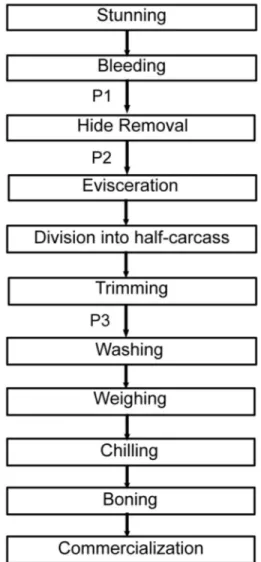
Carcass sampling was carried out at three steps in the processing line: first collection point (P1) was on the hide after bleeding but before hide opening and subsequent hide removal; second collection point (P2) was on the carcass after hide removal but before evisceration; and third collec-tion point (P3) was the half-carcass but before the final washing (Figure 1). The same carcass was sampled at P1, P2 and P3.
Carcass sampling areas
(< 7 °C), within 2 h, to the Food Microbiology and Control Laboratory of Food Science and Technology Institute (ICTA/UFRGS) to be analyzed.
Homogenization of the samples
After the arrival of the samples at the Laboratory, 200 mL of saline peptone solution was added to each plastic bag. The plastic bag was repeatedly squeezed manually for 1 min and further decimal dilutions were made with saline solution to carry outE. colianalysis.
Enumeration ofEscherichia coli
The 3M PetrifilmE. coli/Coliform Count Plates (3M, Sumaré, Brazil) were used to enumerateE.coli. From each appropriate decimal dilution, 1 mL was removed, inocu-lated on a Petrifilm plate and incubated for 48 h at 37 °C. Blue colonies with gas production were counted.
Isolation and enumeration ofSalmonellaspp.
An aliquot of 40 mL of the saline peptone solution was centrifuged at 1000gfor 15 min and the sediment was used to investigate the presence ofSalmonellaspp.
According to the ISO 6579:2002 method (ISO, 2002), each sediment was incubated in 100 mL of buffered pepto-ne water (Oxoid, São Paulo, Brazil) for 18-24 h at 37 °C. Subsequently, 1 mL was transferred to 10 mL of Muller Kauffmann tetrathionate - novobiocin broth (MKTTn, Oxoid) and 0.1 mL was transferred to 10 mL of Rappaport-Vassiliadis Broth with soya (RVS, Oxoid) and incubated at 37 °C and 42.5 °C, respectively, for 24 h. Then, both Xylose Lysine Deoxycholate agar (XLD, Oxoid) and Man-nitol Lysine Crystal Violet Brilliant Green agar (MLCB, Oxoid) plates were inoculated with aliquots of the cultures from RVS and MKTTn. All the plates were incubated at 37 °C for 24 h. Suspect colonies (red with or without a black center from XLD; mauve colored colonies with a black center from MLCB) were purified on a Nutrient agar. Then, biochemical recommended tests and serology by ag-glutination with Poly O antisera (Probac, São Paulo, Brazil) were carried out to confirm the result (MacFaddin, 2000). After these tests,Salmonellaspp. isolates were forwarded to the Laboratório de Enterobactérias of the Instituto Oswaldo Cruz (Rio de Janeiro, RJ, Brazil) for serotyping.
To enumerateSalmonellaspp., 0.1 mL of the first di-lution was spread-plated on XLD agar (Oxoid, São Paulo, Brazil). The plates were incubated for 24 h at 37 °C. Red colonies with or without black centers were counted and later confirmed by biochemical tests.
Antimicrobial susceptibility test
Susceptibility to antimicrobial agents was tested us-ing the disk diffusion method on Muller-Hinton agar (Oxoid, São Paulo, Brazil) plates according to the National Committee for Clinical Laboratory Standards (National Committee for Clinical Laboratory Standards, 2010). The following 15 antimicrobial agents were examined against Salmonellaisolates: ampicillin (10mg), cefoxitin (30mg), cephalothin (30 mg), cefotaxime (30 mg), imipenem (10mg), chloramphenicol (30mg), amikacin (30mg), genta-micin (10mg), kanamycin (30mg), streptomycin (10mg), nalidixic acid (30 mg), ciprofloxacin (5 mg), tetracycline (30mg), trimethoprim-sulphamethoxazole (25mg), and sul-fonamides (300mg). The diameters of the zones of inhibi-tion were recorded to the nearest millimeter and classified as susceptible, intermediate and resistant.
Measurement of the levels of free residual chlorine in the washing animal water
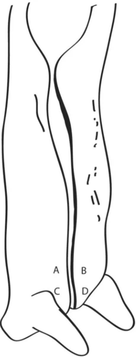
The levels of free residual chlorine in the animal washing water were provided by the Inspection Service of the slaughterhouse. The values were measured at the begin-ning of each slaughter process. The measurement was done Figure 2 - Carcass sampling areas. Each letter indicates a region of
using a C401 colorimeter (Eutech Instruments, Vernon Hills, U.S.A.).
Statistical analysis
E. coliandSalmonella spp. counts were calculated per 100 cm2and converted to log counts before statistical analysis. The mean values were calculated and the analysis of variance (ANOVA) and a Tukey test were carried out to compare the differences between the mean values. The dif-ferences were considered significant with p values less than 0.05. For theSalmonellaspp. analysis, data were reported as the percentage of samples positive for the pathogen. Sal-monellaprevalence distribution was modeled and used as an output to simulate the probability of contamination (P). The simulations were carried out using @Risk program stu-dent version number 5.7, with 10,000 interactions. Preva-lence distribution was simulated using binomial distribu-tion considering positive samples (x) and the sample number (n). Beta distribution was used to calculate the probability of contamination by Salmonella, considering the uncertainties of the sampling (bandafactors). For this purpose we followed the formula below:
p=b(x+ 1,n-x+ 1)
wherepis the probability,xis the number of positive sam-ples forSalmonellaspp.,nis the number of carcasses sam-pled.
Results and Discussion
Enumeration ofEscherichia coli
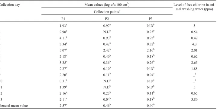
The general mean values ofE. coliwere 2.57, 0.46 and 0.40 log cfu/100 cm2at P1, P2 and P3, respectively. There were significant differences in counts observed in P1 and P2 and P1 and P3, but no significant difference was ob-served between the counts of P2 and P3. The low counts in P2 and P3 could possibly be explained due to proper hide removal and the fact that carcass tissues are considered sterile. The significant reduction in counts when P1 was compared with the other two points and the low counts ob-served in P2 and P3 could indicate that manufacturing prac-tices were adequate during processing in the slaughter-house.
The counts ofE. coli on animal hides (P1) ranged from 0.31 to 5.07 log cfu/100 cm2(Table 1). The high vari-ability inE.colicounts at P1 could be explained by the dif-ferent geographic origin of the animals, the age of animals, cleanliness of hides and breeds of animals (Antic et al., 2010; Davieset al., 2000; Hancocket al., 1997; Mcevoyet al., 2000). Other studies reported higherE.colicounts on hides than the results obtained in this study. For example, Baconet al.(2000) had higher mean values ofE. coli rang-ing from 5.5 to 7.5 log cfu/100 cm2. Arthuret al.(2004) have demonstrated higher mean values ofE.colion animal hides in two commercial fed-beef processing plants. The counts ranged from 6.6 to 8.0 log cfu/100 cm2in one of the
Table 1- Mean values ofE.coliat three points of the slaughter line and levels of free chlorine in animal washing water in one slaughterhouse in Southern Brazil.
Collection day Mean values (log cfu/100 cm2) Level of free chlorine in ani-mal washing water (ppm) Collection pointsd
P1 P2 P3
1 1.93a 0.97a N.Db 5
2 2.98a N.Db 0.25b 0.54
3 4.11a 0.93b 0.93b 0.42
4 3.34a 0.42b 0.32b 4.3
5 5.07a 2.42b 2.10b 2.01
6 2.18a 0.40b 0.18b 0.62
7 3.35a 0.36b 0.26b 2.65
8 2.27a 0.10b N.Db 1.85
9 2.20a 0.11b 0.94c _e
10 0.31a N.Da N.Da _e
11 1.39a N.Db N.Db 5
12 2.16a 0.23b 0.11b 0.65
13 2.11a 0.04b 0.18b 3.80
General mean value 2.57a 0.46b 0.40b
a
Values in a row with the same capital letter are not significantly different (p > 0.05; Tukey Test). d
P1 = first collection point, P2 = second collection point, P3 = third collection point. eThe level of chlorine was not provided by the slaughterhouse.
N.D
plants and 4.9 to 5.8 log cfu/100 cm2in the other plant. The microbiological loads of incoming cattle are important be-cause the external hide is a primary source of fecal contami-nation, which can be eventually transferred to the underly-ing sterile carcass tissue (Baconet al., 2000).
The results obtained forE. coliat P2 showed a maxi-mum value of 2.42 log cfu/100 cm2. Arthuret al.(2004) re-ported similar results examining 288 beef carcasses in the USA. On the other hand, Baconet al.(2000) found higher mean values ranging from 2.6 to 5.3 cfu/100 cm2analyzing beef carcasses in the USA. In the present study, the micro-biological loads at P2 probably reflected the extent of the microbiological contamination originating from the hide, since beef carcass surfaces are generally sterile (Baconet al., 2000).
Analyzing counts obtained on different sampling days, it could be observed that the fifth collection day showed significantly higher counts (P1 = 5.07, P2 = 2.42 and P3 = 2.10 log cfu/100 cm2) for all three points, com-pared to the other days, while the tenth collection day dem-onstrated the lowest mean values. The reasons for these differences were not explored in this study; however they corroborate the idea that the higher the initial contamina-tion, the greater the contamination of the final product. An-other important result observed was the significant increase between P2 and P3 on the ninth collection day. It could pos-sibly be explained as a recontamination after evisceration or another failure in processing.
The results indicated that there was no direct correla-tion between the level of free chlorine in the water used to wash the animals before slaughter and the mean values of E. coliobserved in P1. As an example, on the first sampling day, the mean value of 1.93 log cfu/100cm2was verified with a free chlorine level of 5 ppm in water, however on the twelfth collection day, a mean value of 2.16 log cfu/100cm2 was observed, whilst the level of free chlorine was 0.65 ppm. Even though the chlorine level varied almost 9-fold, the bacterial counts were not significantly different. These results may demonstrate that only washing the ani-mal hide with chlorinated water does not necessarily im-prove the microbial status of the hide. The risk of contami-nation of the beef carcasses may still exist, if only this practice is used. In order to improve microbiological qual-ity, the use of multiple-sequential interventions, like pre-and post-evisceration water washing, organic acid solution rinsing and hot water washing could be possible measures to decontaminate beef carcasses during the slaughtering and dressing process (Baconet al., 2000).
Isolation, enumeration and antimicrobial susceptibility ofSalmonellaspp.
The presence of Salmonella spp. was observed in only four carcasses (3.3%), three sampled on the fifth col-lection day and one carcass sampled on the eighth collec-tion day. Salmonella spp. was only isolated in P1 from
animal hides. Reid et al. (2002) obtained similar results showing a prevalence of 3.3% ofSalmonellain 90 beef cat-tle hides in the south-west of England. A much higher Sal-monella prevalence (94.8%) was found by Arthur et al. (2007) analyzing 288 beef cattle hides in the USA. Con-versely, Anticet al.(2010) were not able to isolate Salmo-nellaspp. in any of the 40 animal hides sampled in one abattoir in Serbia.
The low level ofSalmonellacontamination found in this study could be explained by multiple factors. First, the slaughtered cattle had different origins, coming from dif-ferent farms that could easily have had differences in the prevalence of this pathogen. Secondly, in Brazil, extensive farming is the most prevalent kind of animal exploitation. With this practice, direct contact among animals is very low, which may help to avoid the transmission of Salmo-nella. There are other factors that could explain the Salmo-nellaprevalence on beef carcasses, including those related to the feed, animals, transport and the environment. All those factors vary largely among countries and geograph-ical regions.
The modeling of probability of contamination by Sal-monella on P1 indicated values varying from 0.016% to 0.075% with mean numbers of 0.041%. These numbers were calculated considering the uncertainty of the sam-pling. Such values mean that in 90% of the sampling on beef carcasses in this slaughter line probably at least 1.6% and in the maximum of 7.5% of the carcasses may be posi-tive forSalmonella. These prevalence values were consid-ered low mainly considering thatSalmonellawas isolated on animal hides.
Unlike of the hides, there was no Salmonella spp. found at any of the other two points (P2 and P3) analyzed in the present study, and these results were similar with those found by Meyeret al.(2010) who examined 841 beef car-casses in Germany. The absence ofSalmonellaat P2 and P3 and the lowE.colicounts at these points demonstrated that the hygienic conditions and manufacturing practices of the slaughterhouses were adequate.
Six strains of Salmonella spp. were isolated in the present study, and they were classified as three different serovars. The most prevalent serovar was S. Newport, which was found on three animals. The serovarS.Saintpaul was found on two animals, whileS.Anatum was identified on only one animal.
All theSalmonellaspp. stains were susceptible to the 15 antimicrobials tested. This result is different to those ob-served by Baconet al.(2002) who have reported 69.4% of 49Salmonella strains were resistant at least one antimi-crobial tested. Stevenset al.(2006) obtained 99 isolated of Salmonellaspp. and the percentages of strains resistant to nitrofurans, sulfamethoxazole, streptomycin, chloramphe-nicol and nalidixic acid were 36.7, 21.1, 14.1, 2 and 1%, re-spectively.
The difference in the resistance profile observed in this study and the others reported in the literature could be explained by several factors, especially the type of farming. Extensive farming of cattle has been the most prevalent way of farming in Brazil. In this kind of farming, the use of antimicrobials has been very low; therefore, there is no high selective pressure on bacterial strains. On the other hand, in some animal species, like swine, where animal farming has been done of intensive way, the number of resistant strains has been much higher. In this way of farming, due to the in-tensification of production methods, many diseases related to the introduced technologies have emerged. The control of these diseases has been done by the use of antimicrobials (Castagnaet al., 2001).
A correlation between the presence ofSalmonellaand the quantitative analysis ofE.coliwas not observed in this study. Salmonella was detected on the fifth and in the eighth collection days. However, the mean value ofE. coli on the fifth collection day was significantly higher than the mean value ofE. colion the eighth collection day (p < 0.05). In this study, the percentage of carcasses contaminated with Salmonellaand more than 2 log cfu/100 cm2E.coli was 3.37%. However the percentage of carcasses with Salmo-nellaand less than 2 log cfu/100 cm2E.coli was 3.23%. These values did not present a statistically significant dif-ference and these results indicated that there was no corre-lation between the presence ofSalmonellaand the quantita-tive analysis of E. coli. Ruby and Ingham (2009) have suggested that the correlation would be better if the absence of Salmonella spp. were linked to negative Enterobacteriaceaeresults.
In the present study, low levels ofSalmonellaandE. coliwere found on beef carcasses and also low probability of contamination of the carcasses bySalmonella, suggest-ing that adequate slaughter procedures were carried out in the slaughterhouse analyzed. However, the isolation of these microorganisms on the animal hides indicated that the risk of the contamination still exists.
References
Antic A, Blagojevic B, Ducic M, Nastasijevic I, Mitrovic Ret al.
(2010) Distribution of microflora on cattle hides and its transmission to meat via direct contact. Food Control 21:1025-1029.
Arslan S and Eyi A (2010) Occurrence and antimicrobial resis-tance profiles ofSalmonellaspecies in retail meat products. J Food Prot 73:1613-1617.
Arthur TM, Bosilevac JM, Brichta-Harhay DM, Kalchayanand N, Shackelford SD, Wheeler TLet al.(2007) Effects of a mini-mal hide wash cabinet on the levels and prevalence of Esch-erichia coliO157:H7 andSalmonellaon the hides of beef cattle at slaughter. J Food Prot 70:1076-1079.
Arthur TM, Bosilevac JM, Nou X, Shackelford SD, Wheeler TL, Kent MPet al.(2004)Escherichia coliO157 prevalence and enumeration of aerobic bacteria, Enterobacteriaceae, and
Escherichia coliO157 at various steps in commercial beef processing plants. J Food Prot 67:658-665.
Bacon RT, Belk KE, Sofos JN, Clayton RP, Reagan JOet al.
(2000) Microbial populations on animal hides and beef car-casses at different stages of slaughter in plants employing multiple-sequential interventions for decontamination. J Food Prot 63:1080-1086.
Bacon RT, Sofos JN, Belk KE, Hyatt DRet al.(2002) Prevalence and antibiotic susceptibility of Salmonella isolated from beef animal hides and carcasses. J Food Prot 65:284-290. Braden CR (2006)Salmonella entericaserotypes Enteritidis and
eggs: a national epidemic in the United States. Clin Infect Dis 43:512-517.
Brasil (2009) Ministério da Saúde. Agência Nacional de Vigi-lância Sanitária. Secretaria de VigiVigi-lância em Saúde. Coor-denação de Vigilância das Doenças de Transmissão Hídrica e Alimentar. Análise epidemiológica de surtos de doenças transmitidas por alimentos no Brasil, 1999-2009, Brasília, DF.
Castagna SMF, Bessa MC, Carvalho DA, Cardoso Met al.(2001) Resistência a antimicrobianos de amostras deSalmonellasp. Isoladas de suínos abatidos no estado do Rio Grande do Sul. Arquivos da Faculdade de Veteterinária da UFRGS 29:44-49.
Costalunga S and Tondo EC (2002) Salmonellosis in Rio Grande do Sul, Brazil, 1997 to 1999. Braz J Microbiol 33:342-346. Davies MH, Webster SD, Hadley PJet al.(2000) Production fac-tors that influence the hygienic condition of finished beef cattle. Vet Rec 146:179-183.
Hancock DD, Besser TE, Rice DH, Herriott DEet al.(1997) A longitudinal study ofEscherichia coliO157 in fourteen cat-tle herds. Epidemiol Infect 118:193-195.
ISO (2002) ISO 6579:2002 - Microbiology of food and animal feeding stuffs - Horizontal method for the detection and enu-meration ofSalmonellaspp.
Kimura AC, Reddy V, Marcus R, Cieslak PR, Mohle-Boetani JC, Kassenborg HD et al.(2004) Chicken consumption is a newly identified risk factor for sporadicSalmonella enterica
serotype Enteritidis infections in the United: a case-control study in FoodNet sites. Clin Infect Dis 38 (Suppl 3):244-252.
MacFaddin JF (2000) Biochemical tests for identification of med-ical bacteria. Lippincott Williams & Wilkins, Baltimore. Majowicz SE, Musto J, Scallan E, Angulo FJ, Kirk M, O’Brien SJ,
Jones TFet al.(2010) The global burden of nontyphoidal
Salmonellagastroenteritis. Clin Infect Dis 50:882-889. Mcevoy JM, Doherty AM, Finnerty M, Sheridan JJ, Mcguire L,
Meyer C, Thiel S, Ullrich Uet al.(2010)Salmonellain raw meat and by-products from pork and beef. J Food Prot 73:1780-1784.
National Committee for Clinical Laboratory Standards (NCCLS) (2010) Performance standards for antimicrobial susceptibility testing. Twentieth informational supplement (M100 -S20).CLSI, Wayne.
Norrung B and Buncic S (2008) Microbial safety of meat in the European Union. Meat Sci 78:14-24.
Reid C-A, Small A, Avery SMet al. (2002) Presence of food-borne pathogens on cattle hides. Food Control 13:411-415. Ruby JR and Ingham SC (2009) Use ofEnterobacteriaceae
analy-sis results for predicting absence of Salmonella serovars on beef carcasses. J Food Prot 72:260-266.
Stevens A, Kaboré Y, Perrier-Gros-Claude J-D, Millemann Y, Brisabois A, Catteau Met al.(2006) Prevalence and
antibi-otic-resistance of Salmonella isolated from beef sampled from the slaughterhouse and from retailers in Dakar (Sene-gal). Int J Food Microbiol 110:178-186.
USDA, Food Safety and Inspection Service (1996) Pathogen re-duction; hazard analysis and critical control point (HACCP) systems; final rule. U.S. Fed. Regist., 61, 38806-38989. USDA (2011) Cattle and beef data and statistics. Available at:
http://www.usda.gov. Accessed 21 Feb 2012.
Zhao C, Ge B, De Villena R, Sudler R, Yeh Eet al.(2001) Preva-lence ofCampylobacterspp.,Escherichia coli, and Salmo-nellaserovars in retail chicken, turkey, pork, and beef from the greater Washington, D. C., area. Appl and Environ Microbiol 67:5431-5436.