Ciência Rural, v.47, n.6, 2017.
Dogs poisoned with
Nerium oleander
fresh leaves:
clinical and electrocardiographic findings
Cães intoxicados com folhas frescas de Nerium oleander:
achados clínicos e eletrocardiográficos
Annelise Carla Camplesi1* Carolina Bellodi1 José Javier Mesa Socha1 Mário Roberto Hatayde1
Márcia Ferreira da Rosa Sobreira1 Gustavo Henrique Marques Araujo2 Carla Fredrichsen Moya Araujo3 ISSNe 1678-4596
Received 10.24.16 Approved 03.15.17 Returned by the author 04.07.17
INTRODUCTION
Dog poisoning is becoming more common and frequent in the small animal practice, representing an important component in the clinic casuistry, especially when considering home accidents, involving contact with cleaning chemical products and ornamental toxic plants. Both dogs and cats are potentially exposed to the risk of consuming toxic plants or its parts, especially puppies, which are far more curious. Adult dogs and cats can ingest toxic plants due to behavioral disorders or in cases of alimentary disturbances (LYNN & SAFDAR, 2006).
N. oleander is an ornamental plant and can cause cardiovascular disorders due to the presence of cardiac glycosides in 45 different species of this plant, which are toxic to human and animals (MARTINEZ et al., 2007).
Despite of the plant’s poor palatability, it could be ingested by hungry or bored animals, leading to poisoning symptoms (LYNN & SAFDAR, 2006). Cardiac glycosides are chemically similar to digoxin, and are distributed throughout the plant, either fresh or dried (LANGFORD & BOOR, 1996; SOTO-BLANCO et al., 2006). IBRAHIM et al. (2008)
1Departamento de Clínica Médica Veterinária, Univesidade Estadual Paulista “Júlio de Mesquita Filho” (UNESP), Via de Acesso Prof. Paulo
Donato Castellane, s/n, 14884-900, Jaboticabal, SP, Brasil. E-mail: [email protected]. *Corresponding author. 2Departamento de Clínica Médica Veterinária, Universidade Federal do Goiás (UFG), Jataí, GO, Brasil.
3Departamento de Medicina Veterinária, Universidade Estadual do Centro-Oeste (UNICENTRO), Guarapuava, PR, Brasil.
ABSTRACT: Nerium oleander is distributed worldwide, mainly in tropical and subtropical regions. These shrubs are frequently used as ornamental plants. However, they contain more than 30 cardiac glycosides that can cause serious toxic effects in dogs. The objective of this study was to report the clinical and electrocardiographic alterations in dogs experimentally poisoned with N. oleander. Ten adult, healthy, mixed-breed dogs weighing 10-25kg and aged 3-6 years were selected for the study. We orally administered 0.25g kg-1 of fresh ground leaves of N. oleander to the dogs. No dog died
after the ingestion, but all exhibited signs of poisoning such as vomiting, sialorrhea, nausea, apathy, conjunctiva congestion, dehydration, abdominal pain, tremors, diarrhea, loss of appetite, and tenesmus. Electrocardiogram revealed occurrence of several types of arrhythmias: sinus bradycardia, second-degree atrioventricular block, paroxysmal ventricular tachycardia, and ventricular premature complexes. Systolic blood pressure, as well
as heart rate, decreased in the first 24 hours. The present study concluded that a single dose of 0.25g kg-1 of N. oleander green leaves is sufficient to
cause a moderate intoxication in dogs, with nonspecific clinical changes mainly related to the digestive system and heart rate, thus demonstrating the
importance of this type of intoxication in the list of differential diagnoses of small animals routine.
Key words: blood pressure, arrhythmia, electrocardiography, cardiac glycosides, canine.
RESUMO: O Nerium oleander é uma planta com ampla distribuição mundial, principalmente em regiões tropicais e subtropicais. Esses arbustos são frequentemente usados como plantas ornamentais e possuem mais de 30 glicosídeos cardíacos causadores do quadro clínico de intoxicação
em caninos. Sabendo-se disso, este artigo teve por objetivo a avaliar as alterações clínicas e eletrocardiográficas, nos animais intoxicados experimentalmente com N. oleander. Foram utilizados 10 cães adultos, hígidos, sem raça definida, com 10 a 25kg de peso, de 3 a 6 anos de idade.
Os animais receberam uma única dose de 0,25g kg-1 de peso, de folhas frescas de N. oleander. Nenhum dos animais do experimento veio a óbito. Os
sinais clínicos observados foram vômito, sialorréia, náuseas, apatia, conjuntiva ocular congesta, desidratação, dor abdominal, tremores, diarreia, inapetência e tenesmo. Pela análise do eletrocardiograma encontraram-se arritmias como: bradicardia sinusal, bloqueios atrioventriculares de
segundo grau, taquicardia ventricular paroxística e complexo ventricular prematuro. A pressão arterial sistólica diminui nas primeiras 24 horas,
assim como a frequência cardíaca. Concluiu-se com o presente estudo que uma única dose de 0,25g kg-1 de folhas verdes de N. oleander é suficiente
para causar um quadro moderado de intoxicação em cães, com alterações clínicas inespecíficas principalmente relacionadas ao sistema digestório e
no ritmo cardíaco, mostrando a importância deste tipo de intoxicação na lista de diagnósticos diferenciais da rotina de pequenos animais.
Palavras-chave: pressão sanguínea, arritmia, eletrocardiografia, glicosídeos cardíacos, canino.
described 30 different types of glycosides classified
as oleandrines, digitoxigenines, and folinerines. A simple contact of the plant with the intact skin or mucosa causes mouth erythema, contact dermatitis, sickness, vomiting, sialorrhea, abdominal pain, headache, mental disorders, visual problems, mydriasis, neuritis, and serious cardiovascular disorders (HUGHES et al., 2002; ASLANI et al., 2007). Cardiovascular signs originate due to the activity of the cardiac glycosides. Their mechanism of action is based on the inhibition of sodium-potassium pump (Na/K-ATPase), resulting in depletion of intracellular potassium and increased level of intracellular sodium. These cellular changes lead to cytoplasmic calcium accumulation, responsible for the positive inotropic
heart effect. Electrolytic imbalance modifies the
electrical conduction in the heart (LANGFORD & BOOR, 1996). In these conditions, the sympathetic response is increased, thus sensitizing the myocardium and enhancing the toxicity of the Nerium glycosides (JOUBERT, 1989; ASLANI et al., 2007). Reduced electrical conductivity of the myocardium can lead to conduction blockage such as ventricular arrhythmia and, eventually, a complete loss of contractibility (IBRAHIM et al., 2008).
Diagnosis of poisoning with the plants containing cardiac glycosides starts with the plant
identification and proof of its consumption. This
information could be obtained from anamnesis, clinical history, clinical signs, electrocardiography
findings, clinical-pathologic alterations, and, in case of death, necropsy findings. Nevertheless, sometimes
all these parameters are not enough to conclude a diagnosis (HUGHES et al., 2002).
The treatment of N. oleander poisoning
is based on the oral or intravenous fluid therapy to
reduce the cardiovascular alterations (HUGHES
et al., 2002). Calcium-contained fluids should
be avoided because the extra calcium could increase the effect of the cardiac glycosides on the myocardium (KNIGHT, 1988). Repeated doses of activated carbon are used to prevent enterohepatic recycling of the toxin (LYNN & SAFDAR, 2006). Antiarrhythmic medications like atropine, atenolol, fentoine, procainamide and lidocaine could be used to control cardiac alterations (JOUBERT, 1989; PLUMB, 1999). However, the type of arrhythmia should be determined using the electrocardiographic exam (LANGFORD & BOOR, 1996).
The aim of the present study was to evaluate the clinical and electrocardiographic alterations in dogs fed with 0.25g kg-1 of ground N. oleander leaves
added to the portion in a single dose/serve.
MATERIALS AND METHODS
The study was carried out according to the animal experimental ethical principles adopted by the Brazilian College of Experimentation (COBEA), of the São Paulo State University, UNESP - Jaboticabal, SP-Brazil.
Ten male and female healthy, mixed-breed, dogs, aged 3-6 years and weighing 10-25kg, were acquired from the Animal Research Center of Parasitology and Sanity – CPPAR/UNESP, Jaboticabal/SP-Brazil. Dogs were housed separately in kennels, with individual food and water bowls, at the Veterinary Hospital “Governador Laudo Natel” UNESP, Jaboticabal/SP-Brazil.
To determine the basal values of the electrocardiographic parameters and arterial pressure, the physical examination was performed at the pre-experimental period (T0h), just before the administration of the ground N. oleander fresh leaves. Same parameters were evaluated at the following experimental period: 4 hours (T4h), 24 hours (T24h), and 48 hours (T48h) after the administration.
Systolic arterial pressure (SAP), mean arterial pressure (MAP), and diastolic arterial pressure (DAP) were obtained by a non-invasive technique using oscillometric type device Dixtal®,
model DX2710 (Dixtal Manaus/AM, Brazil). SAP was also measured by another non-invasive method, the Doppler method. The dogs maintained in the right lateral decubitus, with cuff width about 40% of the left thoracic member circumference, placed at the distal portion of radius and ulna bones.
All five parameters were obtained at
each time point (T0h, T4h, T24h, and T48h). Values determined as off the curve were discarded to obtain more exact mean values (TILLEY, 1992). During examinations, all animals were maintained at the same position with cuff position preserved at place for a non-invasive SAP acquiring using the vascular
Doppler method, for five consecutive times, to
posterior comparison with the SAP measured by oscilometric method.
A computer electrocardiography system was used applying peripherical derivations (I, II, II, aVR, aVL, and aVF) and precordial derivations (rV2, V2, V4, and V10). Electrocardiogram tracing was analyzed at bipolar derivation II (DII), with a speed of 50mm second-1, calibrated for 1cm equal to
interval PR and QRS duration, amplitude of R wave, interval QT duration, polarity characteristic of T wave, presence or absence of segment ST depression, and the value in grade of QRS mean axis from I and III derivations. All electrocardiographic data were analyzed as described by TILLEY (1992).
Dogs were subjected to the same diet (commercial ration) during the experimental period. First clinical evaluation was performed 24 hours before the induction of poisoning with a single dose of ground N. oleander fresh leaves (0.25g kg-1) with
the ration. Monitoring continued for 48 hours after the administration. The T0h was established as the basal value measures for each animal before the feeding with the green leaves of N. oleander for posterior analyses and comparisons.
All animals received a bolus intravenous dose of 50mg kg-1 of glucose, followed by continuous
infusion of 10% glucose solution in physiological solution for 60 minutes, to obtain a dose of 10mg kg-1. In addition, the animals received supportive
treatment, such as administration of antiemetic and
fluid therapy, after the evaluation period.
Statistical analyses were performed using statistical program, comparing the mean values of the analyzed parameters after passing the normality test, using ANOVA for repeated measures, and Student Newman’s
Keuls (SNK) when necessary, with 5% of significance
level. Qualitative observations were submitted to descriptive statistical analyses without comparisons.
RESULTS AND DISCUSSION
The presented clinical signs of poisoning with N. oleander were first noted 4 hours after the consumption and included: vomiting at 100% of the dogs (10/10); sialorrhea at 80% (8/10); nausea, apathy, conjunctiva congestion at 70% (7/10);
dehydration 50% (5/10), abdominal pain; tremors 30% (3/10); diarrhea 20% (2/10); and tenesmus at 10% of them (1/10). Loss of appetite was noted in 30% (3/10) of the dogs even after the treatment. The
first presented clinical sign was vomiting, within
27-75 minutes of administration of ground green leaves. Second observed sign was loss of appetite, after the intoxication and after the treatment, with 40% of the animals having these signs for as long as 12 hours after administration.
Other clinical evaluations like weight loss, temperature, time to capillary reperfusion, mucosa appearance, and respiratory frequency did not show
significant alterations and were within the limits for
the canine species.
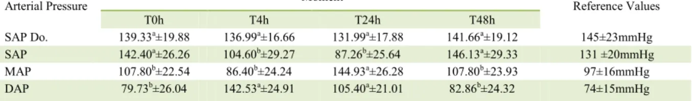
The pressure measured using Doppler
method did not reveal significant variation at the
different time points. On the contrary, the evaluation performed using the oscillometric method demonstrated
significant difference in the pressure (P<0.05) between
the mean values collected at T4h and T24h time points for SAP, MAP, and DAP (Table 1), showing decrease of SAP and elevation of DAP.
At the interpretation and analyses of electrocardiogram tracing were found several alterations. All electrocardiographic alterations were resolved without antiarrhythmic treatment. The observed arrhythmias were: sinusal bradicardia, premature ventricular complex, second grade blockage, ventricular tachycardia, and sinusal tachycardia.
The analyses of the parameters monitored for the 10 dogs indicated that, after the induced poisoning, 40% of the animals (4/10) presented sinusal bradicardia, 20% (2/10) presented intermittent second grade blockage, 20% (2/10) presented premature ventricular complex, 10% (1/10) presented sinusal tachycardia, and 10% of them (1/10) presented paroxistic ventricular tachycardia.
Table 1 - Mean ± standard deviation of SAP Doppler (mmHg), SAP (mmHg), MAP, and DAP (mmHg) measures, at time points T0h, T4h,
T24h and T48h of dogs intoxicated with N. oleander.
Arterial Pressure ---Moment--- Reference Values
T0h T4h T24h T48h
SAP Do. 139.33a±19.88 136.99a±16.66 131.99a±17.88 141.66a±19.12 145±23mmHg
SAP 142.40a±26.26 104.60b±29.27 87.26b±25.64 146.13a±29.33 131 ±20mmHg
MAP 107.80b±22.54 86.40b±24.24 144.93a±26.28 107.80b±23.93 97±16mmHg
DAP 79.73b±26.04 142.53a±24.91 105.40a±21.01 82.86b±24.32 74±15mmHg
SAP Do. = systolic arterial pressure Doppler; SAP = systolic arterial pressure; MAP = mean arterial pressure; DAP = diastolic arterial pressure. Reference values: BROWN et al. (2007). Different letters at the same line indicates a statistical difference with SNK test
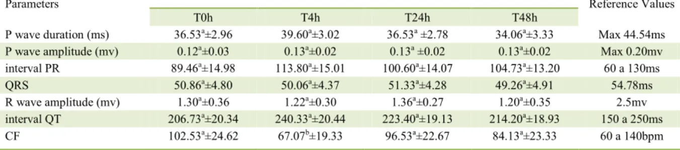
Table 2 demonstrated the mean values and standard deviations of the electrocardiographic variables: duration of Pms wave, amplitude of Pmv, PR interval, QRS complex, Rmv wave amplitude,
and QT interval. The data did not show significant statistical differences (P<0.05). However, the cardiac
frequency has shown a value decrease at T4h. The dose of 0.25g kg-1 of ground N.
oleander fresh leaves applied in the present study was not lethal for any dog. Some studies reported that using dry leaves or extracts with the active principle of the plant (oleandrine) in different concentrations induced intoxication symptoms (CLARCK et al., 1991). GALEY et al. (1996) considered N. oleander as a potentially toxic plant, because ingestion of 0.005% of dog’s weight of dry leaves could lead to death. In the present study, we used fresh leaves to simulate a natural poisoning with this ornamental plant, estimated to contain more than thirty potentially toxic compounds.
The reported clinical signs observed
in the present study were unspecific and could
be confused with other types of poisoning and/or diseases. However, the clinical signs of N. oleander poisoning occur very fast after the ingestion of the leaves. LANGFORD & BOOR (1996) mentioned in their research signs of vomiting, diarrhea, sialorrhea, apathy, central nervous system depression, hypotension, tremors, irregular pulse, and several arrhythmias, also observed in the present study. However, no worse consequences, such as coma or death, were observed in the present study, in contrast with the study of LANGFORD & BOOR (1996). The difference probably came from the amount of the absorbed toxin, as described previously. In this study, all animals showed a clinical improvement
after the treatment, confirmed by the return to the
feed consumption.
The blood pressure measured using the Doppler and oscillometric methods, in the present study, stayed at normal levels during the whole experiment, which is in accordance with the results reported by BROWN et al. (2007) and GAINS et al. (1995). The oscillometric method had shown some differences between the values obtained at T4h and T24h. These alterations are associated with the cardiac glycoside action, similar to the digoxin effect. These compounds increase the vagal tonus with an increase in parasympathetic effect, leading to inhibition of the sympathetic tonus and reduction in cardiac frequency and SAP (MERRETT, 2000). According to HABERMAN (2006), oscillometric method has a high correlation between the indirect measurements and the invasive method, which may have accuracy closer to that of the direct method. However, it is less accurate in case of hypotension.
In a study with anesthetized dogs, SHIH et al. (2010) used the oscillometric monitoring and observed less reliable accuracy compared to the invasive pressure monitoring, during the state of hypotension associated with acute hemorrhage. Doppler method is less accurate and can be used for PAS measuring only, while it appears to be a very poor PAD estimator. The device is further limited with the non-automatic character of the device and the absence of immediate response to sudden changes in pressure (DURHAM, 2005).
Various electrocardiographic changes related to N. oleander poisoning, including
arrhythmias, such as bradycardia, blockages of first
and second grade, premature ventricular complexes, ventricular tachycardia, sinus tachycardia, and
Table 2 - Mean ± standard deviation of: Pms. Pmv. PR. QRS. Rmv. QT and cardiac frequency (CF) measures, at time points T0h, T4h,
T24h and T48h of dogs intoxicated with N. oleander.
Parameters ---Moment--- Reference Values
T0h T4h T24h T48h
P wave duration (ms) 36.53a±2.96 39.60a±3.02 36.53a ±2.78 34.06a±3.33 Max 44.54ms
P wave amplitude (mv) 0.12a±0.03 0.13a±0.02 0.13a ±0.02 0.13a±0.02 Max 0.20mv
interval PR 89.46a±14.98 113.80a±15.01 100.60a±14.07 104.73a±13.20 60 a 130ms
QRS 50.86a±4.80 50.06a±4.37 51.33a±4.28 49.26a±4.91 54.78ms
R wave amplitude (mv) 1.30a±0.36 1.22a±0.30 1.36a±0.27 1.20a±0.35 2.5mv
interval QT 206.73a±20.34 240.33a±20.44 223.40a±19.13 214.20a±18.93 150 a 250ms
CF 102.53a±24.62 67.07b±19.33 96.53a±22.67 84.13a±23.33 60 a 140bpm
fibrillation, were observed in the present study. The
arrhythmias originated due to the high inhibition grade of the Na+/K+ ATPase pump at the cardiac muscular fibers, a condition that causes a reduction
in myocardia electric activity. Inhibition of Na+/K+
ATP ase pump by the cardiac glycosides results in the increased concentration of intracellular sodium,
leading to the influx of calcium in the intracellular
compartment and its release to the sarcoplasm. The increased concentration of free calcium in sarcoplasm augments the contraction force of the myocardium and the positive inotropic effect (JOUBERT, 1989). This event indirectly induces a decreased heart electric conduction by increasing the vagus nerve tonicity (KATZUNG & PARMLEY, 1998); this could explain the decreased heart frequency at T4h reported in the present experiment.
According to KELLERMANN et al. (2005), the isolated toxic principle of N. oleander belongs to the group of cardiac glycosides, known to affect contractility and impulse conduction in the
cardiac fiber. High doses of cardiotoxic glycosides
cause a substantial decrease in conduction rate, called negative dromotropic effect, resulting in severe atrium-ventricular dissociation, with consequent
first-, second-, and third-degree blockade. The
increase in PR interval observed in the present study;
although not significant, is probably due to this action of the toxic principle, indicating a first-degree
atrium-ventricular block.
Intravenous glucose treatment was established in the experimental protocol because of the hypoglycemic characteristics of oleander extracts, which had been studied experimentally for this therapeutic purpose. MWAFY & YASSIN (2011) demonstrated blood glucose levels in diabetic rats treated with N. oleander extract to be lower than those in control rats. PAGE & MURTAUGH (2015) described the successful treatment of a dog poisoned by N. oleander and presenting the classical symptoms, including hypoglycemia, as a pertinent clinical
finding. Blood glucose measurement should be a part
of the treatment protocol for a patient intoxicated by N. oleander. This was not established in the present study, which is a limitation of the research.
Toxic compounds of the plant, due to their chemical and pharmacokinetic similarity to digoxin,
have some properties that define their biological availability and have an influence on the level of
animal intoxication. A less liposoluble substance would have a higher fat deposition and need more time to be eliminated, while the substances could have a lower absorption when administered with the food. The
substances bind to albumin to a lesser extent in animals with hypoalbuminemia, which resulted in higher blood concentrations of the substance. In addition, hypocalcemia could decrease the competition on the binding sites, allowing higher substance concentration in blood (BROWN et al., 2007).
CONCLUSION
The present study demonstrated that dogs can be poisoned with 0.25g/kg single dose of ground N. oleander fresh leaves, and this concentration is
sufficient to cause a moderate intoxication, with nonspecific clinical alterations mainly related
to the digestive system, cardiac rhythm, and blood pressure. This indicated the importance of considering this type of intoxication in the list of differential diagnoses of the routine of small animals, which can be fatal for the patient.
BIOETHICS AND BIOSSECURITY COMMITTEE APPROVAL
The dogs used in this study were from the institution´s kennel. Protocol of the committee of ethics 006357-09.
ACKNOWLEDGEMENTS
Financial support: Conselho Nacional de
Desenvolviment Científico e Tecnológico (CNPq).
REFERENCES
ASLANI, M.R. et al. Experimental oleander (Nerium oleander) poisoning in goats: a clinical and pathological study. Iranian Journal of Veterinary Research, v.8, p.58-63, 2007. Available from: <http://www. sid.ir/en/vewssid/j_pdf/102320070108.pdf>. Accessed: May 20, 2016.
BROWN, S. et al. Guidelines for the identification, evaluation and
management of systemic hypertension in dogs and cats. Journal of Veterinary Internal Medicine, v.21, p.542-558, 2007. Available
from: <http://www.ivis.org/proceedings/acvim/consensus>. Accessed: May 10, 2016. doi: 0891-6640/07/2103-0028/$3.00/0.
CLARCK, R. et al. Digoxin – specific Fab fragments in the treatment of
oleander toxicity in a canine model. Annals of Emergency Medicine,
v.20, p.1073-1077, 1991. Available from: <http://www.sciencedirect. com/science/article/pii/S0196064405813551>. Accessed: May 10, 2016. doi: 10.1016/S0196-0644(05)81355-1.
DURHAM, H.E. Arterial blood pressure measurement. Veterinary Technician, v.26, n.5, p.324-339, 2005. Available from: <http:// www.vetfolio.com/arterial-blood-pressure-measurement>. Accessed: May 20, 2016.
GALEY, F.D. Diagnosis of oleander poisoning in livestock. Journal Veterinary Diagnostic Investigation, v.8, p.358-364, 1996.
Available from: <https://www.ncbi.nlm.nih.gov/pubmed/8844581>.
Accessed: May 13, 2016.
HABERMAN, C.E. et al. Evaluation of oscillometric and Doppler ultrasonic methods of indirect blood pressure estimation in conscious dogs. Canadian Journal of Veterinary Research, v.70,
p.211-217, 2006. Available from: <https://www.ncbi.nlm.nih.gov/
pubmed/16850944>. Accessed: May 10, 2016.
HUGHES, K.J. et al. Suspected Neriumoleander poisoning in a horse. Australian Veterinary Journal, v.80, p.412-415, 2002. Available
from: <https://www.ncbi.nlm.nih.gov/pubmed/12222602>. Accessed:
May 10, 2016. doi: 10.1111/j.1751-0813.2002.tb11000.x.
IBRAHIM, A. et al. A fatal case of oleandrin poisoning. Forensic Science International, v.179, p.31-36, 2008. Available from: <http:// www.sciencedirect.com/science/article/pii/S0379073808002284>. Accessed: May 15, 2016. doi: 10.1016/j.forsciint.2008.05.002.
JOUBERT, J.P.J. Cardiac glicosides. In: CHEEKE, P.R. Toxicants of plant origin. Boca Raton: CRC Press Book, 1989. p.61-96.
KATZUNG, B.G.; PARMLEY, W.W. Cardiac glycosides and other drugs used in congestive heart failure. Basic and Clinical Pharmacology, 1998.
p.197-216. Available from: < https://quizlet.com/5634451/chapt24drugs-for-heart-failure>. Accessed: May 12, 2016.
KELLERMAN, T.S. et al. Plant poisoning and mycotoxicoses of livestock in Southern Africa. 2.ed. Cape Town: Oxford University, 2005. 310p.
KNIGHT, A.P. Oleander poisoning. Compendium on Continuing Education for the Practicing Veterinarian, v.10, p.262-263. 1988.
LANGFORD, S.D.; BOOR, P.J. Oleander toxicity: an examination of human and animal exposures. Toxicology, v.109, p.1-13,
1996. Available from: <http://www.sciencedirect.com/science/ article/pii/0300483X9503296R>. Accessed: May 13, 2016. doi: 10.1016/0300-483X(95)03296-R.
LYNN, M.; SAFDAR, A. An overview of potentially life-threatening poisonous plants in dogs and cats. Journal of
Veterinary Emergency and Critical Care, v.16, p.25-33, 2006.
Available from: <http://onlinelibrary.wiley.com/doi/10.1111/ j.1476-4431.2005.00151.x/abstract>. Acessed: May 13, 2016. doi: 10.1111/j.1476-4431.2005.00151.x.
MARTINEZ, B.R. et al. Intoxicación por Nerium oleander (baladre) de los casos clínicos. Farmacia Hospitalaria, v.31, p.128-136, 2007.
Available from: <http://www.aulamedica.es/gdcr/index.php/fh/article/
view/930>. Accessed: May 13, 2016. doi: 1130-6343/2007/31/2/128.
MERRETT, D. Digoxin therapy. Australian Veterinary Journal,
v.78, p.612-615, 2000. Available from: <http://onlinelibrary.wiley.
com/doi/10.1111/j.1751-0813.2000.tb11932.x/abstract>. Accessed: May 13, 2016. doi: 10.1111/j.1751-0813.2000.tb11932.x.
MWAFY, S.N.; YASSIN, M.M. Antidiabetic activity evaluation of glimepiride and Nerium oleander extract on insulin, glucose levels and some liver enzymes activities in experimental diabetic rat model. Pakistan Journal of Biological Sciences, v.14, n.21, p.984-90, 2011.
Available from: <https://www.ncbi.nlm.nih.gov/pubmed/22514888>.
Accessed: May 10, 2016. doi: 10.3923/pjbs.2011.984.990.
PAGE, C.; MURTAUGH, R.J. Hypoglycemia associated with Oleander toxicity in a dog. Journal of Medical Toxicology, v.11,
p.141-143, 2015. Available from: <https://www.ncbi.nlm.nih.
gov/pubmed/25252802>. Accessed: May 10, 2016. doi: 10.1007/ s13181-014-0436-x.
PLUMB, D.C. Veterinary drug handbook. 3.ed. Iowa: Iowa State University, 1999. 750p.
SHIH, A. et al. Evaluation of an indirect oscillometric blood pressure monitor in normotensive and hypotensive anesthetized dogs. Journal of Veterinary Emergency and Critical Care, v.20, n.3, p.313-318, 2010.
Available from: <https://www.ncbi.nlm.nih.gov/pubmed/20636984>.
Accessed: May 13, 2016. doi: 10.1111/j.1476-4431.2010.00536.x.
SOTO-BLANCO, B. et al. Acute cattle intoxication from Nerium oleander pods. Tropical Animal Health and Production, v.38, p.451-454, 2006.
Available from: <https://www.ncbi.nlm.nih.gov/pubmed/17243471>.
Accessed: May 13, 2016. doi: 10.1007/s11250-006-4400-x.