Cholinesterase as inflammatory markers in a experimental
infection by
Trypanosoma evansi
in rabbits
MÁRCIO M. COSTA1,2, ALEKSANDRO S. DA SILVA2, FRANCINE C. PAIM1, RAQUELI FRANÇA1, GUILHERME L. DORNELLES1, GUSTAVO R. THOMÉ3, JONAS D.S. SERRES3, ROBERTA SCHMATZ3,
ROSÉLIA M. SPANEVELLO3, JAMILE F. GONÇALVES3, MARIA ROSA C. SCHETINGER3, CINTHIA M.A. MAZZANTI1, SONIA T.A. LOPES1 and SILVIA G. MONTEIRO2 1Laboratório de Análises Clínicas Veterinária (LACVet), Universidade Federal de Santa Maria,
Av. Roraima, 1000, Cidade Universitária, Bairro Camobi, 97105-900 Santa Maria, RS, Brasil 2Laboratório de Microbiologia e Parasitologia (LAPAVET), Universidade Federal de Santa Maria,
Av. Roraima, 1000, Cidade Universitária, Bairro Camobi, 97105-900 Santa Maria, RS, Brasil 3Laboratório de Enzimologia Toxicológica - ENZITOX, Universidade Federal de Santa Maria,
Av. Roraima, 1000, Cidade Universitária, Bairro Camobi, 97105-900 Santa Maria, RS, Brasil
Manuscript received on June 13, 2011; accepted for publication on April 14, 2012
ABSTRACT
The aim of this study is to evaluate the role of cholinesterases as an inflammatory marker in acute and chronic infection by Trypanosoma evansi in rabbits experimentally infected. Twelve adult female New Zealand rabbits were used and divided into two groups with 6 animals each: control group (rabbits 1-6) and infected group (rabbits 7-12). Infected group received intraperitoneally 0.5 mL of blood from a rat containing 108 parasites per animal. Blood samples used for cholinesterases evaluation were collected on days 0, 2, 7, 12, 27, 42, 57, 87, 102 and 118 days post-inoculation (PI). Increased activity (P<0.05) of butyrylcholinesterase (BChE) and acetylcholinesterase (AChE) were observed in the blood on days 7 and 27, respectively and no differences were observed in cholinesterase activity in other periods. No significant difference in AChE activity (P>0.05) was observed in the encephalic structures. The increased activities of AChE and BChE probably have a pro-inflammatory purpose, attempting to reduce the concentration of acetylcholine, a neurotransmitter which has an anti-inflammatory property. Therefore, cholinesterase may be inflammatory markers in infection with T. evansi in rabbits.
Key words: acetylcholinesterase, butyrylcholinesterase, inflammation, T. evansi.
Correspondence to: Márcio Machado Costa
E-mail: marmcvet@yahoo.com.br
INTRODUCTION
Cholinesterases are enzymes present in cholinergic
and non-cholinergic tissues, as well as in plasma
and other body fluids. This group of enzymes is
divided into two distinct categories, according to
their substrate-specific, behavior in the presence
of excess substrate and susceptibility to inhibitors
(Çokuğraş 2003). The acetylcholinesterase (AChE)
is a specific cholinesterase, which hydrolyzes esters
of mainly acetylcholine (ACh) and has high levels
in nervous tissue and in erythrocytes (Soreq and
Seidman 2001, Das 2007). Butyrylcholinesterase
(Annals of the Brazilian Academy of Sciences)(BChE) is a non-specific cholinesterase, called
pseudocholinesterase, which catalyzes the
hydro-lysis of other esters such as succinylcholine and
butyrylcholine, as well as ACh (Çokuğraş 2003,
Darvesh et al. 2003). The AChE mainly, but also
the BChE, have the function of hydrolyzing ACh
linked to post-synaptic receptors to regulate the
concentration of transmitter in the nerve synapse
(Soreq and Seidman 2001).
The autonomic nervous system plays an
important role in controlling the immune response,
in terms of regular, partially, the release of
pro-inflammatory cytokines, through the secretion of
adrenaline and noradrenaline, and connection of
these receptors α and β androgenic in the immune
cells (Pavlov and Tracey 2005). In contrast, the
vagal efferent pathway plays an important role in
modulating inflammation and was called cholinergic
anti-inflammatory pathway (Borovikova et al. 2000,
Pavlov and Tracey 2005).
The ACh, the principal neurotransmitter in
vagal, has an important role in attenuating the
release of pro-inflammatory cytokines such as
tumor necrosis factor (TNF), interleukin-1 (IL-1),
interleukin-6 (IL-6) and interleukin-18 (IL-18),
activated macrophage previously by endotoxin,
without affecting the production of interleukin-10
(IL-10), an anti-inflammatory cytokine (Pavlov and
Tracey 2005). This inhibition is due to binding of
ACh at α7 subunit-containing nicotinic acetylcholine
receptor (α7nAChE) present on macrophages, being
the dose-dependent inhibition (Ulloa 2005). Thus,
given the important role of ACh in the suppression
of inflammation, investigate of cholinesterases that
regulate the concentration of ACh can be useful,
even indirectly; the levels of ACh are measured.
The trypanosomosis are diseases that affect
different species of animals such as camels, horses,
cattle, goats, pigs, dogs, elephants, capybaras,
coatis, tapirs, deer and wild rodents (Silva et
al. 2002). Among these diseases, there is the
"Surra" as it is called in India, which is caused
by
Trypanosoma evansi
(
T. evansi
), a digenetic
protozoan section salivate, which is mechanically
transmitted by flies of the genus
Tabanus
and
Stomoxis
(Brun et al. 1998, Taylor and Authié
2004). In Brazil, trypanosomosis affects mainly
horses, being called as "Mal das cadeiras" due to
locomotor and neurological symptoms presented by
infected animals (paralysis of hind limbs), resulting
in incoordination (Herrera et al. 2004).
Among the affected animals, camels, horses,
buffaloes and dogs have the most severe form of the
disease, unlike of cattle and pigs which are moderately
affected or asymptomatic (Taylor and Authié 2004).
The acute phase of the disease mainly affects young
animals and pregnant females (Tailor and Authié
2004) and progresses to death within weeks or months
(Brun et al. 1998). On the other hand, the chronic
stage is more frequent in endemic areas of disease
(Taylor and Authié 2004) and affected animals can
remain infected for many years (Brun et al. 1998).
In equine trypanosomosis presents a chronic phase
with clinical signs of progressive weight loss, fever,
anemia, edema of limbs and lower portions of the
body, incoordination and instability of the hind
limbs and atrophy of muscle mass (Silva et al. 1995,
Conrado et al. 2005, Rodrigues et al. 2005). In other
animals such as rabbits, trypanosomiasis also has a
chronic course, studying with anorexia, apathy, pale
mucous membranes and edema of eyelids and ears.
These animals show irregular peaks of parasitemia
for long periods (Da Silva et al. 2009). Thus, these
animals are good models for chronic infections due
to low peak of parasitemia and long time of infection,
as occurs in cases of natural infection.
in acute and chronic phase of
T. evansi
infection,
using rabbits as an experimental model.
MATERIALS AND METHODS
ANIMALS
Twelve adult female rabbits New Zealand, weighing
between 3.6 to 4.5 kg were used in this study. The
animals were kept in individual cages with controlled
temperature and humidity (23°C and 70% U.R.),
fed with commercial ration and water
ad libitum
.
All animals received a single dose (5mg/kg, orally)
of a commercial product containing pyrantel
pamoate, praziquantel, and fenbendazole 30 days
before beginning the experiment. Hematological
(erythrogram, leukogram and platelet count) and
biochemical (hepatic and renal function) examinations
were performed twice with 15-day intervals. After 30
days (day 0 of the experiment), the evaluated patterns
showed normal values (Campbell 2007).
The procedure was approved by the Animal
Welfare Committee of the Universidade Federal
de Santa Maria, number
23081.020268/2008-76, in accordance to Brazilian laws and ethical
principles published by the Colégio Brasileiro de
Experimentação Animal.
TRYPANOSOME INFECTION
T. evansi
was originally isolated from a naturally
infected dog (Colpo et al. 2005). First, a rat was
intraperitoneally infected with blood cryopreserved
in liquid nitrogen containing 10
6parasites. This
procedure was performed to obtain large amount
parasites for posterior inoculation.
Rabbits were divided into two groups with 6
animals each, a control group (rabbits 1-6) and an
infected group (rabbits 7-12). Infected group were
intraperitoneally inoculated with 0.5 mL of rat blood
containing 10
8trypanosomes (Day 1). Control group
received physiological solution by the same route.
The number of inoculated flagellates was estimated
by Neubauer chamber (Wolkmer et al. 2007).
ESTIMATION OF PARASITEMIA
Parasitemia was estimated daily during 118 days
of infection through microscopic examination of
smears. Each slide was mounted with blood collected
from the ear vein, stained by the panoptic method,
and visualized at a magnification of ×1,000.
BLOOD SAMPLING AND ENCEPHALIC TISSUE
The experimental period reached 118 days. Blood
sampling for hematology and evaluation of
cholinesterases were carried out on days 0, 2, 7, 12,
27, 42, 57, 87, 102 and 118 days post-inoculation
(PI), by cardiac puncture using 5 mL disposable
syringes and 25x7 needles, stored in tubes with
anticoagulant. For this procedure all animals were
anesthetized with ketamine (1 mg/Kg) and xylazine
(0.8 mg/Kg), as recommended by the ethics
committee. Blood with anticoagulant were diluted
in a lytic buffer, proportion of 1:50 (v/v), to evaluate
the activity of AChE. One mL of blood without
anticoagulant was centrifuged for 10 minutes at
3,400 rpm, and serum separated and stored frozen
at -20°C for analysis of BChE activity.
DETERMINATION OF ACHEACTIVITY IN BLOOD AND ENCEPHALON
The AChE enzyme assay in total blood was
determined by the method of Ellman et al.
(1961), modified by Worek et al. (1999). The
specific activity of AChE in blood was calculated
from the quotient between AChE activity and
hemoglobin content, with results expressed in
mU/L¬ mol Hb. The enzyme assay of AChE in
encephalon was determined by a modification
of the spectrophotometric method of Ellman et
al. (1961) as described by Rocha et al. (1993).
All readings were performed in triplicate and
the enzyme activity was expressed in mumoles
AcSCh/h/mg of protein.
DETERMINATION OF BCHEACTIVITY IN SERUM
The methodology used to assess the AChE activity
was also used for determination of BChE activity in
serum, except for the acetylcholine substrate which
was replaced by butyrylthiocholine. Results were
expressed in µmoles BcSCh/h/mg of protein.
DETERMINATION OF HEMATOLOGICAL PARAMETERS
Hematologic parameters were evaluated merely
to monitor the disease in rabbits infected with
T. evansi
. Hematocrit, total leukocytes and
differential leukocyte were performed. The white
blood cells counts were carried out in an automatic
counter Mindray BC 2800 Vet
®. Determination
of hematocrit was obtained from microhematocrit
centrifuge in rotation 19720G, and the differential
count was performed on blood smears, stained with
Panoptic method
®, using light microscopy.
STATISTICAL ANALYSIS
By presenting normal distribution (Kolmogorov
Smirnov), the data were subjected to t test for
inde-pendent samples. Data were considered significantly
different with a probability (P) less than 5%.
RESULTS
PARASITEMIA AND CLINICAL COURSE OF INFECTION
Examination of the peripheral blood smears showed
a prepatency period between 24 and 72 hours in the
infected rabbits. Irregular waves of parasitemia,
ranging from zero to one trypomastigote per
microscopic field, were observed until 35 days PI.
Parasites were no longer observed in blood smears
from the 37
thday onwards (Figure 1).
Clinical signs as hyporexia, fever and weight
loss (8.7%) were observed during the first 30 days
PI in all infected rabbits (acute phase and peak of
parasitemia). Edema of the eyelids, ears and vulva
was observed in all infected animals at determined
periods of the experiment. This clinical signs
disappeared in a few moments, reappearing after a
period that varied among individuals. At days 60, 90
and 118 PI the infected rabbits increased its weights.
Non-infected rabbits showed no clinical changes and
increased its weights throughout the study.
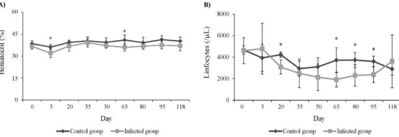
HEMATOCRIT
Rabbits infected with
T. evansi
did not presente
anemia. However, a significant reduction of
hema-tocrit was observed 5 (
P
< 0.01) and 65 (
P
< 0.05)
days PI, when compared to the control group.
During the other days of evaluation this change was
not observed (Figure 2A).
TOTAL LEUKOCYTES AND LYMPHOCYTES
Significant difference in total white cell count were
not observed between the groups (
P
> 0.05). However,
lymphocytes number were reduced in infected
animals on days 20, 65, 80 and 95 PI (
P
< 0.05) when
compared to the non-infected (Figure 2B).
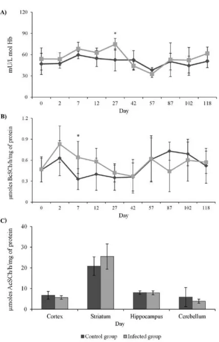
ACTIVITY OF ACHE IN BLOOD AND ENCEPHALIC STRUCTURES
Figure 1 - Parasitemia of T. evansi in infected rabbits along the 118 days post-infection.
Figure 2 - Influences of the infection by T. evansi on hematological parameters of rabbits experimentally infected: A) hematocrit and B) lymphocytes. * indicates statistical difference between infected and control group (*Independent samples T test P < 0.05).
significant differences in enzyme activity were not
detected. No significant difference in AChE activity
(
P
> 0.05) was observed in the brain structures, as
shown in Figure 3C.
ACTIVITY OF BCHE IN SERUM
A significant increase (
P
< 0.05) on BChE activity
was observed on day 7 PI (Figure 3B). During other
days the enzyme activity did not show statistical
difference when compared to the control group.
DISCUSSION
Figure 3 - A) Acetylcholinesterase activity in blood; B) butyrylcholinesterase activity in serum; and C) acetylcholinesterase in encephalic structures of rabbits experimentally infected with Trypanosoma evansi. * indicates statistical difference between infected and control group
(*Independent samples T test P < 0.05).
species, are mainly due to the mean age of animals,
since animals tested by Da Silva et al. (2008a) were
infected during its development phases, unlike the
animals tested in this study, which were adult.
A similar response to this study was found in coati
experimentally infected with
T. evansi
that showed
parasitemia until the first month PI, with subsequent
decrease and absence of parasites in some periods
(Herrera et al. 2001).
In this study, during the initial stage of the
disease, clinical signs were observed such as
appetite loss, fever and weight loss. These findings
are similar to those found in cats (Da Silva et al.
2010), rabbits (Da Silva et al. 2009), dogs (Aquino
et al. 1999, Colpo et al. 2005) and coatis (Herrera et
al. 2001) experimentally or naturally infected with
until euthanasia of animals, swinging during the
experimental period. It was also observed in rabbits
infected by Da Silva et al. (2009), however, after
90 days, the animals showed clinical improvement
with disappearance of clinical signs, feature not
observed in this study. Thus, animals in this study
had a persistent and chronic infection, with absence
of trypomastigotes in the bloodstream.
To the hematocrit, only at 5
thand 65
thdays PI
it was possible to observe a significant reduction.
In the remaining days, the hematocrit of infected
animals had small variations, but significant
differences were not observed when compared
to the control group, differing of results found
by Da Silva et al. (2008b), which demonstrated
a hematocrit reduction in rabbits experimentally
infected with
T. evansi
, which returned to normal
levels after 60 days PI. However, in a
T. evansi
outbreak in horses reported by Zanette et al. (2008),
the animals did not show anemia, even with clinical
signs of swinging gait and incoordination of the
hind limbs, which agrees with the study in question,
since the rabbits showed such clinical signs and
absence of anemia.
Infection with
T. evansi
promoted increase in
BChE activity only on the seventh day PI. These
results are different from those found by Da Silva et al.
(2010) and Wolkmer et al. (2010), as, in both studies,
there was a reduction in BChE activity in cats and
rats infected with
T. evansi
, respectively. Although in
different stages of disease, both studies found similar
results, with reduction of BChE assigned by Wolkmer
et al. (2010) to liver damage, because the liver is
the principal organ of synthesis of plasma BChE
(Chatonnet and Lockridge 1989) or the assigned to
inflammatory process. The period in which BChE
activity increased is classified as acute infection by
T. evansi
, and it is characterized by peak parasitemia,
fever, appetite loss. Within this period, the macrophage
plays a crucial role in the production of an efficient
immune response against African trypanosomes.
This cell is responsible for the initial production of
pro-inflammatory cytokines such as tumor necrosis
factor (TNF) and nitric oxide (NO) (Stijlemans et al.
2007). Thus, BChE could be involved in activation
of macrophages, as it has nicotinic receptors for
acetylcholine. Acetylcholine when linked to these
receptor, produces a dose-dependent inhibition in
cytokine production (Ulloa 2005). Therefore, it is
suggested that the increase in BChE is related to
hydrolysis of acetylcholine by reducing its levels and
preventing its suppressive action.
On the other hand, the increased activity of
AChE in blood was only on the 27
thday PI, within the
chronic phase of infection, which differs from other
studies involving
T. evansi
. Wolkmer et al. (2010)
found blood AChE activity reduced in rats infected
with
T. evansi
on the 3
thand 5
thdays PI. However,
Da Silva et al. (2010) found a reduction in blood
AChE activity of cats experimentally infected with
T. evansi
on days 28
thand 56
thPI, associated with
a reduction in brain activity of AChE. The results
in these two studies were directly associated with
neurological signs presented in both species (rats and
cats), which were not observed in this study, since the
rabbits did not show neurological abnormalities such
as incoordination of hind limbs, and limb muscle
atrophy. The absence of neurological findings is
confirmed, whereas there was no significant statistical
difference between the encephalic structures analyzed
in this study, when compared with the control group.
In a recent study Da Silva et al. (2011a) observed
increase of AChE activity in brains of rats infected
with
T. evansi
, concomitantly with neurological signs,
demonstrating that rabbits has a different response to
infection with
T. evansi
.
mice, rats, rabbits and humans. Nicotinic receptors,
through the binding with acetylcholine, play the
function of suppression of the immune response in
T lymphocytes (Kawashima and Fujii 2003). Thus,
the increase of AChE could reduce the ACh for
nicotinic receptors, enhancing the immune activity of
T lymphocytes, promoting a improved response for
cytokine production and regulation of parasitemia.
Its may compensate the reduction in the total number
of lymphocytes. This hypothesis can be explained
since from the 37th day the presence of parasite in
the bloodstream was no longer observed. Besides,
a recent study from our research group, reached a
positive correlation between increased numbers of
lymphocytes and activation of AChE in lymphocytes
of rats infected with
T. evansi
(Da Silva et al. 2011b).
In this study, the increase of cholinesterase activity
was expected throughout the chronic phase of
infection, but this enzymatic activation did not occur,
despite the clinical signs presented. Possibly, due the
control of parasitemia, mainly by IgG and IgM, as
described by Uche et al. (1992), there are no needs of
a permanent cellular immune response, which could
reduce the levels of AChE close to the normality.
Thus, it is concluded that cholinesterases may
have pro-inflammatory actions during the infection
by
T. evansi
in rabbits, regulating the concentrations
of ACh, which is a potent anti-inflammatory
mediator. These findings demonstrate the role of
rabbits as tolerant to
T. evansi
, reinforcing the idea
that this animal plays an important function as
reservoirs of the parasite.
ACKNOWLEDGMENTS
The authors would like to thank Conselho Nacional
de Desenvolvimento Científico e Tecnológico (CNPq)
and “Universal – CNPq” for the financial support.
RESUMO
O objetivo do presente estudo é avaliar o papel das coli-nesterases como marcadores inflamatórios nas fases aguda
e crônica da infecção por T. evansi em coelhos infectados
experimentalmente. Foram utilizados 12 coelhos adultos,
fêmeas, da raça Nova Zelândia, divididos em dois grupos: um grupo controle, com seis animais (coelhos 1-6), e um grupo infectado, com seis animais (coelhos 7-12). Os
animais pertencentes ao grupo infectados receberam, pela via intraperitoneal, 0,5 mL de sangue de rato contendo
108 tripanossomas por animal. Amostras do sangue
utilizado para avaliação das colinesterases foram coletadas nos dias 0, 2, 7, 12, 27, 42, 57, 87, 102 e 118 pós-inoculação (PI). Aumento (P<0,05) na atividade da butirilcoli-nesterase (BChE) e da acetilcolibutirilcoli-nesterase foi observado no sangue nos dias 7 e 27 (PI), respectivamente e não foram observadas diferenças na atividade da colinesterase em outros períodos. Nenhuma diferença significativa na atividade da AChE (P>0,05) foi observada nas estruturas
encefálicas. O aumento de atividade da AChE e BChE
provavelmente tenha finalidade pró-inflamatória, a fim de reduzir as concentrações de acetilcolina, neurotransmissor que apresenta propriedade anti-inflamatória. Portanto, as colinesterases podem ser marcadores inflamatórios na
infecção por T. evansi em coelhos.
Palavras-chave: acetilcolinesterase, butirilcolinesterase, inflamação, T. evansi.
REFERENCES
AQUINO LP, MACHADO RZ, ALESSI AC,MARQUES LC,DE CASTRO MB AND MALHEIROS EB. 1999. Clinical, parasitological and immunological aspects of experimental
infection with Trypanosoma evansi in dogs. Mem Inst
Oswaldo Cruz 94: 255-260.
BOROVIKOVA LV, IVANOVA S, ZHANG M, YANG H, BOTCHKINA GI, WATKINS LR, WANG H, ABUMRAD N, EATON JW AND TRACEY KJ.2000. Vagus nerve stimulation
atte nuates the systemic inflammatory response to endotoxin. Nature 405: 458-462.
BRUN R,HECKER H AND LUN ZR.1998.Trypanosoma evansi and T. equiperdum: distribution, biology, treatment and
phylogenetic relationship (a review). Vet Parasitol 79: 95-107.
CAMPBELL TW.2007. Hematologia de mamíferos: Animais de
laboratório e espécies variadas. In: Thrall MA, Baker DC, Campbell TW, Denicola D, Fettman MJ, Lassen ED, Rebar A and Weiser G. Hematologia e bioquímica clínica veterinária. Roca: São Paulo, p. 201-214.
COLPO CB, MONTEIRO SG, STAINKI DR, COLPO ET AND
HENRIQUES GB.2005. Infecção natural por Trypanosoma evansi em cães. Ciênc Rural 35: 717-719.
CONRADO AC,LOPES STA, OLIVEIRA LSS, MONTEIRO SG, VARGAS DLB AND BUENO A.2005. Infecção natural por Trypanosoma evansi em cavalos na região central do Rio
Grande do Sul. Ciênc Rural 35: 928-931.
çOKUğRAş AN.2003. Butyrylcholinesterase: structure and
physiological importance. Turk J Biochem 28: 54-61.
DA SILVA AS, COSTA MM, DOYLE RL, LOPES STA AND MONTEIRO SG. 2008a. Infecção experimental por Trypanosoma evansi em coelhos. Ciênc Anim Bras 9: 519-523.
DA SILVA AS,COSTA MM,LOPES STA AND MONTEIRO SG.
2008b. Alterações hematológicas em coelhos infectados
experimentalmente pelo Trypanosoma evansi. Ciênc
Rural 38: 538-542.
DA SILVA AS ET AL. 2011a. Acetylcholinesterase activity and
lipid peroxidation in the brain and spinal cord of rats infected with Trypanosoma evansi. Vet Parasitol 175: 237-244.
DA SILVA AS ET AL. 2011b. Trypanosoma evansi: immune response and acetylcholinesterase activity in lymphocytes
from infected rats. Exp Parasitol 127: 475-480.
DA SILVA AS,PEREIRA PL AND MONTEIRO SG.2009. Achados
patológicos, sinais clínicos e ganho de peso de coelhos
infectados experimentalmente por Trypanosoma evansi.
Semina Ciênc Agrar 30: 93-98.
DA SILVA AS,SPANEVELLO R,STEFANELLO N,WOLKMER P, COSTA MM, ZANETTE RA, LOPES ST, SANTURIO JM, SCHETINGER MR AND MONTEIRO SG. 2010. Influence of Trypanosoma evansi in blood, plasma, and brain cholinesterase of experimentally infected cats. Res Vet
Sci 88: 281-284.
DARVESH S,HOPKINS DA AND GEULA C.2003. Neurobiology
of butyrylcholinesterase. Nat Rev Neurosci 4: 131-138.
DAS UN.2007. Acetilcholinesterase and butyrylcholinesterase
as possible markers of low-grade systemic inflammation.
Med Sci Monit 13: 214-221.
ELLMAN GL,COUTNEY KO,ANDRES V AND FEATHERSTONE RM.1961. A new and rapid colorimetric determination of
acetylcholinesterase activity. Biochem Pharmacol 7: 88-95. HERRERA HM, AQUINO LP, MENEZES RF, MARQUES LC, MORAES MA, WERTHER K AND MACHADO RZ. 2001. Trypanosoma evansi experimental infection in the South American coati (Nasua nasua): clinical, parasitological and
humoral immune response. Vet Parasitol 102: 209-216. HERRERA HM,DÁVILA AM,NOREK A,ABREU UG,SOUZA SS,
D'ANDREA PS AND JANSEN AM.2004. Enzootiology of Tryapanosoma evansi in pantanal, Brasil. Vet Parasitol 125: 263-275.
KAWASHIMA K AND FUJII T.2003. The lymphocytic cholinergic
system and is contribution to the regulation of immune activity. Life Sci 74: 675-696.
PAVLOV VA AND TRACEY KJ. 2005. The cholinergic
anti-inflammtory pathway. Brain Behav Immu 19: 493-499.
ROCHA JBT,EMANUELLI T AND PEREIRA ME.1993. Effects
of early undernutrition on kinetic parameters of brain acetylcholinesterase from adult rats. Acta Neurobiol Exp (Wars) 53: 431-437.
RODRIGUES A, FIGHERA RA, SOUZA TM, SCHILD AN, SOARES MP,MILANO J AND BARROS CSL.2005. Surto de
tripanossomíase por Trypanosoma evansi em equinos no
Rio Grande do Sul: aspectos epidemiológicos, clínicos, hematológicos e parasitológicos. Pesq Vet Bras 25: 239-249.
SILVA RA, AROSEMENA NA, HERRERA HM, SAHIB CA AND FERREIRA MS.1995. Outbreak of trypanosomosis due to Trypanosoma evansi in horses of Pantanal
mato-grossense, Brazil. Vet Parasitol 60: 167-171.
SILVA RAMS,SEIDL A,RAMIREZ L AND DÁVILA AMR.2002. Trypanosoma evansi e Trypanosoma vivax – biologia,
diagnóstico e controle. Corumba: Embrapa Pantanal, 141 p.
SOREQ H AND SEIDMAN S.2001. Acetylcholinesterase – new roles for an old actor. Nat Rev Neurosci 2: 294-302. STIJLEMANS B,GUILLIAMS M,RAES G,BESCHIN A,MAGEZ S
AND DE BAETSELIER P. 2007. African trypanosomosis: from immune escape and immunopathology to immune
intervention. Vet Parasitol 148: 3-13.
TABORDA C,MEHNERT DU AND SILVA CA.2004. Manual de
Normas Técnicas: biotério de experimentação animal do Departamento de Microbiologia - Instituto de Ciências Biomédicas – USP, 24 p.
TAYLOR K AND AUTHIÉ EML.2004. Pathogenesis of animal
trypanosomiasis. In: Maudlin I, Holmes PH and Miles MA. The Trypanosomiases. London: CABI publishing,
p. 331-354.
UCHE UE,JONES TW AND BOID R.1992. Antibody patterns
in rabbits showing different levels of susceptibility to an
experimental Trypanosoma evansi infection. Acta Trop 52: 139-147.
ULLOA L. 2005. The vagus nerve and the nicotinic
anti-inflammatory pathway. Nat Rev Drug Discov 4: 673-684. WOLKMER P, LOPES ST, FRANCISCATO C, DA SILVA AS, TRAESEL CK, SIQUEIRA LC, PEREIRA ME, MONTEIRO SG AND MAZZANTI CM. 2010. Trypanosoma evansi:
cholinesterase activity in acutely infected Wistar rats. Exp Parasitol 125: 251-255.
WOLKMER P,SILVA AS,CARNELUTTI JF,COSTA MM,TRAESEL C, LOPES STA AND MONTEIRO SG. 2007. Resposta eritropoética de ratos em diferentes de graus de parasitemia por Trypanosoma evansi. Ciênc Rural 37: 1682-1687.
WOREK F,MAST U,KIDERLEN D,DIEPOLD C AND EYER P. 1999. Improved determination of acetylcholinestrase
activity in human whole blood. Clin Chim Acta 288: 73-90.
ZANETTE RA, DA SILVA AS, COSTA MM, MONTEIRO, SG, SANTURIO JM AND LOPES STA. 2008. Ocorrência de Trypanosoma evansi em equinos no município de Cruz