online | memorias.ioc.fiocruz.br
Chagas disease, adipose tissue and the metabolic syndrome
Fnu Nagajyothi1, Mahalia S Desruisseaux1,2,3, Louis M Weiss1,2, Streamson Chua1,3, Chris Albanese4, Fabiana S Machado5, Lisia Esper5, Michael P Lisanti6, Mauro M Teixeira5, Philipp E Scherer7,
Herbert B Tanowitz1,2/+
1Department of Pathology 2Department of Medicine 3Department of Neuroscience, Albert Einstein College of Medicine, 1300 Morris Park Avenue, Bronx, NY 10461, USA 4Department of Oncology, Lombardi Comprehensive Cancer Center, Georgetown University
Medical Center, Washington DC, USA 5Departamento de Bioquímica e Imunologia, Instituto de Ciência Biológicas, Universidade Federal de Minas Gerais, Belo Horizonte, MG, Brasil 6Department of Cancer Biology and the Kimmel Cancer Center, Thomas Jefferson University, Philadelphia, PA, USA 7Touchstone Diabetes Center, Departments of Internal Medicine and Cell Biology, University of Texas
Southwestern, Dallas, TX, USA
Trypanosoma cruzi infection of the adipose tissue of mice triggers the local expression of inflammatory mediators and a reduction in the expression of the adipokine adiponectin. T. cruzi can be detected in adipose tissue by PCR 300 days post-infection. Infection of cultured adipocytes results in increased expression of cytokines and chemokines and a reduction in the expression of adiponectin and the peroxisome proliferator-activated receptor γ, both of which are negative regulators of inflammation. Infection also results in the upregulation of cyclin D1, the Notch pathway, and ex-tracellular signal-regulated kinase and a reduction in the expression of caveolin-1. Thus, T. cruzi infection of cultured adipocytes leads to an upregulation of the inflammatory process. Since adiponectin null mice have a cardiomyopathic phenotype, it is possible that the reduction in adiponectin contributes to the pathogenesis of chagasic cardiomyopathy. Adipose tissue may serve as a reservoir for T. cruzi from which parasites can become reactivated during periods of im-munosuppression. T. cruzi infection of mice often results in hypoglycemia. In contrast,hyperglycemia as observed in diabetes results in increased parasitemia and mortality. Adipose tissue is an important target tissue of T. cruzi and the infection of this tissue is associated with a profound impact on systemic metabolism, increasing the risk of metabolic syndrome.
Key words: adipose tissue - adipocyte - adiponectin - Trypanosoma cruzi - Chagas disease
Financial support:NIH AI-06538 (HBT), AI-052739 (HBT), R01-DK55758 (SC), R01-CA112023 (PES)
+ Corresponding author: [email protected] Received 15 March 2009
Accepted 22 May 2009
Chagas disease caused by Trypanosoma cruzi re-mains an important cause of morbidity and mortality in endemic areas of Mexico, Central and South America. Ten to thirty percent of individuals with this infection eventually succumb to chronic manifestations such as cardiomyopathy and/or mega syndromes. This infection is also regarded as an opportunistic infection during cases of immunosuppression, for instance affecting individuals with HIV/AIDS. The pathogenesis of Chagas disease has been explored in detail by many laboratories over the past decades, but the role of the adipocyte and of adipose tis-sue, long ignored, is only now being investigated.
The adipocyte and adipose tissue: general considerations
The adipocyte, or fat cell, contributes to the patho-genesis of diabetes, obesity and the metabolic syndrome (Rajala et al. 2003, Wernstedt-Asterholm et al. 2007) and its secretory products have been implicated in many
physiological processes (Nawrocki & Scherer 2005, At-tie & Scherer 2009). Until recently, adipose tissue was considered to be a mere storage compartment for triglyc-erides. However, adipocytes are active endocrine cells that play a central role in overall energy homeostasis and are important contributors to some aspects of the im-mune system (Halberg et al. 2008). They do so by influ-encing systemic lipid homeostasis and also through the production and release of a host of adipocyte-specific and adipocyte-enriched hormonal factors, inflamma-tory mediators such as cytokines and chemokines and extracellular matrix components also known as adipo- kines. There has been little attention given to the role of adipose tissue and adipocytes in infectious disease (Des-ruisseaux et al. 2007), but the strong pro-inflammatory potential of adipose tissue suggests that it may have an important role in the systemic innate immune response.
hormones. Adipose tissue stores lipid in the form of tri- glycerides. It also stores mostly non-esterified cholesterol on the surface of lipid droplets that act as specialized organelles inside the adipocyte. Since the lipid droplet is such a large component of the adipocyte, changes in the amount of lipid stored within it affect fat cell size (which can range from 25-250 microns).
The potential endocrine function of adipose tissue was first recognized in the 1980s with the report that the serine protease adipsin was secreted by cultured 3T3-L1 adipocytes (Kook et al. 1987). Subsequently, several additional adipokines have been discovered, including adiponectin (Scherer et al. 1995), leptin, resistin, SAA3, omentin, visfatin and RBP4 (Desruisseaux et al. 2007). These adipokines contribute to the regulation of energy homeostasis through effects on both central and periph-eral tissues. Sevperiph-eral of these adipokines also contribute to non-metabolic processes in the body, highlighting the fact that adipokines participate in the coordination of mul-tiple physiological functions in a variety of tissues. The most adipocyte-specific adipokine is adiponectin. Other adipokines can also be synthesized by tissues other than adipose tissue and/or by cells other than adipocytes.
Systemic energy homeostasis is maintained by the competing effects of a number of different hormon-al factors, some of which originate in adipose tissue. These adipocyte-derived factors (adipokines), influ-ence processes such as food intake, energy expenditure and insulin sensitivity in a variety of tissues. Two adi-pokines, resistin and adiponectin have opposing effects on whole-body glucose homeostasis (Combs et al. 2001, Rajala et al. 2003). Pharmacological doses of recom- binant resistin hyper-activate gluconeogenesis through decreased hepatic insulin sensitivity. Adiponectin, a hormone exclusively produced by the adipocytes, is a 30-kDa molecule with three defined domains. The N-terminus contains a hypervariable region, which is commonly used as the antigenic site for species-specific antibody generation. The collagenous stalk containing 22 GXY repeats is followed by a globular domain at the C-terminus. Both intracellularly and extracellularly, adiponectin exists in three different higher-order com-plexes: a high molecular weight form (HMW; 12-36 mer), a low molecular weight form (hexamer), and a tri-meric form. The different complexes have distinct func-tions, and the ratio of HMW to the other forms serves as an independent predicting factor of metabolic disorders. The total level and HMW ratio are decreased in obese patients and obese mouse models. This suggests that adiponectin, especially the HMW form, may be involved in obesity-related disorders. It has been demonstrated that adiponectin increases insulin sensitivity by inhibit-ing hepatic glucose output. Lower levels of circulatinhibit-ing adiponectin are associated with increased susceptibility to a variety of diseases of metabolic dysfunction, in-cluding diabetes, hypertension and obesity.
Many studies have demonstrated an association be-tween circulating adiponectin levels and various meta-bolic parameters that regulate insulin sensitivity in dif-ferent patient populations. For example, Arita et al. (1999) demonstrated decreased plasma adiponectin
concentra-tion in obese humans; follow-up studies confirmed that this finding could be extended to obese rodents and oth-er animal models. The pattoth-ern of decreased adiponectin secretion with increasing adiposity, though contrary to what is observed for the majority of adipospecific se-cretory proteins (i.e., leptin), has been well recognized. There is a reduction in the levels of adiponectin in diabet-ics with coronary artery disease compared to diabetdiabet-ics without coronary artery disease and adiponectin levels in serum are negatively correlated with basal metabolic rate, plasma glucose, insulin and serum triglycerides (Hottoa et al. 2000). Furthermore, a relatively moderate weight loss led to a significant increase in circulating adiponectin levels in both diabetics and non-diabetics. In morbidly obese individuals (Yang et al. 2001) un-dergoing gastric partition surgery, it was demonstrated that post-surgical decreases in basic metabolic rate and fasting glucose and insulin levels were associated with a similar increase in circulating levels of adiponectin, together with an increase in insulin sensitivity. The par-adox of why adiponectin levels tend to increase with de-creasing adiposity has yet to be explained. After weight loss, either through conventional or surgical means, the remaining adipocytes may be correspondingly more in-sulin sensitive and therefore secrete increased amounts of adiponectin. Alternatively, adiponectin expression and/or secretion may be directly or indirectly regulated by plasma insulin levels. Supporting this view, previous studies have demonstrated that insulin treatment of 3T3-L1 adipocytes results in significantly decreased adi-ponectin expression (Fasshauer et al. 2002) and serum adiponectin levels are inversely proportional to fasting insulin levels. A corollary is that an inhibitory feedback pathway must exist to down-regulate the expression and secretion of adiponectin in the obese.
Central adipose pads (intra-abdominal/mesenteric) are the predominant sources of systemic adiponectin in the lean state. The production of adiponectin by this tissue in the obese state is reduced. Individuals with the highest levels of adiponectin had a reduced risk of myocardial infarction compared with those with the low-est adiponectin levels. This relationship persisted even when controlling for several variables. Animal models have corroborated these observations, demonstrating the importance of adiponectin for preventing diet-induced progression of atherosclerosis.
Adiponectin, inflammation and heart disease
bio-activity from the endogenous post-translationally modi-fied and multimerized hormone (Nawrocki & Scherer 2005, Desruisseaux et al. 2007). Although it is unclear how or whether adiponectin itself has anti-inflammatory properties, it is clear that adiponectin production by adi-pose tissue can be inhibited by systemic inflammation in at least some circumstances.
Adiponectin production by cultured adipocytes is inhibited by inflammatory cytokines such as TNF-α (Desruisseaux et al. 2007, Ruan & Lodish 2003). This inhibition may be mediated in part by NFκB signaling. In cultured adipocytes as well as in obese diabetic mice, the inhibition of adipocyte inflammatory NFκB signal-ing by an IκB Kinase inhibitor resulted in a reduction of cytokine levels and increased plasma levels of adi-ponectin. Thus, IκB Kinase inhibition leads to increased plasma adiponectin levels and an improvement in sys-temic insulin sensitivity (Keller et al. 2003). The anti-in-flammatory activity of adiponectin may be mediated by its principal signaling target, the AMP-activated protein kinase (AMPK) as the truncated bacterial form of an adiponectin has anti-inflammatory effects on endothelium via AMPK-mediated modulation of NFκB and Akt/PKB (Ouchi et al. 2000).
Chemokines positively control the secretion of leptin, suggesting a role for these molecules in the regulation of adipose tissue. Targeting chemokines may provide a novel therapeutic basis for the treatment of obesity, dia-betes and cachexia (Gerhardt et al. 2001). A high-fat diet increases the expression of inflammatory genes, includ-ing the early induction of MCP-1 and MCP-3 (Chen et al. 2005). Some of the proven anti-atheromatous effects of adiponectin may be mediated by anti-inflammatory ac-tions directly on the vasculature. Okamoto et al. (2008) recently reported that adiponectin inhibits the produc-tion of CXCR-3 chemokine ligands in macrophages and causes a reduction in T-lymphocyte recruitment.
Adiponectin has a role in protecting against cardiac hypertrophy in cardiac overload states such as hyperten-sion, hypertrophic cardiomyopathy and ischemic heart disease. In mice, adiponectin protects against myo-cardial ischemia-reperfusion injury and overload- and adrenergically-induced cardiac myocyte hypertrophy by inhibiting hypertrophic signals via AMPK (Shibata et al. 2004, 2005, Ouchi et al. 2006). Interestingly, adi-ponectin null mice have a cardiomyopathic phenotype (Ouchi et al. 2006, Shibata et al. 2009). The present in-formation is consistent with the idea that adiponectin is anti-inflammatory and reduced levels of adiponectin are pro-inflammatory.
Adipose tissue adipocytes and infection
The relationship between adipocytes and infection has only recently received attention. For example, there have been several investigations into the infectious eti-ologies of obesity (Pasarica & Dhurandhar 2007). A role for adipose tissue in infection was underscored by the work of the Scherer laboratory, which demonstrated that the injection of LPS into mice that were rendered fatless by manipulation of the apoptotic pathway did not cause the immediate death of the mice, as it did in
control mice with a normal amount of adipose tissue (Pajvani et al. 2005). These data suggested that the in-flammatory mediators associated with adipose tissue play an important role in the inflammatory response to infection. One of the most intensively investigated ar-eas in the interface between infection and adipose tissue has been in HIV/AIDS.
Adipocytes are now regarded as a direct target for HIV infection. In that regard, Hazan et al. (2002) dem-onstrated the receptors for HIV entry, such as CD4, CXCR4 and CCR5, were expressed in vitro on pre-adipocytes and pre-adipocytes. In vivo expression of these receptors was also shown in sections of human white adipose tissue. Maurin et al. (2005) demonstrated HIV receptor expression in freshly isolated pre-adipocytes and in mature adipocytes from subcutaneous fat de-pots. They also verified HIV infection of adipocytes by observing HIV-1 transcriptional activity in these cells. Additionally, they showed that Gag p24 antigen expression was significantly increased upon stimula-tion of these cells with pro-inflammatory cytokines, indicating that HIV-1 infects human adipocytes in vitro.These observationsfurther suggest that the ini-tial infection may be overcome by treatment with pro-inflammatory cytokine stimulation. Alterations in the differentiation and morphology of fat (Jan et al. 2004), as well as increased levels of collagen fibers and vessel density, have been described in HIV patients. Leptin levels are significantly higher in HIV infected patients with lipoatrophy and these levels are negatively correl- ated with insulin resistance. Our laboratory (Scherer) demonstrated that adiponectin levels were lower in HIV-infected individuals with lipoatrophy and that this decrease was positively correlated with insulin re-sistance (Mynarcik et al. 2002). Patients with highly active antiretrovial therapy-associated lipodystrophy have leptin levels that are correlated more closely with nutritional status. Increased levels of TNF-α and IL-6 during HIV infection may directly affect the insu-lin signainsu-ling pathways and induce insuinsu-lin resistance. Moreover, the lipolytic effects of these cytokines may increase free fatty acids and cause insulin resistance through lipotoxic effects in the muscle and liver. Recent evidence suggests that the development of HIV-related lipoatrophy is related to mitochondrial dysfunction in adipose tissue (Mallon et al. 2008).
T. cruzi infection, diabetes and adipose tissue
Diseases caused by nematodes and protozoa have been reported to be associated with nutritional deficien-cies,wasting and diabetes. An association between hu-man T. cruzi infection and obesity and diabetes has been suspected and there has been general belief, although not proven, that the incidence of diabetes may be increased in the chagasic population.
T. cruzi, they had a higher parasitemia and mortality (Tanowitz et al. 1988). The same observation was noted in infected diabetic db/db mice (Tanowitz et al. 1988). The underlying pathophysiological mechanisms of these observations remain unknown.A personal communica-tion from a physician in Argentina (MM Aranda) stated that there are groups of “aboriginal people” where Chagas disease afflicts over 50% of the population and that there is a strong co-existence with the features of the metabolic syndrome in the same population.These observations need to be examined more systematically.
We determined the metabolic consequences of T. cruzi infection on basal glucoselevels and insulin sensitivity. Acute infection of CD-1 mice with the Brazil-strain of T. cruzi is usually associatedwith severe hypoglycemia and generally correlated with mortality (Combs et al. 2005). Interestingly, the metabolic response to bacterial sepsis is often associated with hyperglycemia, insulin resistance, profound negative nitrogenbalance and the diversion of protein from skeletal muscle to splanchnictissues. Thus, the response to this infection differsfrom that generally observed in bacterial sepsis. It is possible that there is an effect on glucose metabolism due toinvasion of the liver by the parasite. During acute infection, glucose lev-els in all of the T. cruzi-infected mice were below those measured in the control mice. Even though the baseline glucose levels in the infected animals were lower, the oralglucose tolerance test indicated a relatively normal ability toclear ingested glucose despite the high degree of inflammationassociated with this infection.
The level of adiponectin was decreased during T. cruzi infection of CD-1 mice (Combs et al. 2005) and FVB mice (unpublished observations). Reduced levels of adiponectin are often associated with insulin resis-tance,hyperglycemia and obesity, i.e., the metabolic syndrome. Decreased levels ofadiponectin are observed in some conditions of inflammation and cardiovascular disease, as noted above. Importantly, acute inflamma-tion induced by endotoxemia does not affect adiponectin levels (Keller et al. 2003). The infection-induced hy-poglycemia cannotbe readily explained by changes in adiponectin. This is an example ofa physiologically rel-evant condition that combines hypoglycemiaand normal glucose tolerance with significantly reduced adiponec-tinlevels. The decreased insulin levels observed 30 days post-infectionin the mouse model of T. cruzi infection are consistent witha physiological response to very low glucose levels duringthat time. In addition,leptin levels were significantly reduced in infected mice comparedto controls. Resistin levels, another fat cell-specific secre-toryfactor with insulin-desensitizing properties, were not affectedby infection. Levels of plasminogen activa-tor inhibiactiva-tor-1, whichis also prominently expressed in adipocytes, were also completelyunaffected by infec-tion. However, proinflammatory markerssuch as cytok-ines (TNF-α, IL-1β, IFN-γ) and chemokcytok-ines were mark-edly elevated in the adipose tissue of acutely infected mice (unpublished observations). This elevation often persisted into the chronic phase.
The significant decrease in leptin levels wasinitially surprising since the infected mice gained more weight
than the control mice. Magnetic resonance imaging studies,as well as body composition studies using an ECHO magnetic resonance spectrometry body com-position instrument, revealed a decrease in abdominal adipose tissue. Mice that had marked right ventricular dilation had a greater loss of fat deposits. The weight gain in infected mice appeared to be related to edema, whichmay have been a consequence of right-sided heart failure (Combs et al. 2005).
CD-1 and FVB mice infected with the Brazil strain of T. cruzi displayed a reduction in plasma levels of adiponectin,suggesting that the infection of adipocytes may also have consequenceson other proteins synthe-sized in adipose tissue. Consistent with the reductionof plasma adiponectin, the level of adiponectin in adipose tissue was reduced during acute infection in a number of fat pads known to be important sources of adiponec-tin. At 30 days post-infection, the acute-phasereactants α-1 acid glycoprotein and SAA3, which are expressedin adipocytes, were upregulated. Consistent with the infec- tion-induced increase in macrophages and inflammatory mediators (cytokines and chemokines) (Figs 1, 2), there was a concomitant reduction in adiponectin and peroxi-some proliferator-activated receptor-γ (PPAR-γ). Both of these proteins are negative regulators of the inflamma-tory pathway (unpublished observations).
We have previously demonstrated by qPCR that T. cruzi DNA in adipose tissue 300 days post-infection was present at the same levels as seen in the heart (Combs et al. 2005) (Fig. 3). This observation suggests that the adipocyte proper may serve as an important target for T. cruzi and in chronic Chagas disease adipocytes may represent an important long-term reservoir for parasites from which relapse of infection can occur.
T. cruzi infection of cultured adipocytes also resulted in the increased expression of cyclin D1. Cyclin D1 is gen-erally associated with cell proliferation; however, cultured adipocytes are usually terminally differentiated. The in-creased expression of cyclin D1 is of interest because it is upregulated by ERK and inversely regulated by caveo-lin-1 (Hulit et al. 2000). Indeed, we have demonstrated that infection resulted in a reduction in the expression of caveolin-1 and the activation of ERK. Both of these events increase the expression of cyclin D1. A reduction in caveolin-1 expression has also been demonstrated to be associated with an increased proinflammatory cytokine response (Cohen et al. 2003, 2004). Interestingly, infec-tion activates the Notch pathway, which regulates, in part, the expression of cyclin D1 (Stahl et al. 2006).
PPAR-γ is expressed in adipose tissue and reduces the inflammatory process, similar to adiponectin (Nawrocki et al. 2006, Kim et al. 2007). As noted, a reduction in the level of adiponectin is associated with an increase in in-flammation (Desruisseaux et al. 2007). In addition, there is an inverse relationship between PPAR-γ and inflamma -tion, as well as between PPAR-γ and cyclin D1 (Wang et al. 2003). It has been demonstrated that increased expres-sion of cyclin D1 is associated with a reduction in PPAR-γ. Recent evidence suggests a similar relationship between adiponectin and PPAR-γ (Nawrocki et al. 2006, Kim et al. 2007). Our observations clearly demonstrated that T. cruzi infection resulted in a reduction in the expression of adiponectin and PPAR-γ (Fig. 5) and an increase in the expression of cyclin D1 and inflammatory mediators.
Interestingly, T. cruzi infection also results in the increased expression of PI3kinase and the activation of AKT/PKB (thymoma viral protoco-oncogene), suggest-ing that this infection may induce components of the in-sulin/IGF-1 receptor cascade. This is surprising since the upregulation of proinflammatory pathways is usually as-sociated with a downregulation of the insulin signal trans-duction pathway. It is not clear what is responsible for this phenomenon. T. cruzi is likely to have an impact on lipid pathways in vivo, yet these issues have not been examined to date. These observations are significant because there is usually a correlation between inflammation and insu-lin resistance. However, the infection of adipocytes with a parasite that resides intracellularly can be viewed as different from exposing adipocytes to an endotoxin. The continued intracellular presence of the parasites clearly has a differential effect on insulin sensitivity, perhaps by lowering the levels of one of the critical lipid mediators of insulin resistance. Although IRS-1 levels are not neces-sarily indicative of activity, it is generally accepted that pAkt levels downstream are an excellent reflection of the cellular insulin signaling activity in 3T3-L1 adipocytes.
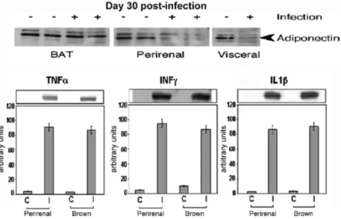
Infection also results in increased expression of PI3kinase and the activation of AKT, suggesting that this infection may induce components of the insulin/ IGF-1 receptor cascade. This would be surprising as the upregulation of proinflammatory pathways is gen-erally associated with a downregulation of the insulin signal transduction pathway (Hotamisligil 2006, Fer-rante 2007). It is not clear what is responsible for this phenomenon, but it can be observed with a high degree Fig. 1:adipose tissue of CD-1 mice infected with the Brazil strain
of Trypanosoma cruzi (for 30 days).Upper panelareimmunoblots demonstrating a reduction in adiponectin expression in perirenal and visceral adipose tissue. Lower panel are immunoblots demonstrating increased expression of cytokines [from Combs et al. (2005), with permission of the Journal of Biological Chemistry].
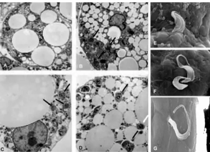
Fig. 2: adipose tissue of CD-1 mice infected with the Brazil strain of Trypanosoma cruzi. For 30 days. F4/80 staining of brown fat (A: uninfected;B:infected). Note the intense staining of macrophages in infected adipose tissue [from Combs et al. (2005) with permission of the Journal of Biological Chemistry].
of reproducibility in these cells. Despite the upregulation of some of the components of the pathway, there were no differences with respect to a dose-response to insulin in infected cells (unpublished observations). It remains to be determined whether other pathways influenced by in-sulin, such as events leading to differences in the rate of lipid accumulation or lipolysis, may be affected. T. cruzi is likely to have an impact on lipid pathways in vivo, yet these issues have not been examined to date.
In summary, fat and glucose metabolism are inter-related and dysregulated in T. cruzi infection. We have clearly demonstrated that adipocytes and adipose tis-sue represent an important target of and reservoir for infection. This is a reservoir from which parasites can be reactivated during periods of immunosuppression. In addition, the infection of adipocytes and adipose tissue create an inflammatory phenotype that affects a variety
of metabolic processes. The reduction in the expression of adiponectin and PPAR-γ perpetuate this inflamma -tory phenotype. Since adiponectin null mice have a car-diomyopathic phenotype, it is tempting to suggest that the reduction in adiponectin and PPAR-γ contributes to the cardiomyopathy of Chagas disease. Recently Coura (2007) has commented on the need for new approaches to the study of Chagas disease. Although the study of adipose tissue was not specifically mentioned, we be-lieve that this is a new and fruitful area of research.
REFERENCES
Andrade ZA, Silva HRA 1995. Parasitism of adipocytes by Trypano-soma cruzi. Mem Inst Oswaldo Cruz 90: 521-522.
Arita Y, Kihara S, Ouchi N, Takahashi M, Maeda K, Miyagawa J, Hotta K, Shimomura I, Nakamura T, Miyaoka K, Kuriyama H, Nishida M, Yamashita S, Okubo K, Matsubara K, Muraguchi M, Ohmo-to Y, Funahashi T, Matsuzawa Y 1999.Paradoxical decrease of an adipose-specific protein, adiponectin, in obesity. BBRC 257: 79-83.
Attie AD, Scherer PE 2009. Adipocyte metabolism and obesity.
J Lipid Res 50: 5395-5399.
Barreto SM, Passos VM, Lima-Costa MF 2003. Obesity and under-weight among Brazilian elderly: the Bambui Health and aging study. Cad Saude Publica 19: 605-612.
Chen A, Mumick S, Zhang C, Lamb J, Dai H, Weingarth D, Mudgett J, Chen H, MacNeil DJ, Reitman ML, Qian S 2005. Diet induc-tion of monocyte chemoattractant protein-1 and its impact on obesity. Obes Res13: 1311-1320.
Cohen AW, Hnasko R, Schubert W, Lisanti MP 2004. Role of caveolae and caveolins in health and disease. Physiol Rev 84: 1341-1379.
Cohen AW, Park DS, Woodman SE, Williams TM, Chandra M, Shi-rani J, Pereira de Souza A, Kitsis RN, Russell RG, Weiss LM, Tang B, Jelicks LA, Factor SM, Shtutin V, Tanowitz HB, Lisanti MP 2003. Caveolin-1 null mice develop cardiac hypertrophy with hyperactivation of p42/44 MAP kinase in cardiac fibroblasts. Am J Physiol Cell Physiol 284: C457-C474.
Combs TP, Berg AH, Obici S, Scherer PE, Rossetti L 2001. Endog-enous glucose production is inhibited by the adipose-derived pro-tein Acrp30. J Clin Inv 108: 1875-1881.
Combs TP, Nagajyothi, Mukherjee S, de Almeida CJ, Jelicks LA, Schubert W, Lin Y, Jayabalan DS, Zhao D, Braunstein VL, Landskroner-Eiger S, Cordero A, Factor SM, Weiss LM, Lisanti MP, Tanowitz HB, Scherer PE 2005. The adipocyte as an impor-tant target cell for Trypanosoma cruzi infection. J Biol Chem 280: 24085-24094.
Coura JR 2007. Chagas disease: what is known and what is needed - a background article. Mem Inst Oswaldo Cruz102: (Suppl. I): 113-122.
Desruisseaux MS, Nagajyothi, Trujillo ME, Tanowitz HB, Scherer PE 2007. Adipocyte and adipose tissue and infectious disease. Inf Immun 75: 1066-1078.
Fasshauer M, Klein J, Neumann S, Eszlinger M, Paschke R 2002. Hormonal regulation of adiponectin gene expression in 3T3-L1 adipocytes. BBRC 290: 1084-1089.
Ferrante AW Jr 2007. Obesity-induced inflammation: a metabolic dia-logue in the language of inflammation. J Intern Med 262: 408-414.
Gerhardt CC, Romero IA, Cancello R, Camoin L, Strosberg AD 2001. Chemokines control fat accumulation and leptin secretion by cul-tured human adipocytes. Mol Cell Endocrinol 175: 81-92.
Goldstein BJ, Scalia R 2004. Adiponectin: a novel adipokine linking adipocytes and vascular function. J Clin Endocrinol Metab 89: 2563-2568.
Fig. 5: mRNA levels of peroxisome proliferator-activated receptor γ (PPAR-γ) and adiponectin in Trypanosoma cruzi infected cultured differentiated 3T3-L1 adipocytes. Note that infection reduces the
ex-pression of both adiponectin and PPAR-γ.
Halberg N, Wernstedt-Asterholm I, Scherer PE 2008. The adipocyte as an endocrine cell. Endocrinol Metab Clin North Am 37: 753-768.
Hazan U, Romero IA, Cancello R, Valente S, Perrin V, Mariot V, Du-monceaux J, Gerhardt CC, Strosberg AD, Couraud PO, Pietri-Rouxel F 2002. Human adipose cells express CD4, CXCR4 and CCR5 [corrected] receptors: a new target cell type for the immu-nodeficiency virus-1? FASEB J16: 1254-1256.
Hotamisligil GS 2006. Inflammation and metabolic disorders. Nature 444: 860-867.
Hotta K, Funahashi T, Arita Y, Takahashi M, Matsuda M, Okamoto Y, Iwahashi H, Kuriyama H, Ouchi N, Maeda K 2000. Plasma concen-trations of a novel, adipose-specific protein, adiponectin, in type 2 diabetic patients. Arterioscler Thromb Vasc Biol 20: 1595-1599.
Hulit J, Bash T, Fu M, Galbiati F, Albanese C, Sage DR, Schlegel A, Zhurinsky J, Shtutman M, Ben-Ze’ev A, Lisanti MP, Pestell RG 2000. The Cyclin D1 gene is transcriptionally repressed by caveolin-1. J Biol Chem 275: 21203-1209.
Jan V, Cervera P, Maachi M, Baudrimont M, Kim M, Vidal H, Girard PM, Levan P, Rozenbaum W, Lombes A, Capeau J, Bastard JP 2004. Altered fat differentiation and adipocytokine expression are inter-related and linked to morphological changes and insulin resistance in HIV-1-infected lipodystrophic patients. Antivir Ther 9: 555-564.
Keller P, Moller K, Krabbe KS, Pedersen BK 2003. Circulating adi-Circulating adi-ponectin levels during human endotoxaemia. Clin Exp Immunol 134: 107-110.
Kim JY, van de Wall E, Laplante M, Azzara A, Trujillo ME, Hofmann SM, Schraw T, Durand JL, Li H, Li G, Jelicks LA, Mehler MF, Hui DY, Deshaies Y, Shulman GI, Schwartz GJ, Scherer PE 2007. Obesity-associated improvements in metabolic profile through expansion of adipose tissue. J Clin Invest 117: 2621-2637.
Kook KS, Min HY, Johnson D, Chaplinsky RJ, Flier JS, Hunt CR, Spiegelman BM 1987. Adipsin: a circulating serine protease homolog secreted by adipose tissue and sciatic nerve. Science 237: 402-405.
Mallon PW, Sedwell R, Rogers G, Nolan D, Unemori P, Hoy J, Sa-maras K, Kelleher A, Emery S, Cooper DA, Carr A 2008. Effect
of rosiglitazone on peroxisome proliferator-activated receptor γ
gene expression in human adipose tissue is limited by antiretro-viral drug-induced mitochondrial dysfunction. J Infect Dis 198: 1794-1803.
Maurin T, Saillan-Barreau C, Cousin B, Casteilla L, Doglio A,
Peni-caud L 2005. Tumor necrosis factor-α stimulates �I�-1 produc-Tumor necrosis factor-α stimulates �I�-1 produc -tion in primary culture of human adipocytes. Exp Cell Res 304: 544-551.
Mynarcik DC, Combs T, McNurlan MA, Scherer PE, Komaroff E, Gelato MC 2002. Adiponectin and leptin levels in HIV-infected subjects with insulin resistance and body fat redistribution. J Ac-quir Immune Defic Syndr 31: 514-520.
Nagajyothi F, Desruisseaux MS, Thiruvur N, Weiss LM, Braunstein VL, Albanese C, Teixeira MM, de Almeida CJ, Lisanti MP, Scherer PE, Tanowitz HB 2008. Trypanosoma cruzi infection of cultured adipocytes results in an inflammatory phenotype. Obe-sity169: 1992-1997.
Nawrocki AR, Rajala MW, Tomas E, Pajvani UB, Saha AK, Trumbauer ME, Pang Z, Chen AS, Ruderman NB, Chen H, Rossetti L, Scherer PE 2006. Mice lacking adiponectin show decreased hepatic insulin sensitivity and reduced responsiveness to peroxisome
proliferator-activated receptor γ agonists. J Biol Chem 281: 2654-2660. Nawrocki AR, Scherer PE 2005. The adipocyte as a drug discovery
target. Drug Discov Today 10: 1219-1230.
Okamoto Y, Folco EJ, Minami M, Wara AK, Feinberg MW, Sukho-va GK, Colvin RA, Kihara S, Funahashi T, Luster AD, Libby
P 2008 Adiponectin inhibits the production of CXC receptor 3 chemokine ligands in macrophages and reduces T-lymphocyte recruitment in atherogenesis. Circ Res 102: 218-225.
Ouchi N, Kihara S, Arita Y, Okamoto Y, Maeda K, Kuriyama H, Hotta K, Nishida M, Takahashi M, Muraguchi M, Ohmoto Y, Nakamura T, Yamashita S, Funahashi T, Matsuzawa Y 2000. Adiponectin, an adipocyte-derived plasma protein, inhibits
en-dothelial NF-κB signaling through a cAMP-dependent pathway. Circulation 102: 1296-1301.
Ouchi N, Kihara S, Funahashi T, Matsuzawa Y, Walsh K 2003. Obe-sity, adiponectin and vascular inflammatory disease. Curr Opin Lipidol 14: 561-566.
Ouchi N, Shibata R, Walsh K 2006. Cardioprotection by adiponectin.
Trends Cardiovasc Med16: 141-146.
Pajvani UB, Trujillo ME, Combs TP, Iyengar P, Jelicks L, Roth KA, Kitsis RN, Scherer PE 2005. Fat apoptosis through targeted acti-vation of caspase 8: a new mouse model of inducible and revers-ible lipoatrophy. Nat Med11: 797-803.
Pasarica M, Dhurandhar NV 2007. Infectobesity: obesity of infec-tious origin. Adv Food Nutr Res 52: 61-102.
Rajala MW, Obici S, Scherer PE, Rossetti L 2003. Adipose-derived
re-sistin and gut-derived rere-sistin-like molecule-β selectively impair
insulin action on glucose production. J Clin Invest 111: 225-230.
Rajala MW, Scherer PE 2003. Minireview: the adipocyte at the cross-roads of energy homeostasis, inflammation and atherosclerosis.
Endocrinology 144: 3765-3773.
Ruan H, Lodish HF 2003. Insulin resistance in adipose tissue: direct
and indirect effects of tumor necrosis factor-α. Cytokine Growth Factor Rev 14: 447-455.
Scherer PE, Williams S, Fogliano M, Baldini G, Lodish HF 1995. A novel serum protein similar to C1q, produced exclusively in adi-pocytes. J Biol Chem 270: 26746-26749.
Shibata R, Ouchi N, Ito M, Kihara S, Shiojima I, Pimentel DR, Ku-mada M, Sato K, Schiekofer S, Ohashi K, Funahashi T, Colucci WS, Walsh K 2004. Adiponectin-mediated modulation of hyper-Adiponectin-mediated modulation of hyper-trophic signals in the heart. Nat Med 10: 1384-1399.
Shibata R, Ouchi N,Murohara T 2009. Adiponectin and cardiovascu-lar disease circulation, in press. Circulation J 73: 608-614.
Shibata R, Sato K, Pimentel DR, Takemura Y, Kihara S, Ohashi K, Funahashi T, Ouchi N, Walsh K 2005. Adiponectin protects against myocardial ischemia-reperfusion injury through AMPK- and COX-2-dependent mechanisms. Nat Med 11: 1096-1103.
Stahl M, Ge C, Shi S, Pestell RG, Stanley P 2006. Notch1-induced transformation of RKE-1 cells requires up-regulation of cyclin D1. Cancer Res 66: 7562-7570.
Tanowitz HB, Amole B, Hewlett D, Wittner M 1988. Trypanosoma cruzi infection in diabetic.Trans R Soc Trop Med Hyg 82: 90-93.
Wang C, Pattabiraman N, Zhou J-F, Fu M, Sakamaki T, Albanese C, Li Z, Wu K, Hulit J, Neumeister P, Novikoff PM, Brownlee M, Scherer PE, Jones JG, Whitney KD, Donehower LA, Harris EL, Rohan T, Johns DC, Pestell RG2003. Cyclin D1 repression
of peroxisome proliferator-activated receptor-γ expression and
transactivation. Mol Cell Biol 23: 6159-6173.
Wernstedt-Asterholm I, Halberg N, Scherer PE 2007. Mouse models of lipodystrophy key reagents for the understanding of the meta-bolic syndrome. Drug Discov Today Dis Models 4: 17-24.