DOI: 10.5935/2359-4802.20180002
Mailing Address: Maynara Leonardi Schuh Martins
Rua Prof. Sebastião Paraná 762. Vila Izabel. Curitiba. Postal Code 80320-070, Novo Mundo, Curitiba, Paraná, PR – Brazil. E-mail: [email protected]; [email protected]
Decrease in the Inflammatory Marker TNF-α after Consumption of Flaxseed by
Hypercholesterolemic Rabbits
Maynara Leonardi Schuh Martins,1 Aniely Bacelar Rocco de Lima,1 Ana Flavia Champoski,1 Pamela Cristiani
Pereira,1 Fernando Martins,2 Carlos Tanizawa,1 Leonardo Précoma,1 Patrícia Campelo,1 Luiz César Guarita-Souza,1
Dalton Bertolim Précoma1
Pontifícia Universidade Católica do Paraná (PUC-PR),1 Paraná, PR; Universidade Federal da Grande Dourados (UFGD),2 Mato Grosso do Sul, MS – Brazil
Manuscript received February 27, 2017; revised manuscript June 22, 2017; accepted July 07, 2017.
Abstract
Background: Functional foods such as flaxseed have been commonly consumed to prevent atherosclerosis.
Objectives: To assess the effects of flaxseed in atherogenesis in rabbits consuming a high-cholesterol diet.
Methods: Thirty male albino rabbits were randomized to three groups based on a 12-week dietary treatment:
control group (G1), standard diet; high-cholesterol diet (G2), standard diet plus 0.25% cholesterol from lyophilized eggs; and high-cholesterol plus flaxseed (G3), similar diet as G2 plus flaxseed. Biochemical (total cholesterol [TC], high-density lipoprotein [HDL-C], low-density lipoprotein cholesterol [LDL-C], and triglycerides) and immunohistochemical (intercellular adhesion molecule 1 [ICAM-1] and tumor necrosis factor alpha [TNF-α]) analyses were performed in all groups. P values < 0.05 indicated statistical significance.
Results: At 12 weeks, serum TC levels increased significantly in G2 and G3 compared with G1. Serum LDL-C levels
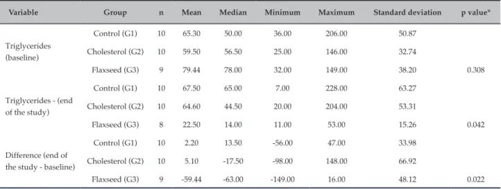
were higher in group G2, and the increase in group G3 was approximately six times lower than that in G2. HDL-C levels increased in all groups, with the highest increase observed in G2. Triglycerides levels in G3 decreased by ~70% and differed significantly in G1 and G3 (p = 0.034) and G2 and G3 (p = 0.015). ICAM-1 levels increased only in aortic segment 4 in G3. TNF-α levels in G3 were similar to those in the control group, while the levels in G2 were greater than twice as those in the control group (p < 0.05).
Conclusions: The group fed with a functional diet (flaxseed) showed decreased development of atherosclerosis, reduced
serum triglycerides levels, and lower TNF-α levels on immunohistochemistry. (Int J Cardiovasc Sci. 2018;31(2)114-122)
Keywords: Rabbits; Flax / seeds; Atherosclerosis / prevention & control; Diet, Atherogenic; Cholesterol, Diet;
Obesity, Sedentary Lifestyle.
Introduction
Atherosclerosis is a chronic inflammatory disease marked by endothelial dysfunction affecting the intimal layer of medium- and large-caliber arteries.1 This process
occurs slowly over the course of decades and has the potential of evolving into more advanced lesions, which may rupture and trigger thrombus formation, potentially leading to acute myocardial infarction and death.2
Several factors are implicated in the etiology of atherosclerosis, including persistent elevation of
atherogenic serum lipoproteins (such as low-density lipoprotein cholesterol [LDL-C]), which are associated with lifestyle factors such as poor eating habits, sedentary lifestyle, and obesity, as well as deposition of these lipoproteins in the inner tunica.2,3
antioxidants, and polyphenols,4 which can significantly
decrease the effect of atherosclerotic factors. One such product is flaxseed (Linum usitatissimum L.).
The nutritional composition of flaxseed comprises fats (41%), dietary fibers (28%), proteins (21%), moisture (7.7%), ashes (3.5%), and soluble sugars (1 – 2%),5,6 and
their lipid content includes 51 – 55% of alpha-linolenic acid.7 Additionally, flaxseed is the richest source of lignan
secoisolariciresinol-diglucoside (SDG), which has potent antioxidant and antiatherogenic properties.
Based on these considerations, the study of the functional properties of flaxseed becomes essential for the scientific community and the general population. The aim of the present study was to assess the potential effects of flaxseed on atherogenesis in rabbits subjected to a high-cholesterol diet (containing 0.25% cholesterol) of lyophilized eggs. For this purpose, we analyzed the lipid levels and performed immunohistochemical analysis of inflammatory markers in the aortic segments of the rabbits.
Methods
The present study was approved by the Animal Research Ethics Committee of PUC-PR under the protocol number 720/2012 and was conducted in the Laboratory of Experimental Surgery at Hospital Angelina Caron (HAC), the Experimental Pathology Laboratory at PUC-PR, and the Central Animal Facility at PUC-PR.
Animals
Thirty male New Zealand white rabbits (Oryctolagus cuniculus) weighing approximately 3 kg and with a mean age of 4 months were selected for the experiment. Calculation of the sample size was based on the study by Prim et al. (2008).8 The rabbits were kept in the
animal facility at PUC-PR in a macroenvironment with light/dark cycles of 12/12 hours, fresh air changes, and temperature controlled between 19°C and 23°C. The animals were identified on an individual basis and maintained in individual cages cleaned daily.
Experimental period and group distribution
The study was conducted over 12 weeks (84 days). The animals were maintained in individual cages and randomized to three groups according to dietary treatment. The control group (G1, n = 10) received
a standard diet for rabbits (Nuvital® Nutrientes S.A., Colombo, PR, Brazil). The high-cholesterol diet group (G2, n = 10) received a standard diet for rabbits (Nuvital®) plus 0.25% cholesterol from lyophilized eggs. The high-cholesterol plus flaxseed diet group (G3, n = 9) received a standard diet for rabbits (Nuvital®), 0.25% cholesterol from lyophilized eggs, and 8 g of ground flaxseed per kg of body weight.
Preparation of supplementary feed
The high-cholesterol diet offered to groups G2 and G3 throughout the experimental period contained 0.25% cholesterol, which was added to induce atherosclerotic lesions in the aorta. In order to achieve that, 5 kg of standard feed (Nuvital®), weighed and ground for each animal group, was added to 1,800 g of egg powder diluted in 2,000 mL of water.
The flaxseed concentration was 8 g per kg of body weight. The seeds were ground in a food processor and stored under refrigeration before added to the feed.
The feed given to groups G2 and G3 was supplemented with lyophilized eggs. This mixture was processed using an industrial meat grinder (Poli®,modelPCP-22LR-N,
feeder #10, Siemsen, Brusque, SC, Brazil). For group G3, the mixture was supplemented with ground flaxseed. After grinding the mixture, the feed was pelleted, and the pellets were heated in an electric oven for 10 min at a temperature of 180°C.
The prepared feed was stored under refrigeration until use.
Animal preparation and sample collection
The animals were anesthetized, and blood was collected according to the method described by Précoma et al.9 and Alessi et al.10 Xylazine (Coopazine;
Schering-Plough-Coopers, Cotia, SP, Brazil) at
5 mg/kg mixed with ketamine (Vetanarcol,
Histological analysis
The thoracic aorta was removed at the level of the aortoiliac bifurcation following a midline thoracotomy and laparotomy, and cross sections with a thickness of 2 to 3 mm were cut from different aortic segments. Each segment was labeled, placed in a microtome (Leica® RM2145, Leica Microsystems Nussloch GmbH,
Germany) for overnight processing, and dehydrated with a graded ethanol series (70%, 80%, and 90%). The tissue samples were subsequently diaphanized for 12 hours in xylol containing serial concentrations of paraffin. Paraffin blocks were obtained by embedding the samples in hot paraffin using a tissue embedding console system (Leica® EG1160, Leica Microsystems Nussloch GmbH,
Germany), according to the standard procedure.
The paraffin blocks were cut into 5 μm sections using the microtome (Leica® RM 2145). Each section was stained
with hematoxylin and eosin (HE) and orcein (elastic tissue staining) according to conventional techniques and mounted on permanent slides.
Blocks with four aortic segments were prepared by selecting one proximal segment near the aortic arch (point 1), one segment located in the thoracic aorta (point 2), one proximal segment in the abdominal aorta (point 3), and one distal segment in the abdominal aorta (point 4). These segments were selected according to the histological findings in HE staining.
Immunohistochemical analysis
The tissue microarray (TMA) technique was used for immunohistochemical analysis. The samples were incubated with intercellular adhesion molecule 1 (ICAM-1) monoclonal antibodies at a dilution of 1:50, and mixed with tumor necrosis factor alpha (TNF-α) monoclonal antibody for 1 hour. The antibodies were detected by incubating the slides with the substrate 3,3'-diaminobenzidine-tetrahydrochloride-dihydrate (DAB; DakoCytomation A/S, Glostrup, Denmark). Counterstaining was performed with Mayer’s hematoxylin, followed by sample dehydration with different concentrations of 100% ethanol and deparaffinization with xylol. The slides were mounted with Canada balsam. The protocol described above was standardized at our department. Positive and negative controls were used for each reaction.
The histological sections were digitized with a scanner (Axio Scan Z1, Zeiss, Jena, Germany) and the images were analyzed using Image-Pro Plus® software, version 4.5.
The color morphometry method was applied, staining
the areas with positive antibody imaging in pink and those with negative imaging in brown. The data were transferred to an Excel spreadsheet and subjected to statistical analysis.
Statistical analysis
The results are expressed as mean, median, minimum and maximum, and standard deviation values. The assumption of normality of the variables was assessed using the Kolmogorov-Smirnov test. The variables not meeting the normality assumption were analyzed using the Kruskal-Wallis nonparametric test. P values below 0.05 were considered statistically significant. The data were analyzed using the IBM SPSS Statistics software, version 20.0.
Results
One death occurred in group G3 at experimental week 12, resulting in a total of 29 rabbits in the final study sample.
Lipid profile
Total cholesterol
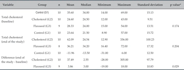
Serum TC levels increased significantly in groups G2 and G3 compared with G1. This result demonstrates that the diet was effective in increasing cholesterol levels in the rabbits (Table 1).
LDL-C and HDL-Cl
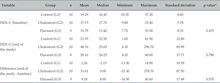
There were no significant differences in the levels of LDL-C and HDL-C among the groups (Tables 2 and 3). However, serum LDL-C levels in the biochemical analysis were higher in group G2 at the end of the experiment. The increase in LDL-C in group G3 was approximately six times lower than that in G2. HDL-C levels increased in groups G1 (12.93 mg/dL), G2 (82.26 mg/dL), and G3 (12.03 mg/dL), with the highest increase observed in G2.
Triglycerides
Table 1 – Mean, median, minimum and maximum, standard deviation, and p values for total cholesterol in all groups
Variable Group n Mean Median Minimum Maximum Standard deviation p value*
Total cholesterol (baseline)
Control (G1) 10 35.60 34.00 14.00 69.00 15.13
Cholesterol (G2) 10 24.60 24.50 12.00 43.00 9.51
Flaxseed (G3) 9 28.33 24.00 15.00 54.00 13.51 0.174
Total cholesterol (end of the study)
Control (G1) 10 23.64 21.50 8.90 57.00 15.72
Cholesterol (G2) 10 62.09 24.54 12.90 336.00 100.23
Flaxseed (G3) 8 36.21 34.20 16.40 72.00 17.32 0.204
Difference (end of the study - baseline)
Control (G1) 10 -11.96 -13.50 -31.00 6.00 12.50
Cholesterol (G2) 10 37.49 2.55 -28.00 305.00 97.79
Flaxseed (G3) 9 3.86 3.00 -19.00 18.00 10.83 0.029
*Nonparametric Kruskal-Wallis test, p < 0.05.
Immunohistochemical analysis
Immunohistochemistry was used to investigate the presence of ICAM-1 and TNF-α in all four aortic segments.
The results indicated that ICAM-1 levels increased only in aortic segment 4 in group G3; however, there was no significant difference in relation to median levels (Figure 1).
A similar result was found for TNF-α, that is, the level of this marker was significantly increased in the aortic segment 4 (Figure 2). TNF-α levels in group G3 were similar to those in the control group, while the levels in group G2 were greater than twice as those in the control group (p < 0.05).
Discussion
Atherosclerosis is a major cardiovascular disease, and the understanding of its development is required for its prevention and treatment. This knowledge can be acquired using experimental models. In this respect, several animal studies have shown that a high-fat diet leads to obesity, hypercholesterolemia, and other complications such as endothelial lesions.11
The experimental protocol in the present study was an early model of atherosclerosis and involved the addition of 0.25% cholesterol from lyophilized eggs to the diet of rabbits aiming at causing atherosclerotic lesions.12
The use of rabbits as an experimental model suits the objectives of this study because these animals develop aortic lesions fast.
T h i s s t u d y a s s e s s e d t h e b i o c h e m i c a l a n d immunochemical changes caused by a high-cholesterol diet with and without flaxseed, which is considered a functional food.
The increase in TC found in the biochemical analysis in groups G2 and G3 was not expected in the group treated with flaxseed. A similar result was obtained by Dupasquier et al.,13 who observed no changes in
TC in animals receiving a diet with 10% of flaxseed. This unexpected result may be explained by several factors, including the concentration of flaxseed used, the form of flaxseed administration (ground seeds), feed heating for the incorporation of components, and the duration of seed intake. Previous results have indicated that alpha-linolenic and linoleic acids are sensitive to light, heating, and oxygen, and undergo oxidation when exposed to temperatures between 120°C and 270°C.14 Therefore, the process of production of feed
containing flaxseed in the present study may have caused a decrease in the level of these acids, thereby compromising their nutraceutical function.
LDL-C levels increased in the groups treated with high cholesterol; however, the levels remained similar between these groups. Similar results were obtained in a study in humans involving the consumption of flaxseed, in which the levels of TC and LDL-C remained unchanged,15 even though the experimental
period of 4 weeks may not have been sufficient to cause this reduction. Zheng et al.16 conducted a
Table 2 – Mean, median, minimum and maximum, standard deviation, and p values for LDL-C in all groups
Variable Group n Mean Median Minimum Maximum Standard deviation p value*
LDL-C (baseline)
Control (G1) 10 8.64 8.45 2.90 18.10 5.61
Cholesterol (G2) 10 7.83 6.30 1.20 27.10 7.60
Flaxseed (G3) 9 11.92 7.40 4.50 27.30 7.94 0.305
LDL- C (end of the study)
Control (G1) 10 21.57 16.45 3.90 81.00 21.49
Cholesterol (G2) 10 90.09 32.00 7.50 348.00 123.47
Flaxseed (G3) 8 26.95 22.00 2.40 93.00 29.82 0.145
Difference (end of the study - baseline)
Control (G1) 10 12.93 11.65 -5.70 63.50 19.71
Cholesterol (G2) 10 82.26 21.95 4.90 341.80 124.45
Flaxseed (G3) 9 12.03 0.50 -27.30 79.40 30.61 0.085
* Nonparametric Kruskal-Wallis test, p < 0.05.
Table 3 – Mean, median, minimum and maximum, standard deviation, and p values for HDL-C in all groups
Variable Group n Mean Median Minimum Maximum Standard deviation p value*
HDL-C (baseline)
Control (G1) 10 19.29 16.45 10.30 37.30 8.60
Cholesterol (G2) 10 17.15 17.70 9.80 25.40 5.78
Flaxseed (G3) 9 15.79 13.40 7.70 33.50 8.35 0.475
HDL-C (end of the study)
Control (G1) 10 21.55 22.50 1.00 41.90 12.80
Cholesterol (G2) 10 48.76 25.65 4.30 290.70 85.99
Flaxseed (G3) 8 28.16 26.25 4.20 60.00 17.71 0.780
Difference (end of the study - baseline)
Control (G1) 10 2.26 -2.15 -13.30 14.90 10.39
Cholesterol (G2) 10 31.61 9.00 -15.30 278.70 87.50
Flaxseed (G3) 9 9.24 8.00 -16.50 46.60 17.48 0.570
* Nonparametric Kruskal-Wallis test, p < 0.05.
hypercholesterolemia and used a lignan extract from flaxseed during an 8-week period. The LDL-C levels in the group that received 600 mg of the extract decreased between weeks 6 and 8. These authors concluded that the lignan extract decreased the concentration of cholesterol in a dose-dependent manner. One study investigated the effect of phytosterol intake and indicated that blood cholesterol levels decreased after 4 weeks.17 In addition,
cholesterol levels remained unchanged between weeks 8 and 12,18,19 which suggested that seasonal factors may
have affected the results.
In the present study, TC increased in all tested groups. Studies have shown that lignans present in flaxseed reduce serum cholesterol. This effect is probably related to their antioxidant properties, provided by secoisolariciresinol lignan (SDG), enterolactone, and enterodiol, which inhibit the peroxidation of polyunsaturated fatty acids in vitro and favor the decrease in cholesterol levels.20
16
14
12
10
8
6
4
2
0
Control Cholesterol Flaxseed
Mean Mean ± SE Mean ± SD
ICAM-1 – segment 4
Group
Figure 1 – Mean intercellular adhesion molecule 1 (ICAM-1) values at the animals’ aortic segment 4 in each group.
Table 4 – Mean, median, minimum and maximum, standard deviation, and p values for triglycerides in all groups
Variable Group n Mean Median Minimum Maximum Standard deviation p value*
Triglycerides (baseline)
Control (G1) 10 65.30 50.00 36.00 206.00 50.87
Cholesterol (G2) 10 59.50 56.50 25.00 146.00 32.74
Flaxseed (G3) 9 79.44 78.00 32.00 149.00 38.20 0.308
Triglycerides - (end of the study)
Control (G1) 10 67.50 65.00 7.00 228.00 63.27
Cholesterol (G2) 10 64.60 44.50 20.00 204.00 53.31
Flaxseed (G3) 8 22.50 14.00 11.00 53.00 15.26 0.042
Difference (end of the study - baseline)
Control (G1) 10 2.20 13.50 -56.00 47.00 33.98
Cholesterol (G2) 10 5.10 -17.50 -98.00 148.00 66.92
Flaxseed (G3) 9 -59.44 -63.00 -149.00 16.00 48.12 0.022
* Nonparametric Kruskal-Wallis test, p < 0.05.
Another hypothesis is the duration of the functional diet (8 weeks), which may have been too short to induce a significant reduction in LDL-C and TC.
In the present study, no differences in HDL-C levels were observed among the three groups. Higher levels of HDL-C increase the protection against the development of atherosclerotic plaques in the heart. Studies have shown that flaxseed intake at a dose of 15% was strongly
associated with protection from inflammation, estrogens, and genotoxicity.21
3.5
3.0
2.5
2.0
1.5
1.0
0.5
0.0
TNF-α – segment 4
Control Cholesterol Group
Flaxseed
Median 25%-75% Min-Max
Figure 2 – Mean tumor necrosis factor alpha (TNF-α) values at the animals’ aortic segment 4 in each group.
effective in decreasing TG levels. This result is in line with those from previous studies and suggests that dietary supplementation with omega-3 via the consumption of flaxseed helps decrease TG levels, possibly via the decrease of remaining chylomicron particles and the inhibition of the synthesis and secretion of very low-density lipoprotein (VLDL-C) by the liver.22
Cintra et al.23 tested the effect of the consumption
of a high-fat flaxseed-based diet in Wistar rats and observed a reduction in TC and TG levels, higher fecal excretion of lipids, and lower deposition of cholesterol in the liver. This result was similar to that obtained by Bhathena (2003).23
Immunohistochemistry was used to investigate the presence of two markers involved in inflammation, a cellular adhesion molecule (ICAM-1) and a proinflammatory cytokine (TNF-α). Several studies have used ICAM-1 as a marker of inflammation. In a study with 60 rabbits (10 in the control group and 50 in the atherosclerotic group), animals fed a high-fat diet exhibited higher levels of ICAM-1 in the aorta compared with those in the control group (p < 0.01), which indicates a role for ICAM-1 as a predictor of cardiovascular disease.25
The importance of the present study lies in the analysis of ICAM-1 in four aortic segments. The results indicated an increase in ICAM-1 levels in all animal groups and all tissue sections; however, the results were not statistically
significant. A similar result was obtained in an animal study conducted by Prim et al.,8 and in human studies,
which also showed nonsignificant reductions in the levels of ICAM-1 and VCAM-1 markers.26
We should emphasize that no antiinflammatory activity was observed in the group that received flaxseed (G3), suggesting that the findings of the antiinflammatory effect of omega-3 fatty acids are contradictory. In addition, the results of ICAM-1 suggest that this activity is dependent on the duration of consumption of omega-3 fatty acids.
The results of the TNF-α analysis indicated a significant difference among the groups. TNF-α levels were higher in G1 than G3. This result demonstrated an effect of flaxseed, but was not statistically significant. Moreover, the levels of TNF-α increased in G2, which indicates that this marker is associated with stress and inflammation. A similar result was observed in a previous study,27 in which the elevated levels of TNF-α
were associated with recurrent coronary events after myocardial infarction.
1. Xavier HT, Izar MC, Faria Neto JR, Assad MH, Rocha VZ, Sposito AC, et al; Sociedade Brasileira de Cardiologia. [V Brazilian Guidelines on Dyslipidemias and Prevention of Atherosclerosis]. Arq Bras Cardiol. 2013;101(4 Suppl 1):1-20. doi: 105935/abc20135010.
2. Castro PS, Oliveira FL. Prevention of atherosclerosis and drug treatment of high-risk lipid abnormalities in children and adolescents. J Pediatr (Rio J). 2009;85(1):6-14. doi:10.2223/JPED.1852.
3. Schoenhagen P, Tuzcu EM. Atherosclerosis imaging in progression/ regression trials: surrogate marker or direct window into the atherosclerotic disease process? Arq Bras Cardiol. 2008;91(6):385-98. PMID: 19142366.
4. Bloedon LT, Szapary PO. Flaxseed and cardiovascular risk. Nutr Rev. 2004;62(1):18-27. PMID: 14995053.
5. Kasote DM. Flaxseed phenolics as natural antioxidants. Int Food Res J. 2013;20(1):27-34.
6. Prasad K. Flaxseed and cardiovascular health. J Cardiovasc Pharmacol. 2009;54(5):369-77. doi: 10.1097/FJC.0b013e3181af04e5.
7. Padilha PC, Pinheiro RL. O papel dos alimentos funcionais na prevenção e controle do câncer de mama. Rev Bras Cancerol. 2004;50(3):251-60.
8. Prim CR, Baroncini LA, Précoma LB, Caron PH, Winter G, Poletti MO, et al. Effects of linseed consumption for a short period of time on lipid profile and atherosclerotic lesions in rabbits fed a hypercholesterolaemic diet. Br J Nutr. 2012;107(5):660-4. doi: 10.1017/ S0007114511003539.
9. Précoma DB, Noronha L, Moura AV, Yamada AS, Knopfholz J, Dusilek CL, et al. Vascular radiolesion as a deleterious effect of high-dose-rate intraarterial brachytherapy with samarium-153 in hypercholesterolemic rabbits. Arq Bras Cardiol. 2006;87(4):512-9. PMID: 17128322.
10. Alessi A, França Neto OR, Brofman PR, Camila Prim C, Noronha L, Kuenzer RF, et al. Use of rosiglitazone before and after vascular injury in hypercholesterolemic rabbits: assessment of neointimal formation. Thromb J. 2008;6:12. doi: 10.1186/1477-9560-6-12.
11. Martins F, Campos SH, Pagan UL, Martinez PF, Okoshi K, Okoshi MP, et al. High-fat diet promotes cardiac remodeling in an experimental model of obesity. Arq Bras Cardiol. 2015;105(5):479-86. doi: 10.5935/abc.20150095.
References
Conclusion
The results of this study are aligned with others in the literature and showed a decreased development of atherosclerosis in the group fed a functional diet.
The results obtained in the flaxseed group showed significant changes in serum TG and decreased TNF-α levels on immunohistochemical analysis. These findings corroborate observations from previous studies that have shown flaxseed as having a strong tendency to act effectively in reducing TNF-α levels, thus preventing injury and development of atherosclerotic plaques in the intima of coronary arteries.
However, several differences may arise when changes are made to the experiment or study protocol, so it we suggest that a larger number of studies should be conducted directly comparing these functional seeds.
Acknowledgments
We are thankful to Jasmine Alimentos Ltda. (Curitiba, PR, Brazil) for donating the flaxseed used in the study. We are also grateful for the medical students (Pontifical Catholic University of Paraná) Carlos Humberto Guilman Tanizawa, Bruno Carnevalli, Alexandre da Silva Phaco Jr., and Eduardo Hubbe Buss for their assistance in feeding the animals.
Author contributions
Conception and design of the research: Martins MLS, Lima ABR, Champoski AF, Précoma L, Precoma DB.
Acquisition of data: Martins MLS, Lima ABR, Tanizawa C. Analysis and interpretation of the data: Martins MLS, Champoski AF, Pereira PC, Martins F, Precoma DB. Statistical analysis: Martins MLS, Campelo P. Obtaining financing: Martins MLS, Lima ABR, Pereira PC. Writing of the manuscript: Martins MLS, Martins F. Critical revision of the manuscript for intellectual content: Martins MLS, Lima ABR, Martins F, Campelo P, Souza LCG. Supervision / as the major investigador: Precoma DB.
Potential Conflict of Interest
No potential conflict of interest relevant to this article was reported.
Sources of Funding
There were no external funding sources for this study.
Study Association
This article is part of the thesis of master submitted by Maynara Leonardi Schuh Martins, from Pontifícia Universidade Católica do Paraná.
Ethics approval and consent to participate
12. Bocan TM, Mueller SB, Mazur MJ, Uhlendorf PD, Brown EQ, Kieft KA. The relationship between the degree of dietary-induced hypercholesterolemia in the rabbit and atherosclerotic lesion formation. Atherosclerosis. 1993;102(1):9-22. PMID: 8257456.
13. Dupasquier CH, Weber AM, Ander BP, Rampersad PP, Steigerwald S, Wigle JT, et al. Effects of dietary flaxseed on vascular contractile function and atherosclerosis during prolonged hypercholesterolemia in rabbits. Am J Physiol Heart Circ Physiol. 2006;291(6):H2987-96, doi: 10.1152/ ajpheart.01179.2005.
14. Marques AC, Hautrive TP, Moura GB, Callegaro MG, Hecktheuer LH. Effect of flaxseed (Linum usitatissimum L.) prepared by different methods on the biological response of rats. Rev Nutr. 2011;24(1):131-41.
15. Stuglin C, Prasad K. Effect of flaxseed consumption on blood pressure, serum lipids, hemopoietic system and liver and kidney enzymes in healthy humans. J Cardiovasc Pharmacol Ther. 2005;10(1):23-7. doi: 10.1177/107424840501000103.
16. Zheng Y, Weisenborn DP, Tostenson K, Kangas N. Energy analysis in the screw pressing of whole and dehulled flaxseed. Journal of Food Engineering. 2005;66(2):193-202. doi: 10.1016/j.jfoodeng.2004.03.005.
17. Kris-Etherton PM, Dietschy J. Design criteria for studies examining individual fatty acid effects on cardiovascular disease risk factors: human and animal studies. Am J Clin Nutr. 1997;65(5 Suppl):1590S-6S. PMID: 9129499.
18. Nguyen TT, Dale LC, Von Bergmann K, Croghan IT. Cholesterollowering effect of stanol ester in a US population of mildly hypercholesterolemic men and women: a randomized controlled trial. Mayo Clin Proc. 1999:74(12):1198-206. doi: 10.4065/74.12.1198.
19. Blair SN, Capuzzi DM, Gottlieb SO, Nguyen T, Morgan JM, Cater NB. Incremental reduction of serum total cholesterol and low-density lipoprotein cholesterol with the addition of plant stanol estercontaining spread to statin therapy. Am J Cardiol. 2000;86(1):46-52. PMID: 10867091.
20. Prasad K. Hypocholesterolemic and antiatherosclerotic effect of flax lignan complex isolated from flaxseed. Atherosclerosis. 2005;179(1):269-75. doi: 10.1016/j.atherosclerosis.2004.11.012.
21. Dikshit MA, Gomes Filho E, Eilati S, McGee C, Small C, Gao T. Flaxseed reduces the pro-carcinogenic micro-environment in the ovaries of normal hens by altering the PG and oestrogen pathways in a dose dependent manner. Br J Nutr. 2015;113(9):1384-95. doi: 10.1017/ S000711451500029X.
22. AdkinsY, Kelley DS. Mechanisms underlying the cardioprotective effects of omega-3 polyunsaturated fatty acids. J Nutr Biochem. 2010:21(9):781-92. doi: 10.1016/j.jnutbio.2009.12.004.
23. Cintra DE, Costa AV, Penuzio Mdo C, Matta SL, Silva MT, Costa, NM. Lipid profile of rats fed high-fat diets based on flaxseed, peanut, trout or chicken skin. Nutrition. 2006:22(2):197-205. doi: 10.1016/j.nut.2005.09.003.
24. Bhathena SJ, Ali AA, Haudenschild C, Latham P, Ranich T, Mohamed AI, et al. Dietary flaxseed mealis more protective than soy protein concentrate against hypertriglyceridemia and steatosis of the liver in an animal model of obesity. J Am Coll Nutr. 2003;22(2):157-64. PMID: 12672712.
25. Li M, Wang X, Yang L, Gao C, Ma Y. Psychological stress increases expression of aortic plaque intercellular adhesion molecule-1 and seruminflammatory cytokines in atherosclerotic rabbit model. J Geriatr Cardiol. 2008;5(4):235-42.
26. Abe Y, El-Masri B, Kimball KT, Pownall H, Reilly CF, Osmundsen K, et al. Soluble cell adhesion molecules in hypertrigliceridemia and potential significance on monocyte adhesion. Aterioscler Thoromb Vasc Biol. 1998;18(5):723-31. PMID: 9598830.
27. Ridker PM, Rifai N, Pfeffer M, Sacks F, Lepage S, Braunwald E. Elevation of tumor necrosis factor-alpha and increased risk of recurrent coronary events after myocardial infarction. Circulation.2000;101(18):2149-53. PMID: 10801754.
28. Grunfeld C, Kotler DP, Shigenaga JK, Doerrler W, Tierney A, Wang J, et al. Circulating interferon-alpha levels and hypertriglyceridaemia in the acquired immunodeficiency syndrome. Am J Med. 1991;90(2):154-62. PMID: 1996584.