Printed version ISSN 0001-3765 / Online version ISSN 1678-2690
www.scielo.br/aabc
Lead tolerance of water hyacinth
(
Eichhornia crassipes
Mart. - Pontederiaceae)
as defined by anatomical and physiological traits
FABRICIO J. PEREIRA1, EVARISTO M. DE CASTRO1, CYNTHIA DE OLIVEIRA1, MARINÊS F. PIRES1, MARCIO P. PEREIRA1, SILVIO J. RAMOS2 and VALDEMAR FAQUIN2
1Universidade Federal de Lavras, Departamento de Biologia, Campus Universitário, 37200-000 Lavras, MG, Brasil 2
Universidade Federal de Lavras, Departamento de Ciência do Solo, Campus Universitário, 37200-000 Lavras, MG, Brasil
Manuscript received on February 17, 2014; accepted for publication on May 13, 2014
ABSTRACT
This study aimed at verifying the lead tolerance of water hyacinth and at looking at consequent anatomical and physiological modifications. Water hyacinth plants were grown on nutrient solutions with five different lead concentrations: 0.00, 0.50, 1.00, 2.00 and 4.00 mg L-1 by 20 days. Photosynthesis, transpiration, stomatal conductance and the Ci/Ca rate were measured at the end of 15 days of experiment. At the end of the experiment, the anatomical modifications in the roots and leaves, and the activity of antioxidant system enzymes, were evaluated. Photosynthetic and Ci/Ca rates were both increased under all lead treatments. Leaf anatomy did not exhibit any evidence of toxicity effects, but showed modifications of the stomata and in the thickness of the palisade and spongy parenchyma in the presence of lead. Likewise, root anatomy did not exhibit any toxicity effects, but the xylem and phloem exhibited favorable modifications as well as increased apoplastic barriers. All antioxidant system enzymes exhibited increased activity in the leaves, and some modifications in roots, in the presence of lead. It is likely, therefore, that water hyacinth tolerance to lead is related to anatomical and physiological modifications such as increased photosynthesis and enhanced anatomical capacity for CO2 assimilation and water conductance.
Key words: heavy metals, ecophysiology, plant anatomy, macrophytes.
Correspondence to: Fabricio José Pereira
E-mail: [email protected]
INTRODUCTION
Environmental contamination by lead (Pb) is a
worldwide problem (Gratão et al. 2005). Lead is one
of the most dangerous pollutants and its deposition in
soil and water is related to effluents, fuels, industries
and agronomical pesticides and fertilizers (Sharma
and Dubey 2005).
Traditional techniques for lead removal are
expensive and often produce new dangerous effluents.
Phytoremediation is an alternative with low cost that
has been utilized for soil and water decontamination
(Gratão et al. 2005, Rahman and Hasegawa 2011).
Eichhornia crassipes showed a hyper-accumulation
capacity for chromium (Faisal and Hasnain 2003),
cadmium (Oliveira et al. 2001) and arsenic (Dhankher
et al. 2002, Pereira et al. 2011). The hyper acumulation
capacity of this plant is related to its large biomass and
the characteristics such as pH and temperature have
little influence on the process (Schoenhals et al. 2009).
There is little information on how lead
may affect photo
synthesis, but it can reduce
chlorophyll content and photosynthetic rate
affecting the photosystem II (Pinchasov et al.
2006, Cenkci et al. 2010). In rice plants lead
promotes an increase in membrane peroxidations
and in the activity of antioxidant enzymes
(Verma and Dubey 2003). In
Phaseolus vulgaris
lead promoted an increase in oxidant compounds
such as phenols (Hamid et al. 2010).
There are few studies of anatomical
modi-fications in
E. crassipes as a consequence of
environmental stresses. Mahmood et al. (2005)
reported that in the presence of textile industry
effluents,
E. crassipes
plants exhibited a reduction
in cell size in leaf tissues, whereas Pereira et al.
(2011) found no deleterious effects in anatomy of
leaves and roots of this species in the presence of
arsenic. In
Plantago major, lead reduced stomatal
density as well as stomatal conductance and
vascular bundle size (Kosobrukhov et al. 2004). In
wheat, plants under lead contamination exhibited
increased antioxidant enzymes activity (Liu et
al. 2010). In this study, we aimed to evaluate the
lead tolerance of E. crassipes plants as related to
modifications in its anatomy, gas exchange and
antioxidant enzymes activities.
MATERIALS AND METHODS
PLANT MATERIALS AND EXPERIMENTAL DESIGN
Water hyacinth plants (
Eichhornia crassipes
Mart.)
were collected and cultivated in a greenhouse at the
Biology Department of the Federal University of
Lavras, state of Minas Gerais, Brazil. Plants were
cultivated in Hoagland and Arnon nutrient solution
(Hoagland and Arnon 1940) at 40% of ionic force
for 30 days in order to obtain individuals free of
endogenous lead and homogeneous clones.
Cloned plants selected by size and the number
of leaves, were transplanted to plastic pots
contain-ing 4 L of Hoagland and Arnon nutrient solution
at 20% of ionic force and the following lead
concentrations: 0, 0.50, 1.00, 2.00 and 4.00 mg L
-1.
The experiment was conducted for 20 days, after
this period, plants were harvested and subsequently
divided into shoots and roots. The experimental
design was completely randomized with five
treatments and five replicates. Data were submitted
to one-way Anova and Scott-Knott test at P<0.05 in
sisvar statistical software.
GAS EXCHANGE EVALUATION
After 15 days, experimental plants were evaluated
for: net photosynthesis (A), stomatal conductance
(g
s), transpiratory rate (E) and the internal and
atmospheric carbon rate (Ci/Ca). Measurements
were conducted with the infrared gas analyzer
(IRGA) model LI-6400 (Li-COR Biosciences,
Lincoln-USA). These evaluations were made with
fully expanded and pathogen-free leaves, in five
replications. Measurements were made at 10
hours and the photon flux of photosynthetic
radia-tion was standardized at 1000 μmol m
-2s
-1in the
equipment chamber.
ANATOMICAL EVALUATIONS
Anatomical evaluations were conducted with
clonal plants at the end of the 20 day experimental
period. Whole Plants were fixed in F.A.A.
70%solution (formaldehyde, acetic acid and ethanol
70%) for 72 hours, and stored in ethanol 70%.
Paradermal sections were prepared for abaxial
and adaxial faces of the leaves. Sections were
cleared with 50% sodium hypochlorite solution,
washed in distilled water for 2 ten-minute periods,
stained with a 1% safranin aqueous solution, and
mounted in 50% glycerol (Johansen 1940). Leaf
portions were removed from the median region
and transverse sections were made with a
cleared with 50% sodium hypochlorite solution,
washed twice in distilled water, and stained with
safrablau solution (safranin 1% and astra blau 0.1%
in the proportion of 7:3), and mounted in glycerol
50% (Johansen 1940). Slides were observed and
photographed with an Olympus light microscope (BX
60 model – Olympus, Tokyo, Japan) and with a digital
camera (Canon A630 – Canon Inc., Tokyo, Japan).
Photomicrographs were evaluated in
UTHSCSA-Imagetool software and quantitative analyses of the
tissues and structures were performed in five sections
by statistical replicate and five fields per section. The
IVC (Carlquist vulnerability index) was calculated
(Carlquist 1975), as well as the aerenchyma proportion
of the root cortex, as described by Pereira et al. (2008),
and the stomatal density and stomatal index according
to Castro et al. (2009).
ANTIOXIDANT ENZYMES ACTIVITY
For biochemical analyses assays, roots and leaves
were collected at 20 days from clones that were
fully developed in the lead solutions. These organs
were frozen in liquid nitrogen and stored at -80°C.
For protein extractions, 0.5 g of roots and leaves
were ground in 2.0 mL of extraction buffer (1.924
μL of potassium phosphate buffer 0.1 M at pH 7; 20
μL of EDTA 0.1 M; 8 μL of DTT 0.5 M; 16 μL of
PMSF 0.1 M and 40 mg of PVPP) modified from
Bor et al.
(2003). The extracts were centrifuged at
14000 g at 4ºC for 20 minutes, and the supernatant
was used for the enzymatic analysis of catalase
(CAT), ascorbate peroxidase (APX) and superoxide
dismutase (SOD). APX activity evaluations were
performed following Nakano and Asada (1981), CAT
activity analysis as described by Madhusudhan et al.
(2003), and the SOD activity evaluated following
Giannopolitis and Ries (1977).
RESULTS
Gas exchange characteristics in
E. crassipes were
modified by the lead treatments. Concentrations at
0.50 and 1.0 mg L
-1increased the photosynthetic
rate of plants by 13.95% and 11.29%, respectively,
compared to the control group; but at higher
concen-trations, only small and non-significant variation
occurred (Figure 1A). The stomatal conductance was
reduced in concentrations of 1.0 mg L
-1and above;
the reduction was of 57.83% in comparison to the
control (Figure 1B). Likewise, the transpiratory
rate was increased by 9.1% only at the highest
concentration (4.0 mg L
-1) (Figure 1C). The Ci/Ca
rate was increased in the 0.50 mg L
-1and maintained
this level in all higher concentrations (Figure 1D).
The different lead concentrations promoted
modifications in the leaf anatomy of
E. crassipes
. Leaf
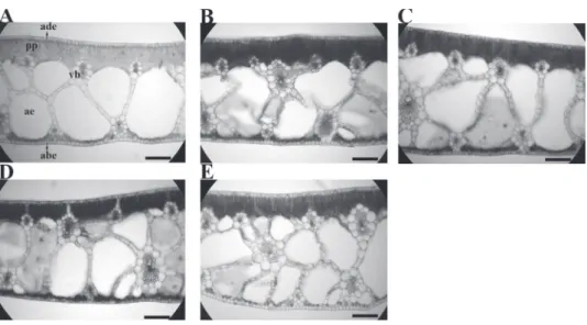
thickness was increased by 19% in the 1.0 mg L
-1in
comparison to the control
(Table I and Figure 2),
but there were no significant modifications to the
leaf epidermis, palisade and spongy parenchyma,
or the palisade/spongy parenchyma rate, in the
presence of Pb (Table I). The distance between
the vascular bundles was reduced in 32.05% in the
0.50 mg L
-1lead concentration and in all higher
concentrations (Table I). The proportion of leaf
aerenchyma did not exhibit any differences related
to lead treatments Pb (Table I).
The abaxial leaf surface showed an increase of
15% in stomatal density in the 0.5 mg L
-1, and an
increase of 8.69% in the 1.0 mg L
-1and in all higher
concentrations (Table II). The number of regular
epidermal cells and the stomatal dimensions were
not modified by lead (Table II). The stomatal polar/
equatorial diameter rate (stomatal functionality)
increased by 17.71% in the 1.0 mg L
-1and in the
higher concentrations (Table II). The stomatal
index increased 13.33% in the 1.00 mg L
-1lead
concentration and this was unaltered in the higher
concentrations (Table II).
On the leaf adaxial surface, the stomatal
density increased by 19.51% in the 0.50 mg L
-1lead concentration, and this was unaltered with
higher concentrations (Table II). There were no
Figure 1 - Gas exchanges characteristics of Eichhornia crassipes grown in nutrient solutions under
different lead concentrations. A = photosynthesis, B = stomatal conductance, C = transpiratory rate, D = Ci/Ca rate. bars= standard error.
PB ADE (μm) ABE (μm) MP (μm) (μm)PP (μm)SP PP/SP VBD(μm) AEP(%) 0.00 09.9 a 12.1a 285.8b 67.61a 236.2a 0.3a 103.7 a 38a 0.50 09.7 a 12.2a 273.7b 66.80a 209.9a 0.3a 082.3 b 26a 1.00 09.3 a 13.2a 325.7a 64.13a 267.2a 0.2a 085.2 b 35a 2.00 09.7 a 12.7a 299.9a 68.16a 214.8a 0.3a 076.4 b 87a 4.00 11.1 a 13.9a 324.6a 69.15a 277.8a 0.2a 069.6 b 32a
TABLE I
Leaves quantitative anatomical characteristics in cross sections of water hyacinth
(Eichhornia crassipes) grown under different lead concentrations (mg L-1).
Means followed by same letters in columns did not differ by Scott-Knott test at P<0.05.
Abaxial surface
Lead SN CN PD (μm) ED (μm) SD SF SI (%)
0.00 08.0 c 63.0 a 44.38a 25.4 a 102.7 c 1.70 b 13 b
0.50 09.2 b 73.0 a 42.36a 23.4 a 118.2 b 1.80 b 13 b
1.00 10.0 a 66.0 a 46.07a 22.8 a 128.4 a 2.03 a 15 a 2.00 10.0 a 64.4 a 48.34a 23.8 a 128.4 a 2.03 a 16 a 4.00 10.0 a 65.4 a 45.71a 22.2 a 128.4 a 2.06 a 15 a
Adaxial surface
Lead SN CN PD (μm) ED (μm) SD SF SI (%)
0.00 8.20 b 65.4 a 44.6 a 26.8 a 105.3 b 1.70 b 13 b
0.50 9.80 a 75.0 a 46.6 a 23.7 b 125.9 a 1.50 b 13 b
1.00 9.60 a 63.4 a 45.0 a 24.6 b 123.3 a 1.80 b 15 a 2.00 9.80 a 65.0 a 45.0 a 21.0 c 125.9 a 2.20 a 15 a 4.00 9.80 a 65.2 a 47.3 a 22.1 c 125.9 a 2.20 a 15 a
Figure 2 - Leaves anatomical modifications of Eichhornia crassipes grown in nutrient solutions
under different lead concentrations. ade= adaxial epidermis, abe = abaxial epidermis, pp = palysade parenchyma, ae = aerenchyma chamber, vb = vascular bundle. A = 0.00 mg L-1, B= 0.50 mg L-1, C =
1.00 mg L-1, D = 2.00 mg.L-1, E = 4.00 mg.L-1. bars = 100 μm.
TABLE II
Leaves quantitative anatomical characteristics in paradermal sections of water hyacinth
(Eichhornia crassipes) grown under different lead concentrations (mg L-1).
Means followed by same letters in columns did not differ by Scott-Knott test at P<0.05.
SN = number of stomata by field; CN = number of regular epidermal cells by field; PD = stomatal polar diameter; ED = stomatal equatorial diameter; SD = stomatal density (stomata by mm2); SF = stomatal
lead (Table II). However, lead promoted reductions
on the stomatal equatorial diameter, which was
reduced by 8.15% in concentrations of 0.50 and
1.00 mg L
-1and 14.52% in the higher concentrations
(Table II). These reductions increased by 40.90%
the stomatal functionality in the 2.00 and 4.00 mg
L
-1concentrations (Table II). The stomatal index
increased by 13.33% in the 1.00 mg L
-1and in
concentrations thereafter (Table II).
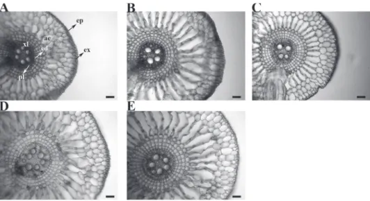
Root epidermal thickness and proportion
of aerenchyma did not exhibit any changes in
the presence of lead (Table III). However, the
endodermal thickness increased 31.52% in the
1.00 mg L
-1and higher concentrations (Table III),
whilst the cortical thickness increased by 52.07%
in the 2.0 mg L
-1and higher concentrations (Table
III). In 0.50 mg L
-1and higher concentrations the
IVC was reduced by 33.91%, this reduction being
related to the increase in the number of tracheary
elements in the xylem that was observed in all lead
concentrations (Table III, Figure 3). The exodermal
thickness increased by 24.36% in the 1.00 mg L
-1and higher concentrations, and phloem thickness
increased 39.12% (Table III).
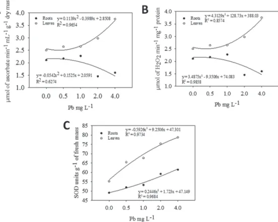
The antioxidant system of
E. crassipes showed
some responses related to the presence of lead
(Figure 4). The APX activity in leaves and roots
was modified only in the 2.00 mg L
-1or higher
lead concentrations (Figure 4A). In the roots, the
CAT activity did not alter, but in the leaves in
all lead concentrations an increased activity was
found (Figure 4B). The SOD activity increased in
leaves and roots of E. crassipes
in the 1.00 mg L
-1concentration or higher, but, in roots, a decrease
was encountered in the 4.00 mg L
-1concentration.
DISCUSSION
The photosynthetic system response encountered
in E. crassipes in the presence of lead was different
from other species described in literature. According
to Pinchasov et al. (2006) lead may promote
reductions in the photosynthetic rate. Lead also
reduces the chlorophyll biosynthesis in some
plants such as Brassica rapa
(Cenkci et al. 2010)
and
Phaseolus vulgaris
(Hamid et al. 2010). In
addition to the damage on chlorophyll biosynthesis,
prejudicial effects on photosynthesis can be related
to the formation of reactive oxygen species (ROS),
which reduce membrane stability in chloroplasts as a
consequence of lipid peroxidation (Stoeva and Bineva
2003). Such effects on chloroplast membranes are
very common in plants under lead stress (Verma and
Dubey 2003). However,
E. crassipes reduced the
effects of lead stress by increasing the antioxidant
enzyme activities. This kind of response is essential
in lead tolerant plants as described by Verma and
Dubey (2003) and Singh et al. (2010). With more
active enzymes in the leaves, E. crassipes plants
were able to cope with lead deleterious effects and
maintain the photosynthetic capacity.
E. crassipes plants were not only able to maintain
photosynthesis in the presence of lead, but they also
increased the photosynthetic rate. This increase in
photosynthesis must be related to regulatory factors.
The photosynthetic rate is regulated by different
factors, but the two most relevant are the radiation
and CO
2(Zhou and Han 2005). Since the radiation
was standardized for all treatments in the IRGA
chamber at 1000 μmol m
-2s
-1, the main factor that
contributed to the increase in photosynthesis was
the CO
2capture capacity of the plants; and the
leaf capacity for CO
2capture is associated with
the modifications in leaf anatomical structure, i.e.
characteristics such as: stomatal density, index,
functionality and the total leaf thickness.
The stomatal density is one of the most
important plant characteristics related to CO
2capture, and in stress conditions such as water
stress, the stomatal density can increase its values
in the most efficient plants (Grisi et al. 2008). An
increase in the stomatal density in the presence
of lead was reported in Plantago major, but with
reduced stomatal conductivity (Kosobrukhov et al.
Figure 3 - Root cross sections in Eichhornia crassipes grown in nutrient solutions containing
different lead concentrations. ep = epidermis, ex = exodermis, er = aerenchyma chamber, ed = endodermis, xl = xylem, pl = phloem. A = 0.00 mg L-1, B = 0.50 mg L-1, C = 1.00 mg L-1, D =
2.00 mg.L-1, E = 4.00 mg.L-1. bars = 100 μm.
Lead AEP
(%)
EP
(μm) (μm)EX (μm)ED IVC (μm)CT (μm)PL
0.00 20 a 19.90 a 15.1 b 542.24 b 2.30 a 26.27 b 26.99 b 0.50 19 a 19.95 a 16.8 b 494.23 b 1.68 b 26.62 b 25.23 b
1.00 14 a 18.92 a 17.9 a 555.22 b 1.52 b 31.79 a 30.29 a 2.00 17 a 19.82 a 18.4 a 722.90 a 1.53 b 32.67 a 35.10 a 4.00 18 a 18.89 a 19.8 a 751.56 a 1.59 b 31.71 a 34.85 a
TABLE III
Root anatomical characteristics of water hyacinth (Eichhornia
crassipes) grown under different lead concentrations (mg L-1).
Means followed by same letters in columns did not differ by Scott-Knott test at P<0.05.
AEP = aerenchyma proportion in cortex, EP = thickness of epidermis, EX = thickness of exodermis, ED = thickness of endoermis, IVC = Carlquist vulnerability index (mean tracheary element diameter/number of tracheary elements), CT = thickness of the cortex, PL = thickness of the phloem.
stomatal density but also the stomatal index in the
presence of lead. Effectively, our study shows the
importance of stomatal characteristics to maintain
photosynthetic capacity of lead tolerant plants by
increasing the CO
2capture capacity and thus permit
an increase in photosynthesis in the presence of lead.
An increase in mesophyll thickness increases
the leaf storage capacity for CO
2. As the aerenchyma
proportion in leaves was the same in the presence
of lead, with higher leaf thickness, the total
aerenchyma was increased. This tissue is directly
related to gas storage in plant organs most likely
with the total CO
2captured by stomata being
stored in aerenchyma and slowly utilized by the
chlorenchyma in the photosynthetic process. This
hypothesis is supported by the increase in the Ci/Ca
rate, showing larger proportions of the CO
2in the
E. crassipes
plants growing under lead influence.
According to Zhou and Han (2005) a higher Ci/Ca
rate represents a larger amount of CO
2in the leaves.
effective for the CO
2uptake and transpiration control.
In our study,
E. crassipes showed an enhanced
capacity to capture and store CO
2, thus increasing the
photosynthetic rate. The absence of modifications in
the leaf epidermis, palisade and spongy parenchyma
in the presence of lead resulted in a good development
of the leaf tissues with no evidence of lead toxicity.
Smaller distances between the vascular bundles
results in increased amounts of vascular tissue and a
higher capacity to conduct water and photoassimilates
from leaves to the sink organs in plants.
One of the most important anatomical
charac-teristic of the roots of aquatic plants is the proportion
of aerenchyma; and the root aerenchyma can increase
in stress tolerant plants (Pereira et al. 2008, Souza et
al. 2009, 2010). Lead can reduce cell growth in plant
roots due to its toxicity (Kozhevnikova et al. 2009),
and this can cause deformations of plant tissues and
structures in roots (Xu et al. 2007). As an apoplastic
barrier, the epidermis is the first tissue in roots
that has to cope with the effects of toxic elements.
The absence modifications to the root epidermis in
the presence of lead in E. crassipes is one of the
characteristics related to the tolerance of this species.
In
E. crassipes
, the antioxidant enzymes
increased both in the roots and leaves, and this
shows the great importance of this system for stress
tolerance and the protection of the photosynthetic
system. This is a common response in plants
growing in the presence of lead, as described
for rice by Verma and Dubey (2003) and for
wheat, by Liu et al. (2010). But, in the roots of
E.
crassipes
this system was only slightly stimulated.
The lysigenous aerenchyma is dependent of the
production of reactive oxygen species (Seago
et al. 2005, Gunawardena 2008), and in water
Figure 4 - Antioxidant enzyme activities of Eichhornia crassipes grown in nutrient solutions containingstress tolerant plants the antioxidant system in the
roots can undergo reduction (Pereira et al. 2010).
Consequently, a reduced antioxidant enzyme activity
in the roots can be related to the maintenance of the
aerenchyma proportion.
The capacity for lead hyper-accumulation
was reviewed by Schoenhals et al. (2009), and
this accumulation is more intense in roots than in
the shoots (Gonçalves Júnior et al. 2008). Lead
accumulation in roots can be important for plant
stress tolerance, because it reduces the effects
on the photosynthetic system in the leaves. The
endodermis is the most important apoplastic barrier
in roots, blocking the translocation of the lead to
shoots. In
E. crassipes,
the endodermis thickness
increased, thus reducing the lead flux to shoots.
The IVC is related to vascular system
ef-ficiency, and a reduction of the IVC increases the
water transport in roots, and has been found to
increase in stress tolerant plants (Carlquist 1975,
Pereira et al. 2008, Souza et al. 2009).
E. crassipes
plants showed a capacity to increase the water and
nutrient transport from roots to shoots, and this
can be related to stress tolerance. The increase in
phloem in the roots under lead effects may also
be a stress tolerance mechanism, because it can
increase the photoassimilate flux to the roots,
leading to higher root growth. Stress tolerance of
E. crassipes to lead may have similar effects to
those reported for arsenic tolerance to this species,
as described by Pereira et al. (2011).
Therefore, the water hyacinth can cope with
lead stress without damage to its structure or
physiology. The presence of lead increased the
photosynthetic rate, which was associated with
an increase in antioxidant system enzymes and
CO
2capture mechanisms. Leaf structure in
E.
crassipes
in the presence of lead increased the
CO
2capture mechanisms and did not show any
toxicity stress. Likewise, the roots in
E. crassipes
did not show toxicity stress but rather exhibited
favorable characteristics in the presence of lead.
ACKNOWLEDGMENTS
We would like to extend our gratitude to Dr. Peter
Edward Gibbs for the critical reading of the work, the
Conselho Nacional de Desenvolvimento Científico
e Tecnológico (CNPq) and Fundação de Amparo à
Pesquisa do Estado de Minas Gerais (FAPEMIG) for
financial support.
RESUMO
Este estudo teve como objetivo verificar a tolerância do aguapé ao chumbo e verificar as modificações anatômicas e fisiológicas decorrentes. As plantas de aguapé foram cultivadas em solução nutritiva com cinco diferentes concentrações de chumbo sendo: 0,00; 0,50; 1,00; 2,00 e 4,00 mg L-1
REFERENCES
BOR M, ÖZDEMIR F AND TÜRKAN I. 2003. The effect of salt
stress on lipid peroxidation and antioxidants in leaves of sugar beet Beta vulgaris L. and wild beet Beta maritima L. Plant Sci164: 77-84.
CARLQUIST S. 1975. Ecological strategies of xylem evolution. University of California, California, 259 p.
CASTRO EM, PEREIRA FJ AND PAIVA R. 2009. Histologia
Vegetal: Estrutura e Função de Órgãos Vegetativos.
UFLA, Lavras, 234 p.
CENKCI S, CIGERCI IH, YILDIZ M, ÖZAY C, BOZDAG A AND
TERZI H. 2010. Lead contamination reduces chlorophyll biosynthesis and genomic template stability in Brassica rapa L. Environ Exp Bot 67: 467-473.
DHANKHER OP, LI Y, ROSEN BP, SHI J, SALT D, SENECOFF
JF, SASHTI NA AND MEAGHER RB. 2002. Engineering tolerance and hyperaccumulation of arsenic in plants by
combining arsenate reductase and γ-glutamylcysteine synthetase expression. Nat Biotechnol 20: 1140-1145.
FAISAL M AND HASNAIN S. 2003. Synergistic removal of Cr
(VI) by Eichhornia crassipes in conjunction with bacterial
strains. Pak J Biol Sci6: 264-268.
GIANNOPOLITIS CN AND RIES SK. 1977. Superoxide dismutases: I. Occurrence in higher plants. Plant Physiol 59: 309-314.
GONÇALVES JÚNIOR AC, LINDINO CA, DA ROSA MF,
BARRICATTI R AND GOMES GD. 2008. Removal of toxic
heavy metals cadmium, lead and chromium from swine biofertilizer, using an aquatic macrophyte (Eichhornia crassipes) as a bioindicator. Acta Sci-Technol 30: 9-14.
GRATÃO PL, PRASAD MNV, CARDOSO PF, LEA PJ AND AZEVEDO RA. 2005. Phytorremediation: Green technology for the clean up of toxic metals in environment. Braz J Plant Physiol 17: 53-64.
GRISI FA, ALVES JD, CASTRO EM, OLIVEIRA C, BIAGIOTTI
G AND MELO LA. 2008. Leaf anatomical evaluations in
'Catuaí' and 'Siriema' coffee seedlings submitted to water stress. Cienc Agrotec 32: 1730-1736.
GUNAWARDENA AH. 2008. Programmed cell death and tissue remodelling in plants. J Exp Bot59: 445-451.
HAMID N, BUKHARI N AND JAWAID F. 2010. Physiological
responses of Phaseolus vulgaris to different lead
concentrations. Pak J Bot 42: 239-246.
HOAGLAND DR AND ARNON DI. 1940. Crop production
in artificial culture solutions and in soils with special reference to factors influencing yield absorption of inorganic nutrients. Soil Sci50: 463-483.
JOHANSEN DA. 1940. Plant Microtechinique. 2nd ed., Mc
Graw-Hill, New York, 523 p.
KOSOBRUKHOV A, KNYAZEVA I AND MUDRIK V. 2004. Plantago major plants responses to increase content of lead in soil: Growth and photosynthesis. Plant Growth Regul 42: 145-151.
KOZHEVNIKOVA AD, SEREGIN IV, BYSTROVA EI, BELYAEVA AI, KATAEVA MN AND IVANOV VB. 2009. The Effects of
Lead, Nickel, and Strontium Nitrates on Cell Division and Elongation in Maize Roots. Russ J Plant Physl56: 242-250.
LIU D, LIU X, CHEN Z, XU H AND DING X. 2010. Bioaccumulation
of Lead and the Effects of Lead on Catalase Activity,
Glutathione Levels, and Chlorophyll Content in the
Leaves of Wheat. Commun. Soil Sci Plan41: 935-944.
MADHUSUDHAN R, ISHIKAWA T, SAWA Y, SHIGEOKA S AND
SHIBATA H. 2003. Characterization of an ascorbate
peroxidase in plastids of tobacco BY-2 cells. Physiol Plantarum 117: 550-557.
MAHMOOD Q, ZHENG P, SIDDIQI MR, ISLAM E, AZIM MR AND
HAYAT Y. 2005. Anatomical studies on water hyacinth
(Eichhornia crassipes (Mart.) Solms) under the influence of textile wastewater. J Zhejiang Univ Sc A 6B: 991-998. NAKANO Y AND ASADA K. 1981. Hidrogen peroxide is
scavenged by ascorbate specific peroxidase in spinach chloroplast. Plant Cell Physiol22: 867-880.
OLIVEIRA JA, CAMBRAIA J, CANO MAO AND JORDÃO CP. 2001.
Cadmium absorption and accumulation and its effects on
the relative growth of water hyacinths and salvinia.Braz J
Plant Physiol 13: 329-341.
PEREIRA FJ, CASTRO EM, OLIVEIRA C, PIRES MF AND PASQUAL
M. 2011. Mecanismos anatômicos e fisiológicos de plantas de aguapé para a tolerância à contaminação por arsênio. Planta Daninha 29: 259-267.
PEREIRA FJ, CASTRO EM, SOUZA TC AND MAGALHÃES PC. 2008. Evolução da anatomia radicular do milho 'Saracura'
em ciclos de seleção sucessivos.Pesqui Agropecu Bras 43: 1649-1656.
PEREIRA FJ, MAGALHÃES PC, SOUZA TC, CASTRO EM AND ALVES JD. 2010. Atividade do sistema antioxidante e desenvolvimento de aerênquima em raízes de milho ‘Saracura’. Pesqui Agropecu Bras 45: 450-456.
PINCHASOV Y, BERNER T AND DUBINSKY Z. 2006. The effect of lead on photosynthesis, as determined by photoacoustics in Sinechococcus leopoliensis (Cyanobacteria).Water air soil poll 175: 117-125.
RAHMAN MA AND HASEGAWA H. 2011. Aquatic arsenic: Phytoremediation using floating macrophytes. Chemosphere 83: 633-646.
SCHOENHALS M, OLIVEIRA VA AND FOLLADOR FAC. 2009.
Lead remotion of automotive batteries recycling industry
wastewater by the aquatic macrofit Eichhornia crassipes. Eng Amb 6: 055-072.
SEAGO JL, MARSH LC, STEVENS KJ, SOUKUP A, VOTRUBOVÁ
O AND ENSTONE DE. 2005. A Re-examination of the Root Cortex in Wetland Flowering Plants With Respect to Aerenchyma. Ann Bot London 96: 565-579.
SHARMA P AND DUBEY RS. 2005. Lead toxicity in plants. Braz J Plant Physiol 17: 35-52.
SINGH R, TRIPATHI RD, DWIVEDI S, KUMAR A, TRIVEDI PK AND
CHACRABARTY D. 2010. Lead bioaccumulation potential of an aquatic macrophyte Najas indica are related to
antioxidant system. Bioresource Technol101: 3025-3032.
SOUZA TC, CASTRO EM, PEREIRA FJ, PARENTONI SN AND
MAGALHÃES PC. 2009. Morpho-anatomical characterization
of root in recurrent selection cycles for flood tolerance of
SOUZA TC, MAGALHÃES PC, PEREIRA FJ, CASTRO EM, SILVA
JÚNIOR JM AND PARENTONI SN. 2010. Leaf plasticity in successive selection cycles of 'Saracura' maize in response
to periodic soil flooding. Pesqui AgropecuBras 45: 16-24.
STOEVA N AND BINEVA T. 2003. Oxidative changes and photosynthesis in oat plants grown in As-contaminated soil. Bulg J Agric Sci 29: 87-95.
VERMA S AND DUBEY RS. 2003. Lead toxicity induces lipid peroxidation and alter the activities of antioxidant enzymes in growing rice plants. Plant Sci 164: 645-655.
XU Y, YAMAJI N, SHEN R AND MA JF. 2007. Sorghum Roots are Inefficient in Uptake of EDTA-chelated Lead. Ann Bot London 99: 869-875.
ZHOU YM AND HAN SJ. 2005. Photosynthetic response
and stomatal behaviour of Pinus koraiensis during the