Influence of carvacrol and thymol on the physiological attributes, enterotoxin
production and surface characteristics of
Staphylococcus aureus
strains
isolated from foods
E.L. Souza
1, C.E.V. Oliveira
1, T.L.M. Stamford
2, M.L. Conceição
1, N.J. Gomes Neto
1 1Laboratório de Microbiologia de Alimentos, Departamento de Nutrição, Centro de Ciências da Saúde, Universidade Federal da Paraíba, João Pessoa, PB, Brazil.
2
Laboratório de Microbiologia de Alimentos, Departamento de Nutrição, Centro de Ciências da Saúde, Universidade Federal de Pernambuco, Recife, PE, Brazil.
Submitted: April 16, 2011; Approved: July 02, 2012.
Abstract
This study evaluated the influence of the phenolic compounds carvacrol (CAR) and thymol (THY) on some physiological characteristics and on the modulation of the secretion of some staphylococcal virulence factors, that is, coagulase and enterotoxin. This study also investigated possible mecha-nisms for the establishment of the anti-staphylococcal activity of these compounds. Sublethal con-centrations (0.3 and 0.15mL/mL) of CAR and THY inhibited the activity of the enzymes coagulase and lipase and led to a decrease in salt tolerance. At the tested sublethal concentrations, both CAR and THY led to a total suppression of enterotoxin production. The loss of a 260-nm-absorbing mate-rial and an efflux of potassium ions occurred immediately after the addition of CAR and THY at 0.6 and 1.2mL/mL and increased up to 120 min of exposure. Electron microscopy of cells exposed to CAR and THY (0.6mL/mL) revealed that individual cells appeared to be deformed, with projections of cellular material. The observations of leakage of cellular material and an altered cell surface sug-gest that gross damage to a cell’s cytoplasmic membrane, which results in a disruption in protein se-cretion, could be responsible for the anti-staphylococcal properties of CAR and THY.
Key words: phenolic compounds, physiological suppression, staphylococcal enterotoxins, viru-lence.
Introduction
Staphylococcus aureusis a common pathogen that is associated with serious community and nosocomial infec-tions and is known as major cause of food poisoning due to the production of enterotoxins (Pereiraet al., 2009). The occurrence ofS. aureustoxicity depends on the capability of the strain to survive, multiply under a variety of condi-tions and produce extracellular toxic compounds. Hemo-lysins, nuclease, coagulase, lipase and enterotoxins are among the extracellular toxins and enzymes produced byS. aureus(Shaeet al., 2005).
Staphylococcal enterotoxins (SEs) comprise a group of heat stable and serologically diverse proteins. Twenty different types of SEs,i.e., SEA-SEE, SEG-SER and SEU,
have already been discovered; however, SEA-SEEs are the most common enterotoxins involved in staphylococcal food poisoning (Jørgensenet al., 2005).
Because staphylococcal foodborne intoxication rep-resents a public health problem worldwide, the develop-ment of strategies to control the survival and growth ofS. aureusin foods is of great interest (Barroset al., 2009). Par-ticularly, the increased demand for safe and natural foods has motivated researchers to investigate the antimicrobial efficacies of many natural compounds against food-related pathogenic microorganisms (Bentoet al., 2009). The cur-rent trend of a negative consumer perception of chemical preservatives has prompted a particularly increased interest in the use of essential oils as antimicrobial compounds in
Send correspondence to E.L. Souza. Departamento de Nutrição, Centro de Ciências da Saúde, Universidade Federal da Paraíba, Campus I, Cidade Universitária, 58051-900 João Pessoa, PB, Brazil. E-mail: [email protected].
food preservation (Souza et al., 2007; Gutierrez et al., 2008).
The essential oil ofOriganum vulgare L. (OVEO), popularly known as oregano, has been found to antagonize several food-related bacteria (Nostroet al., 2004; Souzaet al., 2006). An early study focusing on the antimicrobial properties of OVEO found that this substance strongly in-hibits the growth and some of the metabolic characteristics of food-isolatedS. aureusstrains, including coagulase and lipase activities and salt tolerance (Barros et al., 2009). Some researchers have reported a high content of phenolic compounds in OVEO, mostly carvacrol (CAR) and thymol (THY), which are most likely responsible for the prominent antimicrobial effects (Rhayouret al., 2003; Burt, 2004).
Although previous researchers have described the antimicrobial activities of CAR and THY against food-related microorganisms (Lambertet al., 2001; Ulteeet al., 2002), there is a lack of information about their effect on the physiological characteristics, including some virulence attributes, ofS. aureus. The aim of this study was to verify whether sublethal concentrations of the phenolic com-pounds CAR and THY, the major components of OVEO, exert an influence on some of the physiological characteris-tics, including enterotoxin production, ofS. aureusstrains isolated from foods and to investigate the possible mecha-nisms for the establishment of the anti-staphylococcal ac-tivities of these compounds.
Methods
Test organisms
S. aureusQCD,S. aureusQCE andS. aureusQCF, which were obtained from the Microorganism Collection, Laboratory of Food Microbiology, Health Sciences Center, Federal University of Paraíba (João Pessoa, Brazil), were used as test microorganisms. These strains were isolated from different unripened cheese samples using standard procedures (Bennetet al., 1986; Vnaderzant and Splitts-toesser, 1992). Stock cultures were maintained on Nutrient Agar – NA (Sigma, France) slants under refrigeration (7±1 °C) conditions.
The inocula used in the assays were obtained from cultures grown overnight on NA slants (37 °C). A loopful of the culture was diluted in a sterile saline solution (0.85 g/100 mL) to obtain a final concentration of approxi-mately 108colony forming units per mL (cfu/mL) adjusted to the turbidity of a 0.5 McFarland standard tube.
Phenolic compounds
The phenolic compounds, CAR and THY, were sup-plied by Sigma Aldrich (Sigma, France). Stock solutions were prepared in Nutrient Broth - NB (Sigma, France) us-ing bacteriological agar (0.15 g/100 mL) as a stabilizus-ing agent (Benniset al., 2004). Using the test strains included in this study, previous studies found values for minimum
inhibitory concentrations and minimum bactericidal con-centrations of 0.6 and 1.2mL/mL, respectively, for both CAR and THY (Oliveiraet al., 2010).
Assays for enzymatic activities and salt tolerance
Lipase activity was estimated using Agar Salty Tween - AST (Nostroet al., 2001a), which contained: (g/L) peptone 10.0; NaCl 75.0; CaCl2 H2O 0.10; Tween 80 10 (pH 7.2). Bacterial inocula (100mL) grown overnight in NB (37 °C) and diluted (10-1-10-4) in sterile peptone water (0.1 g/100 mL) were plated onto AST. Sublethal concentra-tions of CAR or THY (0.3 and 0.15 mL/mL) were then added to the AST followed by incubation at 37 °C for 24 h. After the incubation period, the number of lipase positive colonies (cfu/mL) on each AST plate was counted and compared to the number of positive colonies found on AST supplemented with CAR and THY. The results are ex-pressed as a percentage of the inhibition of the lipase activ-ity.
For testing coagulase activity, a 100 mL aliquot of inoculum (approximately 107 cfu/mL) was transferred to Brain Heart Infusion - BHI (Sigma, France) broth supple-mented with sublethal concentrations of CAR or THY (0.3 and 0.15mL/mL) and incubated at 37 °C for 24 h. After the incubation period, coagulase activity (a tube coagulase test) was determined using a standard procedure (Walshet al., 2003), and the results were expressed based on the size (-; + to ++++) of the formed plasma clot [free coagulase forms thrombin, which converts fibrinogen to fibrin and results in plasma clotting (observed after 4 h of incubation at 37 °C)]. Tubes lacking CAR or THY were treated similarly to the positive control.
Preliminary experiments were performed to evaluate the salt tolerance of the strains. For these experiments, 100 mL aliquots of the cultures were spread-plated onto sterile NA and NA supplemented with NaCl (5 to 100 g/L; NA-NaCl) to determine the NaCl concentration that mod-estly inhibited the colony-forming ability of the cultures. Suspensions of bacteria were exposed overnight to sublethal concentrations of CAR or THY (0.3 and 0.15mL/mL) in NB (37 °C). Next, 100mL of culture was di-luted (10-1- 10-4) with sterile peptone water (0.1 g/100 mL) and plated onto NA-NaCl at 37 °C for 24 h (Carsonet al., 2002). Control tubes lacking CAR or THY were similarly treated. After the incubation period, the number of cfu/mL for each NA-NaCl plate was compared to that found for the control assay, and the results are expressed as the percent-age of cells able to form colonies.
Assay for enterotoxin production
-AOAC RI N° 070404. For this assay, a loopful of an over-night NB culture (37 °C) was suspended in BHI broth sup-plemented with sublethal concentrations of CAR or THY (0.3 and 0.15mL/mL) and incubated for 18 h under static conditions at 37 °C (Oliveiraet al., 2010). After the incuba-tion period, the growth media was centrifuged (3000 g, 4 °C), and an aliquot of the supernatant was taken and sub-mitted to an enterotoxin detection test according to proce-dures described by the manufacturer. The results are ex-pressed in terms of positive (+) and (-) negative enterotoxin production. Tubes lacking CAR or THY were treated simi-larly to the positive control assay.
Assay for potassium ion efflux
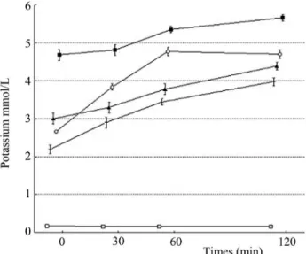
The potassium ion concentration in a cell suspension ofS. aureusQCF was measured after exposure to CAR or THY (0.3 and 0.6 mL/mL) in sterile peptone water (0.1 g/100 mL) for 0, 30, 60 and 120 min. At each pre-established interval, the extracellular potassium concentra-tion was measured using a photometric procedure and a po-tassium kit (Human GmbH, Wiesbaden, Germany) (Oliveiraet al., 2010). Control flasks lacking CAR or THY were treated similarly. The results are expressed as the amount of extracellular free potassium (mmol/L) in the growth medium at each interval of the incubation.
Release of cellular material
The release of cellular material absorbing at 260 nm fromS. aureusQCF was determined in 2 mL aliquots of bacterial inocula containing approximately 108cfu/mL in sterile peptone water (0.1 g/100 mL) supplemented with CAR or THY (0.3 and 0.6mL/mL) at 37 °C. After 0, 30, 60 and 120 min of treatment, the cells were centrifuged (3000 g, 4 °C), and the absorbance (260 nm) of the obtained supernatant was determined using a Biochrom Libra S32/S32 spectrophotometer (Carsonet al., 2002). Control flasks lacking CAR or THY were treated similarly. The re-sults are expressed as the percent of the material absorbing at 260 nm in each interval with respect to the last time inter-val.
The assays for lipase activity, salt tolerance, potas-sium ion efflux and release of cellular material were per-formed in triplicate on three separate occasions, and the results are expressed as averages for each of the assays. The
assays for coagulase activity were performed in triplicate on three separate occasions with consistent results.
Cell morphology using scanning electronic microscopy
After an overnight exposure of S. aureus QCF to CAR or THY (6mL/mL) in BHI broth at 37 °C, the bacterial cells were pre-fixed with glutaraldehyde (2 mL/100 mL) for 2 h at 4 °C and postfixed using an osmium tetroxide so-lution (1 g/100 mL) for 30 min at 30 °C. After each fixation, the cells were washed twice with PBS. The cells were then dried at a critical point in liquid CO2and gold covered by cathodic spraying (fine coat ion sputter JFC-1100, JEOL
Ltd., Tokyo, Japan). Finally, the cells were examined using a scanning electron microscope (JEOL JSM-5600 LV, PAIS) as previously described (Benniset al., 2004). Cells not exposed to CAR and THY were submitted to the same procedures as described for the controls.
Results
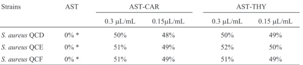
The proportion ofS. aureus cells presenting lipase negative colonies after exposure to sublethal concentra-tions of CAR or THY was monitored (Table 1). Incubation of the cultures with sublethal concentrations of both tested phenolic compounds revealed a strong inhibition of lipase activity. CAR or THY at 0.3 and 0.15mL/mL provided an inhibition of lipase activity of over 50% with all test strains. The effect of CAR and THY on the coagulase activity ofS. aureuscells is shown in Table 2. At 0.3mL/mL, the phenolic compounds led to a large reduction in coagulase activity, that is, no clotting was evident after incubating the cultures in the presence of rabbit plasma. When CAR or THY was added to the growth medium at 0.15mL/mL, a less significant reducing effect was noted in cells, that is, ++ (level 2) or +++ (level 3) levels of coagulase activity.
The growth of S. aureus cells exposed to CAR or THY in salt-supplemented agar was assessed to verify the influence of these compounds on salt tolerance (Table 3). The addition of sublethal concentrations of CAR or THY clearly reduced the colony-forming ability of the cells in the selective medium. Exposure to CAR or THY at 0.3mL/mL totally suppressed the capability of the cells to form colonies on NA-NaCl. At the lower concentration (0.15mL/mL), the tested phenolic compounds also led to a
Table 1- Proportion ofS. aureuscells presenting lipase negative colonies on Salty Tween agar (STA) and Salty Tween agar supplemented with CAR or THY at sublethal concentrations.
Strains AST AST-CAR AST-THY
0.3mL/mL 0.15mL/mL 0.3mL/mL 0.15mL/mL
S. aureusQCD 0% * 50% 48% 50% 49%
S. aureusQCE 0% * 51% 49% 52% 50%
reduction in colony formation on NA-NaCl. However, the reduction was to a lesser extent (8-16%).
The influence of CAR and THY at sublethal concen-trations on the enterotoxin production ofS. aureusQCF was evaluated. Early assays showed that S. aureusQCF presented a greater capability to produce enterotoxins in comparison to the other strains included in this study. Cul-tures ofS. aureusQCF that grew in the presence of CAR or THY at both tested concentrations exhibited a total inhibi-tion of enterotoxin producinhibi-tion.
In an attempt to elucidate the cause of the anti-staphy-lococcal effect, the loss of 260-nm-absorbing material and the release of potassium ions from cells exposed to CAR or THY at 0.3 and 0.6mL/mL was evaluated (Table 4 and Fig-ure 1). The OD260 nmof theS. aureusQCF cell filtrates ex-posed to CAR or THY displayed increasing values during the evaluated times. The efflux of potassium ions from the
S. aureuscells occurred immediately after the addition of the tested phenolic compounds and followed a steady in-creasing release during the evaluated intervals. CAR
pro-Table 2- Effect of sublethal concentrations of CAR or THY on the coagulase activities of theS. aureusstrains.
Strains Control CAR THY
0.3mL/mL 0.15mL/mL 0.3mL/mL 0.15mL/mL
S. aureusQCD ++++ - ++ - +++
S. aureusQCE ++++ - ++ - +++
S. aureusQCF ++++ - ++ - ++
CAR: carvacrol; THY: thymol.
Table 3- Proportion ofS. aureuscells able to form colonies on NA supplemented with 75 g NaCl/L (NA-NaCl) after exposure to CAR or THY at sublethal concentrations.
Strains Control CAR THY
0.3mL/mL 0.15mL/mL 0.3mL/mL 0.15mL/mL
S. aureusQCD 100% 0% 12% 0% 10%
S. aureusQCE 100% 0% 8% 0% 11%
S. aureusQCF 100% 0% 9% 0% 16%
CAR: carvacrol; THY: thymol; AST-C: Agar Salt-Tween supplemented with CAR; AST-T: Agar Salt-Tween supplemented with THY.
Table 4- Rate of release of 260-nm-absorbing material fromS. aureusQCF cells exposed to CAR or THY.
Time (min) CAR THY
0.3mL/mL A260 (%)
0.6mL/mL A260 (%)
0.3mL/mL A260 (%)
0.6mL/mL A260 (%)
0 66.01 93.48 36.87 40.67
30 88.71 98.49 61.11 57.09
60 89.30 100 66.56 69.45
120 100 100 100 100
CAR: carvacrol; THY: thymol; AST-CAR: Agar Salt-Tween supplemented with CAR; AST-THY: Agar Salt-Tween supplemented with THY; * All col-onies grown in AST were lipase positive.
vided a higher loss of 260-nm-absorbing material and leakage of potassium ions in comparison to THY. No leak-age of potassium ions was observed whenS. aureuswas grown in media lacking CAR or THY. These results indi-cate that increased membrane permeability is a factor involved in the establishment of the anti-staphylococcal property of the tested compounds.
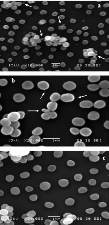
With regards to the fact that the assayed phenolic compounds led to a release of cellular compounds fromS. aureusQCF, the effect of CAR and THY on the bacterial surface cells was investigated. The scanning electron mi-crophotographs showed some morphological damage after exposure to CAR or THY (6mL/mL). The damage caused by CAR (Figure 2a) appeared to be greater than that caused by THY (Figure 2b). Although some background cells did not present alterations, it might be that the surfaces of some cells were consistently deformed, which would reveal the appearance of cellular materials. Cells not exposed to CAR and THY displayed no alterations in cell surface morphol-ogy (Figure 2c). These findings suggest that both tested compounds could exert their anti-staphylococcal activities by damaging the bacterial cell envelope.
Discussion
The effect of the phenolic compounds CAR and THY on some of the physiological characteristics and morpho-logical aspects ofS. aureusstrains isolated from foods was described. It is clear that the assayed compounds strongly interfered with the physiological characteristics, such as cytoplasmic membrane permeability, coagulase activity, salt tolerance and enterotoxin production, of the test strains. Coagulase and lipase assays revealed that CAR and THY suppressed these enzymatic activities inS. aureusand inhibited its production of staphylococcal enterotoxins. Such phenotypic modifications might possibly arise as a re-sult of interactions between the phenolic compounds and enzymes (Nostroet al., 2002). The reductions in the enzy-matic activities (lipase and coagulase) of the cells and in the synthesis of enterotoxins most likely occurred due to a pre-vention of protein secretion, which could have been a con-sequence of changes in the physical nature of the staphylococcal cytoplasmic membrane (Nostroet al., 2002; Shahet al., 2008). Still, it has been reported that the interca-lation of active plant compounds into the cytoplasmic membrane may interfere with the process of membrane-associated signal transduction and impair the function of some membrane proteins, resulting in changes to the archi-tecture and composition of the cell wall (Okubo et al., 1989; Ikigaiet al., 1990; Gustafsonet al., 1998).
Virulence expression in S. aureus is cited as con-trolled by complex regulatory networks, including two-component systems (e.g., AgrAC, SrrAB and SaeRAS) and transcription factors (e.g., SarA and its homologues) (Cheunget al., 2004; Evenet al., 2009). Among these
sential oils and their related compounds towardS. aureus
remains to be studied.
When tested at sublethal concentrations, some plant products may interfere with the secretion of virulence fac-tors in bacteria.Helichrysum italicumextract is effective in inhibiting the enterotoxin production (SEB and SEC) and coagulase and lipase activities ofS. aureuscells (Nostroet al., 2001a; Nostroet al., 2002). Likewise, the extract of
Nepeta catarialeads to a suppression of these enzymatic activities in a number ofS. aureus strains (Nostroet al., 2001b). The extract ofPunica granatumwas found to in-hibit staphylococcal enterotoxin (SEA) production (Braga
et al., 2005).
Sublethal injury of the bacterial cell membrane may interfere with permeability and affect the capability of the cell to adequately osmoregulate or to exclude toxic materi-als (Carsonet al., 2002; Barroset al., 2009). The exposure ofS. aureuscells to CAR or THY strongly reduced the ca-pability of the cells to form colonies on selective media supplemented with salt. A decrease in salt tolerance has been cited as suggesting membrane damage in sublethally injured bacterial cells (Iandolo and Ordal, 1966). These ob-servations are in accordance with the results for the release of 260-nm-absorbing material and the leakage of potassium ions because the addition of CAR or THY to the growth media led to a rapid release of cellular material.
A marked leakage of 260-nm-absorbing material and potassium ions is used as an indication of gross and irre-versible damage to the cytoplasmic membrane (Walshet al., 2003). A significant release of 260-nm-absorbing mate-rial has also been reported as suggesting a loss of nucleic acid through a damaged cytoplasmic membrane (Carsonet al., 2002). Some essential oils and their related compounds induce the leakage of 260-nm-absorbing material (Coxet al., 2000; Coxet al., 2001; Benniset al., 2004).
As expected, the electron microscopy micrographs indicated significant damage toS. aureuscells exposed to CAR or THY. Although background cells did not display apparent surface alterations, the surfaces of some of the in-dividual cells appeared to be deformed, resulting in projec-tions of cellular material. Furthermore, the scanning electron microscopy observations suggest that CAR and THY led to membrane damage, which was accompanied by important surface alterations to theS. aureuscells. These antimicrobial mechanisms could also affect the entire enve-lope of the bacterial cell. Structures such as blebs on the outside surface of cells exposed to CAR or THY may repre-sent a collection of cytoplasmic constituents that were pushed through cracks produced in the cell wall. Our re-sults, which suggest a significant release of OD 260-nm-absorbing material, reinforce this idea. Similar observa-tions have been previously described inEscherichia coli,
Bacillus cereusandSaccharomyces cereviseae(Rhayouret al., 2003; Benniset al., 2004).
Membrane-active compounds are cited as denaturing proteins and as disruptive to membrane structures, leading to cytoplasm leakage, cell lysis and death (Lambertet al., 2001). Earlier studies have revealed that phytochemicals, including phenolic compounds, are able to disrupt and pen-etrate into lipid structures (Rhayouret al., 2003). These findings have reinforced the hypothesis that these com-pounds inhibit microbial viability in a manner similar to membrane-active disinfectants (Evenet al., 2009).
In addition to inhibiting the growth and survival of pathogenic bacteria in foods, new antimicrobials with the capability to inhibit the secretion of virulence attributes could be effective in decreasing the occurrence of food-borne diseases. Our results indicate that CAR and THY strongly interfere with some of the physiological character-istics ofS. aureus, including suppressing enterotoxin pro-duction at sublethal concentrations. The observations of a release of cellular material and an altered cell surface mor-phology suggest that gross damage to a cell’s cytoplasmic membrane could be responsible for the establishment of the anti-staphylococcal effects of CAR and THY. These find-ings indicate that further investigations are required for evaluating the antimicrobial properties of phenolic com-pounds and their influence on the metabolic characteristics of other foodborne pathogenic microorganisms.
Acknowledgments
The authors thank the CNPq (Conselho Nacional de Desenvolvimento Científico, Brazil) for their financial sup-port.
References
Barros JC, Conceição ML, Gomes-Neto NJ, Costa ACV, Si-queira-Júnior JP, Basílio- Júnior ID, Souza EL (2009) Inter-ference ofOriganum vulgareL. essential oil on the growth and some physiological characteristics of Staphylococcus aureus strains isolated from foods. Lebens Winsech u-Technol 42:1139-1143.
Bennett RW, Yeterian M, Smith W, Coles CM, Sassaman M, McClure FD (1986) Staphylococcus aureusidentification characteristics and enterotoxigenicity. J Food Sci 51:1337-1339.
Bennis S, Chami F, Chami N, Bouchikhi T, Remma A (2004) Sur-face alteration ofSaccharomyces cerevisaeinduced by thy-mol and eugenol. Lett Appl Microbiol 38:454-458. Bento RA, Stamford TLM, Campos-Takaki GM, Stamford TCM,
Souza EL (2009) Potential of chitosan fromMucor rouxii
UCP064 as alternative natural compound to inhibitListeria monocytogenes. Braz J Microbiol 40:583-589.
Braga LC, Shupp JW, Cumming C, Jett M, Takahashi JA, Carmo LS, Chartone-Souza E, Nascimento AMA (2005) Pome-grate extract inhibits Staphylococcus aureus growth and subsequent enterotoxins production. J Ethnopharmacol 96:335-339.
Carson CF, Mee BJ, Riley TV (2002) Mechanism of action of
Melaleuca alternifolia (Tea tree) oil on Staphylococcus aureusdetermined by time-kill, lysis, leakage, and salt toler-ance assay and electron microscopy. Antimicrob Agent Chemother 46:191-1920.
Cheung AL, Bayer AS, Zhang G, Gresham H, Xiong YQ (2004) Regulation of virulence determinants in vitro and in vivo
Staphylococcus aureus. FEMS Immunol Med Microbiol 40:1-9.
Cox SD, Mann CM, Markhan JL, Bell HC, Gustafson JE, War-mington JR, Wyllie SG (2000) The mode of antimicrobial action of the essential oil ofMelaleuca alternifolia(tea tree oil). J Appl Microbiol 88:170-175.
Cox SD, Mann CM, Markhan JL, Gustafson JE, Warmington JR, Wyllie SG (2001) Determining the antimicrobial action of tea tree oil. Molecules 6:87-91.
Even S, Charlier C, Nouaille S, Ben Zakour NL, Cretenet M, Cousin FJ, Gautier M, Cocaign-Bouquet M, Loubière P, Le Loir Y (2009)Staphylococcus aureusvirulence expression is impaired byLactococcus lactisin mixed cultures. Appl Environ Microbiol 75:4459-4472.
Gao J, Stewart GC (2004) Regulatory elements of the Staphylo-coccus protein A (Spa) promoter. J Bacteriol 186:3738-3748.
Gustafson JE, Liew YC, Chew S, Markhan J, Bell H, Wyllie HG, Warmington JR (1998) Effects of tea tree oil onEscherichia coli. Lett Appl Microbiol 26:194-198.
Gutierrez J, Barry-Ryan C, Bourke P (2008). The antimicrobial efficacy of plant essential oil combinations and interactions with food ingredients. Int J Food Microbiol 124:91-97. Iandolo JJ, Ordal ZJ (1966) Repair of thermal injury of
Staphylo-coccus aureus. J Bacteriol 91:134-142.
Ikigai H, Toda M, Okubo S, Hara Y, Shimamura T (1990) Rela-tionship between the anti-haemolysin activity and the struc-ture of catechins and theaflavins. Nippon Saikingaku Zasshi 45:913-919.
Jørgensen HJ, Mathisen T, Lovseth A, Omoe K, Qvale KS, Lon-carevic S (2005) An outbreak of staphylococcal food poi-soning caused by enterotoxin H in mashed potato made with raw milk. FEMS Microbiol Lett 252:267-272.
Lambert RJW, Skandamis PN, Coote P, Nychas GJE (2001) A study of the minimum inhibitory concentration and mode of action of oregano essential oil, thymol, and carvacrol. J Appl Microbiol 91:453-462.
Nostro A, Bisignano G, Cannatelli MA, Crisaf G, Germano MP, Alonzo V (2001a) Effects ofHelichrysum italicumextract on growth and enzymatic activity ofStaphylococcus aureus. Int J Antimicrob Agent 17:517-520.
Nostro A, Blanco AR, Cannatelli MA, Enea V, Flamini G, Morelli I, Roccaro AS, Alonzo V (2004) Susceptibility of methi-cillin-resistant staphylococci to oregano essential oil, car-vacrol and thymol. FEMS Microbiol Lett 230:191-195. Nostro A, Cannatelli MA, Crisafi G, Alonzo V (2001b) The effect
of Nepeta cataria extract on adherence and enzyme
produc-tion of Staphylococcus aureus. Int J Antimicrob Agent 18:583-555.
Nostro A, Cannatelli MA, Musolino AD, Procopio F, Alonzo V (2002)Helichrysum italicumextract interferes with the pro-duction of enterotoxins by Staphylococcus aureus. Lett Appl Microbiol 35:181-184.
Nouaille S, Even S, Charlier C, Le Loir Y, Cocaign-Bousquet M, Loubiére P (2009) Transcriptomic responses ofLactococcus lactisin mixed cultures withStaphylococcus aureus. Appl Environ Microbiol 75:4473-4482.
Novick RP (2003) Autoinduction and signal transduction in the regulation of staphylococcal virulence. Mol Microbiol 48:1429-1449.
Okubo S, Ikigai H, Toda M, Shimamura T (1989) The anti-hae-molysin activity of tea and coffee. Lett Appl Microbiol 9:65-66.
Oliveira CEV, Stamford TLM, Gomes-Neto NJ, Souza EL (2010) Inhibition ofStaphylococcus aureusin broth and meat broth using synergies of phenolic compounds and organic acids. Int J Food Microbiol 137:312-316.
Pereira V, Lopes C, Castro A, Silva J, Gibbs P, Teixeira P (2009) Characterization for enterotoxin production, virulence fac-tors, and antibiotic susceptibility of Staphylococcus aureus isolated from various foods in Portugal. Food Microbiol 26:278-282.
Rhayour K, Bouchikhi T, Tantaoui-Elaraki A, Sendide K, Rem-mal A (2003) The mechanism of bactericidal action of oreg-ano and clove essential oils and of their phenolic major com-ponents. J Essent Oil Res 15:286-292.
Shah S, Stapleton PD, Taylor PW (2008) The polyphenol (-)-epi-catechin gallate disrupts the secretion of virulence-related proteins by Staphylococcus aureus. Lett Appl Microbiol 46:181-185.
Shale K, Lues JFR, Venter P, Buys EM (2005) The distribution of
Staphylococcussp. on bovine meat from abattoir deboning rooms. Food Microbiol 22:433-438.
Souza EL, Stamford TLM, Lima EO (2006). Sensitivity of spoil-ing and pathogen-food related bacteria toOriganum vulgare
L. (Lamiaceae) essential oil. Braz J Microbiol 37:527-532. Souza EL, Stamford TLM, Lima EO, Trajano VN (2007)
Effec-tiveness ofOriganum vulgareL. essential oil to inhibit the growth of food spoiling yeasts. Food Cont 18:409-413. Ultee A, Bennik MHJ, Moezelaar R (2002) The phenolic group of
carvacrol is essential for action against the foodborne patho-genBacillus cereus. Appl Environ Microbiol 68:161-1568. Vanderzant C, Splittstoesser DF (1992) Compendium of methods for the microbiological examination of foods. American Public Health Association, Washington D.C., 1219 pp. Walsh SE, Maillard JY, Russel AD, Catrenich CE, Charbonneau
DL, Bartolo RG (2003) Activity and mechanism of action of selected biocidal agents on Gram-positive and negative bac-teria. J Appl Microbiol 94:240-247.