Universidade da Beira Interior
Faculdade de Ciências
Astrocyte-derived GDNF is a potent inhibitor
of microglial activation
Sandra Catarina Moreira Rocha
Covilhã, Julho de 2010
Dissertação de candidatura ao grau de Mestre em
Bioquímica, submetida ao departamento de Química da
faculdade de Ciências da Universidade da Beira Interior
2010
Nome
Sandra Catarina Moreira Rocha
Instituição
Faculdade de Ciências da Saúde da
Universidade da Beira Interior (UBI), Covilhã
Orientadora
Professora Doutora Graça Baltazar, UBI
Co-orientadora Dr. Ana Clara Cristóvão, UBI
Título da Tese
GDNF libertado pelos astrócitos é um potente
inibidor da activação microglial
Thesis Title
Astrocyte-derived GDNF is a potent inhibitor of
microglial activation
2010
Acknowledgment
À Prof. Doutora Graça Baltazar, orientadora deste trabalho, dirijo o meu agradecimento. Obrigada pelo apoio, empenho e espírito crítico, que se tornou fundamental na conclusão dos objectivos propostos. Obrigada também pela enorme disponibilidade manifestada no desenrolar deste trabalho.
Um agradecimento especial à Ana Clara Cristóvão, pela sua exemplar orientação ao longo deste trabalho, o suporte entusiástico, disponibilidade constante nas discussões construtivas e pelo despertar em mim, curiosidade de querer saber mais. Os seus ensinamentos foram decisivos para compreender muitos conceitos. Um grande obrigado pela tua preciosa presença e ajuda nos momentos difíceis.
À Filipa e à Rita, pela transmissão de conhecimento, disponibilidade e acompanhamento na realização deste trabalho. Aos meus colegas do mestrado, o meu muito obrigada por me proporcionarem um ano fabuloso. Para finalizar, gostaria de expressar um sentimento especial ao Cláudio e à minha família. Aos meus pais e irmãos, pela enorme dedicação, estímulo e apoio incondicional ao longo de toda a minha vida. Ao Cláudio pelo incentivo, conforto e motivação a todas as horas. A ti, obrigado pelo constante apoio.
2010
Table of contents
Acknowledgment i Resumo v Abstract viii List of Abbreviations xChapter I
1. Neuroinflammation and neurodegenerative diseases 2
1.1. Parkinson’s disease 3
1.2. The etiology of PD 5
2. Microglia 6
2.1. Microglial Activation 8
2.2. Consequences of microglial activation 10
3. Astrocytes 11
3.1. Factors release by astrocytes 13
3.1.1. GDNF 15
4. Microglia-astrocytes interactions 17
5. Aims of this thesis 19
6. References 20
Chapter II
1. Abstract 25
2. Introduction 26
3. Materials and Methods 27
3.1. Microglial cell cultures 27
2010
3.3. ELISA technique 28
3.4. Transfection of siRNA in astrocytes 28
3.5. Western blot analysis 29
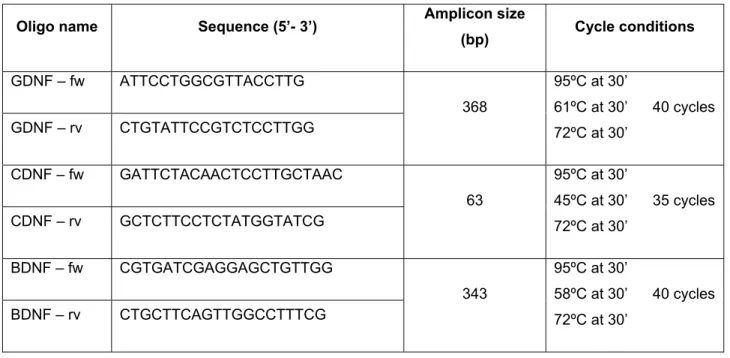
3.6. Total RNA extraction, cDNA synthesis and RT-PCR 29
3.7. Immunocytochemistry 31
3.8. Stimulation of microglia 31
3.9. Phagocytosis assay 31
3.10. Determination of cellular ROS levels 31
3.11. Data analysis and statistics 32
4. Results 32
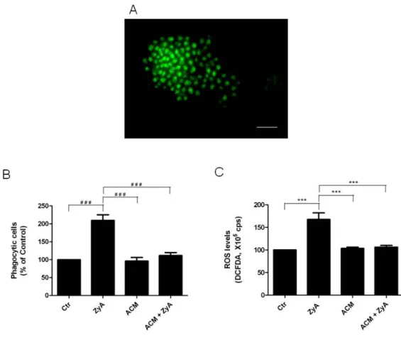
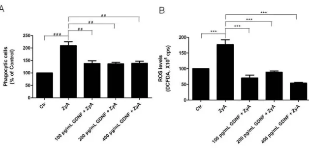
4.1. Effect of ACM on microglial activation induced by Zymosan A 32
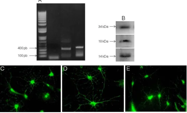
4.2. GDNF, CDNF and BDNF are expressed by astrocytes 33
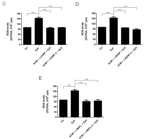
4.3. Effect of neurotrophic factors in microglial activation 34
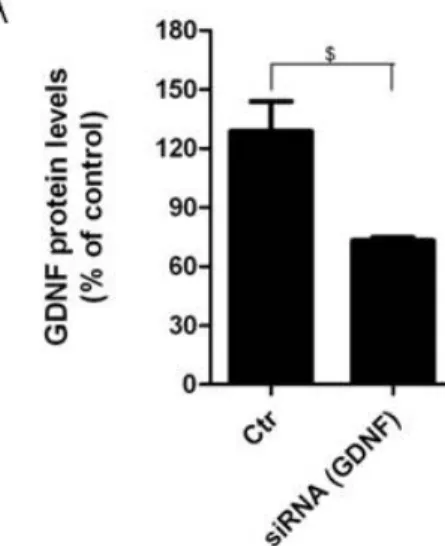
4.4. Conditioned media from astrocytes silenciated for GDNF was
unable to prevent Zymosan A induced microglial activation 36
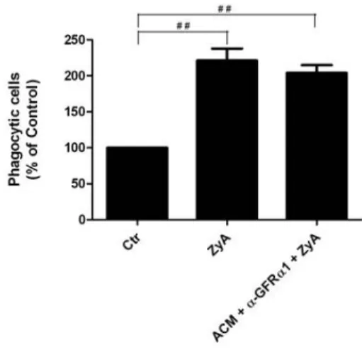
4.5. Protection from Zymosan A induced microglial activation by
ACM: Effect of blocking GFRα1 receptor
39
5. Discussion 40
6. References 44
2010
Resumo
A doença de Parkinson é caracterizada pela perda selectiva de neurónios dopaminérgicos na substantia nigra pars compacta. A origem desta doença não está completamente esclarecida, no entanto têm sido propostas diversas hipóteses em relação aos possíveis factores envolvidos na degeneraração dos neurónios nesta zona. Entre elas, encontra-se a neuroinflamação, que é cada vez mais reconhecida como o principal factor na patogénese da doença de Parkinson, e inúmeras evidências sugerem que as células microgliais são a fonte predominante de inflamação que contribuir para a neurodegeneração dopaminérgica. Os astrócitos desempenham funções vitais na manutenção da função normal do cérebro e diversos estudos sugerem que estes podem actuar como reguladores fisiológicos prevenindo as respostas microgliais inflamatórias excessivas. No entanto, pouco se sabe sobre a forma como os astrócitos modulam a activação microglial. Dada a relevância das interacções astrócitos-microglia na regulação da neuroinflamação, é importante identificar mediadores envolvidos neste processo, os quais podem actuar como agentes anti-inflamatórios naturais no cérebro. Neste trabalho, o principal objectivo foi o estudo dos efeitos de mediadores solúveis libertados pelos astrócitos na activação microglial induzida pelo agente inflamatório Zymosan A, assim como identificar a natureza desses mediadores. Para a determinação do efeito destas moléculas na actividade microglial, culturas primárias de microglia do mesencéfalo ventral foram previamente expostas, a meio condicionado pelos astrócitos (ou meio de cultura – controlo), e posteriormente tratadas com 5 µg/mL de Zymosan A. Estudos anteriormente indicaram que esta concentração de Zymosan provoca um aumento acentuado da actividade fagocítica microglial, bem como da produção de espécies reactivas de oxigénio, sem no entanto induzir a morte destas células. No presente estudo, verificou-se que a pré-incubação das células microgliais com o meio condicionado pelos astrócitos foi capaz de prevenir o aumento da actividade microglial induzida pelo Zymosan A, mantendo a actividade fagocítica bem como os níveis de espécies reactivas de oxigénio em níveis de controlo. Para avaliar a natureza dos mediadores solúveis libertados pelos astrócitos que preveniam a activação
2010
microglial induzida pelo Zymosan A, foram usados anticorpos de forma a bloquear a acção de alguns factores neurotróficos conhecidos pelas suas propriedades neuroprotectoras na substantia nigra. Assim, o meio condicionado pelos astrócitos foi tratado com anti-GDNF, anti-CDNF e anti-BDNF, separadamente, e adicionado às culturas de microglia posteriormente expostas a Zymosan A. O factor neurotrófico derivado de células da glia (GDNF) parece ser um mediador solúvel capaz de prevenir completamente a activação microglial induzida pelo Zymosan A, sendo que os restantes mediadores parecem não exercer qualquer efeito na prevenção da activação microglial. Para confirmar este facto, silenciou-se especificamente o GDNF em culturas de astrócitos, recolhendo-se posteriormente o meio condicionado por estas culturas a aplicando-os a culturas de microglia. Este meio condicionado pelos astrócitos silenciados para o GDNF, não foi capaz de prevenir a activação microglial induzida pelo Zymosan A. Por último, para esclarecer se este efeito era directo e se não haveria outras moléculas a auxiliar o efeito exercido pelo GDNF nas células microgliais, quantificaram-se os níveis de GDNF no meio condicionado pelos astrócitos, e com base nesta quantificação, três concentrações de GDNF, 100 pg/mL, 200 pg/mL e 400 pg/mL, diluído em meio de cultura, foram testadas para avaliar os seu efeitos de prevenção da actividade microglial. Observou-se que todas as concentrações de GDNF suprimem a activação microglial induzida pelo Zymosan A. No entanto, não se verificou um efeito dose – resposta como era de esperar. Os resultados obtidos neste trabalho demonstram que o GDNF é um factor neurotrófico derivado de astrócitos, com capacidade de modular as respostas inflamatórias microgliais. Este efeito do GDNF poderá contribuir para o desenvolvimento de uma possível terapia de prevenção contra a neuroinflamação, e indirectamente reduzir o desenvolvimento da doença de Parkinson.
2010
Abstract
Parkinson’s disease is characterized by the selective loss of dopaminergic neurons in the substantia nigra pars compacts. The aetiology of this disease is not completely clarified; however several hypotheses have been advanced regarding the loss of dopaminergic neurons. Among them, neuroinflammation has been increasingly recognized as a major factor in the pathogenesis of Parkinson’s disease, and increasing evidence suggests that microglial cells are a predominant source of inflammation contributing for the dopaminergic neurodegeneration. Astrocytes play vital roles in the maintenance of the normal brain function and diverse studies suggest that they could act as physiological regulators preventing excessive inflammatory microglial responses. However, little is known regarding how astrocytes may modulate the microglial activation. Due to the relevance of astrocytes-microglia interactions in the regulation of brain inflammation, it is important to identify the mediators involved in this process, which could act as natural anti-inflammatory agents in the brain. In this way, the major goal of the present work was to evaluate of the effect of soluble mediators release by astrocytes on microglial activation induced by the inflammatory agent Zymosan A, as well as to identify the nature of these mediators. For the determination of the effect of these molecules in the microglial activity, ventral midbrain microglial primary cultures were previously exposed to astrocytes conditioned media (or culture medium – control), and then treated with 5 µg/mL ofZymosan. Studies previously made indicated that this concentration of Zymosan provokes an accented increase of the microglia phagocytic activity and increased ROS generation, showing no citotoxici effect to the cells. However, the pre-incubation of the microglial cells with astrocytes conditioned media was capable to prevent the characteristic increase of the phagocytic activity and ROS production induced by Zymosan A, which levels remained at control levels. To evaluate the nature of the soluble mediators release by astrocytes able to prevent the microglial activation, specific antibodies recognizing some neurotrophics factors known by its neuroprotectors properties of substantia nigra. were used to block their action on the astrocytes conditioned media. Those antibodies were: GDNF, CDNF and
anti-2010
BDNF added separately to the astrocytes conditioned medium. Using these condicioned media, we observed that the glial cells line-derived neurotrophic factor (GDNF) seems to be a soluble mediator capable to completely prevent the microglial activation induced by Zymosan A, whereas, the remaining mediators do not exerted an effect on microglial activation. To confirm this fact, specific knockdown of GDNF was achived in astrocytes cell cultures and the resultant condicined medium from this cultures, when applied to microglia cultures before Zymosan A, were not capable to prevent its activation. Finally, to clarify if this effect was an isolated of GDNF or if other molecules were laso involved the levels of GDNF on the astrocytes conditioned media were quntidied by ELISA assay. Based on the obtained values, three concentrations of GDNF (100 pg/mL, 200 pg/mL and 400 pg/mL), diluted in culture medium, were tested to verify its capability to prevent microglia activation induced by Zymosan A. The results have shown that the three concentrations of GDNF were capable to suppress microglial activation induced by Zymosan A. However, the achieved protection was not in a dose dependent manner, as initially expected.
Taken together, the results obtained in this work demonstrate that GDNF a neurotrophic factor expressed by astrócitos, has the capacity to modulate the microglial inflammatory response. In this way, GDNF could be use to develop a potencial therapy to prevent neuroinflammation, and in this way contributing to the reduction of the development of Parkinson’s Disease pathogenesis.
2010
List of Abbreviations
6-OHDA 6-Hydroxydopamine
ACM Astrocytes Conditioned Media
AD Alzheimer’s Disease
AP Parkinson’s Disease
Aβ Amyloid-β
BDNF Brain-derived Neurotrophic Factor
BSA Bovine Serum Albumin
cDNA complementary DNA
CDNF Cerebral Dopamine Neurotrophic Factor
CNS Central Nervous System
CR3 Complement Receptor 3
DCFDA 2’-7’-dichlorodihydrofluorescein diacetato
GDNF Glial-derived Neurotrophic Factor
GFAP Glial Fibrillary Acidic Protein
GFR GDNF family receptor
HIV Human Immunodeficiency Virus
HO-1 Hemeoxygenase-1
IFN Interferon
IL Interleukin
IL-1ra IL-1receptor antagonist
LB Lewy Body
LPS Lipopolysaccharide
MPTP 1-methyl-4-phenyl-1,2,3,6-tetrahydropyridine
mRNA Messenger RNA
MS Multiple Sclerosis
NADPH Nicotinamida Adenine Dinucleotide Phosphate
NGF Nerve Growth Factor
NO Nitric Oxide
NTF Neurotrophic Factor
2010
PFA Paraformaldehyde
RNAi RNA interference
ROS Reactive Oxygen Species
SN Substantia Nigra
SNpc Substantia nigra pars compact
SOD Superoxide Dismutase
TGF Transforming Growth Factor
TH Tyrosine Hydroxylase
TLR Toll-Like Receptor
TNF Tumor Necrosis Factor
Chapter I
INTRODUCTIONIntroduction 2010
1. Neuroinflammation and neurodegenerative diseases
Inflammation is the first response of our body’s immune system to pathogens or irritation. Inflammation is a two-edged sword. In acute conditions, it protects tissue against invading agents and promotes healing. On the other hand, when chronically sustained, it can cause serious damage to host’s own tissue. While the central nervous system (CNS) has been known as an immune privileged organ, increasing evidence demonstrate that inflammation is actively involved in pathogenesis of a number of neurodegenerative diseases including multiple sclerosis (MS), Alzheimer’s disease (AD), Parkinson’s disease (PD), and Human immunodeficiency virus (HIV)-associated dementia (McGeer, Itagaki et al. 1988; Raine 1994; Banati, Daniel et al. 1998).
The hallmark of neuroinflammation is the activation of microglia. Activation of microglia is believed to contribute to neurodegenerative processes through the release of proinflammatory and/or cytotoxic factors, including interleukin (IL)-1, tumor necrosis factor-α (TNF-α), nitric oxide (NO), and reactive oxygen intermediates (Kim, Mohney et al. 2000), that amplify the inflammatory response by activating and recruiting other cells to the site of brain lesion. In addition, microglia can release potent neurotoxins, which may cause neuronal damage. Sustained overactivation of microglia has been observed in multiple neurodegenerative diseases (Kim and Joh 2006).
The most characteristic feature of microglia is their rapid activation in response to pathological changes in the CNS. They respond not only to changes in the brain parenchymal integrity but also to very small alterations in their microenvironment, such as imbalances in ion homeostasis that precede pathological changes (Gehrmann, Banati et al. 1993). Although they have a critical role in host defense by removing invading microorganisms and neoplastic cells, or by secreting neurotrophic factors (NTF), microglia may aggravate the effects of inflammation and cause neuronal degeneration (Kim and Joh 2006).
Introduction 2010
1.1. Parkinson’s disease
PD is the second most common neurodegenerative disorder after AD. PD prevalence is age-associated, with approximately 1% of the population over 65 – 70 years of age, increasing to 4 – 5% at 85 years-old (Lee, Tran et al. 2009). In 90 – 95 % cases, PD occurs in an idiopathic manner, whilst in the remaining 5 – 10 % of cases, a genetic mutation is present (Toulouse and Sullivan 2008). PD primarily affect areas of the brain involved in motor control, and initially manifests clinically as a resting tremor, slowed and reduced amplitude of movement, bradykinesia, absence of normal unconscious movements, postural instability and muscle rigidity (Wolters 2008; Tansey and Goldberg 2009). During the progression of the disease, non-motor areas of the brain become affected (Fahn 2003; Dickson, Fujishiro et al. 2009).
PD is characterized by the degeneration of dopaminergic neurons of the substantia nigra pars compacta (SNpc) in the midbrain, and loss of their ascending projections to the striatum (caudate – putamen). This decrease in dopaminergic tone leads to the loss of control of voluntary movements. By the time a patient has been diagnosed with PD, approximately 80% of striatal dopamine has been lost and the disease is quite advanced. Although the loss of dopaminergic neurons within the SNpc is the primary pathological feature of PD, widespread neuronal loss also occurs in the locus coeruleus, the dorsal motor nucleus of the vagus and glossopharyngeal nerves, the nucleus basalis of Meynert, and in later stages, neuronal loss occurs also in the neocortex (Braak, Del Tredici et al. 2003). However, the loss of dopaminergic neurons in the SNpc is most acute and is responsible for the majority of the clinical manifestations of the disease. It is of note that the lateral SNpc shows more vulnerability than the medial part (Fearnley and Lees 1991), possibly due to differential messenger RNA (mRNA) profiles in cell death-related genes, mitochondrial complex I genes, glutathione genes and pro-inflammatory cytokine genes, amongst others (Duke, Moran et al. 2007).
Post-mortem PD brain, is characterized by the presence of abundant round eosinophilic insoluble cytoplasmic inclusions called Lewy bodies (LBs). These protein aggregates accumulate in neurons and lead to neurotoxicity and loss of
Introduction 2010
dopaminergic neurons (figure 1) (Koo, Lee et al. 2008; Mori, Tanji et al. 2008).
LBs in PD patients have been shown to contain α-synuclein and ubiquitinated proteins (Lees, Hardy et al. 2009) as well as several other proteins. PD is also characterized by the presence of an accumulation of activated microglia within the SNpc (McGeer, Itagaki et al. 1988). However, the exact reasons for the neurodegeneration and specific cellular manifestations of sporadic PD are unknown.
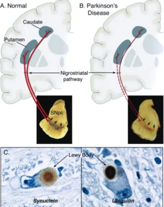
Figure 1: Neuropathology of PD. Schematic representation of the normal (A) and diseased (B)
nigrostriatal pathway. Photograph shows the depigmentation of the SNpc due to dopaminergic neurons degeneration. Immunohistochemistry of LBs in SNpc dopaminergic neurons (C) (Dauer and Przedborski 2003).
1.2. The etiology of PD
For the vast majority of PD cases, the etiology remains unknown even though both genetic and environmental factors are likely to be implicated (Vance, Ali et al. 2010). The environmental hypothesis was strongly suggested about 20 years ago after the report of a parkinsonian syndrome in young adults that were intoxicated by a neurotoxin called 1-methyl-4-phenyl-1,2,3,6-tetrahydropyridine
Introduction 2010
(MPTP) which selectively destroys nigrostriatal dopaminergic neurons (Broussolle and Thobois 2002). Exposure of mice to environmental chemicals such as MPTP and rotenone in mouse produces the symptoms akin to PD and therefore these neurotoxins are commonly used in experimental studies of PD. Another environmental toxin, paraquat (a commonly used herbicide) has also been implicated in the onset of PD (Abdulwahid Arif and Ahmad Khan 2010). The neurotoxicity of these chemicals is accompanied by the blockade of electron flow from NADH dehydrogenase to coenzyme Q. The agents with the ability to improve mitochondrial respiration and ATP production have been shown to exert beneficial effects in PD patients as well as in the animal models of PD (Abdulwahid Arif and Ahmad Khan 2010).
Comparatively, the genetic hypothesis of Parkinson's disease has gained considerable interest during the last decade (Broussolle and Thobois 2002). In rare genetic forms of PD derive in mutations in the genes encoding α-synuclein, leucine-rich repeat kinase 2, Parkin, PTEN-induced putative kinase 1 and DJ-1 (Tsuji 2010). Genetic studies have identified mutations in α-synuclein and ubiquitin C-terminal hydroxylase L1 as rare causes of autosomal dominant PD and mutations in parkin as a cause of autosomal recessive PD. Functional characterization of the identified disease genes implicates the ubiquitin-mediated protein degradation pathway in the hereditary forms of PD and also in the more common sporadic forms of PD (Kruger, Eberhardt et al. 2002).
To date, the most effective treatment for PD remains the administration of a precursor of dopamine, L-dopa, which, by replenishing the brain in dopamine, alleviates almost all PD symptoms. The chronic administration of L-dopa, however, often causes motor and psychiatric side effects which may be as debilitating as PD itself. Moreover, as of yet there is no evidence that L-dopa therapy can impede the neurodegenerative process in PD (Teismann, Tieu et al. 2003).
2
The nerv othe char orga cells Cons throu but n desc Thes (Kur of nu deve activ micr such rece Figur 2009 As a or p chan large rece andu2. Microg
term “mic vous syste er tissues, racteristic “ an system s in the p stitute abo ughout the not touchin cribed as d se cells h rpius, Wilso umerous f eloped den ve phagoc roglia typic h as CD eptors, and re 2: Compa ). a response resence p nges, proli e number eptors, an undergo aglia
croglia” ref m, that sh but that in “ramified” (Rock, G eriphery a out 12% o e normal b ng each o distinct cell ave short on et al. 20 filipodia an nse bodies cytotic acti cally exist i 14, major several ot arison of micr e to alterat pathogenic ferate, bec of molecul nd transcr dramatic t fers to cel hare many n their non morpholog Gekker et a and are re of all glial brain whit t ther (McG lular popul size and 006). Micro nd pseudop s and vac ivity (Kim in a resting r histocom ther marke roglia in rest tions in the agents, m come pha les such a ription fac transforma lls that res y if not all -activated gy not seen al. 2004). M esident ma cells (Zha their ramifi Geer and M ation with a soma t oglial cells podia surro cuoles in t and de V g state an mpatibility ers (Zhanging state (A)
e environm microglial c gocytic, a as cytokine ctors. Mic ation from t side within the prope or resting n in reside Microglia a acrophage ang, Hu et ed proces McGeer 20 specific m that does s are chara ounding th the cytopla Vellis 2005 d express complex , Hu et al. and active s ment, parti cells exhib nd upregu es, adhesio croglial ce their restin Introdu n the pare erties of m state (figu ent macrop are derived es-like cell al. 2010) sses being 008). Micro morphologic not excee acterized b he cell sur asm, sugg 5). In the of cell su molecule 2010). state (B) (Ra cularly neu bit marked ulate the e on molecu ells are ng state int uction 201 nchyma o macrophage ure 2A) ha phages of o d from my s in the C ) and distr close toge oglia has c character ed 5 – 10 by the pres rface, and gestive of mature b rface antig es, chemo ansohoff and uronal dam morpholo expression les, memb readily a to an amoe 0 of the es in ave a other yeloid CNS. ribute ether been ristic. 0 µm ence well-their brain, gens, okine Perry mage ogical of a brane active eboid
Introduction 2010
(round, oval) morphology (figure 2B) (Zhang, Hu et al. 2010). This process, called microglial activation, is a physiological response aimed at protecting the affected neural tissue (Saura, Tusell et al. 2003). However, due to their capacity to produce highly neurotoxic species, chronically activated microglial cells may participate in the pathogenesis of neurodegenerative disorders such as PD or AD (Saura, Tusell et al. 2003). Accumulated evidence suggests that microglial cells are associated not only with brain pathology but also with the normal physiology in the brain (Nakajima and Kohsaka 1993).
2.1. Microglial Activation
Microglia is exquisitely sensitive to disturbance of their microenvironment. The early and rapid response of microglia is entirely consistent whit the role of tissue macrophages as the first line of defense against infection or injury (Ransohoff and Perry 2009). The substances freed from injured cells of the CNS set in motion the microglial activation, and induce the changes in gene expression and reorganization of cellular phenotype (Kreutzberg 1996; Streit 2002). The cells of microglia detect the changes in its environment through the expression of a great number of cellular surface receptors and nuclear receptors that play a critical role in the initiation and/or modulation of its immunitary responses. These receptors recognize factors such as the complement receptor 3 (CR3), immunoglobulin’s, molecules of cellular adhesion, steroids, bacterial products, misfolded proteins and cytokines (Hanisch 2002; Moller 2002; van Rossum and Hanisch 2004).
One of the most commonly used methods of activating microglia both in vitro and in vivo is the application of the endotoxin lipopolysaccharide (LPS). LPS is bacterial endotoxin derived from Gram – negative bacteria and was the first agent to be described in the literature as activator of microglial cells (Hetier, Ayala et al. 1988; Kim, Mohney et al. 2000). Microglia respondes actively to the LPS with the consequent release of a great variety of cytokines, NO and proteases. These effects are mediated by the toll-like receptor (TLR) 4 that has been related with the detection of mycobacterials infections (Takeda, Kaisho et al. 2003). LPS-induced activation of microglia results in the production of
Introduction 2010
cytokines and chemokines such as IL-1β, IL-1 receptor antagonist (IL-1ra), IL-6, IL-8, IL-10, IL-12, IL-18, transforming growth factor-β (TGF-β) and TNF-α by microglia. These cytokines, in turn, potentiate microglial activation by binding to receptoras expressed by microglia (Kim and de Vellis 2005). It was demonstrated that injection a single dose of LPS into the nigrostriatal pathway induces a strong macrophage/microglial reaction that leads to degeneration of dopaminergic neurons in the SN (Herrera, Castano et al. 2000).
One other inflammatory agent that induces selective microglia activation is Zymosan A (ZyA), a substance derived from the cellular wall the Saccharomyces cerevisiae (Fitzpatrick, Haynes et al. 1964). ZyA is capable of stimulating microglial cells through the CD11b of CR3 (also termed Mac1). CR3 is a member of the β2 family of integrins expressed in plasma membranes of mammalian phagocytes and natural killer cells. It is a heterodimeric type I transmembrane glycoprotein, consisting of a CD11b α chain noncovalently associated with the CD18 β subunit (Le Cabec, Cols et al. 2000). The ZyA, when injected in animals, provokes inflammation by inducing of the release of an ample gamma of inflammatory mediators. These include active components of complement system (Pillemer and Ecker 1941), prostaglandins and leukotrienes (Humes, Sadowski et al. 1982), platelet-activating factor (Roubin, Mencia-Huerta et al. 1982), oxidative metabolism (Nauseef, Root et al. 1983), and lysosomal acid hydrolases (Bonney, Wightman et al. 1978). Since ZyA is not degradable, phagocytosis results in a prolonged inflammatory response (Volman, Hendriks et al. 2005).
Mis-folded or aberrant proteins such as amyloid-β (Aβ) are capable of activating microglia via scavenger receptors, which are up-regulated in the brain of AD patients, or by initiating the phagocytosis of the pathological protein by microglia (Rogers, Strohmeyer et al. 2002). It has also been shown that microglial phagocytosis occurs in response to aggregated α-synuclein, the major component of LBs in PD (Zhang, Wang et al. 2005).
The serum factors thrombin and immunoglobulins initiate microglial activation through protease-activated receptor 1 and Fc receptors, respectively (Stangel and Compston 2001; Suo, Wu et al. 2002). ATP released from damaged
Introduction 2010
neurons, has also been demonstrated to activate microglia (Davalos, Grutzendler et al. 2005) by binding to purinergic receptors, expressed by microglia.
2.2. Consequences of microglial activation
Activated microglia has been shown to play key roles in both the developing and adult CNS. Upon activation in the adult CNS, microglia act primarily as scavenger and in brain tissue remodeling to restore and protect brain structures and functions (Nimmerjahn, Kirchhoff et al. 2005).
Local microglia extend out their processes to surround the area of insult (Davalos, Grutzendler et al. 2005) and as a result damaged cells are engulfed by the microglia via phagocytosis, removing any potentially damaging material from the area and protecting the neighbouring cells. Microglial cells share with other cells of the myeloid lineage the ability to secrete a multitude of immunomodulatory molecules such as cytokines, chemokines, neurotrophins and reactive oxygen and nitrogen species that mediate the communication with surrounding cells (Garden and Moller 2006). Cytokines are low molecular-weight proteins that are usually classified as either pro- or anti-inflammatory and are thought to signal both by paracrine and autocrine ways. While pro-inflammatory cytokines have the ability to elicit a sustained immune response, anti-inflammatory cytokines act to down-regulate an immune response by binding to appropriate receptors expressed on microglia and initiating an autocrine signalling process. Cytokines have numerous effects on CNS function including growth promotion, inhibition and proliferation of astrocytes and oligodendrocytes (Hanisch 2002), modulation of neurotransmitter release (Zalcman, Green-Johnson et al. 1994), and behavioral alterations such as memory impairment (Yirmiya, Winocur et al. 2002). Chemokines act primarily as chemoattractants to draw additional microglia to the site of injury, while NTF such as nerve growth factor (NGF), brain-derived neurotrophic factor (BDNF) and glial-derived neurotrophic factor (GDNF) released from microglia have been proposed to participate in the survival and regeneration of neurons (Nagata, Takei et al. 1993; Batchelor, Porritt et al. 2002) and to prolong the existence of
Introduction 2010
microglia and to regulate their function (Elkabes, DiCicco-Bloom et al. 1996). Activated microglia can also produce and release both reactive oxygen and nitrogen species due to catalysis by nicotinamide adenine dinucleotide phosphate (NADPH) oxidase (Babior 1999) and these highly reactive free radicals can kill surrounding pathogens. It has also been reported however, that microglial-derived free radicals can cause neuronal cell death and so they have been implicated in the pathogenesis of neurodegenerative conditions (Chao, Hu et al. 1992).
3. Astrocytes
Astrocytes, also known as astroglia, are the most abundant cells in the CNS constituting 20 – 50% of the cerebral volume (Zhang, Hu et al. 2010). They are characterized by its prolongations and great structural complexity. These cells are also characterized by a dense matrix of ramifications, some of which assists in the formation of the blood brain barrier by contacting and involving local vascular walls (Kim, Kim et al. 2006; Koehler, Gebremedhin et al. 2006). Astrocytes also contribute to brain homeostasis in several ways, including buffering of extracellular K+, regulating neurotransmitter release, releasing
growth factors, and regulating the brain immune response (Gee and Keller 2005). Astrocytes also play a role in capturing through active uptake and neurotransmitters released during synaptic transmission (Nedergaard, Ransom et al. 2003). Some evidence suggest that these cells still execute functions such as neuronal differentiation, regulation of the axonal orientation, formation of synapses, cerebral plasticity and communication (Mosley, Benner et al. 2006). As already it was mentioned, astrocytes are dynamic cells that keep homeostasis in the undamaged SNC. These cells express numerous receptors that allow them to be responsive to neurotransmitters and growth factors, small molecules and toxins. In fact, when astrocytes perceive disturbances of brain homeostasis, its metabolic activity increases, as well as the production of trophics factors, which attributs them a bigger capacity to protect other cells in
Introduction 2010
the brain from energy depletion, and from na overload of toxic free radicals (Mosley, Benner et al. 2006).
The characteristics that are more frequently associated with the astrocytic response to insults involve cellular hypertrophy, astrocyte proliferation, process extension and interdigitation, and increased production of the intermediate filaments glial fibrillary acidic protein (GFAP), vimentin and nestin. The “reactive” astrocytes may also exacerbate tissue damage as they release pro-inflammatory cytokines such as TNF-α, arachidonic acid metabolites, NO and reactive oxygen species (ROS) that can adversely affect cell survival (Liberto, Albrecht et al. 2004). Depending on the disease context, astrogliosis can be viewed as a beneficial event for promotion of neuronal survival by the production of growth factors and neurotrophins that support neuronal growth, or detrimental for neuronal functions by the formation of glial scars (Dong and Benveniste 2001). The function of reactive astrocytes is not well understood, and both harmful and beneficial activities have been attributed to these cells (Sofroniew 2005).
3.1. Factors release by astrocytes
Astrocytes are responsible for the production of cytokines and chemokines, however this expression is scarce in the normal CNS. Aberrant expression of cytokines and chemokines occurs in CNS diseases such as AD, MS, PD and brain injury/trauma (Dong and Benveniste 2001). Both in vitro and in vivo studies have documented the ability of astrocytes to produced interleukins, interferons, colony-stimulating factors and chemokines (table 1) (Dong and Benveniste 2001).
Table 1: Cytokines/ckemokines production by astrocytes (Dong and Benveniste 2001).
Cytokines Chemokines
IL-1α and β, IL-5, IL-6, IL-10 RANTES
IFN-α and β IL-8 TNF-α MCP-1
Introduction 2010
TGF-β IP-10 G-CSF, GM-CSF, M-CSF
Cytokines and chemokines have been implicated in the initiation, propagation and regulation of immune and inflammatory responses by astrocytes (Benveniste 1998).
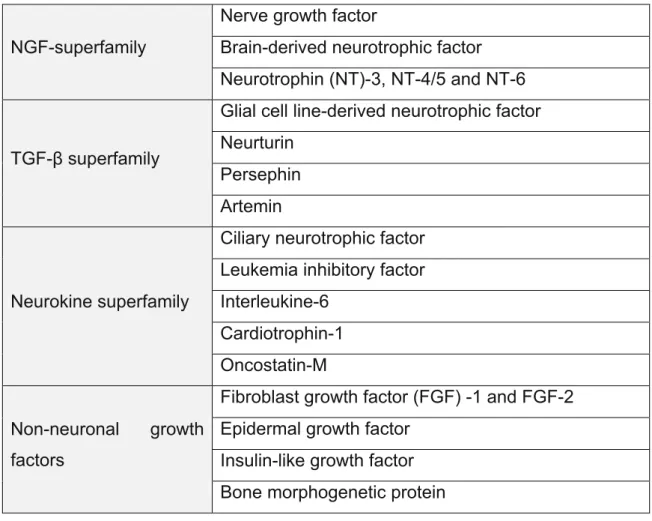
Astrocytes also secrete various soluble factors known as neurotrophic factors. These molecules have pleotropic effect on neurons; being involved in the survival, maturation, differentiation and development of neuronal cells. Accumulating evidences suggests that neurotrophic factors play a role in neurodegeneration as well (Siegel and Chauhan 2000). Neurotrophic factors have been categorized into various families such as NGF superfamily, GDNF family (distantly related to the TGF-β-superfamily), neurokine superfamily and non-neural growth factor superfamily (table 2).
Table 2: Neurotrophic factors and this family (Siegel and Chauhan 2000). NGF-superfamily
Nerve growth factor
Brain-derived neurotrophic factor Neurotrophin (NT)-3, NT-4/5 and NT-6
TGF-β superfamily
Glial cell line-derived neurotrophic factor Neurturin
Persephin Artemin
Neurokine superfamily
Ciliary neurotrophic factor Leukemia inhibitory factor Interleukine-6
Cardiotrophin-1 Oncostatin-M Non-neuronal growth
factors
Fibroblast growth factor (FGF) -1 and FGF-2 Epidermal growth factor
Insulin-like growth factor Bone morphogenetic protein
Introduction 2010
3.1.1. The GDNF
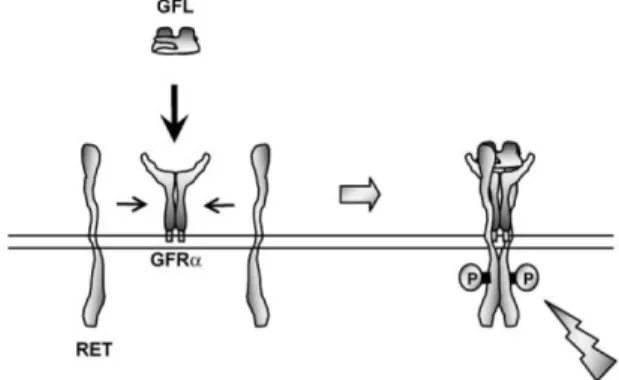
GDNF was originally identified and purified from media conditioned by the B 49 glioma cell line and was characterized by its ability to promote the survival and differentiation of dopaminergic neurons during development (Lin, Zhang et al. 1994). GDNF is a distantly related member of the TGF-β superfamily that is glycosylated, the disulfide-bond homodimer, has a molecular weight 33 – 45 kDa, while the monomer has a molecular weight of 16 kDa after deglycosylation (Lin, Doherty et al. 1993; Lin, Zhang et al. 1994). The actions of GDNF are mediated by the activation of the GDNF family receptor (GFR) alpha, a multi-component receptor complex comprising the transmembrane RET tyrosine kinase. Four members of this family have been identified (GFRα1 – 4) with GDNF binding preferentially to GFRα1. It is believed that the GDNF dimer forms a complex with GFRα and that this complex induces the dimerization of RET (figure 2) (Sariola and Saarma 2003; Enomoto 2005). GDNF-activated RET could also induce different biological responses such as morphological transformation, proliferation, cell migration, neurite elongation and neurite branching. In cells lacking the RET receptor, alternative mechanisms which involve neuronal cells adhesion molecules were suggested for GDNF-mediated activities (Sariola and Saarma 2003; Donatello, Fiorino et al. 2007).
Figure 2: Glial cell line-derived neurotrophic factor (GDNF) family ligand (GFL)-induced
activation of the RET receptor tyrosine kinase. A GFL dimer binds to the GDNF family receptor (GFR) α to form a (GFL) (GFRα)2 complex, which then induces dimerization and successive
phosphorylation of the RET tyrosine kinase (Jing, Wen et al. 1996).
The GDNF is present in many different regions of the developing nervous system, and this localization depends on the local activity and the stadium of
Introduction 2010
development of the individual. Detectable levels of GDNF were observed in the thalamus, hippocampus, cerebellum, cortex, spinal marrow and SN, among others local of the nervous system (Choi-Lundberg and Bohn 1995; Del Fiacco, Quartu et al. 2002).
Studies in primary cultures of midbrain ventral showed that GDNF increases the number tyrosine hydroxylase (TH) positive cells and its morphologic complexity and functional and neuronal maturation (Schaller, Andres et al. 2005). In adult rats, GDNF mRNA levels are significantly higher in the striatum than in the SN suggesting a neuronal maintenance role for GDNF in the adult nervous system, mainly as a target-derived NTF for dopaminergic neurons (Choi-Lundberg and Bohn 1995; Oo, Ries et al. 2005). Several studies strongly suggest that GDNF is an essential player in the development of the dopaminergic nigrostriatal system, and that even slight disturbances in the system maturation due to GDNF deficits might compromise dopaminergic survival in the adulthood (Saavedra, Baltazar et al. 2007). GDNF was been considered the more powerful neurotrophic factor in protecting dopaminergic neurons from the degeneration induced by the toxins MPTP or 6-hydroxydopamine (6-OHDA) in animal models of the PD (Rosenblad, Martinez-Serrano et al. 1998).
Although the neurotrophic effect of the GDNF is well documented, its effect on microglial cells remains is less clear. Nevertheless, there are some results indicating that GDNF is able to regulate microglia activity. In the study development in primary rat microglia, Chang and its collaborators showed that GDNF increased the NO production, the activity of superoxide dismutase (SOD) and the phagocytotic capability but had no effect on the secretion of the pro-inflammatory cytokines TNF-α and IL-1β (Chang, Fang et al. 2006).
Introduction 2010
4. Microglia-astrocytes interactions
The presence of oxidative stress and inflammatory activity is one of the significant pathological features of PD (Kim and Joh 2006). The presence of inflammation is generally indicated by the accumulation of activated microglia in damaged areas of the brain. Particularly high numbers of activated microglia have been found in post-mortem brains of PD patients, predominantly in the SNpc in the vicinity of the degenerating dopaminergic neurons (Long-Smith, Sullivan et al. 2009). Kim and collaborates revealed that the SN had the highest brain density in microglia, thus making post-mortem mesencephalic neurons more sensitive to neurodegeneration-mediated inflammation (Kim, Mohney et al. 2000). The pivotal role of activated microglia-mediated dopaminergic neuronal degeneration has been demonstrated in the rotenone model of PD (Gao, Hong et al. 2002). Wu et al., (2002) and in studies showing that inhibition of microglial activation prevents dopaminergic neuronal loss in MPTP-treated mice (Wu, Jackson-Lewis et al. 2002). Activated microglia has also been implicated in the pathogenesis and progress of PD.
Astrocytes, the most abundant cells in the brain, may have a role in controlling microglial over-activation (Yang, Min et al. 2007). Astrocytes and microglia are the brain representatives of the general immune system, and can, under pathological conditions, act as immune competent cells. Upon activation, the reactive glial cells gain a number of potentially neurotoxic potencies, e.g., via release of inflammation-promoting mediators and oxidative radicals. As long as these factors remain under strict control, reactive glial cells can be seen as having an undoubtedly beneficial role in defense and repair. However, an escalating pathological glial activation which involves both microglial and astrocytes may contribute to secondary neuronal damage (Markiewicz and Lukomska 2006).
The interactions that occur between astrocytes and microglia after a CNS damages are complex and wide unexplored. However, it is known that cytokines and growth factors are important mediators of the glial communication. The discovery of the molecular mechanisms responsible for the microglia-astrocytes interactions has been difficult to attain through the use of of
Introduction 2010
in vivo models. Therefore, the majority of the experimental data has been generated for studies in vitro. It was already demonstrated that microglial morphology is influenced by astrocytes that play an important role in regulating differentiation and microglial down-activation (Streit, Walter et al. 1999). Some authors had described the role of astrocytes in the process microglia ramification, although the process of astrocyte signaling is still controversial (Suzumura, Sawada et al. 1990; Eder, Klee et al. 1997; Eder, Schilling et al. 1999; Liu, Brosnan et al. 1994; Tanaka and Maeda 1996; Tanaka, Toku et al. 1999). Data from literature show that of rat microglial cells modify its morphology from amoeboid to ramified, when cultivated on a top of an astrocytes monolayer (Liu, Brosnan et al. 1994; Tanaka and Maeda 1996). However, they did not detect any alteration in microglial morphology when cells were exposed to astrocytes conditioned media (ACM). Recently, it was reported by Min and its collaborators that soluble factors freed by astrocytes are capable to suppress interferon (IFN)-γ-induced microglial inflammatory responses through the expression of hemeoxygenase-1 (HO-1). These results suggest that astrocytes could cooperate with microglia to prevent excessive inflammatory responses in the brain (Min, Yang et al. 2006).
Introduction 2010
5. Aims of this thesis
The microglial activation is mainly intended for the neuronal protection. However, in several neuropathologies, associated with chronic inflammation, the inflammatory products derived from the activated microglia can also contribute for the neuronal loss. Since astrocytes have been suggested as main regulators of microglial activation, a deeper knowledge of the possible factors freed for these cells that prevent the change of microglia from an anti- to a pro-inflammatory state, can make possible new approaches in the study of the astrocytes-microglia interactions and in the control of the neuroinflammation in pathological situations.
The aim of this study was to characterize the effect of soluble mediators released by astrocytes in controlling microglia activation induced by the pro-inflammatory agent Zymosan A and to identify the molecules responsible for this mechanism.
Introduction 2010
6. References
Abdulwahid Arif, I. and H. Ahmad Khan (2010). "Environmental toxins and Parkinson's disease: putative roles of impaired electron transport chain and oxidative stress." Toxicol Ind Health 26(2): 121-8.
Babior, B. M. (1999). "NADPH oxidase: an update." Blood 93(5): 1464-76.
Banati, R. B., S. E. Daniel, et al. (1998). "Glial pathology but absence of apoptotic nigral neurons in long-standing Parkinson's disease." Mov Disord 13(2): 221-7.
Batchelor, P. E., M. J. Porritt, et al. (2002). "Macrophages and Microglia Produce Local Trophic Gradients That Stimulate Axonal Sprouting Toward but Not beyond the Wound Edge." Mol Cell Neurosci 21(3): 436-53.
Benveniste, E. N. (1998). "Cytokine actions in the central nervous system." Cytokine Growth Factor Rev 9(3-4): 259-75.
Bonney, R. J., P. D. Wightman, et al. (1978). "Regulation of prostaglandin synthesis and of the selective release of lysosomal hydrolases by mouse peritoneal macrophages." Biochem J 176(2): 433-42.
Braak, H., K. Del Tredici, et al. (2003). "Staging of brain pathology related to sporadic Parkinson's disease." Neurobiol Aging 24(2): 197-211.
Broussolle, E. and S. Thobois (2002). "Genetics and environmental factors of Parkinson disease." Rev Neurol (Paris) 158 Spec no 1: S11-23.
Chang, Y. P., K. M. Fang, et al. (2006). "Regulation of microglial activities by glial cell line derived neurotrophic factor." J Cell Biochem 97(3): 501-11.
Chao, C. C., S. Hu, et al. (1992). "Activated microglia mediate neuronal cell injury via a nitric oxide mechanism." J Immunol 149(8): 2736-41.
Choi-Lundberg, D. L. and M. C. Bohn (1995). "Ontogeny and distribution of glial cell line-derived neurotrophic factor (GDNF) mRNA in rat." Dev Brain Res 85(1): 80-8.
Dauer, W. and S. Przedborski (2003). "Parkinson's disease: mechanisms and models." Neuron 39(6): 889-909.
Davalos, D., J. Grutzendler, et al. (2005). "ATP mediates rapid microglial response to local brain injury in vivo." Nat Neurosci 8(6): 752-8.
Del Fiacco, M., M. Quartu, et al. (2002). "Topographical localization of glial cell line-derived neurotrophic factor in the human brain stem: an immunohistochemical study of prenatal, neonatal and adult brains." J Chem Neuroanat 23(1): 29-48.
Dickson, D. W., H. Fujishiro, et al. (2009). "Neuropathology of non-motor features of Parkinson disease." Parkinsonism Relat Disord 15 Suppl 3: S1-5.
Donatello, S., A. Fiorino, et al. (2007). "SH2B1beta adaptor is a key enhancer of RET tyrosine kinase signaling." Oncogene 26(45): 6546-59.
Introduction 2010
Duke, D. C., L. B. Moran, et al. (2007). "The medial and lateral substantia nigra in Parkinson's disease: mRNA profiles associated with higher brain tissue vulnerability." Neurogenetics 8(2): 83-94.
Eder, C., R. Klee, et al. (1997). "Distinct soluble astrocytic factors induce expression of outward K+ currents and ramification of brain macrophages." Neurosci Lett 226(3): 147-50.
Eder, C., T. Schilling, et al. (1999). "Morphological, immunophenotypical and electrophysiological properties of resting microglia in vitro." Eur J Neurosci 11(12): 4251-61.
Elkabes, S., E. M. DiCicco-Bloom, et al. (1996). "Brain microglia/macrophages express neurotrophins that selectively regulate microglial proliferation and function." J Neurosci 16(8): 2508-21.
Enomoto, H. (2005). "Regulation of neural development by glial cell line-derived neurotrophic factor family ligands." Anat Sci Int 80(1): 42-52.
Fahn, S. (2003). "Description of Parkinson's disease as a clinical syndrome." Ann N Y Acad Sci 991: 1-14.
Fearnley, J. M. and A. J. Lees (1991). "Ageing and Parkinson's disease: substantia nigra regional selectivity." Brain 114 ( Pt 5): 2283-301.
Fitzpatrick, F. W., L. J. Haynes, et al. (1964). "Effect of Glucan Derivatives Upon Phagocytosis by Mice." J Reticuloendothel Soc 15: 423-8.
Gao, H. M., J. S. Hong, et al. (2002). "Distinct role for microglia in rotenone-induced degeneration of dopaminergic neurons." J Neurosci 22(3): 782-90.
Garden, G. A. and T. Moller (2006). "Microglia biology in health and disease." J Neuroimmune Pharmacol 1(2): 127-37.
Gee, J. R. and J. N. Keller (2005). "Astrocytes: regulation of brain homeostasis via apolipoprotein E." Int J Biochem Cell Biol 37(6): 1145-50.
Gehrmann, J., R. B. Banati, et al. (1993). "Microglia in the immune surveillance of the brain: human microglia constitutively express HLA-DR molecules." J Neuroimmunol 48(2): 189-98.
Hanisch, U. K. (2002). "Microglia as a source and target of cytokines." Glia 40(2): 140-55. Herrera, A. J., A. Castano, et al. (2000). "The single intranigral injection of LPS as a new model
for studying the selective effects of inflammatory reactions on dopaminergic system." Neurobiol Dis 7(4): 429-47.
Hetier, E., J. Ayala, et al. (1988). "Brain macrophages synthesize interleukin-1 and interleukin-1 mRNAs in vitro." J Neurosci Res 21(2-4): 391-7.
Humes, J. L., S. Sadowski, et al. (1982). "Evidence for two sources of arachidonic acid for oxidative metabolism by mouse peritoneal macrophages." J Biol Chem 257(4): 1591-4. Jing, S., D. Wen, et al. (1996). "GDNF-induced activation of the ret protein tyrosine kinase is
mediated by GDNFR-alpha, a novel receptor for GDNF." Cell 85(7): 1113-24.
Kim, J. H., J. H. Kim, et al. (2006). "Blood-neural barrier: intercellular communication at glio-vascular interface." J Biochem Mol Biol 39(4): 339-45.
Introduction 2010
Kim, S. U. and J. de Vellis (2005). "Microglia in health and disease." J Neurosci Res 81(3): 302-13.
Kim, W. G., R. P. Mohney, et al. (2000). "Regional difference in susceptibility to lipopolysaccharide-induced neurotoxicity in the rat brain: role of microglia." J Neurosci 20(16): 6309-16.
Kim, Y. S. and T. H. Joh (2006). "Microglia, major player in the brain inflammation: their roles in the pathogenesis of Parkinson's disease." Exp Mol Med 38(4): 333-47.
Koehler, R. C., D. Gebremedhin, et al. (2006). "Role of astrocytes in cerebrovascular regulation." J Appl Physiol 100(1): 307-17.
Koo, H. J., H. J. Lee, et al. (2008). "Sequence determinants regulating fibrillation of human alpha-synuclein." Biochem Biophys Res Commun 368(3): 772-8.
Kreutzberg, G. W. (1996). "Microglia: a sensor for pathological events in the CNS." Trends Neurosci 19(8): 312-8.
Kruger, R., O. Eberhardt, et al. (2002). "Parkinson's disease: one biochemical pathway to fit all genes?" Trends Mol Med 8(5): 236-40.
Kurpius, D., N. Wilson, et al. (2006). "Early activation, motility, and homing of neonatal microglia to injured neurons does not require protein synthesis." Glia 54(1): 58-70.
Le Cabec, V., C. Cols, et al. (2000). "Nonopsonic phagocytosis of zymosan and Mycobacterium kansasii by CR3 (CD11b/CD18) involves distinct molecular determinants and is or is not coupled with NADPH oxidase activation." Infect Immun 68(8): 4736-45.
Lee, J. K., T. Tran, et al. (2009). "Neuroinflammation in Parkinson's Disease." J Neuroimm Pharmacol.
Lees, A. J., J. Hardy, et al. (2009). "Parkinson's disease." Lancet 373(9680): 2055-66.
Liberto, C. M., P. J. Albrecht, et al. (2004). "Pro-regenerative properties of cytokine-activated astrocytes." J Neurochem 89(5): 1092-100.
Lin, L. F., D. H. Doherty, et al. (1993). "GDNF: a glial cell line-derived neurotrophic factor for midbrain dopaminergic neurons." Science 260(5111): 1130-2.
Lin, L. F., T. J. Zhang, et al. (1994). "Purification and initial characterization of rat B49 glial cell line-derived neurotrophic factor." J Neurochem 63(2): 758-68.
Liu, W., C. F. Brosnan, et al. (1994). "Macrophage colony-stimulating factor mediates astrocyte-induced microglial ramification in human fetal central nervous system culture." Am J Pathol 145(1): 48-53.
Long-Smith, C. M., A. M. Sullivan, et al. (2009). "The influence of microglia on the pathogenesis of Parkinson's disease." Prog Neurobiol 89(3): 277-87.
Markiewicz, I. and B. Lukomska (2006). "The role of astrocytes in the physiology and pathology of the central nervous system." Acta Neurobiol Exp (Wars) 66(4): 343-58.
McGeer, P. L., S. Itagaki, et al. (1988). "Reactive microglia are positive for HLA-DR in the substantia nigra of Parkinson's and Alzheimer's disease brains." Neurology 38(8): 1285-91.
Introduction 2010
McGeer, P. L. and E. G. McGeer (2008). "Glial reactions in Parkinson's disease." Mov Disord 23(4): 474-83.
Min, K. J., M. S. Yang, et al. (2006). "Astrocytes induce hemeoxygenase-1 expression in microglia: a feasible mechanism for preventing excessive brain inflammation." J Neurosci 26(6): 1880-7.
Moller, T. (2002). "Calcium signaling in microglial cells." Glia 40(2): 184-94.
Mori, F., K. Tanji, et al. (2008). "alpha-Synuclein pathology in the neostriatum in Parkinson's disease." Acta Neuropathol 115(4): 453-9.
Mosley, R. L., E. J. Benner, et al. (2006). "Neuroinflammation, Oxidative Stress and the Pathogenesis of Parkinson's Disease." Clin Neurosci Res 6(5): 261-281.
Nagata, K., N. Takei, et al. (1993). "Microglial conditioned medium promotes survival and development of cultured mesencephalic neurons from embryonic rat brain." J Neurosci Res 34(3): 357-63.
Nakajima, K. and S. Kohsaka (1993). "Functional roles of microglia in the brain." Neurosci Res 17(3): 187-203.
Nauseef, W. M., R. K. Root, et al. (1983). "Inhibition of zymosan activation of human neutrophil oxidative metabolism by a mouse monoclonal antibody." Blood 62(3): 635-44.
Nedergaard, M., B. Ransom, et al. (2003). "New roles for astrocytes: redefining the functional architecture of the brain." Trends Neurosci 26(10): 523-30.
Nimmerjahn, A., F. Kirchhoff, et al. (2005). "Resting microglial cells are highly dynamic surveillants of brain parenchyma in vivo." Science 308(5726): 1314-8.
Oo, T. F., V. Ries, et al. (2005). "Anatomical basis of glial cell line-derived neurotrophic factor expression in the striatum and related basal ganglia during postnatal development of the rat." J Comp Neurol 484(1): 57-67.
Pillemer, L. and E. E. Ecker (1941). "The Terminology of the Components of Complement." Science 94(2445): 437.
Raine, C. S. (1994). "Multiple sclerosis: immune system molecule expression in the central nervous system." J Neuropathol Exp Neurol 53(4): 328-37.
Ransohoff, R. M. and V. H. Perry (2009). "Microglial physiology: unique stimuli, specialized responses." Annu Rev Immunol 27: 119-45.
Rock, R. B., G. Gekker, et al. (2004). "Role of microglia in central nervous system infections." Clin Microbiol Rev 17(4): 942-64, table of contents.
Rogers, J., R. Strohmeyer, et al. (2002). "Microglia and inflammatory mechanisms in the clearance of amyloid beta peptide." Glia 40(2): 260-9.
Rosenblad, C., A. Martinez-Serrano, et al. (1998). "Intrastriatal glial cell line-derived neurotrophic factor promotes sprouting of spared nigrostriatal dopaminergic afferents and induces recovery of function in a rat model of Parkinson's disease." Neuroscience 82(1): 129-37.
Introduction 2010
Roubin, R., J. M. Mencia-Huerta, et al. (1982). "Biosynthesis of platelet-activating factor (PAF-acether). IV. Impairment of acetyl-transferase activity in thioglycollate-elicited mouse macrophages." J Immunol 129(2): 809-13.
Saavedra, A., G. Baltazar, et al. (2007). "Interleukin-1beta mediates GDNF up-regulation upon dopaminergic injury in ventral midbrain cell cultures." Neurobiol Dis 25(1): 92-104. Sariola, H. and M. Saarma (2003). "Novel functions and signalling pathways for GDNF." J Cell
Sci 116(Pt 19): 3855-62.
Saura, J., J. M. Tusell, et al. (2003). "High-yield isolation of murine microglia by mild trypsinization." Glia 44(3): 183-9.
Schaller, B., R. H. Andres, et al. (2005). "Effect of GDNF on differentiation of cultured ventral mesencephalic dopaminergic and non-dopaminergic calretinin-expressing neurons." Brain Res 1036(1-2): 163-72.
Seth, P. and N. Koul (2008). "Astrocyte, the star avatar: redefined." J Biosci 33(3): 405-21. Siegel, G. J. and N. B. Chauhan (2000). "Neurotrophic factors in Alzheimer's and Parkinson's
disease brain." Brain Res Brain Res Rev 33(2-3): 199-227.
Sofroniew, M. V. (2005). "Reactive astrocytes in neural repair and protection." Neuroscientist 11(5): 400-7.
Stangel, M. and A. Compston (2001). "Polyclonal immunoglobulins (IVIg) modulate nitric oxide production and microglial functions in vitro via Fc receptors." J Neuroimmunol 112(1-2): 63-71.
Streit, W. J. (2002). "Microglia as neuroprotective, immunocompetent cells of the CNS." Glia 40(2): 133-9.
Streit, W. J., S. A. Walter, et al. (1999). "Reactive microgliosis." Prog Neurobiol 57(6): 563-81. Suo, Z., M. Wu, et al. (2002). "Participation of protease-activated receptor-1 in thrombin-induced
microglial activation." J Neurochem 80(4): 655-66.
Suzumura, A., M. Sawada, et al. (1990). "Effects of colony stimulating factors on isolated microglia in vitro." J Neuroimmunol 30(2-3): 111-20.
Takeda, K., T. Kaisho, et al. (2003). "Toll-like receptors." Annu Rev Immunol 21: 335-76.
Tanaka, J. and N. Maeda (1996). "Microglial ramification requires nondiffusible factors derived from astrocytes." Exp Neurol 137(2): 367-75.
Tanaka, J., K. Toku, et al. (1999). "Morphological differentiation of microglial cells in culture: involvement of insoluble factors derived from astrocytes." Neurosci Res 34(4): 207-15. Tansey, M. G. and M. S. Goldberg (2009). "Neuroinflammation in Parkinson's disease: Its role in
neuronal death and implications for therapeutic intervention." Neurobiol Dis.
Teismann, P., K. Tieu, et al. (2003). "Pathogenic role of glial cells in Parkinson's disease." Mov Disord 18(2): 121-9.
Toulouse, A. and A. M. Sullivan (2008). "Progress in Parkinson's disease-where do we stand?" Prog Neurobiol 85(4): 376-92.
Tsuji, S. (2010). "Genetics of neurodegenerative diseases: insights from high-throughput resequencing." Hum Mol Genet.
Introduction 2010
van Rossum, D. and U. K. Hanisch (2004). "Microglia." Metab Brain Dis 19(3-4): 393-411. Vance, J. M., S. Ali, et al. (2010). "Gene-environment interactions in Parkinson's disease and
other forms of parkinsonism." Neurotoxicology.
Volman, T. J., T. Hendriks, et al. (2005). "Zymosan-induced generalized inflammation: experimental studies into mechanisms leading to multiple organ dysfunction syndrome." Shock 23(4): 291-7.
Wolters, E. (2008). "Variability in the clinical expression of Parkinson's disease." J Neurol Sci 266(1-2): 197-203.
Wu, D. C., V. Jackson-Lewis, et al. (2002). "Blockade of microglial activation is neuroprotective in the 1-methyl-4-phenyl-1,2,3,6-tetrahydropyridine mouse model of Parkinson disease." J Neurosci 22(5): 1763-71.
Yang, M. S., K. J. Min, et al. (2007). "Multiple mechanisms that prevent excessive brain inflammation." J Neurosci Res 85(11): 2298-305.
Yirmiya, R., G. Winocur, et al. (2002). "Brain interleukin-1 is involved in spatial memory and passive avoidance conditioning." Neurobiol Learn Mem 78(2): 379-89.
Zalcman, S., J. M. Green-Johnson, et al. (1994). "Cytokine-specific central monoamine alterations induced by interleukin-1, -2 and -6." Brain Res 643(1-2): 40-9.
Zhang, D., X. Hu, et al. (2010). "Astrogliosis in CNS Pathologies: Is There A Role for Microglia?" Mol Neurobiol.
Zhang, W., T. Wang, et al. (2005). "Aggregated alpha-synuclein activates microglia: a process leading to disease progression in Parkinson's disease." Faseb J 19(6): 533-42.
Chapter II
ASTROCYTE-DERIVED GDNF IS A POTENT INHIBITOR OF MICROGLIAL ACTIVATION
Astrocyte-derived GDNF is to potent inhibitor of microglial activation
2010
1. Abstract
Neuroinflammation is recognized as a major factor in Parkinson’s disease pathogenesis and increasing evidence suggest that microglia is the main source of inflammation contributing to dopaminergic degeneration. Several indications suggest that astrocytes could act as physiological regulators preventing excessive microglia responses. However, little is known regarding how astrocytes modulate microglia activation. In the present study we shown that astrocytes secrete factors capable of modulating microglial activation by regulating the phagocytic activity and microglial levels of reactive oxygen species induced by Zymosan A. Both parameters were highly diminished in cells incubated, for 24 h, with astrocytes conditioned media (ACM). It is described that glial cell line-derived neurotrophic factor (GDNF), cerebral dopamine neurotrophic factor (CDNF) and brain-derived neurotrophic factor (BDNF) may play a neuroprotective role in the nigrostriatal system. It will be that these neurotrophic factors are among the astrocyte-secreted molecules involved in microglial protection against its activation. Protection provided by ACM against Zymosan A induced microglia activation was abolished when the GDNF present in the ACM was abrogated using an antibody. This effect was not observed when ACM was neutralized with anti-CDNF, anti-BDNF or with a GDNF antibody previously inactivated by heat. In addition media conditioned by astrocytes silenciated for GDNF was not able to prevent microglial activation, whereas supplementation of non-conditioned media with GDNF was able to prevent the activation of microglia evoked by Zymosan A. Taken together the results strongly suggest that astrocyte-derived GDNF has a major contribution to the control of midbrain microglia activation. This effect can have a major contribution to inhibit neuroinflammation, and thus protect from neurodegeneration.
Astrocyte-derived GDNF is to potent inhibitor of microglial activation
2010
2. Introduction
The presence of inflammation is generally indicated by the accumulation of activated microglia in damaged areas of the brain. Particularly high numbers of activated microglia have been found in the brains of post-mortem PD patients, predominantly in the substantia nigra pars compact (SNpc) in the vicinity of the degenerating dopaminergic neurons (Long-Smith, Sullivan et al. 2009). Kim and collaborators revealed that the high density of microglia in the SN, makes mesencephalic neurons more vulnerable to inflammation-induced neurodegeneration (Kim, Mohney et al. 2000). The presence of oxidative stress and inflammatory activity is one of the significant pathological features of PD (Kim and Joh 2006). Astrocytes, the most abundant cells in the brain, may act in preventing microglial over-activation (Yang, Min et al. 2007). However, the interactions that occur between astrocytes and microglia after neuronal damages are complex and wide unexplored. It is known, that cytokines and growth factors are important mediators of the glial communication. The understanding of the molecular mechanisms responsible for the microglia-astrocytes-interactions has been difficult to achieve. However, it was demonstrated that astrocytes play an important role in the regulation of microglia morphology, differentiation, and state of activation (Streit, Walter et.al. 1999). Moreover, it was recently reported by Min and collaborators that soluble factors freed by astrocytes are capable of suppressing interferon (IFN)-γ-induced microglial inflammatory responses through the expression of hemeoxygenase-1 (HO-1). These results suggest that astrocytes could cooperate with microglia to prevent excessive inflammatory responses in the brain (Min, Yang et al. 2006). The microglial activation is mainly directioned for the neuronal protection, making part of regenerative process. However, in several neuropathologies, associated with chronic inflammation, the products derived from the activated microglia can strongly contribute for the neuronal loss. Thus, having in account that astrocytes are main regulators of microglial activation, one great knowledge of the possible anti-inflammatory molecules released by these cells to prevent the microglia in a pro-inflammatory state, can
Astrocyte-derived GDNF is to potent inhibitor of microglial activation
2010
possible new approaches in the study of the astrocytes-microglia-interactions and in the control of the neuroinflammation in pathological situations.
Thus, the aim of this study was to evaluate the role of soluble mediators released by astrocytes on the control of microglia activation status and to identify the mediators involved in this effect.
3. Materials and Methods
3.1 Microglial cell cultures: Animals were handled in accordance with the
national ethical requirements for animal research, and with the European Convention for the Protection of Vertebrate Animals Used for Experimental and Other Scientific Purposes. Postnatal ventral midbrain glia cultures were prepared based on previously reported protocols (McCarthy and de Vellis 1980; Saura, Tusell et al. 2003; Culbert, Skaper et al. 2006). Briefly, the ventral midbrain of postnatal day 4 Wistar rat pups was dissected, carefully stripped of the meninges, and put in iced phosphate buffer saline (PBS: NaCl 140 mM, KCl 2,7 mM, KH2PO4 1,5 mM and Na2HPO4 8,1 mM, pH 7,4). The tissue was then
mechanically dissociated with a 5 ml pipette, followed by 5-10 sequential passes through 20, 22 and 25 gauge needles. Finally, the cells were passed through a 70 µm mesh, pelleted by centrifugation (3K18C Bioblock Scientific; Sigma Laboratory Centrifuges), and suspended in high glucose Dulbecco’s modified Eagle’s medium (DMEM, Life Technologies) with 10% Fetal Bovine Serum (FBS, Biochrom AG), and 100 units/ml penicillin plus 100 µg/ml streptomycin (Sigma), and plated into 175 cm2 poly-D-lysine (Sigma)-coated culture flasks (BD Falcon) at a density of 2 x 10⁶ cells/cm2. The cultures were kept at 37 ºC in a 5% CO2, 95% air atmosphere. The medium was changed
every 4 days. On day 12, culture plates were shacked at 160 rpm (AGITORB200, Aralab) during 4 hours to detach microglial cells to the supernatant, leaving astrocytes in the adherent monolayer. The supernatant was collected and centrifuged for 10 min at 1200 rpm (3K18C Bioblock Scientific; Sigma Laboratory Centrifuges). The pellet was then ressuspended and microglial cells plated into 1,1cm2 or 0,9 cm2 poly-D-lysine-coated