w ith Endemic
M ycoplasma synoviae
Infection
ABSTRACT
There is a need for a better understanding of the epidemiology of M ycoplasma synoviae (M S) infection in broiler breeders in Brazil. M any features of the infection remain unrecognizable, because there are no clinical signs of the disease. A detailed testing w as performed at each 6 to 8 w eeks in three M S-free flocks introduced in farms w ith endemic M S infection for a follow -up epidemiological study. Every flock w as monitored by polymerase chain reaction (PCR), by serum plate agglutination (SPA) and hemagglutination inhibition (HI) for serology studies, and isolation of mycoplasmas from tracheal sw abs. PCR w as found to be the most sensitive test, detecting early M S infection. Serology w as positive in less than 50% of the sera and M S w as isolated only betw een 27 and 28 w eeks of age and in a maximum of 60% positive hens. A similar profile w as seen for M S infection in all three flocks. Infection started at brooding, w hereas laboratory detection of the assymptomatic infection w as more probable in the w eeks of increasing egg production. This predictable profile during rearing may be very useful for the optimization of monitoring M S infection in broiler breeder flocks.
INTRODUCTION
M ycoplasma synoviae (M S) causes a respiratory infection in chickens that is sub-acute most of the times. It also causes assymptomatic synovitis frequently. Progeny might be infected by vertical transmission, due to the contamination of fertile eggs, and thus the control of M S infection includes rearing birds that are obtained preferably from M S-free breeders. These M S-free birds are, consequently, reared w ith rigorous biosecurity measures in order to prevent lateral contamination, w here other birds are the primary source of infection (Kleven, 1997). In some instances, parents and grandparents are slaughtered if they become infected, so that M S dissemination can be prevented.
Free-M S certification in a basic line flock of birds is thus definite for the control of the pathogen. This might not be simple in many cases, since usually no clinical signs are apparent, laboratory tests must be highly reliable. They must be sensitive enough to prevent false-negative results, but also specific enough so as not to result in false-positives (Kleven, 1997). False results, also, may occur if the birds are not tested in a period w hen the agent is active. In that case, even tests w ith a good sensitivity produce unreliable results. As a result, both the maintenance of breeder flocks w ith false negative results and the elimination of a M S-free flock w ith false positive results in monitoring, w ill cause considerable losses to poultry industry.
Laboratory detection of M S infection is made by techniques w ith internationally accepted standardization (Kleven, 1998). Serum plate
Laurimar Fiorentin
Embrapa Suínos e Aves BR 153 Km 110, Caixa Postal 21 89700-000 - Concórdia - SC - Brasil
E-mail: [email protected]
M ail Address
Keyw ords
broiler breeders, M ycoplasmasynoviae, PCR, serology, survey, transmission
Fiorentin L1
M ores M AZ2
Trevisol IM 2
Antunes SC 2
Costa JLA 2
Soncini RA 2
Vieira ND1
1- Embrapa Suínos e Aves
2 - Sadia SA
Aut hor(s)
agglutination (SPA), hemagglutination inhibition (HI) and ELISA are the most common serological techniques. Direct diagnosis requests isolation and identification of the agent in selective culture media (Kleven, 1997) or the demonstration of the DNA of the pathogen in the host using t he polym erase chain react ion (PCR) (Lauerman et al., 1993).
Field observat ions have suggest ed t hat det ailed information about M S infection in broiler breeders in Brazil should be obtained and might be helpful in monitoring the agent and increasing the reliability w hen decisions are to be taken. M any times, breeder flocks are originated from M S-free grandparent flocks and are reared in an infected farm. Although the breeder flocks are kept in isolat ed poult ry houses, t hey becom e serologically reactive. Consequently, the beginning of t he inf ect ion cannot be def ined and biosecurit y measures are more difficult to be implemented. In such cases, it is possible that fertile egg production can be managed as if they w ere from a M ycoplasma-free flock for many w eeks. In other cases, isolation of the pathogen is virtually impossible in flocks that are serologically react ive, even w hen t he laborat ory condit ions are adequate. This suggests that the success of pathogen isolation might depend on the sampling, w hich might have to be done in a short period of time. M S-free farms may also become suddenly positive, and the source of inf ect ion may not be ident if ied in order t o guide biosecurity efforts. Another aspect of diagnosis is that serological tests do not alw ays show the same result and SPA seems to have low sensitivity in some situations (Ew ing et al., 1998).
The results obtained here suggest that M S infection in broiler breeders introduced in endemic farms have a defined profile. Infection during rearing w as predictable and there w ere no signals and lesions, nevertheless laboratory detection w as facilitated in the first w eeks of egg production. These might be important results in order to define control strategies both for the poultry industry and for governmental M S control in Brazil.
M ATERIAL AND M ETHODS
Birds
Grandparents w ere considered negative for M S and M ycoplasma gallisepticum based on many consecutive negative serological results, isolation and PCR attempts, and also because they had been housed in a farm w ith no inf ect ion report . The eggs originat ed f rom t he grandparents hatched in a single hatchery, avoiding any possible contamination from eggs originated from other flocks that might be M S-positive. Three flocks w ere produced w it h approximat ely 12 t housand broiler breeders (Table 1), w hich w ere housed in poultry houses from different places of a rearing farm endemic for M S-infection. Birds w ere reared until the 21st w eek of age and w here then transferred to production farms that, also, had infection report.
Flocks follow -up
Besides routine evaluation for detection of any clinical signs, samples of blood and tracheal sw abs w ere collected from each flock at intervals of approximately 6 to 8 w eeks. Sampling w as maintained till the flocks could be unequivocal ruled as positive, follow ed by a confirmatory sampling 6-8 w eeks later (Table 2).
Serology
Plate serum agglutination (SPA) w as performed in the support laboratory of the farm. Fifteen to thirty undilut ed f resh serum samples w ere t est ed w it h a com m ercial ant igen (Int ervet Int ernat ional, B.V., Boxmeer, Holland), used according to the manufacturer’s instructions (readings w ithin 2 min). Positive sera for SPA w ere taken to the Central Laboratory of Sadia S.A., Concórdia, SC, and submitted to hemagglutination inhibition test (HI) using the antigen produced in the laboratory. Sera w ere considered positive w hen titration w as equal or higher than 80 (Kleven, 1998).
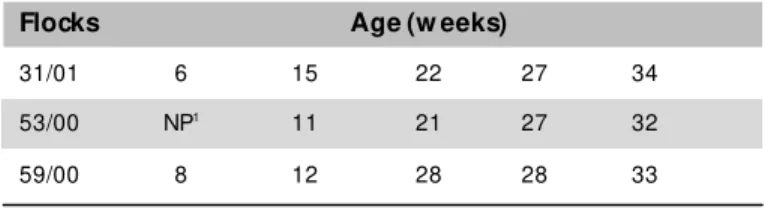
Table 1 – Age of sampling f or each st udied f lock.
Flocks Age (w eeks)
31/01 6 15 22 27 34
53/00 NP1 11 21 27 32
59/00 8 12 28 28 33
1-Not perf ormed.
Table 2 – Flocks age (w eeks) w hen M S-positive tests w ere detected.
Flocks PCR Isolation PSA HI
31/01 27 27 22 27
53/00 11 27 32 32
Polymerase chain reaction (PCR)
Tracheal sw abs w ere obtained from 10 birds per flock in each sampling, at ages indicated in Table 1. The samples w ere used in a polymerase chain reaction (PCR) t o det ect M S, in t he laborat ory of Embrapa Su ín o s A ves, in Concórdia, SC, according t o t he technique described by Lauerman et al. (1993). Sw abs w ith plastic stem and cotton tip w as introduced w ithin the trachea, the cotton tip w as immediately immersed in 0.5mL of phosphate buffered saline (PBS; 150mM NaCl, 2.6mM NaH2PO4, 7.4mM Na2HPO4, pH 7.5) and sen t t o t h e lab o rat o ry u n d er ref rig erat io n . Th is procedure has been described as adequate for sampling and samples w ere sent t o PCR analysis (Silveira et al., 1996). After shaking vigorously and boiling for 5 min, five micro liters of the buffer w as used in the PCR, w i t h 5 µ L o f sam p l e; 1 µ L o f t h e p r i m er (5 p M ) 5 ’ -d[GAGAAGCAAAATAGTGATATCA]3’; 1µL of the primer (5pM ) 5’-d[CAGTCGTCTCCGAAGTTAACAA]3’; and 14µL of a pre-mixture containing 10mM Tris-HCl pH 9.0; 50mM KCl; 1.5mM M gCl2; 200µM of each deoxyribonucleoside triphosphate (dATP, dCTP, dGTP and dTTP), w ater and 1.5 U of Taq polymerase, in a t o t al react io n vo lu m e o f 2 5 µ L. Th e t u b es w ere centrifuged for 30 s at 12,000 x g and denatured for one minute at 94°C, follow ing 40 cycles of 30 seconds at 94°C, 55°C and 72°C, and a final cycle of 5 minutes at 72°C. A positive control (M S culture) and a negative control (all components except DNA) w ere added to each reaction. Amplified fragments w ere submitted to electrophoresis (110V/45 min) in a 2% agarose gel in TE buffer (10mM Tris-HCl, 1mM EDTA, pH 7.5), and stained w ith ethidium bromide (10µg/mL in TE buffer) to compare w ith the pattern of the reference samples and a 100bp-molecular w eight standard. Samples w ere considered posit ive w hen a DNA f ragm ent w it h approximately 206 bp w as obtained.
M ycoplasma isolation
Tracheal sw abs w ere individually collected from 10 birds in each sampling and each age (Table 1). After sam p lin g , t h e sw ab s w ere in t ro d u ced in t u b es containing Frey broth (Frey et al., 1968) with 12% swine serum supplement ed w it h 0.1g/L of nicot inamide adenine dinucleotide (NAD) and 0.1g/L of cysteine hidrochloride hydrate, necessary for M S grow th, besides 1,000,000 IU penicillin G and 0.25g of thalium acetate per liter, to prevent opportunistic bacterial grow th. Tubes w ere vigorously shaken and sw abs w ere discarded
before the samples w ere sent, on ice, to the laboratory. Samples w ere incubated in a microanaerobiosis chamber at 37°C. Aliquots of each culture show ing acidification or turbidity in the broth, indicative of mycoplasma grow th, w ere plated in Frey agar (Frey broth w ith 0.75% agarose), every seven days for three consecutive times. After 21 days, cultures that show ed no colonies on the ag ar w er e d i scar d ed as n eg at i ve. A l l i so l at ed m yco p lasm as w er e su b m it t ed t o co lo n y immunofluorescence, using serum of rabbit immunized w ith the reference sample M S WVU 1853 and labeled w ith fluorescein isotiocyanate (Bradbury, 1998).
Each M S isolate w as cloned three times as follow s: one colony w as collected from the Frey agar plate to produce a liquid culture by incubating for 4 to 5 days in 1mL Frey broth; then, filtered in a 0.45µm filter, diluted and plated in Frey agar to obtain new isolated colonies, and successively for three times (Kleven, 1998). After the third cloning, cultures w ere confirmed as M S using PCR, identified by the lot number or the laboratory protocol and stored at -80°C until necessary.
D N A e x t r a ct i o n a n d Ra n d o m A m p l i f i e d Polym orphic DNA Analysis (RAPD)
of the vaccine sample M S H w as kindly provided by Dr. Phillip F. M arkham, from the University of M elbourne, Australia (M orrow et al., 1998b).
Pathogenicity of M S for SPF birds
A sample obtained from the same farm and w ith a RAPD profile identical to those from the studied flocks (M S 541, Figure 1) w as used for experimental infection. Eight SPF chicks (Spaf as) w ere placed in an isolat or w ith air filter, fed an antibiotic-free diet and inoculated in the 15th day of life w ith 0.1mL of a 24-hour culture in Frey broth, containing approximately 10¹¹ CFU. The inoculum w as sprayed ont o t he choanal clef t f or aspiration. Four w eeks later (sixth w eek of life), all birds w ere sacrificed, and sera w ere submitted to serology, PCR and isolation technique. Independent sw abs w ere collected from the trachea, air sacs and from the tarsus-metatarsial joint for PCR and M S isolation, and tested as described before. Samples from internal organs and t he ext ensor met at arsal t endon w ere collect ed f or histology (Luna, 1968).
RESULTS AND DISCUSSION
Flocks history
The grandparent s used t o produce t he st udied f locks show ed no posit ive result s in any SPA or HI
t est s perf ormed at approximat e int ervals of 6 w eeks t h r o u g h o u t r ear i n g an d p r o d u ct i o n p er i o d s. Nevert heless, 10 t racheal sw abs w ere collect ed f rom t he grandparent s at 19, 33 and 66 w eeks of age an d in d ivid u ally t est ed u sin g PCR. A ll o f t h em conf irm ed t he negat ive result s of t he f locks f or M S. These result s, t oget her w it h t he incubat ion of t he eggs in an single hat chery assured t hat t he chicks w ere M S-f ree w hen housed.
No clinical signs w ere observed in t he breeders t h ro u g h o u t t h e exp erim en t . Dif f eren ces in M S virulence m ay be experim ent ally det ect ed (Fiorent in et al., 1991; Lockaby et al., 1998), suggesting the exist ence of samples w it h higher or low er virulence. The results from the present investigation suggest that t he inf ect ing M S present in t his f arm has a very low virulence f or broiler breeders, or t hat it needs some ext ernal f act or t o f ully express it s pat hogenicit y, and such f act or w as not present .
Serology
All three breeder flocks w ere positive in SPA and HI for M S before egg production w as increasing, i.e., betw een 22 and 32 w eeks of age. Nevertheless, it w as noticed that a low number of sera w ere reactive in SPA, although the same sera w ere confirmed as positive by show ing titration in HI testing (Tables 3, 4 and 5). The delayed serological response detected in tw o flocks, w hich became positive by PCR only after 7 and 16 w eeks, together w ith the low reactive number of sera, indicat es t hat t he sensit ivit y of SPA is below t han expect ed and should be caref ully evaluat ed w hen establishing an epidemiological surveillance program for M S. It h as b een su g g est ed p revio u sly t h at M S surveillance should not be exclusively based in SPA, but it should be supported by PCR and attempts to isolate the agent (Ew ing et al., 1998).
M ycoplasma isolation
M S w as the only microorganism isolated from the tracheal sw abs analyzed. In this study, there w as no occurrence of other mycoplasmas from the trachea of the birds, such as M ycoplasma gallinarum,M ycoplasma gallinaceum and M ycoplasma pullorum, as previously reported (Bencina et al, 1987; Poveda et al, 1990). Other mycoplasmas w ith faster grow th are one of the possible reason for failure in M S isolation. Although no inference can be made on the presence or absence of other mycoplasmas in the trachea of the studied birds, their
apparent absence in t he sw abs w as responsible f or the reasonable levels of isolation (40% to 60% of the sw abs bet w een t he 26th and the 28th w eeks of lif e, Tables 3, 4 and 5).
M S isolation w as obtained only betw een the 27th to 28th w eeks of age suggesting that failures in mycoplasma isolation may be influenced by factors that are not generally considered. Thus, tracheal infection caused by M S can be latent in birds submitted to low stress level, such as that found during brooding or rearing.
During husbandry stressful situations, pre-laying vaccinations and the beginning of egg production, the infection may become acute and the number of viable mycoplasmas in the trachea increases, w hich facilitates the isolation in synthetic media.
M S isolation w as not perfectly correlated to serology data. Isolation w as seen at 27 w eeks in one flock (Flock 53), w hereas serology tests w ere positive at 32 w eeks of age. In another flock, serology w as positive at 22 w eeks of age but isolation w as only seen at 28 w eeks of age. This observation is important to re-evaluate the common practice of trying to isolate M S only if serology results become positive. In turkeys, it has been observed that the serology response to M S infection can be very w eak and that isolation of the agent might be very dif f icult (Kleven et al., 2001). Immunosuppression caused by Infectious Bursal Disease Virus aggravates the lesions caused by M S (Giambrone et al., 1977). Different degrees of temporary immunosuppression might have allow ed a higher invasion of M S, and a consequent positive serological response a posteriori.
PCR
PCR w as the most sensitive test, detecting the infection in tw o flocks (Flocks 59 and 53) 5 and 16 w eeks respectively earlier than positive results w ere seen in the other tests (Tables 3, 4 and 5). Positive results for all 10 sw abs in tw o samplings also suggest a high sensitivity of the PCR. A maximum of 60% sw abs w ere positive for M S-isolation in some w eeks, w hereas 100% of the tested sw abs w ere positive by PCR. There is the possibility that some birds show ing negative results in PCR are positive in the M S isolation (Salisch et al., 1998); how ever, PCR seems to have higher sensitivity w hen samples are considered as a group. M arois et al. (2000) reported that feces samples, feathers and dust collected from poultry houses w ith birds infected w ith M S may be positive by PCR. This finding strongly suggest a great probability of M S detection w hen sw abs w ere collected from the trachea. This information, allied to the results
obtained in this study, indicate for the necessity of ad o p t in g PCR as t h e ro u t in e t ech n iq u e in t h e epidemiological surveillance of M S in broiler breeders.
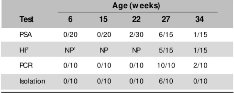
Table 3 – Positive samples/total samples for different tests at each sampling (Flock 31).
Age (w eeks)
Test 6 15 22 27 34
PSA 0/20 0/20 2/30 6/15 1/15 HI2 NP1 NP NP 5/15 1/15
PCR 0/10 0/10 0/10 10/10 2/10 Isolation 0/10 0/10 0/10 6/10 0/10
1- NP: Not performed.
2- HI w as performed only in SPA-positive sera.
Table 4 – Positive samples/total samples for different tests at each sampling (Flock 53).
Age (w eeks)
Test 11 21 27 32
PSA 0/30 0/30 0/30 2/30 HI2 NP1 NP NP 2/2
PCR 6/10 10/10 4/10 6/10 Isolation 0/10 0/10 5/10 1/10
1- NP: Not performed.
2- HI w as performed only in SPA-positive sera.
Table 5 – Positive samples/total samples for different tests at each sampling (Flock 59).
Age (w eeks)
Test 8 12 22 28 33
PSA 0/30 0/30 1/30 0/30 1/30 HI2 NP1 NP 0/1 NP 1/1
PCR 0/10 0/10 3/10 10/10 4/10 Isolation 0/10 0/10 0/10 4/10 0/10
1- NP: Not performed.
2- HI w as performed only in SPA-positive sera.
RAPD
Table 6 – Number of posit ive result s in each t echnique f or samples collect ed f rom SPF birds inoculat ed w it h M S 451.
Test Positive
PSA 8/8
HI 8/81
Trachea - PCR 5/8
Air sac – PCR 3/8
M etatarsal extensor tendon - PCR 1/8 Trachea - isolation 7/8 Air sac - isolation 0/8 M etatarsal extensor tendon - isolation 0/8
1- Geometric mean of titers (GM T): 146.7.
am p lif icat io n p at t ern s in RA PD u sin g t h e p rim er 5 ’ - d [ GTA GA CCCGT] 3 ’ (A m er sh am Ph ar m aci a Bio t ech ) in d icat ed t h at t h e M S sam p les iso lat ed f ro m t h e sam e f arm w ere sim ilar. On t h e o t h er h an d , t h ey w ere d if f eren t f ro m t h o se o b t ain ed in ot her f arm s, f rom t he t herm olabile vaccine sam ple M S H, an d also f ro m t h e ref eren ce sam p le M S W VU 1853, w hich w as used as a diagnosis ant igen (Fi g u r e 1 ). N o co n si d er ab l e d i f f er en ce w as o b served in RA PD p at t ern s am o n g t h e sam p les st u d ied h ere an d f ro m t h e p at t ern sh o w ed b y sam ples obt ained f rom ot her f locks t hat had been h o u sed in t h e sam e f arm in recen t years. Th is su g g est s t h at t h e sam e sam p le o f M S h as b een in t h e f ar m as i n f ect i n g ag en t . A l l M S- p o si t i ve sam p les sh o w ed t h e sam e RA PD p ro f ile an d seem t o b e t h e sam e.
RA PD r esu l t s sh o w ed t h at t h e p r i m er 5 ’ -d [ G TA G A C C C G T] 3 ’ m a y b e v e r y u se f u l i n ep i d em i o l o g i cal st u d i es i n Br azi l , m o st l y i n t rack in g f ield sam p les an d id en t if yin g vaccin e sam ples.
M S inoculation in SPF birds
As seen for the follow -up breeder flocks, no clinical sign w as seen in the SPF birds inoculated w ith a M S isolate. No lesion w as seen during necropsy in the fourth w eek after inoculation, w hen the birds w ere six-w eeks o ld . Ser o lo g ical co n ver sio n w as n ever t h eless considerable, contrary to the results from the field infected flocks. The high infecting dose (1011 per bird) used in the experiment probably caused this difference, but the potential immunogenicity of the studied M S w as evident.
Re-isolation of M S w as possible from the tracheas. PCR w as positive for trachea and air sac sw abs, and also in one sw ab collected from the tarsus-metatarsal joint (Table 6). Since PCR sensitivity is so high, a positive result for a joint sw ab must be carefully considered, because contamination might have occurred during necropsy and material collection.
Histology show ed lymphoid nodules in the trachea and lung, a cuboidal metaplasia and parabronchi lumen stenosis. No lesion w as observed in tendons or air sacs. The lesions and the higher number of M S in the trachea suggest t hat t he st udied M S caused an inf ect ion primarily in that organ. Infections caused by M S w ith these features have been reported (Droual et al., 1992), although the higher number of case reports refers to joint lesions (M orrow et al., 1998a; Lockaby et al., 1998).
CONCLUSIONS
M S infection had a predictable profile w hen free flocks w ere introduced in a farm w ith infection history, beginning at rearing and show ing no clinical signs. There ar e g r eat er ch an ces o f o b t ain in g evid en ce o f assymptomatic infection using PCR in the first w eeks of lif e, or t rying t o isolat e M S in t he onset of egg production. Detection of M S infection based only in serology tests has low reliability and PCR is suggested as the routine technique for epidemiological surveillance.
REFERENCES
Bashiruddin JB. Extraction of DNA from M ycoplasmas. In: R. M iles, R. Nicholas (editors). M ycoplasma Protocols. Humana Press. 1998. pp.141-144.
Bencina D, Dorrer D, Tadina T. M ycoplasma species isolation from six avian species. Avian Pat hology 1987; 16:653-664.
Bradbury J. Identification of M ycoplasmas by Immunofluorescence. In: R. M iles, R. Nicholas (editors). M ycoplasma Protocols. Humana Press. 1998. pp.119-126.
Droual R, Shivaprasad HL, M eteyer CU, Shapiro DP, Walker RL. Severe mortality in broiler chickens associated w ith M ycoplasma synoviae and Pasteurella gallinarum. Avian Diseases 1992; 36:803-807.
Ew ing M L, Cookson KC, Phillips RA , Turner KR, Kleven SH. Experimental infection and transmissibility of M ycoplasma synoviae w it h delayed serologic response chickens. Avian Diseases 1998; 42:230-238.
Frey M LL, Hanson LP, Anderson DP. A medium for the isolation of avian mycoplasmas. American Journal of Veterinary Research 1968; 19:2163-2171.
Giambrone JJ, Eidson CS, Kleven SH. Effects of infectious bursal disease on t he response of chickens t o M ycoplasm a synoviae, New castle disease virus, and infectious bronchitis virus. American Journal of Veterinary Research 1977; 38:251-253.
Kleven SH. M ycoplasma synoviae infection. In: BW Calnek, HJ Barnes, CW Beard, WM Reid and HW Yoder (editors). Diseases of Poultry,
10th edit ion. 1997.pp. 220-228.
Kleven SH. M ycoplasmosis. In: DE Sw ayne (editor). A Laboratory M anual f or t he Isolat ion and t he Ident if icat ion of Avian Pat hogens,
4th edit ion. 1998. pp. 74-80.
Kleven SH, Row land GN, Kumar M C. Poor serologic response to upper respiratory infection w ith M ycoplasma synoviae in turkeys. Avian Diseases 2001; 45:719-23.
Lauerman LH, Hoerr FJ, Sharpt on AR, Shah SM , Van Sant en L. Development and application of a polymerase chain reaction assay for M ycoplasma synoviae. Avian Diseases 1993; 37:829-834.
Ley DH, M cLaren JM , Berkhoff JE, Levisohn S. M ycoplasma synoviae strain identification by Random Amplified Polymorphic DNA (RAPD) Analyses. IOM Let t ers 1998; 5:117.
Lockaby SB, Hoerr FJ, Lauerman HL, Kleven SH. Pathogenicity of M ycoplasma synoviae in broiler chicken. Veterinary Pathology 1998; 35:178-190.
Luna LG. M anual of histologic staining methods of the Armed Forces Institute of Pathology. 3ed. New York (NY): M cGraw -Hill; 1968.
M arois C, Oufour-Gesbert F, Kempf I. Detection of M ycoplasma
synoviaein poultry environment samples by culture and polymerase
chain reaction. Veterinary M icrobiology 2000; 73:311-318.
M orrow CJ, M arkham JF, Whithear G. Production of
temperature-sensit ive clones of M ycoplasma synoviae f or evaluat ion as live
vaccines. Avian Diseases 1998a; 42:667-670.
M orrow CJ, Bell IG, Walkers SB, M arkham PF, Thorp BH, Whitehear KG. Isolat ion of M ycoplasma synoviae f rom inf ect ious synovit is of chickens. Australian Veterinary Journal 1998b; 67:121-124.
Poveda JB, Carranza J, M iranda A, Garrido A, Hermoso M , Fernandez A, Domenech J. An epizootiological study of avian mycoplasmas in Sout hern Spain. Avian Pat hology 1990; 10:627-633.
Salisch H, Hinz KH, Graack HD, Ryll M . A comparison of a commercial PCR-based test to culture methods for detection of M ycoplasma
gallisept icumand M ycoplasma synoviaein concurrently infected
chickens. Avian Pathology 1998; 27:142-147.
Silveira RM , Fiorentin L, M arques EK. Polymerase chain reaction
opt im izat ion f or M ycoplasm a gallisept icumand M ycoplasm a