R E V I E W
Open Access
Alpha-type phospholipase A
2
inhibitors
from snake blood
Norival A. Santos-Filho
1*and Claudia T. Santos
2Abstract
It is of popular and scientific knowledge that toxins from snake venom (among them the PLA2and myotoxins) are neutralized by various compounds, such as antibodies and proteins purified from animal blood. Venomous and nonvenomous snakes have PLA2inhibitory proteins, called PLIs, in their blood serum. One hypothesis that could explain the presence of these PLIs in the serum of venomous snakes would be self-protection against the enzymes of their own venom, which eventually could reach the circulatory system. However, the presence of PLIs in non-venomous snakes suggests that their physiological role might not be restricted to protection against PLA2toxins, but could be extended to other functions, as in the innate immune system and local regulation of PLA2s. The present study aimed to review the currently available literature on PLA2and myotoxin alpha inhibitors present in snake plasma, thus helping to improve the research on these molecules. Furthermore, this review includes current information regarding the mechanism of action of these inhibitors in an attempt to better understand their application, and proposes the use of these molecules as new models in snakebite therapy. These molecules may help in the neutralization of different types of phospholipases A2and myotoxins, complementing the conventional serum therapy.
Keywords:Phospholipases A2, Myotoxin, Myotoxin inhibitor,αPLI, Snake blood
Background
Between 2009 and 2013, the World Health Organization (WHO) included envenomation by snakes among the neglected tropical diseases given the large number of ac-cidents, the complexity of the clinical condition and the fact that the most affected population consists mainly of workers from poor rural communities in tropical coun-tries [1–4]. However, nowadays experts in Toxinology call on WHO and governments to re-establish snakebite as a neglected tropical disease, since each year, approxi-mately 421,000 cases of snakebite occur, of which ap-proximately 20,000 result in death [5].
Generally, the lethality of bites is low, though the fre-quency of sequelae related to local complications is higher, especially when associated with risk factors such as the use of a tourniquet, bite in extremities (fingers and toes) and delayed treatment [6]. It is important to note that some sequelae – especially those that lead to
partial or total limb amputation– despite been a public health problem, also constitute social problems, since they may provoke various disorders, including the dis-ability to work [5]. Snake venoms are a complex mixture of components, and more than 90% of their dry weight consists of proteins with a large variety of enzymes, and a non-protein portion comprising carbohydrates, lipids, metals, free amino acids, nucleotides and others [7]. The protein components of snake venoms include cytotoxins, cardiotoxins, nerve growth factors, lectins, enzyme in-hibitors and various enzymes, such as phospholipase A2 (PLA2), metalloproteases, serine proteases, phosphodies-terases, cholinesphosphodies-terases, aminotransferases, L-amino acid oxidases, catalases, ATPases, hyaluronidases, etc. [8].
Thus, considering the search for natural inhibitors that neutralize snake venom toxins is of extreme importance for the production of more efficient antivenoms, the present study aims to review the currently available lit-erature on alpha inhibitors present in snake plasma, thus helping to improve the current knowledge about these molecules.
* Correspondence:[email protected]
1Institute of Chemistry, São Paulo State University (UNESP
–Univ Estadual Paulista), Araraquara, SP, Brazil
Full list of author information is available at the end of the article
Phospholipases A2(PLA2)
Phospholipases are a superfamily of enzymes that act on phospholipids in the cell membrane leading to their cleav-age in fatty acids and lysophospholipids. Phospholipases A2(PLA2) (EC 3.1.1.4) were the first phospholipases to be known and their discovery was based on observation of the action of pancreatic fluid of mammals and snake venom in the hydrolysis of phosphatidylcholine [9].
These enzymes play an important role in several cellular functions including maintenance of cellular phospholipids, generation of prostaglandins (PGs) and leukotrienes, cell proliferation and muscle contraction. Furthermore, it is known that these enzymes are involved in human inflamma-tory processes and due to their central role in many cellular processes, they have been extensively studied [7, 10–12].
The PLA2s are a superfamily of enzymes belonging to 16 groups and subgroups that can also be divided into six distinct types: the secreted PLA2 (sPLA2), among them PLA2s found in snake venoms; the cytosolic PLA2 (cPLA2); the Ca2+ independent PLA2s (iPLA2); the acetyl-hydrolases activating factors of platelets (PAF-AH); lysosomal PLA2and the lipoprotein-associated phospho-lipase A2(Lp-PLA2) [13, 14].
According to Schaloske and Dennis [13] and Dennis et al. [14], the sPLA2s are enzymes with a molecular weight between 14,000 and 18,000 Da, usually containing from 5 to 8 disulfide bridges. These enzymes have a histidine in their active site and require the presence of Ca2+ion for catalysis. The phospholipase A2 from groups IA, IB, IIA, IIB, IIC, IID, IIE, IIF, III, V, IX, X, XIA, XIB, XII, XIII and XIV are representatives of sPLA2s.
The PLA2s from snake venoms (svPLA2s) are classified into groups I and II, and those from the Viperidae family belong to group IIA [11, 13–15]. The svPLA2s belonging to group IIA are subdivided into subgroups based on the presence of a conserved residue on position 49, being the most studied: (i) PLA2s Asp49, enzymes that usually have high catalytic activity, and (ii) homologous PLA2s (or PLA2-like) Lys49, which have no enzymatic activity [16, 17]. It is important to point out that other variants in snake venom group II PLA2s have been reported, e.g., Ser49, Asn49 and Arg49 [18–23].
Interestingly, despite having no catalytic activity, the homologous PLA2s Lys49 have a wide variety of pharma-cological and/or toxic effects, including myotoxicity, cyto-toxicity, antibacterial, antifungal, muscle necrotic and anticoagulant activities [7, 24–27]. According to some au-thors, the main structural domain responsible for the toxic effect, particularly cytotoxic, in homologous Lys49-PLA2 is the C-terminal region (amino acids 115–129) [27].
PLA2inhibitory proteins (PLIs) from snake blood
Venomous and non-venomous snakes have PLA2 inhibi-tory proteins, called PLIs, in their blood serum [28–30].
These PLA2 inhibitory proteins are produced by the liver, as indicated by Northern blot analysis and RT-PCR analysis of genetic material extracted from different tis-sues. This PLI production by the liver (and not by the venom glands or other organ) makes it possible for these proteins to enter the bloodstream, since the liver is the main organ producing plasma proteins, thus improving and accelerating the protection mechanism against poi-soning [31–33]. Furthermore, it has been known that some secreted PLA2receptors, which have structural simi-larity with PLIs, also exist in soluble forms, showing that PLIs, as well as PLA2endogenous receptors, could have a regulatory role of proinflammatory activity of sPLA2s [34]. Several PLIs were purified from the plasma of different species of snakes, and their structures have been deter-mined [28–30, 34, 35]. So far, for the isolation of PLA2 in-hibitors described in the literature, two different methods were used. One of these purification methods is the bioaffi-nity chromatography, which is based on the immobilization of different proteins, PLA2 in this case (for example BthTX-I and BthTX-II, from Bothrops jararacussu), on a stationary phase [32, 36–40]. Another method used in purification of PLIs from snake plasma is a sequence of chromatographic steps such as gel filtration, ion ex-change and hydrophobic chromatography [35, 41, 42].
The blood used for plasma separation is typically col-lected by cardiac puncture, by puncturing the tail vein or after decapitation of the snake. It is noteworthy that in re-cent years concern about the ethics in the use animals for experimentation is growing and therefore the least aggres-sive method that does not require animal death is the blood collection from the tail vein of the snake, being the most indicated. After collecting the blood, plasma and serum are separated, then plasma is lyophilized and stored. During purification, the inhibitory activity of these PLIs is monitored by biological assays based on inhibition activity of PLA2 and myotoxins, depending on the inhibitor of interest.
The PLA2and myotoxin inhibitors from the blood of snakes are globular, acid and oligomeric proteins, which form soluble complexes with PLA2and myotoxins, thus inhibiting the action of these molecules [34, 43–46]. Blood inhibitors found in snakes are classified into types alpha (α), beta (β) and gamma (γ) according to structural aspects [30, 47, 48].
One of the PLIs classes, the βPLIs, have repeated leucine-rich structures and show similarity to humanα 2-glycoprotein [49].βPLIs inhibit only basic group II PLA2s isolated from snake venoms and have been isolated from plasma ofAgkistrodon blomhoffii siniticus,Elaphe quadri-virgata andE. climacophora snakes, which belong to the Viperidae and Colubridae family [33, 49, 50].
with a mass of 90–130 kDa consisting of 3 to 6 noncova-lent subunits. Their amino acid sequences contain two sets of standards cysteine residues, responsible for the formation of the three-finger motif [51]. This type of in-hibitor has been reported in different snakes, asCrotalus durissus terrificus [52–54],Naja naja kaouthia [55, 56],
Agkistrodon blomhoffii siniticus [57],Trimeresurus flavo-viridis[58], Laticauda semifasciata [59],Elaphe quadri-virgata [60], E. climacophora [50], Cerrophidion godmani[32], Notechis ater, Notechis ater serventyi[61],
Oxyuranus scutellatusand O. microlepidotus [61], Pseu-donaja textilis[61],Python reticulates[62],Notechis scu-tatus [63], Lachesis muta muta [64], Protobothrops flavoviridis[65], Bothrops alternatus, B. erythromelas, B. jararaca, B. moojeni, B. neuwiedi [51], Bothrops jarara-cussu [39] and Crotalus durissus collilineatus [66] and these γPLIs appear to be less specific, since they inhibit PLA2from groups I, II and III.
Alpha-type PLA2inhibitor
The alpha-type PLA2 inhibitors (αPLIs) from the snake blood are found mainly as trimers in solution and have a region with high similarity with the carbohydrate recog-nition domain (CRD) of C-type lectins and pulmonary surfactant protein [30, 36, 37, 40, 67–70]. This region covers approximately 67% of the primary sequence of the monomers ofαPLIs and is the most conserved por-tion of these molecules, with approximately 46% of se-quence identity between species [30]. The CRD ofαPLIs lacks the amino acid residues involved in Ca2+ binding, making the interaction with their respective ligands Ca2 +
-independent [40, 42]. Moreover, several studies have
shown that the carbohydrate motif present in αPLIs is not necessary for the connection with PLA2[32, 38].
αPLIs studied to date
Various αPLIs were purified to date (Table 1), such as the plasma PLI from the snakeTrimeresurus flavoviridis, which was purified by a combination of chromatographic steps through Sephadex gel filtration column G-200, DEAE-cellulose anion exchange and Blue Sepharose CL-6B [41]. The purified inhibitor was found as a glycoprotein with an approximately molecular weight of 100,000 Da, with non-homologous subunits of approxi-mately 20,000 to 24,000 Da. Subsequently, it was verified the ability of this inhibitor to interact with venom phospholipase A2ofT. flavoviridis, andAgkistrodon halys blomhoffii, besides the enzyme and the porcine pancreatic phospholipase C ofBacillus cereus. According to Kogaki et al. [41], this inhibitor showed specificity toT. flavoviri-disPLA2, and an independent inhibitory activity of Ca2+.
Afterward, Inoue et al. [67] purified two distinct but homologous subunits (PLIα-A and PLIα-B) of the PLI fromTrimeresurus flavoviridis. These subunits were sep-arated by reversed-phase HPLC and showed molecular weights around 21,000–22,000 Da when glycosylated and 17,000 after deglycosylation. Furthermore, the se-quences were significantly homologous to CRD portions of pulmonary surfactant apoprotein and animal lectins. Then, Shimada et al. [71] studied this αPLI, which was purified into different subspecies of two homologous subunits. Before this work, it was expected that this
αPLI was a tetramer, composed of two molecules of
αPLI-A and two molecules of αPLI-B [67]. However, in
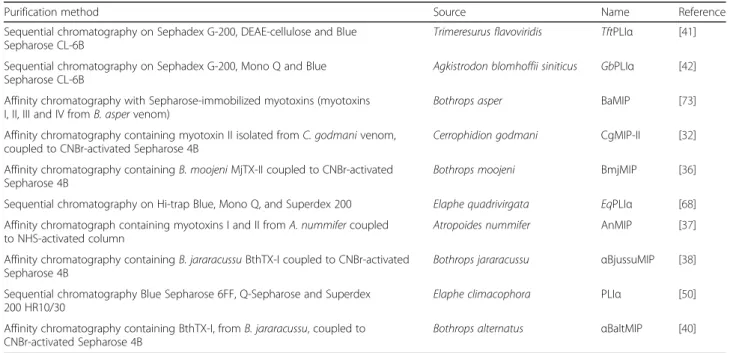
Table 1Alpha-type PLA2inhibitors (αPLIs) studied to date
Purification method Source Name Reference
Sequential chromatography on Sephadex G-200, DEAE-cellulose and Blue Sepharose CL-6B
Trimeresurus flavoviridis TftPLIα [41]
Sequential chromatography on Sephadex G-200, Mono Q and Blue Sepharose CL-6B
Agkistrodon blomhoffii siniticus GbPLIα [42]
Affinity chromatography with Sepharose-immobilized myotoxins (myotoxins I, II, III and IV fromB. aspervenom)
Bothrops asper BaMIP [73]
Affinity chromatography containing myotoxin II isolated fromC. godmanivenom, coupled to CNBr-activated Sepharose 4B
Cerrophidion godmani CgMIP-II [32]
Affinity chromatography containingB. moojeniMjTX-II coupled to CNBr-activated Sepharose 4B
Bothrops moojeni BmjMIP [36]
Sequential chromatography on Hi-trap Blue, Mono Q, and Superdex 200 Elaphe quadrivirgata EqPLIα [68]
Affinity chromatograph containing myotoxins I and II fromA. nummifercoupled to NHS-activated column
Atropoides nummifer AnMIP [37]
Affinity chromatography containingB. jararacussuBthTX-I coupled to CNBr-activated Sepharose 4B
Bothrops jararacussu αBjussuMIP [38]
Sequential chromatography Blue Sepharose 6FF, Q-Sepharose and Superdex 200 HR10/30
Elaphe climacophora PLIα [50]
Affinity chromatography containing BthTX-I, fromB. jararacussu, coupled to CNBr-activated Sepharose 4B
this last study, it was showed that thisαPLI is a trimeric protein. Curiously, all the αPLIs except that from P. flavoviridisare multimers composed of a single subunit.
Ohkura et al. [42] purified an alpha inhibitor from the snake Agkistrodon blomhoffii siniticus, using a similar method described by Kogaki et al. [41]. In this case, this
αPLI purification was performed by sequential chroma-tography through Sephadex G-200 column, Mono Q and Sepharose Blue CL-6B. The purified PLI showed up as a glycoprotein with a molecular mass from 75,000 to 24,000 Da for the trimer and the monomer.
After, Inoue et al. [72] studied the specificity of the two previously purified (and cited above) PLA2inhibitors from
T. flavoviridis and A. b. siniticus plasma, purified by Kogaki et al. [41], and Ohkura et al. [42], respectively. Both αPLI showed a high specificity for group II acidic PLA2s from their own venom. In this work, the authors draw a parallel between PLI from snake plasma and PLA2 receptors of rabbit, bovine, and human, suggesting that the CRD-like domain would be involved in the binding to the PLA2molecule.
Regarding theαPLI fromBothrops genus, otherα inhibi-tors were purified, for example, BaMIP, a PLI isolated from the plasma ofBothrops asper by affinity chromatog-raphy in Sepharose 4B CNBr-activated with myotoxins immobilized [73]. BaMIP presented monomers with a mo-lecular weight of approximately 24,000 Da and a structure in solution composed of five subunits. The BaMIP showed inhibition on myotoxic, edema and cytolytic activity of the myotoxins I and III ofB. asper snake. Structural studies have also shown that BaMIP, as well as allαphospholipase A2inhibitors has a homologous domain to CRD of C-type lectins.
Another snake inhibitor studied is CgMIP-II, anαPLI, purified from plasma ofCerrophidion (Bothrops) godmani
snake by affinity column containing myotoxins [32]. The inhibitor is an acidic protein (pI 4.0), glycosylated, the monomeric subunits with a molecular weight between 20,000 Da and 25,000 Da, forming a polymer of about 180,000 Da.
Soares et al. [36] purified a protein that neutralizes the enzymatic, toxic and pharmacological activity of a variety of toxins (acidic or basic) of different venoms. This inhibi-tor, called BmjMIP, was isolated from the plasma of the snake Bothrops Moojeni, by affinity chromatography. BmjMIP presented similar biochemical and structural characteristics to those already described forαPLIs, besides being stable at a wide range of pH and temperature.
Okumura et al. [68] purified the αPLI-like protein (PLIα-LP) from a non-venomous snake E. quadrivirgata
serum by sequential chromatography on Hi-trap Blue, Mono Q and Superdex 200 columns. The PLIα-LP showed the highly conserved C-type lectin-like domain (CTLD) and 51 kDa, being a trimer. Although this protein has
about 70% similarity with other inhibitors previously stud-ied, this protein did not demonstrate any inhibitory activity against different PLA2s. It is important to cite that Shirai et al. [50] also purified anαPLI-like protein (PLIα-LP) from
E. climacophora snake. According to Okumura et al. [68], the high homology with αPLIs and the lack of inhibitory activity on αPLI-like proteins may provide important information concerning the structure/function of these
αPLIs.
Quirós et al. [37], purified anαPLI (AnMIP) from the plasma of Atropoides nummifer by affinity matrix, pre-pared by coupling a mixture of myotoxins I and II from
A. nummiferto an NHS-activated column. According to the work, this trimeric inhibitor neutralized the activity of basic PLA2myotoxins and showed specificity towards group II PLA2, either belonging to the catalytically active (Asp49 PLA2) or inactive (Lys49 PLA2-like) subtypes.
Oliveira et al. [38] and Santos-Filho et al. [40] purified two different αPLIs (namedαBjussuMIP andαBaltMIP), fromB. jararacussuandB. alternatus, respectively. These molecules were purified through affinity chromatography using BthTX-I immobilized on Sepharose gel and neutralize enzymatic, toxic and pharmacological activ-ities of several phospholipases A2. Santos-Filho et al. [74, 75] subsequently expressed an active recombinant alpha inhibitor, named rBaltMIP, inPichia pastoris heter-ologous system. According to these works, heterheter-ologous expression would enable large-scale obtainment of these
αPLI, thus allowing further investigations for the elucida-tion of possible mechanisms of inhibielucida-tion of PLA2s, which have not yet been fully clarified.
Mechanism of action ofαPLIs
In the last 30 years, several studies have been published aiming to biochemically, structurally and functionally characterize αPLIs. However, the mechanism of action of these αPLIs is still unknown. Some authors have suggested that the αPLI/PLA2binding site is probably related to the CRD region of the molecule, which recog-nizes and binds to the enzyme, preventing its toxic activ-ity. One factor that supports this idea is that these CRD domains are present in endogenous PLA2receptors, such as the human receptor of group I pancreatic PLA2and receptors of group II secretory PLA2 from rabbits, mice, cattle and humans [38, 73, 76–78]. Nevertheless, the molecular nature of the interaction between the CRD region and PLA2is still unknown and efforts towards the elucidation of the structure ofαPLIs and their complexes are being performed [30].
hydrophobic tripeptides and Tyr144 residue appear to be involved in the interaction PLI/PLA2[37, 69, 79].
Thereafter, Okumura et al. [69] studied the relationship of the structure/function of the αPLI previously purified from the snake Agkistrodon blomhoffii siniticus, named GbPLIα, and the αPLI-like protein EqPLIα-LP, purified from the nonvenomous snake Elaphe quadrivirgata, and which does not show inhibitory activity against PLA2s [42, 68]. In that work, by constructing chimeric proteins, they mapped important residues to the inhibitory activity of theαPLIs; for example, the region 13-36 of the neck C-terminal portion of the trimer. Interestingly, the re-gion found as the responsible for PLA2 inhibition was distinct from the carbohydrate-binding site. Furthermore, other residues were pointed as candidate, including Asn26, Lys28, Asp29, and Tyr144 [69].
According to Okumura et al. [69], the trimer is formed through the interactions of the helical neck regions, forming a central pore, responsible for PLA2 binding. Furthermore, as Tyr144 is expected to be located in this central pore, this residue may be one of the responsibles for the direct interaction to the PLA2 molecule. In a complementary study, Nishida et al. [70] created hetero-trimers ofαPLI composed of two different subunits de-rived from the recombinant GbPLIα, EqPLIα-LP, and chimeras of GbPLIα-EqPLIα-LP homotrimers, in order to estimate the contribution of each subunit to the total inhibitory activity as a trimeric PLA2inhibitory protein. Summing up, in this work, it was observed, once more, the importance of the residues 13–36 for the trimer for-mation, and consequently for theαPLI inhibitory activity. Furthermore, the interactions between residues Glu23 and
Lys28 of GbPLIαwere also suggested to be important to stabilize the trimeric structure.
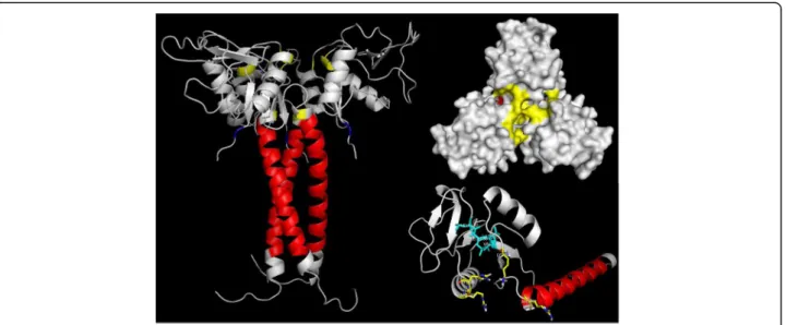
Lastly, in a recent study, Estevão-Costa et al. [80] stud-ied the importance ofαPLI trimerization for the binding and inhibition to acidic PLA2s. Furthermore, they sug-gested that the central pore, which is composed by posi-tive charged residues, especially Arg57, Lys71, Arg108 and His109, could be a significant part of the binding site of
αPLIs to acidic PLA2s. In addition, these authors pointed the importance of the hydrophobic core (Leu158 to Val161), which may be the responsible for the central pore structural integrity. However, the positive surface of the basic PLA2could prevent the PLA2/PLI interaction at the central pore and according to these authors, the mechan-ism of inhibition of basic PLA2by αPLIs remains to be understood. It is interesting to point out that, considering the sequence of the native protein, obtained through Edman degradation sequencing [40], the numbering of central pore important residues should be Arg38, Lys52, Arg89 and His90 (Fig. 1).
So far, it is possible to observe that the mechanism of action of these inhibitors and the region responsible for their inhibitory properties are not yet fully elucidated in the literature, requiring further study concerning these macromolecules and their interactions with PLA2s.
Potential complement of antiophidic serum therapy
Currently, antiserum composed of specific immunoglob-ulins is the only treatment for snake envenomation, but there are ongoing issues with availability, effectiveness and dosing [81–83]. These antivenoms neutralize the toxicity and lethality of specific venoms, but their administration
is often related with significant clinical side effects [84, 85]. Additionally, the production of antivenoms is associ-ated with high costs relassoci-ated to animal maintenance and also comes across animal welfare concerns, which insti-gates the search for innovative products for snakebite therapy [82, 86].
Interestingly, the production of specific antivenom was started by Vital Brazil in the 1900’s and it was Vital Brazil who also discovered the effectiveness of the poly-valent antivenom [87, 88]. At that time, antivenom was prepared with crude plasma of hyperimmunized animals. However, it was thereafter discovered that antibodies (immunoglobulins) were the active therapeutic molecules responsible for the action of the antivenom. Therefore, only the antibodies started to be purified and used in anti-venom therapy.
Nowadays, despite advances in the production of anti-venoms, this production is still similar to the methods originally described by Vital Brazil [87, 88]. Currently, immunoglobulins or immunoglobulin fragments [F(ab’)2 or Fab] purified from serum are used in antivenom [2]. Other innovations have been proposed on traditional antiserum, as the use of the single chain variable fragment (scFv) or the use of recombinant antigen binding domains derived from camelid heavy chain antibodies (VHH) [82, 89–91]. However, there are numerous challenges on antivenom improvement, for example, the high cost of monoclonal antibodies production or the lower affinity and the short serum half-life profiles of some immuno-globulin fragments [82, 92].
Although serum therapy effectively reverses the systemic effects of venom into the victim’s body, avoiding death many times, it has some disadvantages including a number of side effects (anaphylactic shock, renal failure and serum sickness, for example). The inefficiency to combat the local effects of the envenomation (increasing the chances of sequelae in the stricken member), the need for careful storage and the short shelf life of the serum are also other limiting factors.
PLA2enzymes and PLA2-like myotoxins are the main responsible for myonecrosis, an important medical com-plication of snake envenomation, and which, in severe cases can lead to drastic consequences such as perman-ent loss of tissue or limb amputation. These outcomes provoke severe problems for both the affected individual and public health, since the victim may become incap-able of working and lose life quality. In addition, these sequelae burden the public health once they increase the length of hospitalization and surgeries and, in some cases, can lead to early retirement of the individual af-fected by the envenomation.
The search for natural inhibitors that neutralize snake venom toxins is of extreme importance for the produc-tion of more efficient antivenoms, especially considering
that several toxins induce weak immunogenic responses, making traditional serum therapy unable to inhibit local effects such as the myotoxicity induced by phospholi-pases A2and PLA2-like enzymes [46, 93].
Conclusions
In conclusion, the traditional antivenom is not completely able to inhibit local effects of envenomation, mainly caused by myotoxins. Thus, the search for proteins, such as αPLIs, that neutralize myotoxins present in snake venom is extremely important for the production of a more efficient treatment.
Abbreviations
cPLA2:Cytosolic PLA2; CRD: Carbohydrate recognition domain; CTLD: C-type lectin-like domain; iPLA2: Ca2+ independent PLA2s; Lp-PLA2: Lipoprotein-associated phospholipase A2; PAF-AH: Acetyl-hydrolases activating factors of platelets; PG: Prostaglandin; PLA2: Phospholipase A2; PLI: PLA2 inhibitory proteins; sPLA2: Secreted PLA2;αPLI: Alpha-type PLA2 inhibitor
Acknowledgements
The authors are grateful to Dr. Lucas Blundi Silveira for the careful and meticulous correction of this paper, scientific support and essential discussion upon the completion of the manuscript. Thanks are also due to the Center for the Study of Venoms and Venomous Animals (CEVAP) of UNESP for enabling the publication of this paper (Edital Toxinologia CAPES no. 063/2010, Process no. 230.38.006285/2011-21, AUXPE Toxinologia 1219/2011).
Funding
This work was supported by the São Paulo Research Foundation (FAPESP).
Authors’contributions
Both authors contributed with data compilation, writing and critical discussion of the manuscript. Both authors read and approved the final manuscript.
Competing interests
The authors declare that they have no competing interests.
Consent for publication Not applicable.
Ethics approval and consent to participate Not applicable.
Publisher’s Note
Springer Nature remains neutral with regard to jurisdictional claims in published maps and institutional affiliations.
Author details
1Institute of Chemistry, São Paulo State University (UNESP
–Univ Estadual Paulista), Araraquara, SP, Brazil.2School of Pharmaceutical Sciences, São Paulo State University (UNESP–Univ Estadual Paulista), Araraquara, SP, Brazil.
Received: 21 October 2016 Accepted: 16 March 2017
References
1. Harrison RA, Hargreaves A, Wagstaff SC, Faragher B, Lalloo DG. Snake envenoming: a disease of poverty. PLoS Negl Trop Dis. 2009;3(12):e569. 2. WHO. WHO Expert Committee on Biological Standardization. 59threport.
WHO Technical Report Series, no. 964. 2012. Available: http://www.who.int/ biologicals/WHO_TRS_964_web.pdf.
4. Chippaux JP. Epidemiology of envenomations by terrestrial venomous animals in Brazil based on case reporting: from obvious facts to contingencies. J Venom Anim Toxins Incl Trop Dis. 2015;21:13. 5. Bagcchi S. Experts call for snakebite to be re-established as a neglected
tropical disease. BMJ. 2015;351:h5313.
6. Warrell DA. Venomous bites, stings, and poisoning. Infect Dis Clin North Am. 2012;26(2):207–23.
7. Santos-Filho NA, Silveira LB, Oliveira CZ, Bernardes CP, Menaldo DL, Fuly AL, et al. A new acidic myotoxic, anti-platelet and prostaglandin I2inductor phospholipase A2isolated fromBothrops moojenisnake venom. Toxicon. 2008;52(8):908–17.
8. Tu AT. Overview of snake venom chemistry. Adv Exp Med Biol. 1996;391:37–62. 9. Barros GA, Pereira AV, Barros LC Jr AL, Calvi SA, Santos LD, et al.In vitro
activity of phospholipase A2and of peptides fromCrotalus durissus terrificus venom against amastigote and promastigote forms ofLeishmania(L.) infantum chagasi. J Venom Anim Toxins Incl Trop Dis. 2015;21:48. 10. Dennis EA, Rhee SG, Billah MM, Hannun YA. Role of phospholipase in generating
lipid second messengers in signal transduction. FASEB J. 1991;5(7):2068–77. 11. Six DA, Dennis EA. The expanding superfamily of phospholipase A2enzymes:
classification and characterization. Biochim Biophys Acta. 2000;1488(1–2):1–19. 12. Murakami M, Kudo I. Diversity and regulatory functions of mammalian
secretory phospholipase A2s. Adv Immunol. 2001;77:163–94.
13. Schaloske RH, Dennis EA. The phospholipase A2superfamily and its group numbering system. Biochim Biophys Acta. 2006;1761(11):1246–59. 14. Dennis EA, Cao J, Hsu YH, Magrioti V, Kokotos G. Phospholipase A2enzymes:
physical structure, biological function, disease implication, chemical inhibition, and therapeutic intervention. Chem Rev. 2011;111(10):6130–85.
15. Rodrigues VM, Marcussi S, Cambraia RS, de Araújo AL, Malta-Neto NR, Hamaguchi A, et al. Bactericidal and neurotoxic activities of two myotoxic phospholipases A2fromBothrops neuwiedi pauloensissnake venom. Toxicon. 2004;44(3):305–14.
16. Ownby CL, Selistre de Araujo HS, White SP, Fletcher JE. Lysine 49 phospholipase A2proteins. Toxicon. 1999;37(3):411–45.
17. Soares AM, Sestito WP, Marcussi S, Stábeli RG, Andrião-Escarso SH, Cunha OA, et al. Alkylation of myotoxic phospholipases A2inBothrops moojenivenom: a promising approach to an enhanced antivenom production. Int J Biochem Cell Biol. 2004;36(2):258–70.
18. Krizaj I, Bieber AL, Ritonja A, Gubensek F. The primary structure of ammodytin L, a myotoxic phospholipase A2homologue fromVipera ammodytesvenom. Eur J Biochem. 1991;202(3):1165–8.
19. Polgár J, Magnenat EM, Peitsch MC, Wells TN, Clemetson KJ. Asp-49 is not an absolute prerequisite for the enzymic activity of low-M(r) phospholipases A2: purification, characterization and computer modelling of an enzymically active Ser-49 phospholipase A2, ecarpholin S, from the venom ofEchis carinatus sochureki(saw-scaled viper). Biochem J. 1996;319(Pt 3):961–8. 20. Pan H, Liu X, Yang G, Zhou Y, Wu X. Research on the diversity of PLA2gene
fromAgkistrodon halys pallas. Acta Biochim Biophys Sin Shanghai. 1998;30(1):91–5. 21. Tsai IH, Wang YM, Chen YH, Tsai TS, Tu MC. Venom phospholipases A2of
bamboo viper (Trimeresurus stejnegeri): molecular characterization, geographic variations and evidence of multiple ancestries. Biochem J. 2004;377(Pt 1):215–23. 22. Mebs D, Kuch U, Coronas FI, Batista CV, Gumprecht A, Possani LD.
Biochemical and biological activities of the venom of the Chinese pitviper Zhaoermia mangshanensis, with the complete amino acid sequence and phylogenetic analysis of a novel Arg49 phospholipase A2myotoxin. Toxicon. 2006;47(7):797–811.
23. Murakami MT, Kuch U, Betzel C, Mebs D, Arni RK. Crystal structure of a novel myotoxic Arg49 phospholipase A2homolog (zhaoermiatoxin) fromZhaoermia mangshanensissnake venom: insights into Arg49 coordination and the role of Lys122 in the polarization of the C-terminus. Toxicon. 2008;51(5):723–35. 24. Koduri RS, Grönroos JO, Laine VJ, Le Calvez C, Lambeau G, Nevalainen TJ, et
al. Bactericidal properties of human and murine groups I, II, V, X, and XII secreted phospholipases A2. J Biol Chem. 2002;277(8):5849–57.
25. Murillo LA, Lan CY, Agabian NM, Larios S, Lomonte B. Fungicidal activity of a phospholipase-A2-derived synthetic peptide variant againstCandida albicans. Rev Esp Quimioter. 2007;20(3):330–3.
26. Costa TR, Menaldo DL, Oliveira CZ, Santos-Filho NA, Teixeira SS, Nomizo A, et al. Myotoxic phospholipases A2isolated fromBothrops brazilisnake venom and synthetic peptides derived from their C-terminal region: cytotoxic effect on microorganism and tumor cells. Peptides. 2008;29(10):1645–56. 27. Lomonte B, Angulo Y, Moreno E. Synthetic peptides derived from the
C-terminal region of Lys49 phospholipase A2homologues from viperidae
snake venoms: biomimetic activities and potential applications. Curr Pharm Des. 2010;16(28):3224–30.
28. Fortes-Dias CL. Endogenous inhibitors of snake venom phospholipases A2in the blood plasma of snakes. Toxicon. 2002;40(5):481–4.
29. Marcussi S, Sant’Ana CD, Oliveira CZ, Rueda AQ, Menaldo DL, Beleboni RO, et al. Snake venom phospholipase A2inhibitors: medicinal chemistry and therapeutic potential. Curr Top Med Chem. 2007;7(8):743–56.
30. Lizano S, Domont G, Perales J. Natural phospholipase A2myotoxin inhibitor proteins from snakes, mammals and plants. Toxicon. 2003;42(8):963–77. 31. Nobuhisa I, Deshimaru M, Chijiwa T, Nakashima K, Ogawa T, Shimohigashi Y,
et al. Structures of genes encoding phospholipase A2inhibitors from the serum ofTrimeresurus flavoviridissnake. Gene. 1997;191(1):31–7.
32. Lizano S, Angulo Y, Lomonte B, Fox JW, Lambeau G, Lazdunski M, et al. Two phospholipase A2inhibitors from the plasma ofCerrophidion(Bothrops) godmaniwhich selectively inhibit two different group-II phospholipase A2 myotoxins from its own venom: isolation, molecular cloning and biological properties. Biochem J. 2000;346(Pt 3):631–9.
33. Okumura K, Inoue S, Ikeda K, Hayashi K. Identification of beta-type phospholipase A2inhibitor in a nonvenomous snake,Elaphe quadrivirgata. Arch Biochem Biophys. 2002;408(1):124–30.
34. Dunn RD, Broady KW. Snake inhibitors of phospholipase A2enzymes. Biochim Biophys Acta. 2001;1533(1):29–37.
35. Faure G. Natural inhibitors of toxic phospholipases A2. Biochimie. 2000;82(9–10):833–40.
36. Soares AM, Marcussi S, Stábeli RG, França SC, Giglio JR, Ward RJ, et al. Structural and functional analysis of BmjMIP, a phospholipase A2myotoxin inhibitor protein fromBothrops moojenisnake plasma. Biochem Biophys Res Commun. 2003;302(2):193–200.
37. Quirós S, Alape-Girón A, Angulo Y, Lomonte B. Isolation, characterization and molecular cloning of AnMIP, a new alpha-type phospholipase A2 myotoxin inhibitor from the plasma of the snakeAtropoides nummifer (Viperidae: Crotalinae). Comp Biochem Physiol B Biochem Mol Biol. 2007; 146(1):60–8.
38. Oliveira CZ, Menaldo DL, Marcussi S, Santos-Filho NA, Silveira LB, Boldrini-Franca J, et al. An alpha-type phospholipase A2inhibitor fromBothrops jararacussusnake plasma: structural and functional characterization. Biochimie. 2008;90(10):1506–14. 39. Oliveira CZ, Santos-Filho NA, Menaldo DL, Boldrini-Franca J, Giglio JR,
Calderon LA, et al. Structural and functional characterization of gamma-type phospholipase A2inhibitor fromBothrops jararacussusnake plasma. Curr Top Med Chem. 2011;11(20):2509–19.
40. Santos-Filho NA, Fernandes CA, Menaldo DL, Magro AJ, Fortes-Dias CL, Estevao-Costa MI, et al. Molecular cloning and biochemical characterization of a myotoxin inhibitor fromBothrops alternatussnake plasma. Biochimie. 2011;93(3):583–92.
41. Kogaki H, Inoue S, Ikeda K, Samejima Y, Omori-Satoh T, Hamaguchi K. Isolation and fundamental properties of a phospholipase A2inhibitor from the blood plasma ofTrimeresurus flavoviridis. J Biochem. 1989;106(6):966–71. 42. Ohkura N, Inoue S, Ikeda K, Hayashi K. Isolation and amino acid sequence of a phospholipase A2inhibitor from the blood plasma ofAgkistrodon blomhoffii siniticus. J Biochem. 1993;113(4):413–9.
43. Ovadia M, Kochva E. Neutralization of Viperidae and Elapidae snake venoms by sera of different animals. Toxicon. 1977;15(6):541–7.
44. Domont GB, Perales J, Moussatché H. Natural anti-snake venom proteins. Toxicon. 1991;29(10):1183–94.
45. Thwin MM, Gopalakrishnakone P. Snake envenomation and protective natural endogenous proteins: a mini review of the recent developments (1991–1997). Toxicon. 1998;36(11):1471–82.
46. Santos-Filho N, Silveira LB, Boldrini-França J. Myotoxin inhibitors. In: Toxins and drug discovery. 1st ed. Heidelberg: Chapter: Myotoxin Inhibitors. Springer Netherlands; 2015. p. 1–24.
47. Thwin MM, Satish RL, Chan ST, Gopalakrishnakone P. Functional site of endogenous phospholipase A2inhibitor frompythonserum. Eur J Biochem. 2002;269(2):719–27.
48. Thwin MM, Samy RP, Satyanarayanajois SD, Gopalakrishnakone P. Venom neutralization by purified bioactive molecules: synthetic peptide derivatives of the endogenous PLA2inhibitory protein PIP (a mini-review). Toxicon. 2010;56(7):1275–83.
50. Shirai R, Toriba M, Hayashi K, Ikeda K, Inoue S. Identification and characterization of phospholipase A2inhibitors from the serum of the Japanese rat snake,Elaphe climacophora. Toxicon. 2009;53(6):685–92. 51. Estevão-Costa MI, Rocha BC, de Alvarenga MM, Redondo R, Franco GR,
Fortes-Dias CL. Prospection, structural analysis and phylogenetic relationships of endogenous gamma-phospholipase A2inhibitors in BrazilianBothropssnakes (Viperidae, Crotalinae). Toxicon. 2008;52(1):122–9. 52. Fortes-Dias CL, Jannotti ML, Franco FJ, Magalhães A, Diniz CR. Studies on
the specificity of CNF, a phospholipase A2inhibitor isolated from the blood plasma of the South American rattlesnake (Crotalus durissus terrificus). I. Interaction with PLA2fromLachesis muta mutasnake venom. Toxicon. 1999;37(12):1747–59.
53. Fortes-Dias CL, Lin Y, Ewell J, Diniz CR, Liu TY. A phospholipase A2inhibitor from the plasma of the South American rattlesnake (Crotalus durissus terrificus). Protein structure, genomic structure, and mechanism of action. J Biol Chem. 1994;269(22):15646–51.
54. Perales J, Villela C, Domont GB, Choumet V, Saliou B, Moussatché H, et al. Molecular structure and mechanism of action of the crotoxin inhibitor from Crotalus durissus terrificusserum. Eur J Biochem. 1995;227(1–2):19–26. 55. Ohkura N, Inoue S, Ikeda K, Hayashi K. The two subunits of a phospholipase
A2inhibitor from the plasma of Thailand cobra having structural similarity to urokinase-type plasminogen activator receptor and LY-6 related proteins. Biochem Biophys Res Commun. 1994;204(3):1212–8.
56. Ohkura N, Inoue S, Ikeda K, Hayashi K. Isolation and characterization of a phospholipase A2inhibitor from the blood plasma of the Thailand cobra Naja naja kaouthia. Biochem Biophys Res Commun. 1994;200(2):784–8. 57. Ohkura N, Okuhara H, Inoue S, Ikeda K, Hayashi K. Purification and
characterization of three distinct types of phospholipase A2inhibitors from the blood plasma of the Chinese mamushi,Agkistrodon blomhoffii siniticus. Biochem J. 1997;325(Pt 2):527–31.
58. Nobuhisa I, Inamasu S, Nakai M, Tatsui A, Mimori T, Ogawa T, et al. Characterization and evolution of a gene encoding aTrimeresurus flavoviridis serum protein that inhibits basic phospholipase A2isozymes in the snake’s venom. Eur J Biochem. 1997;249(3):838–45.
59. Ohkura N, Kitahara Y, Inoue S, Ikeda K, Hayashi K. Isolation and amino acid sequence of a phospholipase A2inhibitor from the blood plasma of the sea krait,Laticauda semifasciata. J Biochem. 1999;125(2):375–82.
60. Okumura K, Masui K, Inoue S, Ikeda K, Hayashi K. Purification, characterization and cDNA cloning of a phospholipase A2inhibitor from the serum of the non-venomous snakeElaphe quadrivirgata. Biochem J. 1999;341(Pt 1):165–71. 61. Hains PG, Broady KW. Purification and inhibitory profile of phospholipase A2
inhibitors from Australian elapid sera. Biochem J. 2000;346(Pt 1):139–46. 62. Thwin MM, Gopalakrishnakone P, Kini RM, Armugam A, Jeyaseelan K.
Recombinant antitoxic and antiinflammatory factor from the nonvenomous snakePython reticulatus: phospholipase A2inhibition and venom neutralizing potential. Biochemistry. 2000;39(31):9604–11.
63. Hains PG, Nield B, Sekuloski S, Dunn R, Broady K. Sequencing and two-dimensional structure prediction of a phospholipase A2inhibitor from the serum of the common tiger snake (Notechis scutatus). J Mol Biol. 2001;312(4):875–84. 64. Fortes-Dias CL, Barcellos CJ, Estevão-Costa MI. Molecular cloning of a
gamma-phospholipase A2inhibitor fromLachesis muta muta(the bushmaster snake). Toxicon. 2003;41(7):909–17.
65. So S, Chijiwa T, Ikeda N, Nobuhisa I, Oda-Ueda N, Hattori S, et al. Identification of the B subtype of gamma-phospholipase A2inhibitor from Protobothrops flavoviridisserum and molecular evolution of snake serum phospholipase A2inhibitors. J Mol Evol. 2008;66(3):298–307.
66. Gimenes SN, Ferreira FB, Silveira AC, Rodrigues RS, Yoneyama KA, Izabel dos Santos J, et al. Isolation and biochemical characterization of aγ-type phospholipase A2inhibitor fromCrotalus durissus collilineatussnake serum. Toxicon. 2014;81:58–66.
67. Inoue S, Kogaki H, Ikeda K, Samejima Y, Omori-Satoh T. Amino acid sequences of the two subunits of a phospholipase A2inhibitor from the blood plasma of Trimeresurus flavoviridis. Sequence homologies with pulmonary surfactant apoprotein and animal lectins. J Biol Chem. 1991;266(2):1001–7.
68. Okumura K, Inoue S, Ikeda K, Hayashi K. Identification and characterization of a serum protein homologous to alpha-type phospholipase A2inhibitor (PLIalpha) from a nonvenomous snake,Elaphe quadrivirgata. IUBMB Life. 2003;55(9):539–45.
69. Okumura K, Ohno A, Nishida M, Hayashi K, Ikeda K, Inoue S. Mapping the region of the alpha-type phospholipase A2inhibitor responsible for its inhibitory activity. J Biol Chem. 2005;280(45):37651–9.
70. Nishida M, Okamoto M, Ohno A, Okumura K, Hayashi K, Ikeda K, et al. Inhibitory activities of the heterotrimers formed from twoα-type phospholipase A2 inhibitory proteins with different enzyme affinities and importance of the intersubunit electrostatic interaction in trimer formation. Biochim Biophys Acta. 2010;1804(11):2121–7.
71. Shimada A, Ohkura N, Hayashi K, Samejima Y, Omori-Satoh T, Inoue S, et al. Subunit structure and inhibition specificity of alpha-type phospholipase A2 inhibitor fromProtobothrops flavoviridis. Toxicon. 2008;51(5):787–96. 72. Inoue S, Shimada A, Ohkura N, Ikeda K, Samejima Y, Omori-Satoh T, et al.
Specificity of two types of phospholipase A2inhibitors from the plasma of venomous snakes. Biochem Mol Biol Int. 1997;41(3):529–37.
73. Lizano S, Lomonte B, Fox JW, Gutiérrez JM. Biochemical characterization and pharmacological properties of a phospholipase A2myotoxin inhibitor from the plasma of the snakeBothrops asper. Biochem J. 1997;326(Pt 3):853–9.
74. Santos-Filho NA, Boldrini-França J, Santos-Silva LK, Menaldo DL, Henrique-Silva F, Sousa TS, et al. Heterologous expression and biochemical and functional characterization of a recombinant alpha-type myotoxin inhibitor fromBothrops alternatussnake. Biochimie. 2014;105:119–28.
75. Santos-Filho NA, Sousa TS, Boldrini-França J, Santos-Silva LK, Menaldo DL, Henrique-Silva F, et al. rBaltMIP, a recombinant alpha-type myotoxin inhibitor from Bothrops alternatus (Rhinocerophis alternatus) snake, as a potential candidate to complement the antivenom therapy. Toxicon. 2016;124:53-62. 76. Nicolas JP, Lambeau G, Lazdunski M. Identification of the binding domain
for secretory phospholipases A2on their M-type 180-kDa membrane receptor. J Biol Chem. 1995;270(48):28869–73.
77. Lambeau G, Ancian P, Nicolas JP, Beiboer SH, Moinier D, Verheij H, et al. Structural elements of secretory phospholipases A2involved in the binding to M-type receptors. J Biol Chem. 1995;270(10):5534–40.
78. Valentin E, Lambeau G. Increasing molecular diversity of secreted phospholipases A2and their receptors and binding proteins. Biochim Biophys Acta. 2000;1488(1-2):59–70.
79. Nobuhisa I, Chiwata T, Fukumaki Y, Hattori S, Shimohigashi Y, Ohno M. Structural elements ofTrimeresurus flavoviridisserum inhibitors for recognition of its venom phospholipase A2isozymes. FEBS Lett. 1998;429(3):385–9. 80. Estevão-Costa MI, Fernandes CA, Mudadu M de A, Franco GR, Fontes MR,
Fortes-Dias CL. Structural and evolutionary insights into endogenous alpha-phospholipase A2inhibitors of Latin American pit vipers. Toxicon. 2016;112:35–44.
81. Scheske L, Ruitenberg J, Bissumbhar B. Needs and availability of snake antivenoms: relevance and application of international guidelines. Int J Health Policy Manag. 2015;4(7):447–57.
82. Prado ND, Pereira SS, da Silva MP, Morais MS, Kayano AM, Moreira-Dill LS, et al. Inhibition of the myotoxicity induced byBothrops jararacussuvenom and isolated Phospholipases A2by specific camelid single-domain antibody fragments. PLoS One. 2016;11(3):e0151363.
83. Bochner R. The international view of envenoming in Brazil: myths and realities. J Venom Anim Toxins Incl Trop Dis. 2013;19(1):29.
84. Morais VM, Massaldi H. Snake antivenoms: adverse reactions and production technology. J Venom Anim Toxins Incl Trop Dis. 2009;15(1):2–18.
85. Zolfagharian H, Dounighi NM. Study on development ofVipera lebetina snake anti-venom in chicken egg yolk for passive immunization. Hum Vaccin Immunother. 2015;11(11):2734–9.
86. Krifi MN, El Ayeb M, Dellagi K. The improvement and standardization of antivenom production in developing countries: comparing antivenom quality, therapeutical efficiency, and cost. J Venom Anim Toxins. 1999;5(2):128–41.
87. Brazil V. Dosagem do valor antitóxico dos serums antipeçonhentos. Brasil: Trib Med. 1908;14(3):39–44.
88. Bochner R. Paths to the discovery of antivenom serotherapy in France. J Venom Anim Toxins Incl Trop Dis. 2016;22:20.
89. Cardoso DF, Nato F, England P, Ferreira ML, Vaughan TJ, Mota I, et al. Neutralizing human anti crotoxin scFv isolated from a nonimmunized phage library. Scand J Immunol. 2000;51(4):337–44.
90. Fernandes I, Assumpção GG, Silveira CR, Faquim-Mauro EL, Tanjoni I, Carmona AK, et al. Immunochemical and biological characterization of monoclonal antibodies against BaP1, a metalloproteinase fromBothrops aspersnake venom. Toxicon. 2010;56(6):1059–65.
92. Chames P, Van Regenmortel M, Weiss E, Baty D. Therapeutic antibodies: successes, limitations and hopes for the future. Br J Pharmacol. 2009;157(2):220–33.
93. Kulkeaw K, Chaicumpa W, Sakolvaree Y, Tongtawe P, Tapchaisri P. Proteome and immunome of the venom of the Thai cobra,Naja kaouthia. Toxicon. 2007;49(7):1026–41.
• We accept pre-submission inquiries
• Our selector tool helps you to find the most relevant journal
• We provide round the clock customer support
• Convenient online submission
• Thorough peer review
• Inclusion in PubMed and all major indexing services
• Maximum visibility for your research
Submit your manuscript at www.biomedcentral.com/submit