R E S E A R C H
Open Access
Preparation of monoclonal antibodies
against gamma-type phospholipase A
2
inhibitors and immunodetection of these
proteins in snake blood
Jingjing Li
1†, Ying Xiong
2†, Shimin Sun
1, Lehan Yu
1and Chunhong Huang
3*Abstract
Background:The gamma-type phospholipase A2inhibitor (PLIγ) is a natural protein commonly found in snake serum, which can neutralize pathophysiological effects of snake venom phospholipases A2. Therefore, this protein is a potential
candidate to the development of a novel antivenom. To the best of our knowledge, there is no antibody currently available for PLIγidentification and characterization.
Methods:Bioinformatics prediction of epitope using DNAStar software was performed based on the sequence of Sinonatrix annularisPLIγ(SaPLIγ). The best epitope151CPVLRLSNRTHEANRNDLIKVA172was chosen and synthesized, and then conjugated to keyhole limpet hemocyanin and bovine serum albumin for use as an immunogen and plate-coating antigen, respectively.
Results:Eighteen IgG anti-PLIγmAb hybridoma cell strains were obtained, and all the mAbs had positive interaction with recombinant His6-PLIγand natural SaPLIγ. Moreover, the mAb from 10E9 strain was also successfully used for the immunodetection of other snake serum PLIγs. cDNA sequence alignment of those PLIγs from different snake species showed that their epitope segments were highly homologous.
Conclusions:The successful preparation of anti-PLIγmAb is significant for further investigation on the relationship between the structure and function of PLIγs, as well as the interaction between PLIγs and PLA2s.
Keywords:Monoclonal antibody, Phospholipase A2inhibitor, Epitope prediction
Background
Although appeals to improve the prevention and care of snakebites date back more than a century, they remain a neglected medical problem, leading annually to more than 125,000 deaths [1, 2]. Snake venom phospholipase A2s (svPLA2) play a critical role in the early morbidity
and mortality due to snakebites, causing paralysis and death, as well as tissue destruction and disorders of homeostatic mechanisms [3–5]. PLA2s are commonly
found in the venom of all snakes including the ones from the families Hydrophiidae, Viperidae, Elapidae and
Crotalinae. However, it is rarely found in Colubridae family (non-venomous snakes), which indicates that PLA2s are vital toxic components of snake venoms [6].
Phospholipase A2 or PLA2(E.C. 3.1.1.4) is a class of
enzyme that hydrolyzes the sn-2-acyl groups on phospholipid molecules, releasing free fatty acids and lysophospholipids as products. SvPLA2s are known to
belong to group I (Elapidae/Hydrophidae) and II (Viperidae/Crotalidae) of PLA2s [7]. However, unlike
other mammalian PLA2s, svPLA2s have strong
charac-teristics of neurotoxicity, myotoxicity, hemorrhage, cardiac toxicity, platelet aggregation, presynaptic and postsynaptic activities [5, 8, 9]. Victims of snakebite are also subjected to systematic inflammation response syndrome (SIRS) and local pain and necrosis, due to the activation of the arachidonic acid pathway and
* Correspondence:chhuang@ncu.edu.cn
†
Equal contributors
3Jiangxi Province Key Laboratory of Tumor Pathogens and Molecular
Pathology, Nanchang University, 461 Bayi Avenue, Nanchang 330006, China Full list of author information is available at the end of the article
accumulation of prostaglandins and leukotrienes, a process induced by increasing PLA2enzymatic activity
[10]. In summary, svPLA2s are key toxic components of
snake venom and, therefore, comprise a good target for antivenom development.
Clinically, antivenom is the current most effective medicine for clinical treatment of snakebites. However, the shortage of antivenom has been increasingly worry-ing physicians and scientists in recent years. Initially, a correct diagnosis of snakebite is experience dependent and critical for victims who can be treated with appro-priate antivenoms. Second, the burden of snakebite is worldwide heavy, especially in Africa, Asia and Latin America [11–13]. The categories of antivenoms are far less than the number of venomous snake species. For example, there are about 60 venomous snakes in China, while there are only four antivenoms produced by the sole manufacturer in China (Shanghai Serum Biological Technology). Even worse, due to the low profitability, the French pharmaceutical company Sanofi Pasteur had ceased the production of Fav-Afrique, the most effective antivenom against African vipers, mambas and cobras. This scenario has been leading the rural Africa into a major snakebite crisis [11]. These alarming situations appeal to appropriate replacements or new antivenom drug candidates. Finally, the common side effects of antivenom are also unfavorable for clinical application, i.e., the high risk of anaphylaxis and serum reactions [14, 15]. There is an urgent need for an effective and less costly replacement drug that can be administered in medical limited areas.
Due to these deficiencies in current snakebite treat-ment, there has been increasing interest in the search for naturally occurring molecules, which are able to inhibit the main pathophysiological effects induced by svPLA2s. These antidotes are mainly obtained from plant
extracts, marine organisms and animal blood and are well summarized in the literature [16–18]. Some of these compounds have been studied for several years, but the treatment of snakebite has not met the necessary efficiency and effectiveness. Nevertheless, endogenous resistant proteins of svPLA2 in snake blood are often
found effective in mitigating venom toxicity. Snakes gen-erally produce svPLA2 inhibitors (PLI) as an innate
immunity product for survival of their own species [19]. According to the structural features, snake PLI can be divided into α, β and γ types [20]. PLI-α and PLI-βare mainly distributed in Viperidae, and their inhibitory activities are mainly against their venom type II sPLA2
[21–23]. PLI-γis a relatively nonspecific inhibitor that can inhibit svPLA2subtypes I and II, bee venom, type III PLA2
from lizard venom, and mammalian sPLA2 (IB and IIA).
PLIγs are widely found in the Elapidae, Hydrophiidae, Boidae and Colubridae snake families [20, 24–28].
To date, although there are more than 20 snake blood PLIγs described [20], there is no antibody against PLIγs available. In our previous study, we identified a novel PLIγ from a Chinese endemic non-venomous snake
Sinonatrix annularis by ion exchange chromatography [29]. The PLIγ showed good anti-hemorrhagic effects against venoms of Deinagkistrodon acutus, Agkistrodon hylas and Naja atra [28]. In order to discover more natural PLIγs, as well as to investigate the interaction of PLIγwith svPLA2s, we used bioinformatics tools to
pre-dict B cells epitopes of S. annularis PLIγ. The resulted mAb is applicable for PLIγ immunodetection of a wide range of snake species.
Methods
Materials
Freund’s complete and incomplete adjuvants, bovine serum albumin (BSA), keyhole limpet hemocyanin (KLH), HAT medium supplement and HT medium sup-plement were purchased from Sigma Aldrich (USA). Protein G resin and the peroxidase conjugated goat anti-mouse IgG (IgG-HRP) were purchased from TransGen Biotech (China). Other reagents were of chemical grade from Beijing Solarbio Science & Technology Co. Ltd. (China). Female Balb/c mice were purchased from the Animal Research Institute of Nanchang University (China). Animal manipulation was performed in compliance with institutional guidelines approved by the Nanchang University (Nanchang, China).
Epitopes prediction
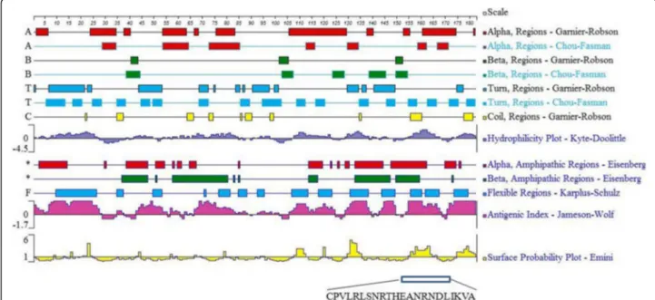
The amino acid sequence of the PLIγ protein ofS. annu-laris was used for epitope prediction. The linear B-cell epitopes of PLIγ were analyzed using DNASTar® protein sequence and structure analysis software. The secondary structure of the PLIγ – including α-helixes, β-sheets,
β-turns and random coils – was construed with Chou-Fasman and Garnier-Robson parameters of Protean 3D software. The surface probability, flexibility and antigenic-ity of PLIγwere analyzed using Emini, Karplus-Schulz and Jameson-Wolf algorithm. Potential B cell epitopes were selected based on the following parameters: the epitope region containingβ-turn or random coil, and fewα-helix and β-sheet; the epitope peptides should display good hydrophilicity, high accessibility, high flexibility and strong antigenicity.
Preparation of immunogen
Following the prediction results, the best epitope sequence was chosen for further study. The peptide was synthesized by Qiang Yao Biotechnology Company (China). The synthetic peptide was called SHE.
sulfo-GMBS (10 mg/mL) were mixed at 5:1 mass ratio and shaken at room temperature for 30 min, followed by 5 min of centrifugation at 12,000 rpm. The resulted super-natant was applied on a Sephadex G25 column to remove excess sulfo-GMBS. The mcKLH eluate was added drop-wise into SHE peptide solution (6 mg/mL) and incubated at room temperature for 3 h. The coupling process was monitored using DTNB assay (Ellman’s reagent) and stopped when OD412 value decreased 2. The conjugated product was named SHE-KLH.
Bovine serum albumin (BSA) was used to prepare the coating antigen (SHE-BSA) using the same protocol as described above.
mAb preparation
Mouse immunization
Four six-week-old BALB/c female mice were subcutane-ously injected with 60μg of SHE-KLH conjugate emulsi-fied with Freund’s complete adjuvant as the primary immunization. Subsequently, 30μg of SHE-KLH conjugate emulsified by Freund’s incomplete adjuvant was injected subcutaneously on days 14, 28, and 42, respectively, for a total of four immunizations. Seven days after the last immunization, serum from each mouse was verified for ability to bind SHE-BSA by indirect ELISA (iELISA). The mouse with the highest antibody titer was injected intra-peritoneally with 50 μg of SHE-KLH conjugate and chal-lenged with booster immunization. Three days later, the spleen was removed for hybridoma cell fusion.
Indirect ELISA
iELISA was used to assess epitope specific antibody titers of mouse sera or ascites fluid. SHE-BSA diluted with bicarbonate buffer (pH 9.6) (2μg/mL) was added to 96-well microtiter plates at 100μL/well and incubated at 4 °C overnight. The plate was washed three times with 300μL/ well of PBST (0.05% Tween-20 in 0.01 M PBS) and then blocked using 2% skim milk blocking buffer (200μL/well) for 2 h at 37 °C followed by three washes. The immunized mouse serum was diluted 200 times in assay buffer, added into each well (100μL/well) in a series of two-fold dilu-tions, and incubated at 37 °C for 1 h. The plate was then washed three times and subsequently incubated with goat anti-mouse IgG/HRP (1:20,000 dilutions in PBS) for 1 h at 37 °C (100 μL/well). After washing, coloring solution (100μL/well) was added and incubated in dark for 5 min. The reaction was terminated by adding 50 μL/well of 2 mol/L H2SO4. The absorbance values were measured at
two wavelengths (450, 630 nm) by an automatic ELISA plate reader.
Cell fusion and subtype analysis
The mouse with the highest antibody titers was selected from four mice, and was injected with 50 g of immunogen
SHE-KLH intraperitoneally to obtain higher titers of antisera. The mouse was sacrificed through cervical vertebra dislocation, then soaked in 75% alcohol for 5 min. Splenocytes isolated from the immunized mouse and murine SP2/0 myeloma cells were mixed with PEG1500 and cultured in IMDM complete medium (with 15% serum) containing HAT and plated at 37 °C with 5% CO2. On the fifth day of fusion, the complete
medium containing HT was used. Eight days later, culture supernatants and hybridoma screening were performed by iELISA. The antibody isotypes were determined using a Mouse Immunoglobulin Panel kit (Southern Biotech. USA) following the manufacturer’s instructions. The hybridoma cell line producing the highest binding capacity of immunoglobulins was injected intraperitoneally into mice to produce a higher amount of monoclonal antibody.
Preparation of mAb
Ascites fluid was collected 7 to 10 days after injection of hybridoma cells. The ascites fluid was diluted 1:3 with equilibration buffer (20 mmol/L PB, 0.15 mmol/L KCl, pH 7.0) and centrifuged at 10,000 rpm for 20 min. The supernatant was further filtered through a 0.22-μm mem-brane to remove fat, cell debris and small particles. Ascites mAb was purified on a protein G chromatography column, which was pre-exhilarated by equilibration buffer and eluted by five times of bed volume elution buffer (0.1 mol/L glycine, pH 3.0) at a flow rate of 0.6 mL/min. The eluted mAb was immediately neutralized with an alkaline buffer (1 mol/L Tris-HCl, pH 9.0). The protein concentration and purity was determined following BCA assay kit and SDS-PAGE, respectively.
Western blot analysis
Recombinant SaPLIγ was expressed in 6-histidine tag form by pET28c vector in our laboratory [28]. The His6-PLIγand natural saPLIγ were separated under reducing conditions on 12% SDS-PAGE gel, and transferred to different PVDF membranes. Immunodetection was per-formed by incubating the membranes overnight at 4 °C, stirring, with the PLIγmAbs at a dilution of 1:500. The reaction was developed using goat anti-mouse peroxid-ase IgG at a dilution of 1:10,000 and the chemilumines-cent substrate Lumi-light (Roche). Images were captured with ChemiDoc XRS+system (Bio-Rad). Parallel Western blot using anti-His6 antibody was conducted to detect His6-PLIγ fusion protein and validate the specificity of PLIγmAbs.
PLIγscreening in snake sera
Serum from four venomous snake–includingBungarus multicinctus, Naja naja, Deinagkistrodon acutus and
namelySinonatrix annularis,Zaocys dhumnades,Elaphe carinata, Dinodon rufozonatum, Macropis thodonrudis,
Elaphe rufodorsata, Elaphe taeniura and Achalinus rufescens – was collected in local market. Western blot analysis of snake sera was performed using the same protocol the abovementioned one. PLIγ mAb was pro-duced by the highest titer hybridoma cell line and puri-fied by protein G affinity chromatography.
Gene cloning and sequence alignment
In order to validate PLIγexpression in different snakes, gene cloning and DNA sequencing were performed. Due to incomplete samples, snake species did not entirely match those used in PLIγ screening. In brief, we collected fresh liver samples of four venomous snakes–namelyB. multicinctus, N. naja, D. acutusand
A. halys –and five non-venomous snakes –including S. annularis, Z. dhumnades, E. carinata, D. rufozonatum
andE. rufodorsata.
Total RNA was extracted from snake liver by using of Trizol reagent (Invitrogen, USA). cDNA was synthesized with SuperScript First Strand Synthesis System (Invitrogen Corp., USA). A pair of degenerate primer was designed based on cDNA sequence of PLIγ as follows: forward: 5’CRCTCATGTAMWTTTGTCACAA3’, reverse primer: 5’TTATTCAGAAGGTGTARTTTTGG3’(where R = A + G; M = A + C; W = A + T). PCR amplification was con-ducted in 0.2 mL PCR tubes containing 12.5 μL of 2× EasyTaq PCR SuperMix (Transgen, Beijing, China), 1 μL (10 μM) each of forward and reverse primers, 20 ng of cDNA and autoclaved Milli-Q water to make a volume up to 25 μL. Thirty-five cycles of amplification were run as
follows: 95 °C denaturation for 30 s, 54 °C annealing for 30 s and 72 °C extension for 30 s. PCR products were sequenced directly with a DYEnamic ET terminator cycle sequencing premix kit (GE Healthcare) on an ABI Prism310 genetic analyzer (Applied Biosystems). Multiple sequence alignment was performed by DNAMAN soft-ware (Lynnon Biosoft, USA).
Results
Prediction of epitope
The secondary structure, flexible regions, hydrophilicity, surface accessibility and antigenic index were predicted using DNAStar Protean program (Fig. 1). Ideal B cell linear epitopes should be located on the surface of a pro-tein where hydrophilic regions usually exist. Epitopes should contain at least eight amino acids, where the secondary structure should be flexible likeβturn and ran-dom coil [30]. There are several segments with good hydrophilic and antigenic index in position of 8-27, 68-84, 108-122, 131-136, 151-172 and 176-182, but most of them are poor surface accessible or short than eight amino acids. Based on those rules, a better candidate peptide was selected:151CPVLRLSNRTHEANRNDLIKVA172(Fig. 1).
Antigen preparation
The peptide (CPVLRLSNRTHEANRNDLIKVA) was called SHE and synthesized by Qiang Yao Biotechnology Com-pany (China). The purity of synthetic SHE was 95% by HPLC assay. In order to increase its immunogenicity, SHE was coupled to KLH (360 kDa) for usage as an immunogen and to BSA (67 kDa) to be used as a coating antigen.
Immunization and determination of anti-serum titer
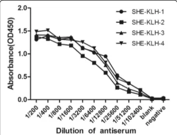
After four immunization protocols, serum from each mouse was tested for its ability to bind to SHE-BSA by indirect ELISA (iELISA). The serum titers of the immunized Balb/c mice were significantly higher than negative control (injected with PBS buffer only), indi-cating that SHE-KLH complete antigen induced the production of antibodies. Mouse 1 showed the highest titer and therefore was chosen for further cell fusion with SP2/0 myeloma cells (Fig. 2).
Cell fusion and selection of positive clones
The fused cells were screened in HAT selective culture medium (only hybridomas from spleen cells and from myeloma cells that successfully fused could grow). At the eighth day of fusion, the hybridoma culture super-natant was detected by indirect ELISA. A total of 44 positive strains were obtained, and the mean positive rate of fusion cells was 5.90%. After the second iELISA screening, 21 positive monoclonal hybridoma cell lines were obtained (Table 1).
Ten plates (93 wells per plate) were picked and the cell fusion rate were 100%. A total of 44 positive cells were obtained with an average rate of 5.90%. After the second iELISA screening, 21 positive monoclonal hybridoma cell lines were obtained. The nomenclature of positive cell clone was “plate number + row + column”, i.e. 1C7 stands for plate 1, the well of row C (third) and the seventh column of the plate.
Subtype and immunoblotting analysis of anti-PLIγmAb
Eighteen IgG positive hybridoma cells belonging to three isotypes–IgG1 (5E10, 5G1, 6G11, 7A10); IgG 2a (6C12, 6D8, 6E11, 8E12, 8G1, 8G2, 9H4, 10E9); and IgG2b
(1C11, 5D9, 5E9, 6B2, 8C7, 9A10) – were obtained, while the other three were IgA subtype.
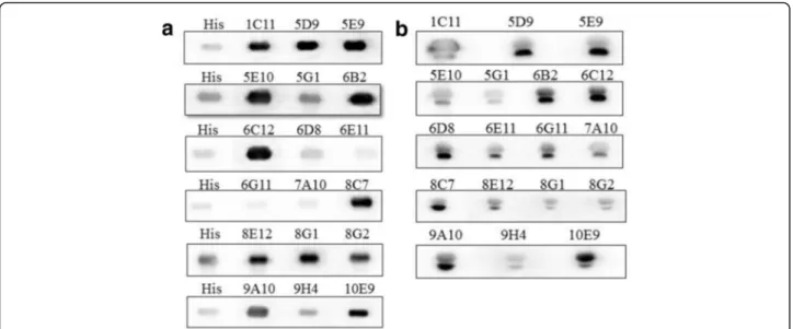
A fusion recombinant His6-PLIγwas expressed in pro-karyotic system and used for PLIγ mAb validation through regular Western blot method [28]. The immu-nodetection result is shown in Fig. 3a. His6-PLIγ bands that were detected by His6 antibody or the 18 mAb sub-types migrated to the same position, which indicated that PLIγs were recognized by all the 18 mAbs.
NaturalS. annularisserum was also used for immuno-detection of the 18 mAbs following the same protocol. All the natural PLIγshowed two close bands by immunoblot-ting, which indicated natural PLIγ is composed of two subunits (Fig. 3b). The result also suggested that the two subunit might be homologous in amino acid sequence. Due to the specificity and sensitivity, we choose 10E9 strain for further amplification of PLIγmAb preparation.
Purification of anti-PLIγmAb
The mAbs were purified from ascites fluid by protein G resin affinity chromatography. The peak value of the antibody at 280 nm was 1.65. The electrophoretic graph shows that the protein band results presented the expected size (lane 4, Fig. 4a) under natural PAGE con-ditions. The peak under reduced PAGE conditions showed two distinct bands, the 54 kDa heavy chain and the 25 kDa light chain (lane 2, Fig. 4a). Indirect ELISA indicated that the titer of purified anti-PLIγ mAb secreted by 10E9 clones was above 1.0 × 10⁵(Fig. 4b).
PLIγscreening in snake sera
In order to investigate the application of PLIγmAb, 12 serum samples from different snake species were col-lected from a local market and underwent immunode-tection using anti-PLIγ mAb. Eleven samples showed bands on the film except Bungarus fasciatus (lane 3, Fig. 5). Interestingly, the molecular weight of PLIγ in venomous species was usually higher than that of non-venomous snakes. PLIγ levels among these snake sera
Fig. 2Determination of mouse serum titers by iELISA. KLH1 ~ SHE-KLH4 represented the four mice that were immunized with SHE-KLH conjugate. The blank received PBS buffer, whereas the negative control received normal mouse serum (1:200)
Table 1The result of cell fusion and positive hybridoma screening
Plate no. Fusion rate (%) Positive rate (%) Monoclonal hybridomas 1 100% (93/93) 3.23% (3/93) 1C7, 1C11
4 100% (93/93) 5.38% (5/93) –
5 100% (93/93) 5.38% (5/93) 5D6, 5E9,5E10,5G1 6 100% (93/93) 16.12% (15/93) 6B2,6C12, 6D8, 6E11, 6G11 7 100% (93/93) 1.08% (1/93) 7A10
were also different, there were only 15 μg of in total serum protein loaded in lane 1 (S. annularis) and lane 2 (B. multicinctus), while other lanes loaded 150 μg serum protein.
Gene cloning and sequence alignment
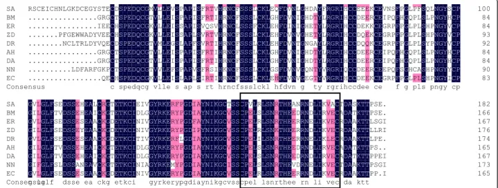
The amino acid sequence and alignment of the nine snake PLIγ was shown in Fig. 6. The result indicated a high consistency among different PLIγs (partial CDS), espe-cially in the epitope region (CPVLRLSNRTHEANRN DLIKVA). In addition, the identity of an intraclass PLIγ
(i.e. venomous snake only) is higher than interclass. For
instance, the amino acids at 155 and 164 of the PLIγ
epitope are R/I and R/N in non-venomous snakes PLIγs, whereas they are S and D in venomous snakes.
Discussion
Snake venom PLA2s (svPLA2) are closely related to
toxicity and comprise an important target for the development of new antivenom drugs. Snake and/or mammals sera are repositories of svPLA2 inhibitors
(PLIs) due to protective benefits. Immunodetection is an essential technique commonly employed for pro-tein discovery, quantification and investigation. Thus,
Fig. 3Western blot analysis of the culture supernatant of 18 IgG positive hybridoma clones.aImmunoblotting of 18 mAbs to identify recombinant His6-PLIγ. The first lane was blotted with anti-His tagged antibody as positive control, while others were the 18 IgG positive hybridoma culture supernatant. The same position of all lanes indicated that all the 18 mAbs reacted with recombinantS. annularisPLIγ.bImmunoblotting of the 18 mAbs to identify nativeS. annularisserum.The positive bands in all lanes indicated the availability of the 18 mAbs in natural PLIγ. All the natural PLIγ showed two close bands by the immunoblotting, which indicated natural PLIγis composed of two subunits
mAb development of PLIγ is technically significant for antivenom studies.
The classical routine of monoclonal antibody prepar-ation is time-consuming and laborious; the resulted mAbs are generally high specific. Protein-specific antibodies can be generated by immunization of animals with peptides, if the peptide is an effective epitope of the protein. Bioinfor-matics prediction followed by concrete experimental validation is both economical and effective. For epitope prediction, bioinformatics software can reduce the experi-mental workload by 95% and increase the efficiency of new epitope location by 10 to 20 folds [31]. In this study, we used DNAStar Protean program to predict epitopes of saPLIγ by comprehensively analyzing many parameters such as hydrophilicity, surface accessibility, antigenic index, secondary structure and flexibility. Finally, we choose 151CPVLRLSNRTHEANRNDLIKVA172 as a hapten and obtained 18 IgG mAb cell strains. The resulted PLIγmAb could recognize a broad range of snake sera including venomous and non-venomous snake spe-cies, because the epitope peptide is highly homologous among snake PLIγs.
As predicted by Protean program, the saPLIγ has six segments with high antigenic index: 8-27, 68-84, 108-122, 131-136, 151-172 and 176-182. The peptide 8-27aa was good in hydrophilicity, flexibility, antigenicity and had a loose secondary structure (mainly in β-turn), but its surface probability was low, which indicates the epi-tope may be hidden inside the 3D structure of a protein and unable to act as an antigenic determinant. The pep-tide 108-122aa was ideal in hydrophilicity, flexibility, an-tigenicity and surface accessibility, but this segment was a rigidα-helix which might influence its interaction with antibodies. Other segments were too short to be effect-ive epitopes. The best peptide (151-172) generated 18 IgG mAbs that had the ability of binding with recombin-ant and natural PLIγs.
Epitope prediction and design should also avoid the active amino acid/peptide, otherwise its binding to mAb will block the activity of the protein of interest. The seg-ment 107PGLPLSLQNG116 in Python’s PLIγ (with N terminal signal peptide) had active sites, as described by Thwin et al. [25]. The saPLIγ also had homologous ac-tive peptide as 87PGLPFSQLNG96, therefore the mAb
Fig. 5Immunoblot analysis of PLIγfrom different snake sera. 1–Sinonatrix annularis,2–Bungarus multicinctus,3–Bungarus fasciatus, 4–Naja naja, 5–Deinagkistrodon acutus,6–Macropisthodon rudis,7–Elaphe carinata, 8–Zaocys dhumnades, 9–Elaphe taeniura,10–Dinodon
rufozonatum, 11–Elaphe rufodorsata, 12–Achalinus rufescens
resulted from 151CPVLRLSNRTHEANRNDLIKVA172 did not affect the binding of PLIγ to svPLA2s and mammal
secretory PLA2s and is available for coIP investigation
(data not shown).
This study showed a wide range distribution of PLIγs in various snake sera. The molecular size of PLIγin venom-ous species was generally higher than in non-venomvenom-ous snakes. Nevertheless, their cDNA sequences were quite identical in length, which indicated that these venomous PLIγs probably undergo more post-translational modifica-tion such as glycosylamodifica-tion. The natural PLIγ had been reported to be N-glycosylated on asparagin residues in the sequence Asn-Xaa-Ser/Thr (where Xaa represents any amino acid except proline) [24, 27] at position 178. The modification requires further investigation such as mass spectrum analysis.
Although PLIγs were found in many snake species, their activities were quite different. For example, the saPLIγ have strong anti-hemorrhagic effect and inhibi-tory activity against venom PLA2s fromD .acutusandA.
halys.However, the inhibitory effect of PLIγwas weaker against venom PLA2s ofB. multicinctus, E. carinataand
N. naja[29]. Thus, more efforts should be made on the elaboration of interaction between PLIγs and PLA2s,
including analysis of active sites, modification and spatial structure difference. The successful preparation of anti-PLIγantibody is of great impetus for the investigation of the key sequences of natural PLIγs. Effective natural inhibitors have a potential part in the treatment of many diseases involving the PLA2 enzyme and also play an
important role in bridging the gap between snakebite victims and their successful treatment [23, 32]. It is worth mentioning that PLA2also exists in other animal
toxins, such as bee and scorpion venoms [33, 34]. There-fore, inhibition of PLA2 is considered a promising lead
molecule for a wide spectrum of drugs, including antive-noms and anti-inflammatories.
Conclusion
A monoclonal antibody against saPLIγ was success-fully prepared using epitope prediction with DNAStar Protean program. The resulted anti-PLIγ mAb can be utilized for new PLIγ discovery, quantification and investigation, as well as for the study of interactions between PLIγ and PLA2.
Abbreviations
BSA:Bovine serum albumin; iELISA: Indirect ELISA; KLH: Keyhole limpet hemocyanin; mAb: Monoclonal antibody; PLA2: Phospholipase A2; PLIγ:
Gamma type phospholipase A2inhibitor; SaPLIγ:Sinonatrix annularisPLIγ;
SIRS: Systematic inflammation response syndrome; svPLA2: Snake venom
phospholipase A2
Acknowledgments
Not applicable.
Funding
The authors would like to thank the support by the National Natural Science Foundation of China (No. 31260209) and (No. 31460227); and by the Jiangxi Science and Technology Development Funds (No. 20122BBG70091).
Availability of data and materials
All data (table and figures) were obtained by standard operation and determination. All the materials (hybridomas and mAb cell lines) were available, the PLIγmAb has been validated as useful by means of serum screening.
Authors’contributions
JJ and XY contributed equally to his work. JJ performed the animal immunization and western blot. XY took part in hybridoma screening. SH collected the sera of snakes. LH was responsible for the PLIγgenes. CH conducted the experimental design and writing of the manuscript. All the authors read and approved the final manuscript.
Ethics approval and consent to participate
The present study was approved by the Ethics Committee of Nanchang University (SYXX-2015-0001).
Consent for publication
Not applicable.
Competing interests
The authors declare that they have no competing interests.
Publisher’s Note
Springer Nature remains neutral with regard to jurisdictional claims in published maps and institutional affiliations.
Author details
1Department of Biochemistry, College of Basic Medical Science, Nanchang
University, Nanchang 330006, China.2Second Affiliated Hospital to Nanchang
University, Nanchang University, Nanchang 330006, China.3Jiangxi Province
Key Laboratory of Tumor Pathogens and Molecular Pathology, Nanchang University, 461 Bayi Avenue, Nanchang 330006, China.
Received: 31 March 2017 Accepted: 25 July 2017
References
1. Kasturiratne A, Wickremasinghe AR, de Silva N, Gunawardena NK, Pathmeswaran A, Premaratna R, et al. The global burden of snakebite: a literature analysis and modelling based on regional estimates of envenoming and deaths. PLoS Med. 2008;5(11):e218.
2. Chippaux JP. Snake-bites: appraisal of the global situation. Bull World Health Organ. 1998;76(5):515–24.
3. Herrera M, Fernández J, Vargas M, Villalta M, Segura Á, León G, et al. Comparative proteomic analysis of the venom of the taipan snake,Oxyuranus scutellatus, from Papua New Guinea and Australia: role of neurotoxic and procoagulant effects in venom toxicity. J Proteome. 2012;75(7):2128–40.
4. Del Brutto OH, Del Brutto VJ. Neurological complications of venomous snake bites: a review. Acta Neurol Scand. 2012;125(6):363–72.
5. Reeks TA, Fry BG, Alewood PF. Privileged frameworks from snake venom. Cell Mol Life Sci. 2015;72(10):1939–58.
6. Stephen M. The field of reptile toxinology snakes, lizards and their venoms. In: Stephen Mackessy, editor. Handbook of venoms and toxins of reptile. Boca Raton: CRC press; 2010. http://www.academia.edu/634557/The_Field_ of_Reptile_Toxinology_Snakes_Lizards_and_Their_Venoms.
7. Nunes DC, Rodrigues RS, Lucena MN, Cologna CT, Oliveira AC, Hamaguchi A, et al. Isolation and functional characterization of proinflammatory acidic phospholipase A2 fromBothrops leucurussnake venom. Comp Biochem Physiol C Toxicol Pharmacol. 2011;154(3):226–33.
8. Dennis EA, Cao J, Hsu YH, Magrioti V, Kokotos G. Phospholipase A2 enzymes: physical structure, biological function, disease implication, chemical inhibition, and therapeutic intervention. Chem Rev. 2011; 111(10):6130–85.
10. Murakami M, Sato H, Miki Y, Yamamoto K, Taketomi Y. A new era of secreted phospholipase A2. J Lipid Res. 2015;56(7):1248–61.
11. Schiermeier Q. Africa braced for snakebite crisis. Nature. 2015;525(7569):299. 12. Gutiérrez JM. Current challenges for confronting the public health problem
of snakebite envenoming in central America. J Venom Anim Toxins incl Trop Dis. 2014;20(1):7–15.
13. Chippaux JP. Epidemiology of envenomations by terrestrial venomous animals in Brazil based on case reporting: from obvious facts to contingencies. J Venom Anim Toxins incl Trop Dis. 2015;21:13.
14. Gao JF, Wang J, He Y, Qu YF, Lin LH, Ma XM, et al. Proteomic and biochemical analyses of short-tailed pit viper (Gloydius brevicaudus) venom: age-related variation and composition-activity correlation. J Proteome. 2014;105:307–22.
15. de Silva HA, Ryan NM, de Silva HJ. Adverse reactions to snake antivenom, and their prevention and treatment. Br J Clin Pharmacol. 2016;81(3):446–52. 16. Marcussi S, Sant'Ana CD, Oliveira CZ, Rueda AQ, Menaldo DL, Beleboni RO,
et al. Snake venom phospholipase A2 inhibitors: medicinal chemistry and therapeutic potential. Curr Top Med Chem. 2007;7(8):743–56.
17. Faure G. Natural inhibitors of toxic phospholipases A2. Biochimie. 2000;82(9-10):833–40.
18. Sergio L, Gilberto D, Jonas P. Natural phospholipase a(2) myotoxin inhibitor proteins from snakes, mammals and plants. Toxicon. 2003;42(8):963–77.
19. Neves-Ferreira RHV, Perales GB, editors. Natural inhibitors: innate immunity to snake venoms. In: Stephan M, editor. Handbook of Venoms and Toxins of Reptiles; 2010. p. 259-84.
20. Campos PC, Melo LA, Dias GLF, Fortes-Dias CL. Endogenous phospholipase A2 inhibitors in snakes: a brief overview. J Venom Anim Toxins incl Trop Dis. 2016;22:37. 21. Nishida M, Okamoto M, Ohno A, Okumura K, Hayashi K. Ikeda Kiyoshi, et al.
inhibitory activities of the heterotrimers formed from two alpha-type phospholipase A2 inhibitory proteins with different enzyme affinities and importance of the intersubunit electrostatic interaction in trimer formation. Biochim Biophys Acta. 2010;1804(11):2121–7.
22. Santos-Filho NA, Santos CT. Alpha-type phospholipase A2 inhibitors from snake blood. J Venom Anim Toxins incl Trop Dis. 2017;23:19.
23. Lima RM, Estevao-Costa MI, Junqueira-de-Azevedo ILM, Ho PL, Diniz MRV, Fortes-Dias LF. Phospholipase A2 inhibitors (betaPLIs) are encoded in the venom glands ofLachesis muta(Crotalinae, Viperidae) snakes. Toxicon. 2011;57(1):172–5. 24. Estevão-Costa MI, Rocha BC, de Alvarenga MM, Redondo R, Franco GR,
Fortes-Dias CL, et al. Prospection, structural analysis and phylogenetic relationships of endogenous gamma-phospholipase a(2) inhibitors in BrazilianBothropssnakes (Viperidae, Crotalinae). Toxicon. 2008;52(1):122–9. 25. Thwin MM, Satish RL, Chan ST, Gopalakrishnakone P. Functional site of
endogenous phospholipase A2 inhibitor from python serum. Eur J Biochem. 2002;269(2):719–27.
26. So S, Chijiwa T, Ikeda N, Nobuhisa I, Oda-Ueda N, Hattori S, et al. Identification of the B subtype of gamma-phospholipase A2 inhibitor from Protobothrops flavoviridis serum and molecular evolution of snake serum phospholipase A2 inhibitors. J Mol Evol. 2008;66(3):298–307.
27. Shirai R, Toriba M, Hayashi K, Ikeda K, Inoue S. Identification and characterization of phospholipase A2 inhibitors from the serum of the Japanese rat snake,Elaphe climacophora. Toxicon. 2009;53(6):685–92.
28. Le Z, Li XZ, Yuan P, Liu P, Huang C. Orthogonal optimization of prokaryotic expression of a natural snake venom phospholipase A2 inhibitor from
Sinonatrix annularis. Toxicon. 2015;108:264–71.
29. Chen K, Zhong LP, Li X, Xu X, Huang CH. Investigation and purification of snake venom secretory phospholipase A2 inhibitors from sera of some common snake species in Jiangxi province. Pharm Biotechnol. 2011;18(3):220–3. 30. Bannon GA, Ogawa T. Evaluation of available IgE-binding epitope data and
its utility in bioinformatics. Mol Nutr Food Res. 2006;50(7):638–44.
31. DeGroot AS, Sbai H, Aubin CS, McMurry J, Martin W. Immuno-informatics: mining genomes for vaccine components. Immunol Cell Biol. 2002;80:255–69. 32. Gimenes SN, Ferreira FB, Silveira AC, Rodrigues RS, Yoneyama KA. Izabel dos
Santos J, et al. isolation and biochemical characterization of a gamma-type phospholipase A2 inhibitor fromCrotalus durissus collilineatussnake serum. Toxicon. 2014;81:58–66.
33. Ferreira RS Jr, Sciani JM, Marques-Porto R, Junior AL, de Orsi RO, Barraviera B, et al. Africanized honey bee (Apis mellifera) venom profiling: seasonal variation of melittin and phospholipase A2 levels. Toxicon. 2010;56(3):355–62. 34. Valdez-Cruz NA, Segovia L, Corona M, Possani LD. Sequence analysis and
phylogenetic relationship of genes encoding heterodimeric phospholipases A2 from the venom of the scorpionAnuroctonus phaiodactylus. Gene. 2007; 396(1):149–58.
• We accept pre-submission inquiries
• Our selector tool helps you to find the most relevant journal
• We provide round the clock customer support
• Convenient online submission
• Thorough peer review
• Inclusion in PubMed and all major indexing services
• Maximum visibility for your research
Submit your manuscript at www.biomedcentral.com/submit