Roberto Lanna Filho(1), Reginaldo da Silva Romeiro(2) and Eduardo Alves(1)
(1)Universidade Federal de Lavras, Departamento de Fitopatologia, Caixa Postal 3037, CEP 37200‑000 Lavras, MG, Brazil. E‑mail: robertolanna@yahoo.com.br, ealves@ufla.br (2)In memoriam
Abstract – The objective of this work was to evaluate in vitro and in vivo biocontrol of bacterial spot (Xanthomonas vesicatoria)and early blight (Alternaria solani) by the epiphytic bacteria Paenibacillus macerans and Bacillus pumilus. Tomato plants were previously sprayed with epiphytic bacteria, benzalkonium chloride and PBS buffer and, after four days, they were inoculated with A. solani and X.vesicatoria. To determine the phytopathogenic
bacteria population, leaflet samples were collected from each treatment every 24 hours, for seven days, and
plated on semi-selective medium. The effect of epiphytic bacteria over phytopathogens was performed by the antibiosis test and antagonistic activity measured by inhibition zone diameter. The epiphytic and benzalkonium chloride drastically reduced the severity of early blight and bacterial spot in comparison to the control (PBS).
In detached leaflets, the epiphytic bacteria reduced in 70% the number of phytopathogenic bacteria cells in the phylloplane. The antibiosis test showed that the epiphytic bacteria efficiently inhibit the phytopathogens
growth. In all the bioassays, the epiphytic bacteria protect tomato plants against the phytopathogens.
Index terms:Alternaria solani, Bacillus pumilus, Paenibacillus macerans, Xanthomonas vesicatoria, biological control, epiphytic bacteria.
Biocontrole da mancha‑bacteriana e da pinta‑preta
por bactérias epifíticas em tomateiro
Resumo – O objetivo deste trabalho foi avaliar o biocontrole in vitro e in vivo dos fitopatógenos
mancha-bacteriana (Xanthomonas vesicatoria) e pinta-preta (Alternaria solani)pelas bactérias epifíticas Paenibacillus macerans e Bacillus pumilus. Plantas de tomate foram previamente pulverizadas com as bactérias epifíticas,
cloreto de benzalcônio e tampão PBS e, após quatro dias, receberam inoculação com A. solani e X. vesicatoria.
A fim de determinar a população da bactéria fitopatogênica, amostras de folíolos foram coletadas de cada
tratamento em intervalos de 24 horas, durante sete dias, e inoculadas em meio semisseletivo. O efeito das
bactérias epifíticas sobre os fitopatógenos foi realizado pelo teste de antibiose e atividade antagônica avaliada pelo diâmetro da zona de inibição. As bactérias epifíticas e o cloreto de benzalcônico reduziram drasticamente
a severidade da pinta-preta e da mancha-bacteriana, comparado com o controle (PBS). Em folíolos destacados,
as bactérias epifíticas reduziram em até 70% o número de células da bactéria fitopatogênica no filoplano. As bactérias epifíticas inibem eficientemente o crescimento dos fitopatógenos em meio de cultura. Em todos os bioensaios, as bactérias epifíticas protegem as plantas de tomate contra os fitopatógenos.
Termos para indexação: Alternaria solani, Bacillus pumilus, Paenibacillus macerans, Xanthomonas vesicatoria,
controle biológico, bactéria epifítica.
Introduction
Epiphytic bacteria have been defined as populations
that can survive and multiply on the surface of plants (Hirano et al., 1982). Thus, they develop survival strategies in protected positions such as the trichomes base, inside substomatal chambers, hydathodes, and, especially, in between the depressions along the junctions of adjacent epithelial cells (Beattie &
Lindow, 1999; Lindow & Brandl, 2003; Monier & Lindow, 2004, 2005a, 2005b).
occupy ecological niches on the phylloplane that could be occupied by pathogens (Monier & Lindow, 2005a), and to their broad antagonistic effect against pathogens. Biosurfactants, antibiotics, bacteriocins and volatile organic compounds (VOCs) synthesis, siderophores and competition for space and nutrients are related to the antagonistic effects of epiphytic bacteria on the phytopathogen growth (Beattie & Lindow, 1999; Lindow & Brandl, 2003). Recent surveys demonstrate that epiphytic bacteria also act as elicitors of the induced systemic resistance (ISR) in plants (Halfeld-Vieira et al., 2006).
The objective of this work was to evaluate in vitro and in vivo biocontrol of bacterial spot (Xanthomonas vesicatoria) and early blight (Alternaria solani) by the epiphytic bacteria Paenibacillus macerans and Bacillus pumilus.
Materials and Methods
Two epiphytic bacteria were obtained from leaves
of healthy tomato plants, and identified by fatty-acid
analysis (FAA) (Lanna Filho, 2006) as Paenibacillus macerans and Bacillus pumilus. Afterward, bacteria were grown in medium 523 (Kado & Heskett, 1970) [10 g L-1 of sucrose, 8 g L-1 of casein acid hydrolysate,
4 g L-1 of yeast extract, 2 g L-1 of K
2HPO4 (anhydrous),
0,3 g L-1 of MgSO
4.7H2O and 18 g L-1 of agar], and
preserved in deep freezer at -80oC. They were also
emulsified once in 30% (v/v) glycerin.
The tomato pathogens were obtained from the Plant Pathology Department collection of the Universidade
Federal de Viçosa, Brazil. The fungus Alternaria solani (Jones & Grout) was grown in potato dextrose agar (PDA) and maintained at 4oC under mineral oil
(Smith & Onions, 1994). The bacterium Xanthomonas vesicatoria (Doidge) Vauterin et al. was grown in medium 523 (Kado & Heskett, 1970), preserved at -80oC, and emulsified once in 30% glycerin.
The antagonistic activity was tested by the overlay diffusion method (Vidaver et al., 1972). A drop of
15 μL of cell suspension (optical density at 540 nm
wavelength, OD540 = 0.3) of the P. macerans and
B. pumilus epiphytic bacteria was placed in the center of the solid medium 523 (Kado & Heskett, 1970), in Petri dishes, and incubated at 28°C for 24 hours. As control,
a drop of 15 μL of phosphate-buffered saline (PBS)
was placed in the center of the solid medium of the same culture.
Subsequently, the colonies were killed by exposure to ultraviolet (UV) and chloroform vapors for 1 hour.
Then, melted semisolid culture media [0.8% (w/v)
agar, 45oC] containing propagules of the pathogens
was placed over the basal layer, and incubated at 28ºC for 24 hours (X. vesicatoria) and at 25ºC for seven days (A. solani). After incubation, the inhibition zones were measured and antimicrobial activity was expressed by the diameter of the inhibition zone (mm). The percentage of the inhibition zone was calculated in relation to the diameter of the Petri dish (90 mm),
considered 100%. The bioassay was repeated three
times for each treatment (P. macerans, B. pumilus and PBS), and three zones of inhibition were measured for each epiphytic bacterium.
The epiphytics were observed on phylloplane by scanning electron microscopy. For that, seeds of
Santa Cruz 'Kada' tomato were disinfected in 70% ethanol for 2 min, sodium hypochlorite solution (2%
available Cl-) for 2 min, and two washes were carried
out in sterilized distilled water. Then, they were transferred to 20 mL plastic cups with the epiphytic bacteria cell suspensions, adjusted to OD540 = 0.3,
corresponding to approximately 108 colony forming
units per mL (CFU mL-1). Cell suspension volume
was onlysufficient to cover the seeds. After 24 hours,
seeds were placed on filter paper for drying. Seeds
immersed only in PBS (1 mol L-1; pH 7.0) were used
as controls. After drying, seeds were transferred to three tubes containing MS medium per treatment (Murashige & Skoog, 1962), and germinated at 25oC
with 12-hours photoperiod. In fifteen-day-old plants,
three leaves per treatment were cut, and submitted to
a fixative procedure in a modified Karnovsky solution (glutaraldehyde 2.5% and paraformaldehyde 2.5% in
sodium cacodylate buffer 0.05 mol L-1, pH 7.2, CaCl 2
0.001 mol L-1), for 24 hours (at 4ºC), infiltrated with
a cryoprotection solution (glycerol 30% in water) for
30 min, and cross-sectioned with a scalpel blade after being immersed in liquid nitrogen.
Sections were transferred to a 1% aqueous solution
of osmium tetroxide for 1 hour at room temperature, and subsequently dehydrated for 10 min each in a crescent series of acetone solutions (25, 50, 75, 90 and
100%). After that, they were dried in a critical-point
Processed materials were mounted on aluminum stubs, fractured side up, sputter coated with gold SCD 050, (Balzers, Jundiaí, SP, Brazil) and observed in a scanning electron microscopy (SEM) LEO EVO 40 XVP (K. E. Developments, Cambridge, England). Leaves of healthy tomato plants exposed to PBS were used as controls. Two images were generated and
three leaflets were used for each treatment. Images of
the phylloplane region were generated at random for
each sample, at several magnifications, and digitally
recorded. Images were processed using the software Corel Draw 12, with which comparisons among treatments were done.
The antagonistic effect of the epiphytic bacteria over the phytopathogenic one was carried out in twenty-day-old tomato plants, previously exposed to the P. macerans, B. pumilus, benzalkonium chloride and PBS treatments. After four days, the X.vesicatoria suspension (OD540 = 0.3)was sprayed.
For each treatment, four replicates were used; with three grams of leaves per pot being considered as one replicate. Leaves were collected randomly and placed
in flasks containing 50 mL of sterile phosphate buffer
(0.1 mol-1, pH 7.0, containing 0.05% Tween-80), and
sonicated for 10 min in an ultrasonic cleaning bath (Ultrasonic Cleaner 1440D, Odontobrás, Ribeirão Preto, SP, Brazil) in order to recover bacterial cells. Bacterial populations were estimated from three
grams symptomless leaflets randomly sampled from
each plant pot. The obtained suspensions of washed leaves were submitted to serial dilution (factor = 1:1,000) and were inoculated in the semi-selective medium propose by Lanna Filho & Romeiro (2009),
containing cycloheximide (50 μg mL-1), cephalexin
(50 μg mL-1) and streptomycin sulfate (50 μg mL-1).
Petri dishes were incubated for 48 hours at 28°C, and then the CFU count was made per gram of leaf tissue. The leaf samples were collected every 24 hours, for seven days. For each evaluation day, three Petri dishes were used for each dilution, with three replicates per dishes. The mean viable bacterial population size was derived from the log10-transformed bacterial
population.
In greenhouse experiments, seeds of Santa Cruz 'Kada' tomato were planted in plastic pots containing non-sterilized mixture of soil, sand and manure
(2:1:1), maintained in greenhouse at 25°C and 70%
relative humidity. In each treatment, four replicates
were used, with one plant per pot considered as a replicate. Plants were sprayed with live cells of the P. macerans and B. pumilus (OD540 = 0.3) 30 days
after planting. For the positive control, plants were sprayed with benzalkonium chloride sanitizer (2.5 g L-1 a.i.), and PBS as negative control. Four days later,
plants were inoculated spraying the A. solani (1.0 × 10⁵ conidia mL-1) and X. vesicatoria (OD
540 = 0.3)
pathogen suspensions. Inoculated plants were kept in greenhouse, and after the symptoms of the disease were fully developed, the number of lesions per leaf
was counted within all the leaflets. The bioassay was
repeated three times.
All experiments were carried out in a completely randomized design. The results were subjected to analysis of variance (ANOVA) and means were
compared by Tukey test at 5% probability, using
the software Statistica, version 7.0 (Statsoft, 2005).
Regression equations, coefficient of determination (R2)
and significance levels of the curves were calculated
in order to determine the bacterial population growth according to time.
Results and Discussion
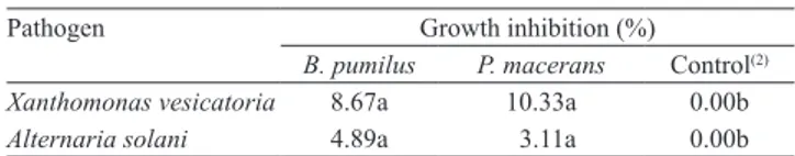
The in vitro antibiosis test showed that the epiphytic bacteria presented direct activity against the pathogens X. vesicatoria and A. solani, inhibiting their growth (Table 1). Paenibacillus macerans and B. pumilus inhibited X. vesicatoria growth at 10.33 and 8.67%, respectively, and A. solani growth at 3.11 and 4.89%. Additionally, the control treatment with PBS showed
no zone of inhibition (0%) against the pathogens. These results confirmed that the antagonists produce
some type of toxic substance with antimicrobial effect against the pathogens, causing the antibiosis phenomenon.
Possibly, these substances are bioactive compounds derived from lipopeptides of the surfactin, iturin and
Table 1. In vitro antimicrobial activity of Paenibacillus macerans and Bacillus pumilus against tomato pathogens(1).
(1)Means followed by the same letter, in the lines, do not differ by Tukey’s test at 5% probability. (2)Phosphate-buffered saline (PBS).
Pathogen Growth inhibition (%)
B. pumilus P. macerans Control(2)
Xanthomonas vesicatoria 8.67a 10.33a 0.00b
fengycin families, frequently reported as toxic to pathogens (Peypoux et al., 1999; Ongena et al., 2005). Magnet-Dana et al. (1992) reported that the iturins and fengycins exhibit a strong antifungal activity and are inhibitory for the growth of a wide range of pathogens. Additionally, the synthesis of antimicrobials by epiphytic bacteria provides higher adaptability of those microbes on the phylloplane, impeding the growth and establishment of pathogenic populations (Beattie & Lindow, 1999).
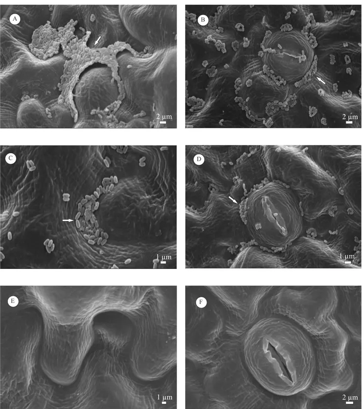
The observation of the colonization pattern of the epiphytic bacteria on the phylloplane by SEM
confirmed their ability in establishing at specific sites
on the foliar surface (Figure 1). This demonstrates the capacity of these bacteria to migrate from spermosphere to the aerial part, their habitat. Notably, the formation of P.macerans cell aggregates embedded in an exopolymeric matrix could be observed in the depressions along the junctions of adjacent epithelial cells (Figure 1 A), besides the colonization of the stomatal region (Figure 1 B).Numerous studies report the capacity of epiphytic bacteria to form aggregates on the foliar surface of different species of plants (Morris et al., 1997; Fett, 2000), which gives them protection against environmental stress and larger survival on the phylloplane, as reported by Monier & Lindow (2004). Bacilluspumilus also formed small aggregates between the depressions along the junctions of adjacent epithelial cells (Figure 1 C), as well as in the stomatal region (Figure 1 D). As expected, the treatment with phosphate-buffered saline did not present bacterial colonization (Figures 1 E and F). Monier & Lindow (2005a, 2005b) report the importance of the aggregate formation for the protection of epiphytic population against desiccation, UV radiation, high temperatures
and environmental fluctuations, besides the higher efficiency in nutrient acquisition and migration
to regions of the phylloplane more favorable for survival. This work demonstrated that bacterial cells of P. macerans and B. pumilus form large aggregates on the tomato phylloplane. This confirms the natural habitat of these bacteria, since both were exposed to previously disinfested seeds and, after 15 days of
germination, they were established at specific sites on
the foliar surface.
Tomato leaves previously exposed to the epiphytic bacteria and to benzalkonium chloride drastically reduced the X. vesicatoria population on the
phylloplane, in comparison with the control, treated with PBS (Figure 2). The sanitizer (benzalkonium chloride) reduced the pathogen population on
the phylloplane by 80.4%, while the antagonists
P.macerans and B. pumilus reduced it by 68 and 70%, respectively, compared to PBS.These results confirm the antibacterial activity of the sanitizer against the phytobacteria, which was already expected, since quaternary ammonium based compounds possess fungicidal, bactericidal, algicidal and virucidal action (Mcbain et al., 2004; Oosterhof et al., 2006; Abreu et al., 2008). The antagonists probably acted on the reduction of the pathogen population by multiple mechanisms, such as competition for space and nutrients, antibiosis, and, possibly, resistance induction (Lindow & Brandl, 2003; Monier & Lindow, 2004, 2005ab; Halfeld-Vieira et al., 2006).
Although ISR has not been investigated in this research, Halfeld-Vieira et al. (2006) relate the phenomenon for the epiphytic Bacillus cereus in tomato plants, which controlled the bacterium Pseudomonas syringae pv. tomato. Besides ISR, synthesis of antimicrobial compounds and competition for niches important to pathogen establishment are reported by Lindow & Brandl (2003) as one of the most important mechanisms for antagonistic effect on the foliar surface. Leveau & Lindow (2001) reported
efficient use of nutrients by epiphytics as an important
mechanism for the antagonistic activity.
Effective control of bacterial spot and early blight was observed in the greenhouse bioassay, in the treatments using P. macerans, B. pumilus and benzalkonium chloride (Figure 3). The epiphytics and the sanitizer differed from the control treatment,
reducing the severity by over 50% in some cases.
These results demonstrate the use potential of those biocontrol agents against two important tomato diseases. Halfeld-Vieira et al. (2008) proved the
efficiency of the epiphyte Bacillus cereus UFV-IEA6 against Phytophthora infestans.
in Brazil, few researches report the importance of epiphytic bacteria to control diseases.
Conclusions
1. Paenibacillus macerans and Bacillus pumilus epiphytic bacteria and benzalkonium chloride reduce Xanthomonas vesicatoria and Alternaria solani disease severity in tomato plants.
2. Epiphytic bacteria are able to inhibit the growth of
tested phytopathogens in vitro, and efficiently colonize
the phylloplane of tomato plants.
Acknowledgments
To Fundação de Amparo à Pesquisa de Minas Gerais
(Fapemig), for financial support; and to the Laboratório
de Microscopia Eletrônica e Análise Ultraestrutural of the Universidade Federal de Lavras, for the scanning electron microscopy analysis.
References
ABREU, F.M. de; LOURENÇO, S.A.; BASSETTO, E.; GONÇALVES, F.P.; MARTINS, M.C.; AMORIM, L. Efeito de
sanificantes no controle pós-colheita da podridão parda (Monilinia
fructicola) e da podridão mole (Rhizopus stolonifer) em pêssegos.
Summa Phytopathologica, v.34, p.86-88, 2008.
BEATTIE, G.A.; LINDOW, S.E. Bacterial colonization of leaves: a spectrum of strategies. Phytopathology, v.89, p.353-359, 1999.
FETT, W.F. Naturally occurring biofilms on alfalfa and other types
of sprouts. Journal of Food Protection, v.63, p.625-632, 2000. GNANAMANICKAM, S.S.; IMMANUEL, J.E. Epiphytic bacteria, their ecology and functions. In: GNANAMANICKAM, S.S. (Ed.). Plant‑associated bacteria. Dordrecht: Springer, 2006. p.131-154.
HALFELD-VIEIRA, B. de A.; ROMEIRO, R. da S.; MOUNTEER, A.; MIZUBUTI, E.S.G. Efficiency of phylloplane bacteria in
controlling aerial tomato diseases under field conditions. Summa
Phytopathologica, v.34, p.86-87, 2008.
HALFELD-VIEIRA, B. de A.; VIEIRA JÚNIOR, J.R.; ROMEIRO, R. da S.; SILVA, H.S.A.; BARACAT-PEREIRA, M.C. Induction of systemic resistance in tomato by the autochthonous phylloplane resident Bacillus cereus. Pesquisa Agropecuária Brasileira, v.41, p.1247-1252, 2006.
HIRANO, S.S.; NORDHEIM, M.E.V.; ARNY, D.C.; UPPER, C.D. Lognormal distribution of epiphytic bacterial populations on leaf surfaces. Applied and Environmental Microbiology, v.44, p.695-700, 1982.
KADO, C.I.; HESKETT, M.G. Selective media for isolation of
Agrobacterium, Corynebacterium, Erwinia, Pseudomonas, and
Xanthomonas. Phytopathology, v.60, p.969-976, 1970.
Figure 2. Population dynamics of plant pathogenic bacterium Xanthomonas vesicatoria (Log10 CFU g-1 leaflet) on tomato
leaves after prior exposure to treatments with Paenibacillus macerans (▲), Bacillus pumilus (
■)
, benzalkonium chloride(BC) (●) and PBS buffer (control) (
◊
). Each point indicates the mean of the log10-transformed bacterial population.Figure 3. Severity of bacterial spot (A), and early blight (B), in Santa Cruz 'Kada' tomato plants, four days after exposure to treatmentswith Bacillus pumilus, Paenibacillus macerans, benzalkonium chloride (BC) and PBS buffer (control). The white, light grey, and dark grey bars represent the experiment replicates. Means followed by same letter do
LANNA-FILHO, R. Isolamento e seleção de procariotos
residentes de filoplano do tomateiro com potencial para o
controle de doenças da cultura. 2006. 52p. Tese (Mestrado) –
Universidade Federal de Viçosa, Viçosa.
LANNA FILHO, R.; ROMEIRO, R. da S. Sensibilidade de
Xanthomonas vesicatoria a antibióticos para desenvolvimento
de um meio semi-seletivo. Revista Trópica: Ciências Agrárias e
Biológicas, v.3, p.28-39, 2009.
LEVEAU, J.H.J.; LINDOW, S.E. Appetite of an epiphyte: quantitative monitoring of bacterial sugar consumption in the phyllosphere. Proceedings of the National Academy of Sciences of the United States of America, v.98, p.3446-3453, 2001. LINDOW, S.E.; BRANDL, M.T. Microbiology of the phyllosphere.
Applied and Environmental Microbiology, v.69, p.1875-1883, 2003.
MAGNET-DANA, R.; THIMON, L.; PEYPOUX, F.; PTAK, M.
Surfactin/iturin A interactions may explain the synergistic effect of
surfactin on the biological properties of iturin A. Biochimie, v.74, p.1047-1051, 1992.
MCBAIN, A.J.; LEDDER, R.G.;MOORE, L.E.; CATRENICH, C.E.; GILBERT, P. Effects of quaternary-ammonium-based formulations on bacterial community dynamics and antimicrobial susceptibility. Applied and Environmental Microbiology, v.70, p.3449-3456, 2004.
MONIER, J.M.; LINDOW, S.E. Aggregates of resident bacteria facilitate survival of immigrant bacteria on leaf surfaces. Microbial Ecology, v.49, p.343-352, 2005a.
MONIER, J.M.; LINDOW, S.E. Frequency, size, and localization of bacterial aggregates on bean leaf surfaces. Applied and Environmental Microbiology, v.70, p.346-355, 2004.
MONIER, J.M.; LINDOW, S.E. Spatial organization of dual-species bacterial aggregates on leaf surfaces. Applied and Environmental Microbiology, v.71, p.5484-5493. 2005b.
MORRIS, C.E.; MONIER, J.M.; JACQUES, M. Methods for
observing microbial biofilms directly on leaf surfaces and recovering
them for isolation of culturable microorganisms. Applied and Environmental Microbiology, v.63, p.1570-1576, 1997.
MURASHIGE, T.; SKOOG, F. A revised medium for rapid growth and bioassays with tobacco tissue cultures. Physiology Plant, v.15, 473-497, 1962.
ONGENA, M.; DUBY, F.; JOURDAN, E.; BEAUDRY, T.; JADIN, V.; DOMMES, J.; THONART, P. Bacillus subtilis M4 decreases plant susceptibility towards fungal pathogens by increasing host resistance associated with differential gene expression. Applied Microbiology and Biotechnology, v.67, p. 692-698, 2005.
OOSTERHOF, J.J.H.; BUIJSSEN, K.J.D.A.; BUSSCHER, H.J.; LAAN, B.F.A.M. van der; MEI, H.C. van der. Effects of quaternary ammonium silane coatings on mixed fungal and
bacterial biofilms on tracheoesophageal shunt prostheses.
Applied and Environmental Microbiology, v.72, p.3673-3677, 2006.
PEYPOUX, F.; BONMATIN, J.M.; WALLACH, J. Recent trends in the biochemistry of surfactin. Applied Microbiology and Biotechnology, v.51, p.553-563, 1999.
SMITH, D.; ONIONS. A.H.S. The preservation and maintenance of living fungi. Surrey: International Mycological Institute, 1994. 122p.
STATSOFT. Statistica for Windows: user’s manual. Tulsa Oklahoma: Statsoft Incorporation, 2005. 293p. Disponível em:
<http://www.statsoft.com>. Acesso em: 23 jul. 2010.
VASUDEVAN, P.; KAVITHA, S.; PRIYADARISINI, S.B.; BABUJEE, L.; GNANAMANICKAM, S.S. Biological control of rice diseases. In: GNANAMANICKAM, S.S. (Ed.). Biological control of crop diseases. New York: Marcel Dekker, 2002. p.11-32.
VELUSAMY, P.; DEFAGO, G.; THOMASHOW, L.S.; GNANAMANICKAM. S.S. Role of 2,4-diacetylphloroglucinol (DAPG) for plant disease control: its importance to rice bacterial blight suppression in India. In: MAYEE, C.D.; MANOHARACHARY, C.; TILAK, K.V.B.R.; MUKADAM, D.S.; DESHPANDE, J. (Ed.). Biotechnological approaches for the integrated management of crop diseases. New Delhi: Daya Publishing House, 2004. p.182-191.
VELUSAMY, P.; GNANAMANICKAM, S.S. Identification of
2,4-diacetylphloroglucinol production by plant-associated bacteria and its role in suppression of rice bacterial blight in India. Current Science, v.85, p.1270-1273, 2003.
VIDAVER, A.K.; MATHYS, M.L.; THOMAS, M.E.; SCHUSTER, M.L. Bacteriocins of the phytopathogens Pseudomonas syringae
pv. glycinea and Pseudomonas phaseolicola. Canadian Journal of Microbiology, v.18, p.705-713, 1972.