UNIVERSIDADE DE LISBOA FACULDADE DE FARMÁCIA
AKT2 siRNA-NANOPARTICULATE SYSTEM AS A NEW TOOL TO
RESTORATION OF E-CADHERIN AND ERADICATE TUMOR METASTIC
PHENOTYPE
DIANA FERNANDES DE SOUSA RAFAEL
Orientadores: Professora Doutora Mafalda Ascenção Marques Videira
Professora Doutora Helena Fialho Florindo
Professor Doutor Simó Schwartz Navarro
Tese especialmente elaborada para a obtenção do grau de Doutor em Farmácia, especialidade Tecnologia Farmacêutica
Júri
Presidente: Doutora Matilde da Luz dos Santos Duque da Fonseca e Castro, Professora Catedrática e Directora da Faculdade de Farmácia da Universidade de Lisboa.
Vogais:
- Doutor Luís Fernando Morgado Pereira Almeida, Professor Auxiliar da Faculdade de Farmácia da Universidade de Coimbra;
- Doutora Fernanda Raquel da Silva Andrade, Research Scientist do Biomedical Research
Networking Center in Bioengineering, Biomaterial and Nanomedicine, Espanha;
- Doutora Luísa Maria Ferreira Romão Loison, Investigadora Principal do Instituto Nacional de Saúde Dr. Ricardo Jorge;
- Doutor Sérgio Jerónimo Rodrigues Dias, Professor Associado Convidado da Faculdade de Medicina da Universidade de Lisboa;
- Doutora Maria Beatriz da Silva Lima, Professora Catedrática da Faculdade de Farmácia da Universidade de Lisboa;
- Doutor Luís Filipe Batista Pleno de Gouveia, Professor Auxiliar da Faculdade de Farmácia da Universidade de Lisboa;
- Doutora Mafalda de Castro Ascensão Marques Videira, Professora Auxiliar da Faculdade de Farmácia da Universidade de Lisboa.
Acknowledgements
Starting with the institutional acknowledgments, I would like to thank to Fundação para a Ciência e a Tecnologia (FCT) for financial support through the grant SFRH/BD/76270/2011 financed by the Programa Operacional Potencial Humano (POPH) do Quadro de Referência Estratégico Nacional (QREN) Portugal 2007-2013, and by funds from the Ministério da Ciência, Tecnologia e Ensino Superior (MCTES).
To Instituto de Investigação do Medicamento da Faculdade de Farmácia, Universidade de Lisboa (iMed.ULisboa) for had receive me as a PhD student.
To Faculdade de Farmácia da Univeridade de Lisboa, all the Professors and non-teaching staff of this institution that directly or indirectly make part of my life since 2005.
To my supervisor Professor Mafalda Videira, I am grateful for all the opportunities given in the last 6 years. They allow me to learn a lot, to have remarkable experiences, to travel around the world, to know incredible people, and to growth as a person.
A deep acknowledgment to Dr. Simó Schwartz Jr. for receive me at CIBBIM-Nanomedicine, for all the support, for the trust and for the wonderful opportunities. Truly, thank you very much for all the help.
To the innumerous colleagues and friends that I found during this long way at the different laboratories where I have been. Thank you for always receive me well and make the work easier and funniest.
To Petra and Fernanda because without them this thesis would never be possible. You know how much I will be always grateful for everything.
To my closest friends with whom I shared fantastic moments, support me in the not so good moments, and advised me when I must needed.
To my family, you are everything I am, my biggest support, my safe harbor and the reason of being who I am. My beloved parents and grandparents you will be always the most important thing in my life.
Abstract
Cancer has currently a major impact in the worldwide health, with breast and colon carcinomas presenting the second and third highest incidence rates, respectively. Despite the important advances in understanding the mechanism of carcinogenesis, disease progression, and development of new and more effective therapies, advanced cancer is still generally an incurable disease. The currently used treatments have improved both the survival and cure rates of patients; however there are some serious drawbacks, mostly concerning high toxicity and serious side-effects, which may cause even the death of a small percentage of patients. Furthermore, many patients suffer from tumors that are resistant to the conventional treatments, thus not taking profit from any therapeutic benefit. The resistance seen in the aggressive forms of the disease remains to be the biggest challenge of current treatments. More than half of the treated patients suffer from a disease relapse, most of them with distant metastases. Cancer maintenance, resistance to therapy, and metastatic disease seem to be sustained by the presence of cancer stem cells (CSC) within the tumors. These cells retain the capacity of repopulating the tumor after the treatment, while being insensitive to conventional anticancer therapies, antimitotic agents or radiation. It is still not entirely clear if CSC originate from the epithelial stem cells which are essential to maintain proliferative homeostasis or result by accumulation of mutations from differentiated cells. However it is known that only a few number of CSC are necessary and sufficient for tumor regeneration. In this scenario, new cancer therapies under development should fulfil two conditions: (i) reduce the toxicity displayed in current therapies and most importantly, (ii) specifically target and eliminate CSC to overcome drug resistance and tumor recurrence.
Both criteria could be accomplished using innovative targeted nanomedicines for drug and/or gene delivery, which is widely expected to bring promise and create novel therapeutics in advanced cancer therapy. Nanoparticles have shown, in both preclinical and clinical practice, to improve the therapeutic window of the therapeutic agents by specifically delivering higher concentrations to the tumor lesions, while reducing the systemic toxicity. This behavior explains the increased interest of research groups and pharmaceutical companies in nanomedicine-based systems to treat cancer, resulting in an increase in the approved and clinically tested products in the last decades.
The advances observed in molecular biology and biotechnology have been contributing for the identification and understanding of the complex biological pathways associated with cancer development, and consequently for the emergence of gene therapy as a promising
therapeutic alternative in diseases with a strong genetic component. New biomarkers and therapeutic targets have been identified. AKT2 is one of the three Protein (serine/threonine) Kinase B (PKB or AKT) isoforms that is amplified in solid tumors. This oncogene is increased in response to apoptosis and its activation is also associated with phosphoinositide 3-kinase (PI3K) related effects as well as TWIST-promoted metastatic process by enhancing cell migration and invasion. Activation of TWIST/AKT2 signaling pathway is involved in E-cadherin silencing in the population of CSC, conferring them high tumorigenic potential. Thus, a therapeutic RNAi-mediated strategy using AKT2 as a downstream target represents an enormous step forward CSC eradication and anti-cancer therapy.
The effects of AKT2 silencing in different breast and colon cancer cells, expressing and non-expressing TWIST are discussed in the present work. The obtained results demonstrated that the AKT2 inhibition is effective in terms of reducing cells migration, invasion, and transformation via different mechanisms and do not depend on the presence of TWIST within the tumor. We were able to observe that for cell lines expressing TWIST, AKT2 acts via Epithelial-Mesenchymal Transition (EMT) reversion, while for cells not expressing TWIST, AKT2 acts via mTOR pathway promoting the reduction of stemness markers and alteration in the expression of apoptotic genes such as the Bcl-2 and p53. In summary, the aim of this work was to silence the AKT2 expression using an small interfering RNA (siRNA) against the AKT2 (siAKT2) in different cancer cells lines in order to prove that, not only in bulk cancer cells but also in the sub-population of CSC, the AKT2 knockdown is able to impair their increased tumorigenic ability, reverting their mesenchymal phenotype, reducing cell invasion, inhibiting colony formation or inducing apoptosis. Due to the effect of AKT2 in both bulk cells and CSC, the therapy with siAKT2 could be used as two different strategies: i) to revert the EMT and maintain the tumors in its primary stage for easier chirurgical removal, and ii) prevent the tumor recurrence through the inhibition of CSC tumorigenic and metastatic potential.
Despite the enormous outbreak of gene therapy in the last years, its clinical use is still limited mainly due to genetic material-associated delivery difficulties, such as fast degradation, insufficient transfection efficiency or dose limiting vectors toxicity. In order to overcome these problems, we pursued the development and validation of a nanocarrier composed by Pluronic® F127 micelles associated with polyethylenimine (PEI)-based polyplexes to deliver a siAKT2. The proposed system for the delivery of siAKT2 seems to gather the requirements for an efficient and safe transport of siRNA in terms of their physicochemical characteristics, internalization capacity, biological efficacy and toxicity profile. These results make us believe that this new formulation will constitute a
technological platform for the development of systems to encapsulate different siRNAs, as well as other types of genetic material. We are putting efforts in the improvement of these formulation in order to achieve a potential gene or multifunctional delivery system, approaching the so increasingly required personalized and combined therapies and bringing new hope into the field of cancer therapy.
Keywords: Breast and Colon Cancer, AKT2, TWIST, Epithelial-Mesenchymal Transition, mTOR pathway, Cancer Stem Cells, Gene Delivery, siRNA, Nanoparticles, Polymeric Micelles.
Resumo
As doenças oncológicas possuem um enorme impacto na saúde das populações a nível mundial, apresentando o cancro da mama e o do cólon a segunda e terceira maior taxa de incidência, respetivamente. Apesar dos importantes avanços observados nos últimos anos na compreensão dos mecanismos de carcinogénese e progressão da doença, bem como no desenvolvimento de terapias mais eficazes, o cancro em estadios mais avançados é ainda geralmente uma doença incurável. Os tratamentos usados atualmente têm vindo a melhorar as taxas de sobrevivência e de cura dos doentes, contudo existem ainda algumas dificuldades especialmente relativas à elevada toxicidade e aos efeitos secundários severos, responsáveis até pela morte de uma pequena percentagem dos doentes. Adicionalmente, muitos doentes apresentam tumores que são resistentes aos tratamentos convencionais, não alcançando assim nenhum benefício terapêutico. A resistência à terapêutica observada nas formas mais agressivas da doença continua a ser o maior desafio dos tratamentos atuais. Mais de metade dos doentes tratados com terapias convencionais sofrem de uma recidiva da doença, na sua maioria com metástases distantes. A manutenção do cancro, a resistência à terapia e o potencial metastático da doença parece ser sustentado pela presença de células estaminais cancerígenas (CEC) no microambiente do tumor. Estas células retêm a capacidade de repopular o tumor após o tratamento e são insensíveis aos tratamentos convencionais com compostos antimitóticos ou à radiação.
Ainda não está absolutamente esclarecido se as CEC são originadas a partir das células estaminais epiteliais, essenciais à manutenção da proliferação homeostática, ou se resultam da acumulação de mutações de células diferenciadas. No entanto, estudos demonstram que apenas um pequeno número de CEC é suficiente para provocar a regeneração do tumor. Perante este cenário, as novas terapêuticas para o cancro em desenvolvimento devem satisfazer duas condições: 1) reduzir a toxicidade causada pelas terapias atuais e, mais importante ainda, 2) atingir e eliminar especificamente as CEC de forma a evitar a resistência à terapêutica e a recorrência do tumor. Ambas as condições podem ser alcançadas usando terapêuticas inovadoras baseadas em nanomedicina direcionada para a veiculação de fármacos e/ou material genético, nas quais se deposita uma nova esperança no campo da terapia contra o cancro avançado. As nanopartículas têm demonstrado, tanto na prática clínica como na pré-clínica, serem capazes de melhorar a janela terapêutica de um determinado fármaco ao permitir a entrega de maiores
concentrações do mesmo às lesões tumorais, reduzindo ao mesmo tempo a sua toxicidade sistémica. Este comportamento explica o interesse crescente que se tem verificado por parte de diversos grupos de investigação e indústrias farmacêuticas na nanomedicina para o tratamento do cancro. Consequentemente, nos últimos anos, tem-se verificado também um aumento na aprovação e entrada no mercado, bem como a aplicação clínica de produtos baseados em nanopartículas.
Os avanços na biologia molecular e biotecnologia têm contribuído para a identificação e compreensão das complexas vias biológicas associadas ao desenvolvimento do cancro e, consequentemente têm contribuído para o crescimento da terapia génica como uma alternativa terapêutica promissora para o tratamento de doenças com um forte componente genético. Novos biomarcadores e agentes terapêuticos têm sido identificados nos últimos anos.
O AKT2 é uma das três isoformas da proteína quinase B (PKB ou AKT) que está amplificada em tumores sólidos. Este oncogene está aumentado em resposta à apoptose e a sua ativação está também associada com os efeitos da fosfoinositídeo 3-quinase (PI3K) e com o processo metastático promovido pelo TWIST, nomeadamente o aumento da migração e invasão celular. A activação da via biológica TWIST/AKT2 está envolvida no silenciamento da E-caderina na população de CEC, o que lhes confere um elevado potencial tumorogénico. Desta forma, uma estratégia terapêutica baseada na tecnologia de RNA de interferência, usando a AKT2 como alvo, surge como uma alternativa importante na terapia oncológica e, mais importante, na erradicação das CEC. Os efeitos do silenciamento do AKT2 em diferentes linhas celulares de cancro da mama e do cólon, com e sem expressão de TWIST, são discutidos no presente trabalho. Os resultados obtidos demonstram que a inibição do AKT2 é eficaz na redução da migração, invasão e transformação celular via diferentes mecanismos e que não depende da presença de TWIST no tumor. Foi possível observar que para as linhas celulares que expressam TWIST, o AKT2 atua via redução da transição epitelial-mesenquimal (EMT), enquanto que em células que não expressam TWIST, o AKT2 atua pela via do mTOR promovendo a redução de marcadores de malignidade e alteração de genes apoptóticos como o Bcl-2 e o p53. Em resumo, o objetivo deste trabalho consiste no silenciamento da expressão do AKT2 usando pequenos fragmentos de RNA de interferência (siRNA) contra o AKT2 (siAKT2) com o objetivo de provar que, não apenas em linhas de cancro parentais mas também nas sub-populações de CEC, o silenciamento do AKT2 é capaz de comprometer as suas características tumorigénicas através da reversão do seu fenótipo mesenquimal, redução da invasão celular, inibição da formação de colónias ou indução da apoptose. Devido ao efeito observado do AKT2 nas células de cancro parentais e também nas CEC,
a terapia com siAKT2 poderia ser usada em duas possíveis estratégias: 1) para reverter o EMT e manter os tumores no seu estadio inicial, e como tal mais facilmente removíveis cirurgicamente, e ii) prevenir a recorrência do tumor através da inibição do potencial tumorigénico e metastático das CEC.
Apesar da enorme eclosão da terapia génica nos últimos anos, o seu uso clínico é ainda bastante limitado, maioritariamente devido às dificuldades relacionadas com a veiculação do material genético tais como a rápida degradação dos oligonucleótidos, a insuficiente eficiência de transfeção ou a toxicidade associada aos vetores usualmente utilizados. Com o objetivo de ultrapassar esses problemas, este trabalho teve como segundo objetivo o desenvolvimento e validação de uma formulação para a veiculação de siAKT2, consistindo em micelas de Pluronic® F127 associadas com poliplexos de polietilenoimina (PEI). O nanosistema proposto parece preencher os requisitos necessários a um transportador de siRNA seguro e eficiente em termos das suas características físico-químicas, capacidade de sofrer internalização pelas células, eficácia biológica, e perfil de toxicidade. Estes resultados promissores fazem-nos acreditar que esta nova formulação pode servir como uma plataforma tecnológica ao desenvolvimento de sistemas para encapsular diferentes siRNAs, bem como outros tipos de material genético. Reunimos esforços no sentido de melhorar esta formulação com o objetivo de alcançar um sistema de veiculação genético e/ou multifuncional promissor. Desta forma, será possível alcançar mais facilmente a desejada terapia combinada e personalizada e com o objetivo final de trazer uma nova esperança no campo da terapia contra o cancro.
Palavras-chave: Cancro da Mama e Colon, AKT2, TWIST, Transição Epitelial-Mesenquimal, via do mTOR, Células Estaminais Cancerígenas, Veiculação de Genes, siRNA, Nanopartículas, Micelas Poliméricas.
Table of Contents
Acknowledgements ... v
Abstract ... vii
Resumo ... xi
List of Figures ... xxi
List of Tables ... xxiv
Abbreviations ... xxv
Aims and Organization of the Thesis ... xxxi
CHAPTER 1
... 1State of Art – Epithelial-Mesenchymal Transition as the Motor for Stemness: AKT2 Emerges as a Potential Therapeutic Target ... 1
1.1. Cancer Facts ... 3
1.1.1. Cancer Scenario ... 4
1.1.2. Cancer Treatment ... 5
1.2. The Cancer Stem Cells Theory ... 6
1.2.1. Cancer Stem Cell Models ... 6
1.2.2. Cancer Stem Cell Properties ... 9
1.2.3. Targeting CSC ... 9
1.3. Epithelial-Mesenchymal Transition (EMT) ... 12
1.3.1. Adhesion-related Proteins: E-cadherin ... 13
1.3.2. Tumor-associated Proteins in EMT Activation ... 16
1.3.2.1. The influence of tumor microenvironment ... 16
1.3.2.2. Players at the cell membrane level ... 18
1.3.2.3. Intracellular transcriptional regulation of EMT ... 19 1.4. The Importance of TWIST-based Signaling Pathways in the EMT Program 19
1.4.1. TWIST Mechanism of Action: AKT2 and PI3K as the Leading Players ... 21
1.5. RNA Interference Regulation of EMT... 24
1.5.1. miRNA and Cancer ... 27
1.5.2. siRNA-mediated Silencing of AKT Isoforms ... 27
1.6. Conclusions ... 29
1.7. References ... 30
CHAPTER 2
... 41State of Art – Nanotechnology for Gene Delivery ... 41
2.1. The Problematic Choice of a Vector for Gene Therapy ... 43
2.2. Non-viral Gene Delivery Systems ... 45
2.2.1. Polymer-based Delivery Systems (Polyplexes) ... 49
2.2.1.1. Cationic polymers ... 49
2.2.1.2. Amphiphilic polymers ... 51
2.2.1.3. Characteristics of amphiphilic copolymers and copolymer-based structures ... 53
a) Self-assembly ... 53
b) Surface hydrophilicity and functionalization ... 55
c) Stimuli-responsive properties ... 55
2.2.1.4. Amphiphilic polymers-based gene delivery systems ... 56
2.2.1.5. Pluronic® and its role as biological response modifier ... 57
2.2.1.6. Polymer-based non-viral vectors on the way to clinical trials ... 58
2.2.2. Lipid-based Delivery Systems (Lipoplexes) ... 60
2.2.2.1. Cationic lipoplexes ... 61
2.2.2.2. Anionic and neutral lipids ... 62
2.2.3. Lipid Nanoparticles ... 62
2.2.4. Combination of Polymers and Lipids: does it meet the ideal system? ... 65
2.3. Conclusions ... 68
CHAPTER 3
... 89Formulation Studies and Efficacy Assessment of Different siRNA Delivery Systems ... 89
3.1. Introduction ... 91
3.2. Materials and Methods ... 92
3.2.1. Materials ... 92
3.2.2. Polyplexes Preparation ... 93
3.2.3. PEI- and CS-based Micelles Preparation ... 94
3.2.4. Association Efficiency (AE) ... 94
3.2.5. Particles Physicochemical Properties Characterization ... 95
3.2.6. Serum Stability ... 95
3.2.7. Cell lines Culture Conditions ... 95
3.2.8. GFP Reporter Gene Silencing Assay ... 95
3.2.9. In vitro Cytotoxicity Assay ... 96
3.2.10. Conjugation of F127 with 5-DTAF ... 96
3.2.11. Internalization Assays ... 97
3.2.12. Statistical Analysis ... 98
3.3. Results ... 98
3.3.1. CS-based Polyplexes ... 98
3.3.2. Pluronic®-based Micelles Containing CS-polyplexes ... 102
3.3.3. PEI-based Systems ... 103
3.4. Discussion ... 107
3.5. Conclusions ... 110
3.6. References ... 111
CHAPTER 4
... 117AKT2-related Biological Pathway Characterization and Validation in Breast and Colon Cancer Stem Cells ... 117
4.2. Materials and Methods ... 120
4.2.1. Materials ... 120
4.2.2. Cell Lines and Culture Conditions ... 120
4.2.3. Fluorescence-Activated Cell Sorting (FACS) ... 121
4.2.4. Cell Transfection ... 121
4.2.5. RNA Extraction and Quantitative RT-PCR (qRT-PCR)... 122
4.2.6. Protein Extraction and Western Blotting (WB) ... 123
4.2.7. Proliferation Assay ... 123
4.2.8. Wound Healing Migration Assay ... 123
4.2.9. Cell Transformation Assay (Anchorage-independent Growth Assay) ... 124
4.2.10. Invasion Assay ... 124
4.2.11. Statistical Analysis ... 124
4.3. Results... 125
4.3.1. AKT2 Silencing Impairs Proliferation and Migration of Cancer Cells ... 125
4.3.2. AKT2 Inhibits Anchorage-independent Growth and Invasion of CSC ... 125
4.3.3. AKT2 Inhibition is Achieved via Diverse Signaling Pathways ... 129
4.4. Discussion ... 134
4.5. Conclusions ... 136
4.6. References ... 136
CHAPTER 5
... 141Functional Validation of Amphiphilic-based Polymeric Micelles for siRNA Delivery and Cancer Stem Cells Genes Inhibition ... 141
5.1. Introduction ... 143
5.2. Material and Methods ... 143
5.2.1. Materials ... 143
5.2.2. Polyplexes PEI-siRNA Production... 144
5.2.3. Production of Micelles ... 145
5.2.5. Cell Lines and Culture Conditions ... 145
5.2.6. Cell Transfection with the PM ... 146
5.2.7. Serum Stability Assay ... 146
5.2.8. Assessment of Micelles Toxicity ... 147
5.2.9. Fluorescence-Activated Cell Sorting (FACS) ... 147
5.2.10. Micelles Internalization ... 147
a) Confocal microscopy (qualitative analysis) ... 147
b) Flow cytometry (quantitative analysis) ... 148
5.2.11. Green Fluorescent Protein Silencing Efficacy ... 148
5.2.12. RNA Extraction and Quantitative RT-PCR (qRT-PCR) ... 149
5.2.13. Cell Transformation Assay (Anchorage-independent Growth Assay) ... 149
5.2.14. Invasion Assay ... 149
5.2.15. In vivo Maximum Tolerated Dose (MTD) Determination ... 150
5.2.16. Statistical Analysis ... 150
5.3. Results ... 150
5.3.1. PM are Technologically Favorable for siRNA Delivery ... 150
5.3.2. Complete PM Internalization Occurs After 4 hours of Incubation ... 152
5.3.3. PM do not Present in vitro or in vivo Toxicity ... 155
5.3.4. PM-siGFP Efficiently Silence the GFP Reporter Gene Expression ... 157
5.3.5. PM-siAKT2 Reduce the Metastatic Potential of CSC ... 157
5.4. Discussion ... 160
5.5. Conclusions ... 162
5.6. References ... 163
CHAPTER 6
... 167List of Figures
CHAPTER 1
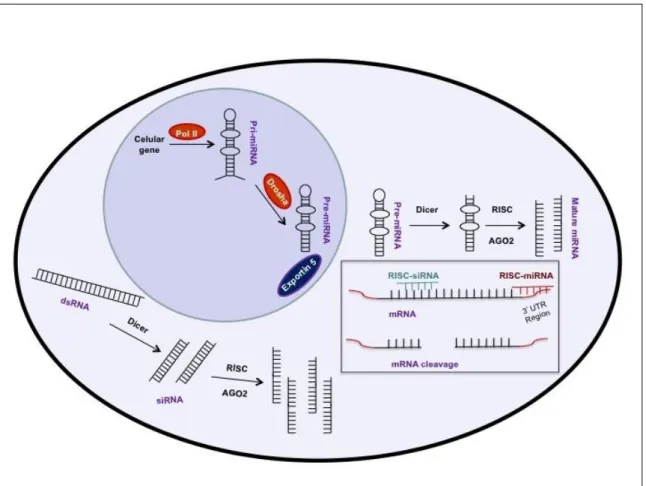
Figure 1.1. The complexity of cancer ... 3 Figure 1. 2. Cancer statistics ... 4 Figure 1. 3. CSC therapy resistance accordingly the different models ... 7 Figure 1. 4. Schematic representation of the EMT process as well as some of the most representative affected proteins ... 12 Figure 1. 5. Schematic representation of E-cadherin ... 14 Figure 1. 6. Schematic representation of the most important biological pathways involved in cancer and EMT activation ... 17 Figure 1. 7. Schematic representation of PI3K/AKT2 mechanism of action ... 21 Figure 1. 8. RNA interference mechanism ... 26
CHAPTER 2
Figure 2. 1. In vitro barriers for non-viral vectors based gene delivery ... 45 Figure 2. 2. Different types of nanotechnology-based systems for gene delivery ... 46 Figure 2. 3. Advantages and limitations in nucleic acid nanosystems delivery ... 47 Figure 2. 4. Schematic representation of the mechanisms of nanoDDS for CSC targeting. ... 48 Figure 2. 5. Diagram of an amphiphilic polymers-based micelle ... 52 Figure 2. 6. The main features of amphiphilic copolymers and their based structures ... 52 Figure 2. 7. Schematic representation of a SNALP and its main components ... 63 Figure 2. 8. Schematic representation of a type of lipopolyplex and its main components. ... 66
CHAPTER 3
Figure 3. 2. Reaction schematic for the conjugation of F127 with 5-DTAF via nucleophilic aromatic substitution by an addition-elimination mechanism ... 97 Figure 3. 3. Polyplexes association efficiency ... 99 Figure 3. 4. In vitro cytotoxicity of CS-siRNA polyplexes at different N/P ration in MDA-MB-231 cells ... 99 Figure 3. 5. Polyplexes CS-siRNA silencing efficacy 72 hours after transfection in RXO-C colon cancer cells expressing GFP... 101 Figure 3. 6. Polyplexes physicochemical characterization using CL213 CS ... 101 Figure 3. 7. Comparative cytotoxicity and IC50 values of Pluronic® F127 and F108 in MDA-MB-231 cells ... 103 Figure 3. 8. CS-siRNA-Pluronic® micelles silencing efficacy 72 hours after transfection with the siGFP and siC in RXO-C colon cancer cells ... 103 Figure 3. 9. PEI-siRNA polyplexes and PEI-siRNA-Pluronic® micelles (obtained by DM) silencing efficacy in GFP expressing RXO-C cells ... 104 Figure 3. 10. PEI-siRNA-Pluronic® internalization behavior ... 105 Figure 3. 11. Physicochemical characterization of PEI-siRNA-Pluronic® micelles .. 106 Figure 3. 12. Serum stability of PEI-siRNA-Pluronic® formulation ... 107 Figure 3. 13. Diagram resuming the different developed tested formulations for gene therapy and the main results. ... 108
CHAPTER 4
Figure 4. 1. Effect of AKT2 silencing in breast and colon cancer cell lines ... 126 Figure 4. 2. siAKT2 transfection reduces cells transformation ability of MDA-MB-231, HCT8 and MCF7 CSC and non-CSC ... 127 Figure 4. 3. Effects of siAKT2 in cells invasive capability of MDA-MB-231, HCT8 and MCF7 CSC and non-CSC ... 128 Figure 4. 4. Effects of siAKT2 in cells transformation ability and invasive capability of MDA-MB-468 and SKBR3 ... 129 Figure 4. 5. Cell lines phenotypes based in the EMT/stemness markers ... 130 Figure 4. 6. Impact of AKT2 silencing in key regulators of EMT reversion and mTOR-dependent signaling pathways. Expression levels of different genes were quantified by
qPCR in different cell lines after the treatment with siAKT2 and siC. Results are expressed as mean±SD (n≥3). ... 131 Figure 4. 7. Effects of AKT2 silencing in different signaling pathways. A) Efects of AKT2 silencing in the mTOR pathway for TWIST+ cells. B) Efffects of AKT2 silencing in the EMT reversion for TWIST- cells. Results are expressed as mean±SD (n≥3). ... 132 Figure 4. 8. Summary of AKT2 silencing effects in cancer development in the different cell lines, accordingly their phenotype and TWIST expression level. ... 133
CHAPTER 5
Figure 5. 1. Schematic representation of micelles production ... 144 Figure 5. 2. Micelles physicochemical characterization ... 151 Figure 5. 3. FACS quantification of micelles uptake by MDA-MB-231 and MCF7 ALDH1A1 tdTomato+ versus tdTomato- ... 153 Figure 5. 4. PM internalization visualization throught confocal microscopy ... 154 Figure 5. 5. In vitro cytotoxicity of PM and its isolated components ... 154 Figure 5. 6. Serum stability assay PM-siRNA ... 155 Figure 5. 7. PM toxicity in vivo ... 156 Figure 5. 8. GFP reporter assay for PM-siGFP biological efficacy assessment ... 158 Figure 5. 9. PM-siAKT2 effects in MDA-MB-231 and MCF7 cells ... 159
CHAPTER 6
Figure 6. 1. Schematic representation of an amphiphilic polymer based multifunctional nanoparticle for gene and drug delivery combination. ... 173
List of Tables
CHAPTER 1
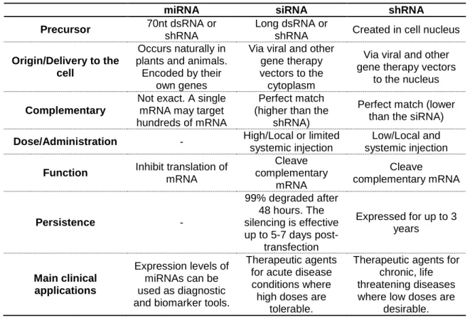
Table 1. 1. Drugs in clinical development targeting CSC* ... 11 Table 1. 2. Different types of EMT and their main functions. ... 13 Table 1. 3. Main characteristics and differences between miRNA, siRNA, and shRNA. .. 25
CHAPTER 2
Table 2. 1. Examples of self-assembled particles under clinical trials evaluation. ... 54 Table 2. 2. Examples of polymeric nanoparticles for gene delivery at different stages of development. ... 59 Table 2. 3. Examples of lipidic nanoparticles for gene delivery at different stages of development. ... 65
CHAPTER 3
Table 3. 1. Poly(ethylene glycol) and polypropylene oxide units, molecular weight and critical micelle concentration values of the different Pluronic® used ... 92 Table 3. 2. Summary of the different tested conditions regarding the CS-based polyplexes. ... 100 Table 3. 3. Summary of the different tested conditions regarding the branched PEI-based systems ... 104 Table 3. 4. Physicochemical characterization of different Pluronic®-based micelles ... 102
CHAPTER 4
Table 4. 1. List of the cell lines used in this study and their main phenotype ... 121 Table 4. 2. List of the primers used in the study and their sequences ... 122 Table 4. 3. List of primary antibodies used in the study and their specifications. ... 123
CHAPTER 5
Abbreviations
3’UTR Untranslated region
5-DTAF 5-8[4,6-dichlorotriazin-2-yl]amino)fluorescein hydrocloride
ABC ATP-binding cassette
AE Association efficiency
AGO2 Argonaute 2
AKT Protein (serine/threorine) kinase B (PKB)
ALDH1A1 Aldehyde dehydrogenase 1
Alox5 Arachidone 5-lipoxigenase
ATCC American type cell colection
ATP Adenosine triphosphate
BAD Bcl2-associated death promoter
BCA Bicinchoninic acid
Bcl-2 B-cell lymphoma 2
Bcl-xL B-cell lymphoma extra large
BCRP Breast cancer resistance protein
BH3 Bcl-2 Homology 3 domain
bHLH basic helix-loop-helix
BLMH Bleomycin hydrolase
CAMs Cell adhesion molecules
CD Cyclodextrin
CHEMS Cholesteryl hemisuccinate
CMC Critical miccelar concentration
CML Chronic myeloid leukemia
CMT Critical micellar temperature
CRISPR Clustered Regularly Interspaced Short Palindromic Repeats
CryoSEM Cyro-scanning electron microscopy
CS Chitosan
CSC Cancer stem cells
CTAB Cetyltrimethylammonium bromide
CTLA4 CytotoxicT-lymphocyte antigen 4
CXCR4 C-X-C chemokine receptor type 4
DC Denditric cells
DD Degree of deacetylation
DDS Drug delivery system
DLL Delta-like ligand
DLS Dynamic light scattering
DM Dissolution method
DMEM Dulbecco’s modified Eagle medium
DMRIE 2,3-di(tetradecoxy)proply-2hydroxyethyl)-dimethylazanium bromide
DMSO dimethyl sulfoxide
DOPC 1,2-dioleoyl-sn-glycero-3-phosphocholine
DOPE 1,2-di-(9Z-octadecenoyl)-sn-glycero-3-phosphoethanolamine
DOPG 1,2-Dioleoyl-sn-glycero-3-phosphoglycerol
DOSPA 2,3dioleyloxy-N-[2(sperminecarboxamido ethyl]-N,N-dimethyl-1-propanaminium trifluoroacetate
DOSPER 1,3-dioleoyloxy-2-. (6-carboxyspermyl)-propyl amide
DOTAP 1,2-dioleoyl-3-trimethylammonium-propane
DOTMA 1,2-di-O-octadecenyl-3-trimethylammonium propane
DSPC 1,2-distearoyl-sn-glycero-3phosphocholine
dsRNA Double-stranded RNA
DTA Diphteria toxin
ECM Extracellular matrix
EGFR Epidermal grownth factor recptor
EMT Epithelial-mesenchymal transition
EO Ethylene oxide
EPR Enhanced permeability and retention effect
ERK Extracellular signal–regulated kinases
FACS Fluorescence-activated cell sorting
FBS Fetal bovine serum
FDA Food and drug administration
FGFR Fibroblast growth factor receptors
FH Thin-film hydration
FR Folate Receptor
GADPH Glyceraldehyde 3-phosphate dehydrogenase
GFP Green fluorescent protein
GSK3 Glycogen synthase kinase 3 beta
HER2 Hormone Estrogen Receptor 2
HES Hydroxyethyl starch
HIF-2α Hypoxia-inducible factor 2 alpha
HLB Hydrophilic lipophilic balances
HPMA N-(2-hydroxypropyl)methacrylamide
IC50 Inhibitory concentration 50
IFN-y Interferon Y
IGF-1 Insulin-like growth factor 1
IGFR Insulin-like growth factor receptor
IL-3 Interleukin-3
IL-6 Interleukin-6
ILK Integrin-linked kinase
LinOS N4-linoneolyl-N9-oleoyl-1,12-diamino-4,9-diazadodecane Lipoplexes Lipid-based Delivery Systems
LPP Lipoplexes
MAPK Mitogen-activated protein kinase
Md Mean diameter
MDM2 Mouse double minute 2 homolog
MDR Multidrug resistance
MDT Maximum tolerated dose
MEK Methyl ethyl ketone
MET Mesenchymal-epithelial transition
miRNA MicroRNA
MRD Minimal residual disease
MRP Multidrug resistance-associated proteins
MTD Maximum tolerated dosis
mTOR Mechanistic target of rapamycin
m-TORC1/2 Mammalian target of rapamycin complex 1 and 2
MTT 3-(4,5-dimethythiazol-2-yl)-2,5 diphenyl tetrazolium bromide
MW Molecular weight
N/P ratio Nitrogen/Phosphate ratio
NF-kB Nuclear factor kappa B
Nio-AU Gold niosomes
NK Natural killer
OGN Phosphate groups
PAEs Poly(b-amino ester)s
PAMAM Polyamidoamine
PARP Poly (ADP-ribose) polymerase
PBS Phosphate buffered saline
PCL Poly(-caprolactone)
PCSK9 Proprotein convertase subtilisin/kexin type 9
Pdi Poydispersity index
PDK1 Phosphoinositide-dependent protein kinase 1
PDMAEMA Poly(2-(N,N-dimethylamino)ethyl methacrylate)
PE Phosphoethanolamine
PEG/PEO Polyethylene glycol/ Polyethyleneoxide
PEI Polyethylenimine
PCL Polycaprolactone
PFA Paraforlmaldehyde
PGE2 Prostaglandin E2
P-gp P-glycoprotein
PH Pleckstrin homology domain
PHML-b-PLLA-b-PHML Poly(hydroxyletheyl methacrylate-L-lysine)-b-poly)L-lactide)— poly(hydroxyletheyl methacrylate-L-lysine
PI3K Phosphoinositide 3-kinase
PILP Pegylated immuno-lipopolyplexes
PKB Protein kinase B
PLA Poly(d,l-lactide)
PLAS PEGylated lipoplex-entrapped alginate scaffold
PLGA Poly(d,l-lactide-co-glycolide)
PLK1 Polo-like kinase
PLL Poly(L-lysine)
PM Polymeric micelles
PO Propylene oxide
Poliplexes Polymer-based Delivery Systems
Poll II Polymerase II
PPEEA Poly(2-aminoethyl ethylene phosphate)
PPO Polypropylene oxice
PTEN Phosphatase and tensin homolog
qRT-PCR Quantitative real time polymerase chain reaction
RFP Red Fluorescent Protein
RISC RNA-induced silencing complex
RRM2 Ribonucleoside-diphosphate reductase subunit M2
RTK Receptor tirosine kinase
SCID Severe combined immunodeficiency
SD Mean±standard desviation
SDS-PAGE sodium dodecyl sulfate polyacrylamide gel electrophoresis
Ser Serine
shRNA Short hairpin RNA
SLN Solid lipid nanoparticules
SLUG Zinc finger protein SNAIL2
SNAIL Zinc finger protein SNAIL1
SNALP Stable nucleic acid lipid particles
SPARC Secreted protein acidic and rich in cysteine STAT3 Signal transducer and actibvator of transcription 3
SV40 Simian vacuolating virus 40
TBE Tris/Borate/EDTA
TEM Transmission electron microscopy
Tf Human transferrin
TfR Transferrin receptors
TGF- Tumor growth factor beta
TGFR Tumor growth factor beta receptor
Thr Threonine
TNF Tumor necrosis factor
TORC2 Target of rapamycin complex 2
TQ Thymoquinone
UV Ultra violet
VEGF Vascular endothelial growth factor
WB Western Blotting
WNT Wingless-related integration site
ZEB Zinc finger E-box-binding homeobox
Aims and Organization of the Thesis
The high complexity of cancer diseases and their still unknown and incontrollable pattern, has made prioritary the study and a better understanding of the wide range of biological pathways involved in their development and progress. Worstly, the heterogeneity of cancer cell populations within the tumor difficults the complete remission of the disease, being the cancer recurrence one of the major challenges for the current therapies. The small subpopulation of CSC within a certain tumor have been reported as the responsible for the so feared tumor recurrence and resistance to therapy.This work could be divided in two main research areas and general objectives:
i) Molecular biology of cancer: study and identification of AKT2 as a good target for cancer therapy;
ii) Pharmaceutical technology and development: design and characterization of a Nanotechnology-based system for delivery of a siRNA against the AKT2.
Based on the previous, the specific objectives of this work are the follows:
1) Due to its recognized role as a putative oncogene, we aim to understand and characterize the biological pathway of AKT2 and assess the effects of AKT2 silencing in different breast and colon cancer cell lines in terms of cell migration, proliferation, and gene expression pattern;
2) Isolate the subpopulations of CSC from breast and colon cancer cells and assess the effects of AKT2 silencing specifically in this subpolation in terms of cell invasion, cell anchorage-independent growth, and gene expression pattern;
3) Understand the downstreams effectors of AKT2 for each cell line (TWIST or mTOR); 4) Design a nanoparticle-based system able to complex siRNA and silence the gene
of interest using classic reporter gene assays;
5) Characterize the new nanosystem in terms of their physicochemical features and stability, in vitro and in vivo toxicity, and cellular internalization;
6) Assess the biological effects of nanoparticles complexing siRNA against the AKT2 in subpopulations of both breast CSC and non-CSC in terms of invasion and cell anchorage-independent growth abilities.
The present thesis is organized in six chapters. In the first two chapters are presented the main theoretical concepts and the state of art for each of research areas:
Chapter 1 – State of Art – Epithelial-Mesenchymal Transition as the Motor for Stemness: AKT2 Emerges as a Potential Therapeutic Target
Chapter 2 – State of Art – Nanotechnology for Gene Delivery
After the introductory section is presented the research work developed and the main results obtained:
Chapter 3 (Formulation Studies and Efficacy Assessment of Different siRNA Delivery Systems) describes the way paved through the design and development of a nanoparticulate system based in polymeric micelles (PM) for siRNA delivery until reach the final selected formulation presented in Chapter 5;
Chapter 4 (AKT2-related Biological Pathway Characterization and Validation in Breast and Colon Cancer Stem Cells) presents the study and characterization of the biological pathway and the role of AKT2 in the development and progression of cancer both in breast and colon CSC and non-CSC;
Chapter 5 (Functional Validation of Amphiphilic-based Polymeric Micelles for siRNA Delivery and Cancer Stem Cells Genes Inhibition) presents the results obtained with the chosen PM for the delivery of siAKT2.
In the final chapter (Chapter 6) are presented the Main Conclusions and Future Perspectives of the work.
CHAPTER 1
State of Art – Epithelial-Mesenchymal Transition as the
Motor for Stemness: AKT2 Emerges as a Potential
Therapeutic Target
The information presented in this chapter was partially published in the following publications:
1) P Gener, D Rafael, Y Fernández, J Sayos, D Arango, I Abasolo, M Videira, S Schwartz Jr., Cancer Stem Cells and Personalized Cancer Nanomedicine. Nanomedicine (Lond), 11(3):307-20, 2016.
2) D Rafael, S Doktorovová, H Florindo, P Gener, I Abasolo, S Schwartz Jr., M Videira, EMT Blockage Strategies: Targeting Akt Dependent Mechanisms for Breast Cancer Metastatic Behaviour Modulation, Current Gene Therapy, 15(3) 2015.
Table of contents
1.1. Cancer Facts ...3
1.1.1. Cancer Scenario ...4 1.1.2. Cancer Treatment ...5 1.2. The Cancer Stem Cells Theory ...6
1.2.1. Cancer Stem Cell Models ...6 1.2.2. Cancer Stem Cell Properties...9 1.2.3. Targeting CSC ...9 1.3. Epithelial-Mesenchymal Transition ... 12
1.3.1. Adhesion-related Proteins: E-cadherin ... 13 1.3.2. Tumor-associated Proteins in EMT Activation ... 16 1.3.2.1. The influence of tumor microenvironment ... 16 1.3.2.2. Players at the cell membrane level ... 18 1.3.2.3. Intracellular transcriptional regulation of EMT ... 19 1.4. The Importance of TWIST-based Signaling Pathways in the EMT Program……… ... 19 1.4.1. TWIST Mechanism of Action: AKT2 and PI3K as the Leading Players ... 21 1.5. RNA Interference Regulation of EMT... 24
1.5.1. miRNA and Cancer ... 27 1.5.2. siRNA-mediated Silencing of AKT Isoforms ... 27 1.6. Conclusions ... 29
1.1. Cancer Facts
According to World Health Organization (WHO), cancer is a generic term used to describe a large and heterogeneous group of diseases that can affect any part of the body. A key feature is the genesis of abnormal cells that rapidly grow beyond their normal boundaries invading adjacent parts of the body and spreading to other organs (metastasis) (Figure 1.1). Metastization is of major impact in the malignancy of the disease and clinical outcome (1). Due to its high diversity, complexity, and unpredictable and uncontrollable character, cancer has become one of the most feared diseases by humans in the last decades. With the arise of molecular biology and biotechnology, tremendous advances have been observed in the last decades in the oncology field leading to the identification of biological pathways involved in cell growth and dissemination, as well as disease players and therapeutic targets/agents (Figure 1.1).
Figure 1.1. The complexity of cancer. The figure represents different phases of the disease
evolution and the problematic of cancer mutations that increase exponentially along with the tumor development originating a highly heterogeneous compartment. Circles point out the main cancer hallmarks as well as the associated most common therapeutic strategies. BH3: Bcl-2 homology 3 domain; CTLA4: cytotoxic T-lymphocyte antigen 4; EGFR: epidermal growth factor receptor; PARP: poly (ADP-ribose) polymerase; VEGF: Vascular endothelial growth factor.
1.1.1. Cancer Scenario
The progresses referred before enables researchers and pharmaceutical industry with the information necessary to develop a variety of drugs to treat the different types of cancer, resulting in an important decrease of the cancer related deaths (superior to 20% since 1990s) and an increase in the patient’s overall survival (2, 3). The incidence and mortality rates of this disease are superior in developed countries and are expected to increase in the next years, with an estimated 9 million cancer deaths in 2015 and 11.4 million in 2030 (3). Just in the USA, 1.7 million new cancer cases and 0.6 million cancer deaths are projected to occur in 2017 (4). Nowadays, the seven most common types of cancers worldwide are lung cancer, breast cancer, colorectal cancer, prostate cancer, stomach cancer, liver cancer, and cervical cancer (Figure 1.2) (5). Cancers typically associated with infections such as the cervical cancer, stomach cancer, liver cancer and Kaposi's sarcoma are declining in the developing countries as infections become better controlled with the programs implemented by WHO. On the contrary, lung, breast, prostate, and colorectal cancers present a faster rate of incidence, being the world cancer incidence predicted to rise 75% by 2030, mainly associated with the increasing bad lifestyle practices, population growth and aging (3, 5).
Figure 1. 2. Cancer statistics. Estimated incidence (A) and mortality (B) worldwide of the most
common types of cancer in both sexes. Estimated age-standardized rates of incident cases (C) and deaths (D) in both sexes for all cancers excluding non-melanoma skin cancer. Data Source: GLOBOCAN 2012. Map Production IARC (2016) (5).
In the last years, due to the growing incidence of the disease, the global market share of medicines to treat cancer has been growing. In 2014 oncology remained as the largest therapeutic area worldwide regarding the market share, presenting an increase of 8% compared to 2013 and counting for more than 10% of the total year sales (6). As a consequence of the expected growth in the prevalence and incidence of oncological diseases in the upcoming years, it is also predicted an increase in the prescription of anticancer drugs, with an escalation of their global market share up to almost 15% in 2020 (6). This disease profile entails high costs to the health care systems related to the patients’ cancer treatment and care. For example, only in USA, cancer care is projected to cost around 174 billion US dollars in 2020 (7). This scenario boosts researchers, health care agencies, and pharmaceutical companies to pursuit the urgent need for the development of new and more cost-effective treatments.
1.1.2. Cancer Treatment
Despite the enormous efforts employed at different fields of science and medicine, the real cure and eradication of cancer remains a desire far to be achieved and stands as a huge challenge to researchers around the world.
Major challenges surrounding the administration of current anticancer treatments are their systemic toxicity, associated with serious side-effects owing this drugs lack of specificity, and the therapeutic resistance that cancer cells often acquire during treatment. Indeed, because of the use of chemotherapeutics at an early stage of the disease that renders tumor resistance, treatment options for late metastatic disease are often reduced (8). The ability of cancer cells to activate alternative molecular pathways allowing them to escape from the common drug therapeutic mechanism of action is one of the major mechanisms involved in the development of drug resistance (9, 10). Further, drug resistance also occurs through the increased expression of ATP-dependent drug efflux transporters on the surface of cancer cells (11). This causes a significant decrease of intracellular drug accumulation, which results into severe limitation of drug’s efficacy (12). Besides, cells with different phenotypes and proliferative abilities co-exist within a tumor, generating tumor heterogeneity and contributing to specific selection of resistant clones and disappointing therapeutic responses, in particular when a single drug treatment is applied.
Special interest has been paid to the use of biopharmaceuticals and gene therapy as therapeutic strategies to treat cancer owing to its capacity to target the specific antigen or signaling pathway involved in cancer progression (13). Among the different types of gene therapy, the silencing of particular genes through the RNA interference (RNAi) therapy has
been intensively investigated as a encouraging strategy in the field of oncology, given the possibility of targeting oncogenes involved in proliferation, survival, angiogenesis, metastasis, apoptosis suppression or drug-resistance (14).
Nowadays, more than 800 medicines and vaccines to treat cancer are under development and clinical assessment (2), with more than 73% of the medicines in the pipeline being studied based on biomarkers, presenting potential for personalized treatment (15). Some of the medicines under development are based on biopharmaceuticals, namely monoclonal antibodies, vaccines, cell or gene therapy. In 2013, the majority of biopharmaceuticals under development are intended to treat cancer and related diseases (more than 300), being the monoclonal antibodies the most studied ones with 170 medicines (16). Also, seventeen products under development/clinical trials were based on gene therapy such as ALN-VSP from Alnylam Pharmaceuticals (Phase I), B7-2/GM-CSF from NuVax Therapeutics (Phase I), BC-819 from BioCancell Therapeutics (Phase II), CALAA-01 from Calando Pharmaceuticals (Phase I), EGEN-001 from EGEN (Phase II) or GliAtak™ from Advantagene (Phase II) (16).
Unfortunately, even though important clinical breakthroughs in the fight against cancer have been achieved by using combined protocols such as conventional chemotherapy with hormonal therapy or therapeutic antibodies, recurrence and metastasis are still observed. Thus, the development of treatments able to reduce the formation of metastasis and to avoid or at least reduce the recurrence of the disease is required.
1.2. The Cancer Stem Cells Theory
As referred previously, despite progresses in cancer management, advanced cancers have still low clinical response rates and high recurrence and mortality rates. Regarding this, a main issue that should be considered is the fact that cancer cells within a tumor are not equal in terms of characteristics and tumorigenic potential.
1.2.1. Cancer Stem Cell Models
Before the 1990s, cancer initiation and progression was explained by a clonal cancer model. It was considered that all tumor cells have similar characteristics and equal tumor formation capacity and that tumors expansion depended on clonal selection advantages (17-20). The first evidence regarding the existence of cancer stem cells (CSC) was obtained in acute myeloid leukemia (21). Subsequently, CSC were identified also in other hematopoietic
cancers and in many solid tumors (breast, brain, colon, prostate, lung, head & neck, among others) (22-24). It was also shown that the small CSC sub-population has the ability to generate and maintain the tumor progression (20-22, 25). Based on these observations, a new hierarchical model describing cancer propagation was postulated (21); CSC can self-renew their own population and have long-term propagating capacity contrariwise to normal cells whose division finish by clonal exhaustion (Figure 1.3A) (26, 27).
Figure 1. 3. CSC therapy resistance accordingly the different models. (A) CSC are more
resistant than tumor bulk cells to most conventional therapeutic interventions. Therefore, after treatment, CSC remain in the tumors or in circulation inducing a rapid recurrence. (B) As for the hierarchic model, CSC specific therapy results in loss of proliferative capacity and decline of the malignancy with tumor regression. (C) Considering the novel dynamic model, the non-CSC can acquire CSC-features through signals from the environment and the stromal cells, thus even with specific CSC eradication a rapid restoration of CSC features occurs, with a subsequent recurrence when therapy is discontinued. (D) The best strategy to tumor remission should target not only the CSC but also the bulk tumor cells as well as the interconversion capacity between them. Stromal cells represent myofibroblasts, endothelial cells, mesenchymal stem cells, or infiltrating immune cells. CSC: cancer stem cells.
In summary, CSC have been considered the major critical player in tumor malignancy and recurrence, thus strong efforts have been recently done to specifically target CSC subpopulation in order to improve therapy efficacy and avoid recurring tumors. However,
recent data suggest that targeting exclusively the CSC sub-populations may not be sufficient because acquisition of stemeness phenotype by other cells within tumors seems to be a bidirectional dynamic process (Figure 1.3B) (26, 27). Because the hierarchical model of cancer cannot explain the dynamic behavior of CSC, a new interconversion cancer model has been very recently postulated (Figure 1.3C) (26-28). According to the model, the amount of CSC within a tumor or cancer cell line seems to be inconstant and finely tuned, to maintain a specific equilibrium between CSC and non-CSC populations. In one hand, CSC can differentiate to non-CSC which in turn can de-differentiate and revert into cells with CSC properties, like resistance and self-renewal capacity with typical stemness gene expression signature. This cell de-differentiation process was first predicted by Marcov mathematical model (28) and recently confirmed experimentally in various CSC models (28-30). In melanoma, a subpopulation of slow-cycling cells that express histone demethylase JARD1B was identified. As expected from CSC, during propagation of purified JARD1B positive cells appeared a JARD1B negative, non-CSC population. Conversely, a single JARD1B negative cell originates a heterogeneous progeny including JARD1B positive cells, suggesting a bidirectional dynamic of the CSC phenotype (31). Similarly, normal somatic basal-like mammary epithelial cells are able to spontaneously de-differentiate into stem-like cells (32). Further, regeneration of stem-like cells is observed as well in vivo after sorting, suggesting that stable equilibrium of CSC/non-CSC cells within a tumor occurs due to cell-state interconversion (28). These findings reinforce the hypothesis of the existence of a controlled balance between CSC and non-CSC populations. It seems that cancer cells might survive to stress conditions by entering de-differentiation as survival mechanism. Several external factors and paracrine communications as well as cell-to-cell interactions are involved in the regulation of this process. As an example, it has been shown that apoptotic cells excrete prostaglandin E2 (PGE2) which promotes proliferation of neighbouring CSC after chemotherapy, whereas PGE2-neutralizing antibodies abrogate CSC repopulation after treatment (33). Similarly, the matrix cellular protein SPARC (secreted protein acidic and rich in cysteine, also known as osteonectin or BM-40) secreted by non-CSC is able to modulates the metastatic capacity of CSC in prostate cancer models (34), and Interleukin-6 (IL-6) secreted by non-CSC is reported to induce formation of breast CSC in association with octamer-binding transcription factor 4 (OCT4) expression (35). Furthermore, OCT4 overexpression is induced via IL-6-JAK1-STAT3 signaling pathway in low attachment conditions as well as in vivo to maintain dynamic equilibrium (36).
Therefore, the dynamic phenotype of CSC represents an important challenge for targeted cancer therapies, as tumor cell populations are continuously evolving and therapeutic eradication of existing CSC populations might be followed by their regeneration from
non-CSC. Regarding this, the major goal is to find a way that allows to targeting the CSC but also stopping the interconversion between CSC and non-CSC (Figure 1.3D).
1.2.2. Cancer Stem Cell Properties
Even though the intratumoral amount of CSC as well as the expression of stemness markers among this sub-population differs, depending mostly on tumor types, the essential stemness properties of CSC like self-renewal, tumor initiation capacity and long-term repopulation potential are common features independently of tumor type. Owing their ability to survive in non-attachment conditions showing capacity to grow as tridimensional tumorspheres, CSC have increased capacity to initiate tumor growth in vivo, migrate and intravasate the blood stream generating distant metastasis at specific sites. CSC are substantially insensitive to most conventional anticancer therapies, antimitotic agents, and/or radiation (37). Such aggressive behavior results from a particular CSC phenotype as well as their characteristic gene expression and epigenetic modifications. In particular, CSC have a potent machinery of anti-apoptotic proteins and a efficient activation of the DNA damage sensor and repair machinery for maintenance of genome integrity (9, 37). Moreover, CSC overexpress a great number of detoxifying enzymes (aldehyde dehydrogenase 1 (ALDH1A1), bleomycin hydrolase (BLMH)), drug efflux transporters (P-glycoprotein (P-gp)), breast cancer resistance protein (BCRP), and the multidrug resistance-associated proteins (MRP) at the cell membrane, which pump-out chemotherapeutic drugs from the intracellular space (10, 38). In addition, induction of quiescence in stress conditions and higher stability under hypoxic conditions provide CSC with further protection against anticancer therapies (10). Accordingly, the percentage of CSC within a tumor often increases after treatment (33, 37, 39, 40). This often leads to cancer recurrence and metastatic growth since only few CSC are necessary and sufficient for tumor regeneration in vivo (41). For all these reasons, if cancer has to be eradicated, new therapies should specifically target the CSC fraction.
1.2.3. Targeting CSC
In agreement with the interconversion cancer model, traditional cancer treatments appear to be effective at reducing tumor mass but often fail to produce desired long-term outcomes, and remission of metastatic disease is common. Moreover, current treatments often render
the tumor resistant at the time of remission, possibly due to their inability to eliminate CSC (8, 37).
Therapeutic targeting of CSC may have the potential to remove residual disease and become an important component of any given cancer treatment. Currently, there are multiple anti-CSC agents in pre-clinical and clinical trials (Table 1.1) (42). The likelihood of their clinical success will depend on many aspects, including safety, specificity, pharmacokinetics etc., but also on their ability to effectively target CSC. In fact, most drugs specifically designed to target CSC are based on the effective inhibition of crucial CSC signaling pathways as example: i) cell survival and proliferation pathways; including PI3K-AKT, JAK/STAT, and NF-kB signaling, and ii) signaling linked to the stemness properties of CSC like self-renewal and pluripotency (Notch pathway, WNT pathway and Hedgehog signaling). Of note, one of the risks associated to drugs targeting commonly shared cellular pathways is the difficulty to distinguish normal cells and/or stem cells from CSC. High throughput screening methodologies are reliable approaches to reveal new therapeutic targets that effectively eliminate CSC without compromise the normal somatic stem cells. One of the hopeful targets identified by high throughput screening is Arachidone 5-lipoxigenase (Alox5). Alox5 was found over-expressed in chronic myeloid leukemia CSC but not in normal hematopoietic stem cells (43). It was shown that the absence of Alox5 due to genetic modification or its inhibition with a specific inhibitor, Zileuton, impaired the induction of leukemia in the BCR-ABL inducible cancer model of chronic myeloid leukemia (CML) (43). The inhibition of Alox5 leads to an upregulation of the tumor suppressor gene Msr1 and consequently, to the inhibition of PI3K-AKT and β-catenin pathways (44). Currently, the use of Zileuton in combination with Imatinib is in phase I clinical trials for first line therapy in acute myeloid leukemia. Of note, Imatinib alone does not cure CML because it is not able to kill leukemia stem cells (45) . Another example of a successful high through screening is the chemical screen method for agents with CSC-specific toxicity. As a result, salinomycin was identified as a drug showing selective toxicity for CSC (46).
Previous results from preclinical trials in human xenograft mice models and clinical pilot studies reveal that salinomycin is able to effectively eliminate CSC and to induce partial clinical regression of heavily pre-treated and therapy-resistant cancers, particularly in combination with novel tumor-targeted drugs (47). However, a very important drawback for the potential clinical application of Salinomycin is its marked neural and muscular toxicity (48). In this context, incorporation of cytotoxic drugs within different particulate nanocarrier systems, i.e. liposomes and polymeric particles, is a useful approach to modify the biodistribution of drugs and target tumors. Nanomedicines can in fact address not only the biodistribution profile of CSC-targeting drugs but also reduce their overall toxicity and
enhance their therapeutic efficacy by promoting higher local drug exposure and improving their transport properties across biological membranes (49).
Table 1. 1. Drugs under clinical development with therapeutic activity against CSC* Therapeutic
Target Drug Cancer Type Clinical phase Trial reference
PI3K,
mTORC1/2 VS-5584 Solid tumors I NCT01991938
AKT
MK2206 Breast cancer II NCT01277757 GSK2141795 Breast cancer II NCT01964924 AZD5363 Breast cancer I/II NCT01625286
STAT3 OPB-31121 Solid tumors I NCT00955812
NF-κB Parthenolid
analog Leukemia II/III ISRTN40571019
Multiple
kinases BBI503 Solid tumors I/II NCT01781455
STAT3, β-catenine, Nanog
BBI608
Gastric cancer III NCT02178956 Esophageal
cancer III NCT02178956 Colon cancer III NCT01830621 BBI503+ BBI608 Solid tumors I NCT02432326
WNT pathway
Vantictumab Solid tumors I NCT0 1345201 Ipafricept Pancreatic cancer I NCT02050178 Notch 2,3 (receptor) Tarextumab Pancreatic cancer II NCT01647828 Lung cancer II NCT01859741 DLL (Notch ligand) Demcizumab Ovarian cancer II NCT01952249 Pancreatic cancer II NCT02289898 Lung cancer II NCT02259582
Hedgehog GDC-449 Solid tumors II NCT00739661
ABC transporters MS-209 Ovarian cancer II NCT00739661 Tariquidar Ovarian cancer II NCT00069160 Cervical cancer II NCT00069160 Lung cancer II NCT00069160 Kidney cancer II NCT00069160 Chemokine
receptors 1,2 Reparixin Breast cancer II NCT01861054
IL-3 receptor SL-401 Leukemia I/II NCT02268253
Alox5 Zileuton Leukemia I NCT02047149
*Details including dosage of the chemical agents under trial, duration of the trial, inclusion and exclusion criteria for recruiting patients, contact and locations where the trial is being conducted, and the current status of the trial can be obtained by searching the ‘Trial reference’ in the US National Institutes of Health Registry (www.clinicaltrials.gov) and EU Clinical Trials Register (www.clinicaltrialsregister.eu). ABC: ATP-binding cassette; AKT: protein (serine/threonine) kinase B (PKB); Alox5: arachidone 5-lipoxigenase; DLL: delta-like ligand; IL-3: interleukin-3; mTORC1/2: mammalian target of rapamycin complex 1 and 2; NF-κB: nuclear factor kappa B; PI3K: phosphoinositide 3-kinase; STAT3: signal transducer and activator of transcription 3; WNT: wingless-related integration site.
Figure 1. 4. Schematic representation of the EMT process as well as some of the most representative affected proteins. EMT: epithelial-mesenchymal transition.
1.3. Epithelial-Mesenchymal Transition (EMT)
As referred previously, tumor invasion and subsequent metastization are the major causes of morbidity and mortality in patients with cancer (50). Most of malignant tumors are carcinomas originated from various epithelial tissues. EMT is the biological process that triggers the evolution from a well-differentiated adenocarcinoma to an invasive epithelial carcinoma (Figure 1.4) (51). It is defined as a phenomenon during which cells lose their characteristic epithelial traits: columnar or polygonal shape, apico-basolateral polarization, organization in cell layers with strong cell-cell adhesion and limited migratory potential. During this event, cells acquire mesenchymal features such as, spindle-shaped morphology, anterior-posterior polarization, focal cell-cell contacts and strong migratory potential, becoming able to detach from their original site, penetrate the bloodstream, and subsequently create metastasis at different sites. These morphological and functional changes, demands several intracellular molecular alterations such as the loss of epithelial markers including E-cadherin, certain cytokeratins, occludin, and claudin, while mesenchymal markers, namely N-cadherin, vimentin and fibronectin are upregulated (52, 53).
EMT is an essential process for appropriate embryonic development. In adults, EMT occurs during wound healing, tissue regeneration, organ fibrosis, and cancer progression (8). Based on the biological context in which EMT occur, this mechanism can be classified into three different subtypes presented in Table 1.2 (53). During cancer progression, the genetic and epigenetic changes that mainly affect oncogenes and tumor suppressor genes of epithelial carcinoma cells seem to be responsible for the activation of an EMT program (54, 55). Thus, preventing epithelial carcinoma cells to undergo such transition in order to keep the tumor in its primary stage and, therefore, treatable should be a future goal of therapeutic intervention.