Using Retrovirus-Mediated RNA Interference
Ni Xie1., Lisha Mou1., Jianhui Yuan2*, Wenlan Liu1, Tingting Deng2, Zigang Li3, Yi Jin1, Zhangli Hu4 1Shenzhen Second People’s Hospital, The First Affiliated Hospital of Shenzhen University, Shenzhen, Guangdong, China,2The Shenzhen Center for Disease Control and Prevention, Shenzhen, Guangdong, China,3The Shenzhen Graduate School, Peking University, Shenzhen, Guangdong, China,4College of Life Science, Shenzhen University, Shenzhen, Guangdong, China
Abstract
Background:The BCRP/ABCG2 transporter, which mediates drug resistance in many types of cells, depends on energy provided by ATP hydrolysis. Here, a retrovirus encoding a shRNA targeting the ATP-binding domain of this protein was used to screen for highly efficient agents that could reverse drug resistance and improve cell sensitivity to drugs, thus laying the foundation for further studies and applications.
Methodology/Principal Findings:To target the ATP-binding domain ofBCRP/ABCG2, pLenti6/BCRPsi shRNA recombinant retroviruses, with 20 bp target sequences starting from the 270th, 745thand 939thbps of the 6thexon, were constructed and packaged. The pLenti6/BCRPsi retroviruses (V-BCRPi) that conferred significant knockdown effects were screened using a drug-sensitivity experiment and flow cytometry. The human choriocarcinoma cell line JAR, which highly expresses endogenousBCRP/ABCG2, was injected under the dorsal skin of a hairless mouse to initiate a JAR cytoma. After injecting V-BCRPi-infected JAR tumor cells into the dorsal skin of hairless mice, BCRP/ABCG2 expression in the tumor tissue was determined using immunohistochemistry, fluorescent quantitative RT-PCR and Western blot analyses. After intraperitoneal injection ofBCRP/ABCG2-tolerant 5-FU, the tumor volume, weight change, and apoptosis rate of the tumor tissue were determined using in situ hybridization. V-BCRPi increased the sensitivity of the tumor histiocytes to 5-FU and improved the cell apoptosis-promoting effects of 5-FU in the tumor.
Conclusions/Significance:The goal of thein vivoandin vitrostudies was to screen for an RNA interference recombinant retrovirus capable of stably targeting the ATP-binding domain of BCRP/ABCG2(V-BCRPi) to inhibit its function. A new method to improve the chemo-sensitivity of breast cancer and other tumor cells was discovered, and this method could be used for gene therapy and functional studies of malignant tumors.
Citation: Xie N, Mou L, Yuan J, Liu W, Deng T, et al. (2014) Modulating Drug Resistance by TargetingBCRP/ABCG2 Using Retrovirus-Mediated RNA Interference. PLoS ONE 9(7): e103463. doi:10.1371/journal.pone.0103463
Editor:Javier S. Castresana, University of Navarra, Spain
ReceivedMay 30, 2014;AcceptedJune 30, 2014;PublishedJuly 30, 2014
Copyright:ß2014 Xie et al. This is an open-access article distributed under the terms of the Creative Commons Attribution License, which permits unrestricted use, distribution, and reproduction in any medium, provided the original author and source are credited.
Data Availability:The authors confirm that all data underlying the findings are fully available without restriction. All relevant data are within the paper.
Funding:Funding provided by National Natural Science Fund (30500599, 81200465); Guangdong Natural Science Fund Project (9151503102000019); Shenzhen Science and Technology Plan (medical treatment and public health) key project (201201028); Shenzhen Technical Research and Development Fund Project (GJHZ20130412153906740, JCYJ20120615085512920, JCYJ20120613171430264, ZYC201006180477A, SW201110060, SW201110018); Shenzhen Peacock Plan (KQTD201103). The funders had no role in study design, data collection and analysis, decision to publish, or preparation of the manuscript.
Competing Interests:The authors have declared that no competing interests exist.
* Email: [email protected]
.These authors contributed equally to this work.
Introduction
The ultimate goals of oncology drug resistance mechanism research are to find targets of drug resistance and screen for specific agents that can reverse these phenotypes, to improve the curative effects of chemotherapy for prevention and clinical treatment, and to reduce drug toxicity. Several papers have demonstrated the reversal of BCRP/ABCG2 multidrug resistance as well as specific, non-specific and chemical drug resistance [1]. The first BCRP/ABCG2 inhibitor was fumitremorgin C (FTC) [2]. The same reversal effects have been observed after targeting other membrane transporter proteins due to the structural similarities of these proteins [3]. For example, one inhibitor, isoquinoline acridorex (GF120918), and an analogue of dipyri-damole, BIB-E, helped to inhibit the drug resistance phenotype
mediated by BCRP/ABCG2 [4,5]. Typically reversal-agent studies are performed for specific targets and unitary target structures. However, these studies have unclear clinical application prospects and weak feasibility. Studying drug-resistance mecha-nisms and reversal agents with the same inhibitory effects is both valuable and important for understanding the multidrug-resistance phenotype.
shRNA-encoding retrovirus with high infection efficiency was used to stably express a hairpin structure targeting the ABC domain of BCRP/ABCG2, and cell and animal experiments were conducted. The goal of the study was to screen for constructs that could efficiently target the ATP-binding domain and for agents that could reverse the drug-resistant phenotypes, which would ideally improve tumor cell sensitivity to drugs and lay a solid foundation for further studies and applications.
Materials and Methods
Cells, strains, plasmids and reagents
The 293FT virus-packaging cells,Escherichia coliTOP10 cells, Stbl3 competent cells, pENTR/U6, and the shRNA retrovirus vector pLenti6/BLOCK-iT-DEST were purchased from Invitro-gen (Carlsbad, CA, USA). The human choriocarcinoma cell line JAR was purchased from the Shanghai Institute of Cellular Biology of the Chinese Academy of Sciences. BALB/c-nu/nu hairless mice (3- to 5-week-old females) were provided by the Laboratory Animal Center at the Guangzhou University of Traditional Chinese Medicine. The mice were raised in a specific pathogen-free environment. All animal experiments were con-ducted in accordance with the NIH Guide for the Care and Use of Laboratory Animals, and the methods were approved by the Ethics Committee at the Shenzhen Center for Disease Control and Prevention. The DNA loading buffer, DNA molecular weight marker, and PCR kit were purchased from Takara (TaKaRa, Dalian, China), and the agarose was obtained from BBI (Toronto, Ontario, Canada). RPMI-1640 medium, DMEM, penicillin/ streptomycin double-resistant fluid, 0.25% trypsin-EDTA and fetal calf serum were obtained from Gibco (Grand Island, NY, USA). The single-stranded oligonucleotides and primers were synthesized by Takara. The HRP-conjugated goat anti-mouse secondary antibody was obtained from Sigma (Shanghai, China). The tris (hydroxymethyl) aminomethane (Tris), acrylamide, dithiothreitol (DTT) and tetramethylethylenediamine (TEMED) were purchased from Sangon Biotech Co., Ltd (Shanghai, China). The Western blotting prestain was obtained from Fermentas (Hanover, MD, USA). The rabbit anti-b-actin primary antibody was obtained from Biovision ((Mountain View, CA, USA), and the mouse anti-BCRP primary antibody (BXP-21) was purchased from Alexis (San Diego, CA, USA). The ECL and 2-D quantification kits were obtained from GE (Uppsala. Sweden). Trizol reagent was obtained from Invitrogen, and the reverse transcription and fluorogenic quantitative PCR kits were pur-chased from Takara. The DAPI was obtained from APPLICHEM (Darmstadt, Germany). The paraformaldehyde was domestic and analytically pure, and the 5-fluorouracil (5-FU) was domestic and obtained from SunnyHope Pharmaceutical Co., Ltd (Chengdu, China). The Cellular Orthotopic apoptosis detection kit was obtained from Shanghai KeyGEN Biotech Co., Ltd (Shanghai, China). All other chemical reagents were domestic, analytical reagents.
Design and synthesis ofBCRP/ABCG2RNA interference constructs
The BLOCK-iT RNAi Designer at Invitrogen was used to design DNA oligonucleotides complementary to the ABC region of the BCRP/ABCG2 gene, beginning at the 207th, 745th, and 939thinitiation sites of the 6thexon. These oligonucleotides were then synthesized, and the sequences of each pair of DNA oligonucleotides included the following sequences: upstream sequence - target sense sequence of 59-CACCG-20 bases and antisense sequence of 39CGAA20 bases; downstream sequence
-antisense sequence of -C-39 and 59-AAAA-44, which is comple-mentary to the upstream sequence.
Construction of the pENTR/U6-BCRPi entry vector At room temperature, 5ml of ‘‘Top strand’’ DNA oligo (200mM), 5ml of ‘‘Bottom strand’’ DNA oligo (200mM), 2ml of 106 Oligo Annealing Buffer, and 8ml of DNase/RNAse-free water (for a total volume of 20ml) was placed in a 0.5-ml sterile EP centrifuge tube. The reaction mixture was incubated at 95uC for 4 minutes. In total, 99ml of DNase/RNAse-free water was added to 1ml of each of the double-strand products (500 nM) to dilute them 100-fold, resulting in a final concentration of 5 nM. The samples were then mixed with 4ml of 56Ligation Buffer, 2ml of pENTR/U6 (0.5 ng/ml), 1ml of double-stranded oligo (5 nM), 12ml of DNase/RNAse-free water, and 1ml of T4 DNA Ligase (1 U/ml), which resulted in 20ml of reactive liquid that was then incubated for two hours at room temperature. CompetentE. coli cells were thawed and added to the recombinant plasmid; this mixture was then incubated on ice for 10 minutes, heat shocked for 30 seconds at 42uC, and then immediately placed on ice. In total, 250ml of room-temperature S.O.C. medium was then added to the cells, and the culture was shaken slightly for one hour at 37uC. Ampicillin was added to a sterile LA plate to serve as the negative control, and kanamycin was added to the other plates. Each E. coli transformation mixture consisted of a volume of 250ml, and 125ml was used to coat the plate. The plates were then incubated for 20 minutes in a 37uC incubator, and the plate was then turned over and incubated for an additional 16 hours. Positive clones were selected for sequencing.
Construction of the pLenti6/BCRPi RNAi expression vector and preparation of recombinant retrovirus
The pLenti6/DEST vector (6mg) was dissolved in 40ml of TE buffer (pH 8.0) at a final concentration of 150 ng/ml. A 30-ml recombination mixture was established, and 2ml ofLR Clonase II enzyme was added. The mixture was then incubated for 18 hours at 25uC. Proteinase K (1ml) was added, and the mixture was incubated for 1 hour; 5ml of the reaction product was mixed with competent E. coli cells. The bacterial mixtures were then incubated on ice for 30 minutes, heat shocked for 45 seconds at 42uC, and placed on ice for 2 minutes. In total, 250ml of S.O.C. medium was then added to the cells, and the culture was incubated for one hour at 37uC. The transformation reactions were plated, and the bacterial colonies were observed after incubation at 37uC. Positive clones were then selected for sequencing. 293FT cells were cultured at 37uC in 5% CO2 until they reached 90%
confluency. Next, 195mg of Virapower was placed in 195ml of sterile TE buffer, and 9mg of Virapower Packing Mix was mixed with 3mg of pLenti6/BLOCK-iT-DEST Lentiviral RNAi. Lipofectamine 2000 was added, and the solution was incubated for 20 minutes at room temperature. The 293FT cells were incubated overnight at 37uC in 5% CO2. After cotransfection for
Cell culture
The culture solution was RPMI-1640 medium supplemented with 10% fetal calf serum and a pH value of 7.2 to 7.4. The cells were cultured in a 37uC incubator with 5% CO2. Once the cells
adhered to the plate, the medium was exchanged every 2 to 3 days. When the cells reached 80–90% confluency, they were washed once with PBS, Next, 1 ml of 0.25% pancreatin was added slowly to the plates until the entire cell surface was covered, and the cells were incubated with the digestive fluid at room temperature for 5 to 10 minutes. Cytoplasm retraction was observed under a microscope, and new solution was added until the cell space enlarged. The cells were manually detached, inoculated into 2 to 3 culture flasks and moved to a 37uC incubator with 5% CO2to allow for growth.
Drug sensitivity after V-BCRPi infection, measured by cell survival
After JAR cells infected with each recombinant V-BCRPi retrovirus were incubated in 96-well cell culture plates for 48 hours, the IC50concentration of 5-FU (60 mg/l) was added to the
culture, and the cells were incubated for 72 hours. A blank cell group and cells incubated with the BCRP/ABCG2-specific inhibitor Ko143 (final concentration: 10mmol/l) were used as experimental control groups. The survival ratios of the cells were then determined.
BCRP/ABCG2expression in JAR cells measured by fluorescent quantitative RT-PCR and Western blot analyses
After infecting JAR cells with each recombinant V-BCRPi vector for 48 hours, RNA was extracted from the cells to determine the gene expression levels using fluorescent quantitative RT-PCR. Blank cell groups were used as controls in these experiments. In addition, whole cell extracts were collected. After the quantitative measurements, SDS-PAGE protein electrophore-sis, transfer, primary and secondary antibody incubation, ECL chemiluminescence detection and photographic fixing, the protein expression levels were determined. The blank cell groups were used as controls in these experiments.
BCRP/ABCG2expression in JAR cells measured by V-BCRPi immunofluorescence
After infecting JAR cells with each recombinant V-BCRPi retrovirus for 48 hours, the cells were removed from the incubator. Groups of uninfected cells incubated with primary antibody or PBS were used as controls in this set of experiments. The cells were washed three times with warm 16PBS for 10 minutes; then, 4% cold paraformaldehyde was added, and the cells were incubated at room temperature for 20 to 30 minutes. The cells were then washed three times with 16PBS for 10 minutes each. Next, 0.2% Triton X-100 was added for 10 minutes to permeabilize the cells, and the cells were washed three times with 16PBS for 10 minutes each. The cells were incubated with serum for blocking at room temperature for 30 minutes. The cells were then incubated with primary antibody (diluted in 1% BSA) in a humidified box for 4 nights and washed three times with 16PBS for 10 minutes each. The secondary antibody, which was labeled with fluorescein (diluted in 1% BSA), was added to the cells for 30 minutes in the absence of light, and the cells were then washed three times with 16PBS for 10 minutes each. DAPI staining was performed and observed under a fluorescence microscope after the cells were mounted on slides.
FCM analysis of the influence of V-BCRPi onBCRP/ABCG2 function
JAR cells were plated in a six-well plate and infected with each recombinant V-BCRPi retrovirus for 48 hours with 70% confluent. The medium was exchanged, mitoxantrone was added at a final concentration of 3mM, and the cells were cultured in an incubator for an additional 2 hours. The cells were washed with PBS, the media were exchanged, and the cells were cultured for an additional hour. The cells were then trypsinized and resuspended in PBS. The amount of mitoxantrone fluorescence that was retained in 5,000 cells was then determined using flow cytometry. An excitation wavelength of 488 nm and an emission wavelength of 675 nm were used.
The JAR transplant subcutaneous sarcoma model The cell-grade malignancy and tumor-formation ratios of the JAR cells were high, and endogenousBCRP/ABCG2was highly expressed. The JAR cells were cultured until they reached 80– 90% confluency. The cells were detached using 0.25% trypsin and were collected in a single-cell suspension in serum-free RPMI-1640 medium; 0.2 ml of the suspension (4–86106JAR cells) was injected under the dorsal skin of a hairless mouse to form a subcutaneous tumor. The mice were observed every day for tumor growth, swelling, and diabrosis. The following experiment was conducted after the tumors reached 15–75 mm3in size.
In vivoanalysis of tumor-bearing hairless mice
The hairless mice were divided into four groups, each containing 15 mice: 1: a blank control group that was injected with 200ml of sterile PBS; 2: a group injected with 200ml of sterile PBS into the tumor body and 200ml of 106IC50of the drug into
the abdominal cavity; 3: a group injected with 200ml of V-BCRP3i viral suspension into the tumor body; and 4: a group injected with 200ml of V-BCRP3i viral suspension into the tumor body and 200ml of 106IC50 of the drug into the abdominal
cavity. The intraperitoneal injections took place 48 hours after the injection of the viral suspension into the tumor body. The mice were observed for 14 days and then euthanized by cervical dislocation on the 15thday and reserved as specimens. Both tumor volume (V) and tumor weight (g) were both measured to calculate the tumor inhibition ratio: tumor inhibition ratio = (12G/ g)6100%, where G is the tumor weight of the treatment group and g is the tumor weight of the control group. The tumor volume (V) was calculated using the following equation: Tumor volume (V) = length60.5 width. After 10 blocks of each group were quickly soaked in liquid nitrogen, the samples were stored at270uC in a cryogenic refrigerator until they were used to measure variations in the mRNA and protein levels. The 5 remaining blocks from each group were fixed with 10% formalin.
Immunohistochemistry
re-stained with hematoxylin and stripped of color with ethanol. Dimethylbenzene was used to clean and seal the samples, which were then observed under a microscope. The section staining was repeated three times.
In situ apoptosis analysis using the TUNEL assay
The paraffin-embedded tissue slices were washed with dimethylbenzene, dehydrated with ethanol, fixed with formalin, treated with proteinase K, fixed with formaldehyde solution, and washed with PBS. In total, 50ml of TdT was added to the samples for 5 minutes at room temperature, and the samples were then incubated at 37uC in a humid environment for 90 minutes. The samples were washed three times with PBS, bathed in H2O2for 3
to 5 minutes, and then washed again with PBS. The samples were then incubated with streptavidin-HRP solution for 3 to 5 minutes at room temperature and then washed with PBS three times. Next, 100ml of DAB working solution was applied until a light brown color appeared; the samples were then rinsed with deionized water and cleaned with ethanol for 3 minutes. The samples were fixed with dimethylbenzene, sealed with resin, and observed under a microscope; photographs of the samples were taken. Six random views were selected for each slice. The number of brown positive cells for every 50 cells within each view was counted, and the apoptosis index (AI), which reflects the degree of apoptosis, was calculated using the following equation: AI (TUNEL) = (total positive cells/300)6100%.
Statistical analysis
The data are represented as the mean6standard deviation (SD) and were analyzed using one-way ANOVA analysis.P,0.05 was considered statistically significant.
Results
Sequencing results of the RNA interference
oligonucleotide construct targeting theBCRP/ABCG2 ATP-binding domain
Three groups of pLenti6/BLOCK-iT constructs bearing unique restriction sites and DNA sequences complementary to the ABC domain of the BCRP/ABCG2 gene were designed using the BLOCK-iT RNAi Designer (Invitrogen). These fragments began from the 207th, 745thand 939thinitiation sites of the 6thexon and were named BCRP1i, BCRP2i, and BCRP3i, respectively. The sequences of each pair of DNA oligonucleotides are as follows:
Beginning at the 207thbp of the BCRP1i ATP-binding domain (20 bp):
59CACCGCAGGATAAGCCACTCATAGACGAATCTAT-GAGTGGCTTATCCTGC39
59 AAAAGCAGATGCCTTCTTCGTTATGTTCGTCTAT-GAGTGGCTTATCCTGC39
Beginning at the 745thbp of the BCRP2i ATP-binding domain (20 bp):
59CACCGCTTCAGTACTTCAGCATTCCCGAACATAAC-GAAGAAGGCATCTGC39
59 AAAAGCAGATGCCTTCTTCGTTATGTTCGCATAAC-GAAGAAGGCATCTGC39
Beginning at the 939thbp of the BCRP3i ATP-binding domain (20 bp):
59C A C C G C T T C A G T A C T T C A G C A T T C C C G A A G -GAATGCTGAAGTACTGAAGC39
59A A A A G C T T C A G T A C T T C A G C A T T C C T T C G G -GAATGCTGAAGTACTGAAGC39
Preparation of the pENTR/U6-BCRPi entry vectors and recombinant pLenti6/BCRPi retroviruses
After plasmid extraction, sequencing and purification, three groups of positive pENTR/BCRPi recombinant plasmids were obtained and named as follows: pENTR/BCRP1i, pENTR/ BCRP2i, and pENTR/BCRP3i. Additionally, one clone of each base mutation was selected to serve as a control group, and these constructs were named pENTR/BCRP1i-C, pENTR/BCRP2i-C, and pENTR/BCRP3i-C. After the LR enzymatic reaction, six groups of positive pLenti6/BCRPi recombinants were obtained: pLenti6/BCRP1i, pLenti6/BCRP1i-c, pLenti6/BCRP2i, pLenti6/BCRP2i-c, pLenti6/BCRP3i, and pLenti6/BCRP3i-c. After transfection of the 293FT cells, retrovirus preparation, and titering, each group of retroviruses achieved a 106titer, and these samples were used for the subsequent in vitro and in vivo infection experiments.
Drug sensitivity of V-BCRPi-enriched cells measured by cell survival and the JAR cell inhibition ratio
The inhibition ratio of each V-BCRPi-treated cell group greatly improved. However, the ratio of the V-BCRP3i treatment group was significantly higher than those of the virus-free treatment groups (P,0.01). Moreover, there was little difference between the specific inhibitor Ko143 group. In addition, it was discovered that the cell inhibition ratios of the V-BCRPi-c and V-BCRPi groups were reduced (Fig. 1).
Knockdown ofBCRP/ABCG2expression by V-BCRPi in JAR cells, measured by fluorescent quantitative RT-PCR
The expression ofBCRP/ABCG2in each V-BCRPi treatment group was lower than that of the blank cell group; however, the levels ofBCRP/ABCG2expression in the no expression vector and blank cell groups were similar. Among the V-BCRPi groups, the expression of BCRP/ABCG2 in the V-BCRPi-c group was the highest. The inhibition ratio of the expression ofBCRP/ABCG2in the V-BCRP3i treatment group was the lowest but was still significantly different from the blank cell group (P,0.01).
Figure 1. Inhibition ratio of 5-FU against JAR cells infected with V-BCRPi using cell survival analysis.Experimental groups 1 to 9, which were submitted to the following treatments, are shown: 1: 5-FU, 2: Ko143+5-FU, 3: pLenti6/vector+5-FU, 4: V-BCRP1i+5-FU, 5: V-BCRP1ic+ 5-FU, 6: V-BCRP2i+5-FU, 7: V-BCRP2ic+5-FU, 8: V-BCRP3i+5-FU, and 9: V-BCRP3ic+5-FU. The inhibition ratio of the V-BCRP3i treatment group was significantly higher than those of the other virus-free treatment groups. Each inhibition ratio represents the mean value of three independent experiments. *P,0.01.
Knockdown of BCRP/ABCG2 expression by V-BCRPi in JAR cells, as determined by Western blot analysis
After 48 hours of infection by V-BCRPi, total cellular protein was extracted and analyzed. The BCRP/ABCG2 protein expres-sion in each V-BCRPi-treated group was lower than that of the blank cell group. Of all of the V-BCRPi-treated groups, the expression of BCRP/ABCG2 was highest in the V-BCRPi-c-treated group and lowest in the V-BCRP3i treatment group (Fig. 2).
Knockdown of BCRP/ABCG2 by V-BCRPi in JAR cells, as measured by immunofluorescence
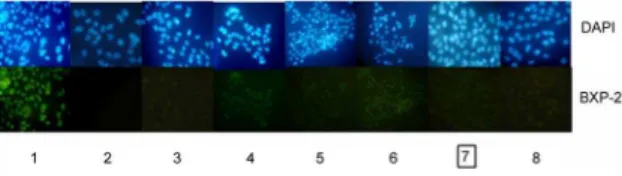
After 48 hours of infection by V-BCRPi, immunofluorescence was used to analyze the cell morphology. Using DAPI staining for comparison, the fluorescence intensity of BCRP/ABCG2 in each V-BCRPi treatment group was reduced; the fluorescence intensity of uninfected cells incubated with the primary antibody was the strongest, and the fluorescence intensity of uninfected cells with PBS instead of the primary antibody was weakest. The fluores-cence intensity of the V-BCRPc treatment group was higher than those of all the other V-BCRPi groups, and the fluorescence intensity of the V-BCRP3i treatment group was the lowest (Fig. 3).
Influence of V-BCRPi on the BCRP/ABCG2 drug pump, as measured by FCM
The results indicated that, after infection with the recombinant V-BCRpi retroviruses, the amount of mitoxantrone that was retained by the cells was increased but was similar to that of the blank control group. The amount of drug retained after infection with V-BCRP3i was greatly improved, with a mean fluorescence intensity of 4.9 (P,0.01). Infection with the recombinant retroviruses with single base variations resulted in lower retention than that observed in the V-BCRPi-infected cells, but the retention was higher than that of the blank cell control group. The mean value of the fluorescence intensity for the Ko143 group (positive control) was 5.3 (Fig. 4). It was therefore concluded that the increased amount of drug within the V-BCRP3i-infected JAR cells would increase the effect of the drug on the cell.
Results of tumor growth after V-BCRPi and 5-FU injection On the 10thand 12thdays of inoculation, we observed a grain-sized node at the injection site of the JAR cells. A transplantation tumor was formed in mice from each group, with a 100% success rate. We observed the tumors on the 14thday after injecting V-BCRPi into the tumor and 106IC50of 5-FU into the peritoneal
cavity. The transplantation tumors grew quickly in elliptical shapes during the early stages and transformed into smooth and irregular forms with nodositas during the later stages. The covering skin was purplish-black. When the diameter of the tumor was greater than 1 cm, the ulceration began to feel soft and cystic. When the skin
was removed from the transplantation tumor, the surface of the solid tumor tissue was red-brown. The hairless mice injected with 5-FU alone did not eat and appeared dispirited at an early stage. At the later stages, the mental state and appetites of the mice improved. After injecting 5-FU, the tumors in the hairless mice previously injected with V-BCRPi were smaller than those of hairless mice that were not injected with the virus. The anti-tumor rate was approximately 10 times as slow (P,0.01; Fig. 5 and Table 1). It was therefore concluded that V-BCRPi increases the 5-FU inhibition effects on tumor growth.
Knockdown ofBCRP/ABCG2and drug sensitivity by V-BCRPi in hairless mice bearing tumors, as shown using immunohistochemical analysis
The expression of BCRP/ABCG2 in tumors injected with V-BCRPi was lower than that in tumors not injected with V-V-BCRPi (including tumors injected with PBS and 5-FU alone). The membranes of theBCRP/ABCG2-positive histiocytes were brown, and the nuclei were blue, with larger karyoplasm and altered morphologies in different cells. Of the groups injected with 5-FU, more dying cells were evident in the groups injected with V-BCRPi than in the uninjected group. Specifically, cellular morphology was lost, the karyoplasm was large, and necrosis was increased in the virus-infected cells (Fig. 6). These results indicate that V-BCRPi inhibits BCRP/ABCG2 expression in tumor cells and improves their sensitivity to 5-FU.
Figure 2. Knockdown of BCRP/ABCG2 expression by V-BCRPi in JAR cells, shown by Western blot analysis. The results for experimental groups 1 to 7 are shown: 1: mock cells or cells infected with 2: V-BCRP1i, 3: V-BCRP1i-c, 4: V-BCRP2i, 5: V-BCRP2i-c, 6: V-BCRP3i, or 7: V-BCRP3i-c. The BCRP/ABCG2 protein expression of the V-BCRP3i treatment group was the lowest (group 6).
doi:10.1371/journal.pone.0103463.g002
Figure 3. Knockdown of BCRP/ABCG2 expression by V-BCRPi in JAR cells using immunofluorescence analysis (6100).The results for experimental groups 1 to 8 are shown: 1: mock cells with MoAb or 2: PBS or cells infected with 3: BCRP1i, 4: BCRP1i-c, 5: BCRP2i, 6: V-BCRP2i-c, 7: V-BCRP3i, or 8: V-BCRP3i-c. The fluorescence intensity of cells subjected to V-BCRP3i treatment was the lowest.
doi:10.1371/journal.pone.0103463.g003
Figure 4. Residual drug volumes after infection of JAR cells with V-BCRPi according to flow cytometry analysis.The results of experimental groups 1 to 9 are shown: 1: mock cells, 2: cells with Mit, 3: cells with Mit and Ko143, or cells with Mit and infected with 4: BCRP1i, 5: BCRP2i, 6: BCRP3i, 7: BCRP1i-c, 8: BCRP2i-c, or 9: V-BCRP3i-c. Each residual drug volume represents the mean value of three independent experiments. *P,0.01.
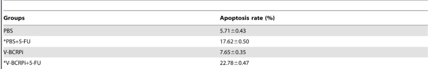
Apoptosis of V-BCRPi-treated tumor cells in hairless mice, as demonstrated by in situ hybridization analysis
Each group of tumor tissue slices was stained with TUNEL and re-stained with hematoxylin. After staining, the cells with brown nuclei of different sizes and irregular forms were considered TUNEL-positive, apoptotic cells. The single blue cells were non-apoptotic chorion cancer cells. The results indicated that the amount of apoptotic tumor cells in mice injected with V-BCRPi and 5-FU was greater than that in mice injected with 5-FU alone (Fig. 7). According to the calculation results, the number of apoptotic tumor cells following injection with V-BCRPi and 5-FU was larger than the number of apoptotic tumor cells following injection with 5-FU alone (Table 2).
Knockdown ofBCRP/ABCG2in hairless mice bearing V-BCRPi-treated tumors, as shown by Western blot analysis
Based on the results, the following conclusions were made. The expression of BCRP/ABCG2 in V-BCRPi-injected tumors (in-cluding those injected with 5-FU) was lower than that of uninjected tumors (including those injected with PBS and 5-FU). There were no differences between the groups injected with V-BCRPi and 5-FU. The BCRP/ABCG2 protein expression level was lower in the non-injected group (Fig. 8).
Knockdown ofBCRP/ABCG2in hairless mice bearing V-BCRPi-treated tumors, as shown by fluorescent quantitative RT-PCR analysis
The results showed that theBCRP/ABCG2mRNA level in the V-BCRPi-injected group was lower than that in the group injected with 5-FU alone (P,0.01). The mRNA expression in the group injected with V-BCRPi alone was not different from that of the group injected with 5-FU alone (Table 3). It was therefore concluded that V-BCRPi could more efficiently downregulate BCRP/ABCG2mRNA in tumor histiocytes.
Discussion
Since the first report of RNA interference (RNAi) in 1998, its feasibility and innovation have been continuously improved. RNAi has shown the advantages of high efficiency, specificity, simple operation and a short experimental period [14,15], and it has become a powerful tool for gene function and gene therapy research [16].In vitrochemical synthesis has been used from the early stages of the development of the technique. The techniques of in vitro transcription, siRNA expression cassettes, andin vivo transcription for PCR preparation were middle stage develop-ments [17]. More recently, stable expression DNA carriers have proven advantageous for the in vivo transcription of siRNA. Targeted gene inverted repeat sequences have been placed downstream of the promoters of retroviral expression vectors to create recombinant viruses with short hairpin loops (shRNA), and these constructs have resulted in the best inhibition effects to date [18,19]. This type of viral vector has long-lasting RNAi effects that can infect a wide range of cells, thus increasing the potential Table 1.Changes in tumor weight and volume and the anti-tumor rate in transplantation tumors in nude mice after virus infection and drug treatment (x6s, n = 15).
Group Tumor weight (g) Tumor volume (cm3) Anti-tumor rate (%)
PBS 2.4660.56 3.7160.73
-*PBS+5-FU 2.3760.78 3.6260.56 3.6
V-BCRPi 2.3160.49 3.6560.53
-*V-BCRPi+5-FU 1.7560.65 2.8960.57 24.2
*: PBS+5-FU vs. V-BCRPi+5-FU, p,0.01. doi:10.1371/journal.pone.0103463.t001
Figure 5. Tumor bodies of hairless mice after injection of JAR cancer cells infected with V-BCRPi and treatment with 5-FU.A1: Tumor body injected with PBS alone; A2: Tumor body injected with PBS and 5-FU; B1: Tumor body injected with V-BCRPi alone; and B2: Tumor body injected with V-BCRPi and 5-FU. After injecting 5-FU, the tumors in the hairless mice injected with V-BCRPi were smaller than those in the un-injected hairless mice. The anti-tumor rate was approximately a factor of 10 (P,0.01). It was concluded that V-BCRPi increases the inhibition effects of 5-FU on tumor growth.
doi:10.1371/journal.pone.0103463.g005
Figure 6. Immunohistochemical staining of tumor bodies in hairless mice bearing JAR cancer cells after injection with V-BCRPi and 5-FU (6100). The results indicate that V-BCRPi inhibits
BCRP/ABCG2 expression and improves drug sensitivity to 5-FU in tumors. The red arrows indicate the dead cells.
applications of this technology.
Some researchers have applied thein vitrochemical synthesis siRNA method to explore the reversal of P-gp-mediated multidrug resistance, which has provided a basis for the operability of this technique [20]. The pLenti6/BLOCK-iT lentiviral RNAi expres-sion system is an RNAi system expressed by a 3rd generation, replication-defective retrovirus that was developed by Invitrogen. This system has been widely applied in scientific research on gene functions and clinical gene biotherapy due to its efficient expression and infection and high biosafety and operability. One goal of researchers is to counter the drug resistance of tumor cells and improve drug sensitivity by modifying the BCRP/ABCG2-mediated, drug-resistant phenotype and the drug-resistant pheno-types of ABC membrane transporter families. Therefore, we first used the pLenti6/BLOCK-iT lentiviral RNAi expression system to construct vectors targeting conserved regions of the ATP-binding domain of BCRP/ABCG2and then conducted in vitro and in vivo RNA interference studies [21]. In this study, three DNA constructs that were complementary to the ABC domain of theBCRP/ABCG2gene, beginning at the 207th, 745thand 939th
bps of the 6th exon of the BCRP/ABCG2 gene [22–24], were successfully designed. Subsequently, these fragments were used to construct pENTR/BCRPi recombinant plasmids through anneal-ing, recombination, transformation, sequencing and comparison using BLAST. The pLenti6/BLOCK-iT Lentiviral RNAi expres-sion vectors were then constructed. After 293T virus packaging, highly infective retroviruses were acquired with titers of 106. In the subsequentin vivoand in vitroinfection studies, we expected to screen for biotherapy reversal agents that could reverse drug resistance by efficiently targeting the ATP-binding domain. This
method will lay a solid foundation for future studies and applications.
BCRP/ABCG2, P-gp and multidrug resistance-associated proteins (MRPs) all have ABC domains, which use ATP hydrolysis to ‘‘pump’’ drugs stored inside cancer cells outside of the cell membrane, thus facilitating drug resistance [25–32]. Determining how to reverse the multidrug resistance mediated by drug-resistance proteins is a problem that must be solved. RNAi using DNA constructs encoding siRNAs that are complementary to the ABC domain ofBCRP/ABCG2has shown that gene silencing of multidrug-resistant proteins is possible [33].
In this study, multiple RNA hairpins were designed to target the ABC domain of BCRP/ABCG2, and stable shRNA-expressing recombinant retroviruses with high infection rates were construct-ed [34–36]. Using this RNA interference system within vitrocell studies, the siRNA recombinant retroviruses were screened for efficient knockdown of BCRP/ABCG2 mRNA and protein expression levels and alterations in the drug susceptibility phenotype using immunofluorescence and drug pump functional tests. It was concluded that all three groups of recombinant retroviruses that targeted the ABC domain (including recombinant retroviruses with single base mutations) efficiently knocked down BCRP/ABCG2. The 3rd group, V-BCRP3i, exhibited the best knockdown effects. Additionally, the results indicated that a single base change in the target sequence resulted in an siRNA interference efficiency that was lower than that of the correspond-ing non-mutated experimental group. This findcorrespond-ing further demonstrates the efficiency and sequence specificity of siRNA interference. Therefor, this study screened for cells with better in vitro interference effects caused by the V-BCRPi retroviruses targeting theBCRP/ABCG2-mediated drug-resistant phenotypes and other ABC transporter protein-mediated drug-resistant phenotypes. This study also demonstrated how to conductin vivo Table 2.Changes in the apoptosis rate of transplantation tumors in nude mice injected with virus and 5-FU (x6s, n = 5).
Groups Apoptosis rate (%)
PBS 5.7160.43
*PBS+5-FU 17.6260.50
V-BCRPi 7.6560.35
*V-BCRPi+5-FU 22.7860.47
*: PBS+5-FU vs. V-BCRPi+5-FU, p,0.01. doi:10.1371/journal.pone.0103463.t002
Figure 7. TUNEL staining of the tumor bodies of hairless mice bearing JAR cancer cells injected with V-BCRPi and 5-FU (6200). The number of apoptotic cells in tumors injected with
V-BCRPi and 5-FU was greater than that of the tumors injected with 5-FU alone. The red arrows indicate the apoptotic cells.
doi:10.1371/journal.pone.0103463.g007
Figure 8. Knockdown of BCRP/ABCG2 expression by V-BCRPi in tumor cell bodies, as shown by Western blot analysis.1: Tumor body injected with PBS alone; 2: Tumor body injected with PBS and 5-FU; 3: Tumor body injected with V-BCRPi alone; and 4: Tumor body injected with V-BCRPi and 5-FU. It was concluded from the experimental results that the expression of BCRP/ABCG2 in tumors injected with V-BCRPi (with 5-FU treatment) was lower than that of the un-injected tumors (with PBS and 5-FU). There was no difference among the groups injected with the various V-BCRPi retroviruses and 5-FU.
studies and overall assessments in animals, as well as how to design efficient, specific siRNAs targeting ABC domains to reverse drug resistance and improve cell sensitivity to drugs.
Heterotransplantation in hairless mice is a favorable in vivo animal model to study cancer treatment because experimental results using transplantation tumors in these mice feasibly predict clinical effects. When exploring drug resistance mechanisms and the reversal of drug resistance, the hairless mouse heterotrans-plantation tumor model has important implications for further studies of the prognosis and survival rates for patients with drug-resistant tumors.
The growth of transplanted tumors in hairless mice is affected by the biological characteristics of the tumors and the body features of the hairless mice. This study inoculated 106 highly malignant JAR cells expressing high levels of endogenousBCRP/ ABCG2 into hairless mice. The results showed strong cell reproductive capacity, early formation of the transplantation tumors and a tumor formation rate of 100%, which meant that the transplantation tumor model was successfully established.
Overcoming multidrug resistance in tumors would improve chemotherapy and comprehensive treatment effects, survival rates and quality of life. In recent years, RNAi interference techniques have provided favorable strategies for improving the multidrug resistance of tumors, especially when using siRNA retroviral vectors with high infectability, specificity and stability. In this study, the pLenti6/BLOCK-iT Lentiviral RNAi expression system was used to construct recombinant retroviruses (V-BCRPi) that were then usedin vitro to determine which retrovirus conferred the best knockdown effects and improved the BCRP/ABCG2 -mediated drug-resistant phenotype. An animalin vivostudy was
then conducted using transplanted tumors in hairless mice. V-BCRPi injection into tumor histiocytes followed by mRNA and protein level analysis showed thatBCRP/ABCG2expression was inhibited. This study also revealed the improved function of the drugs, the induction of cell death and the reversal of drug resistance in the transplantation tumors, which were demonstrated by decreased transplantation tumor growth and drug sensitivity.
The study results provide a favorable experimental animal model for the reversal of ABC membrane transporter protein family-mediated drug resistance and a scientific basis for targeting the ABC domain by V-BCRPi with high efficiency and specificity. Althoughin vitro andin vivoexperiments have demonstrated the favorable prospects of applying RNAi interference in gene treatment, functional gene research and drug sensitization, the safe administration of the siRNAs to humans and the expression of these constructs in specific organs and tissues in clinical applications still need to be perfected. In addition, the expression of BCRP/ABCG2 in normal tissues, such as placenta, brain, prostate, small intestine, testicles, ovary, colon and liver, is high. BCRP/ABCG2plays a fundamental role in maintaining normal physiological functions and preventing damage from external toxins. Determining how to improve the drug sensitivity ofBCRP/ ABCG2in selected RNAi-targeted tumor cells still remains to be elucidated.
Author Contributions
Conceived and designed the experiments: JY. Performed the experiments: NX LM JY. Analyzed the data: TD ZL. Contributed reagents/materials/ analysis tools: WL YJ. Contributed to the writing of the manuscript: ZH.
References
1. Schuurhuis GJ, Broxterman HJ, de Lange JH, Pinedo HM, van Heijningen TH, et al. (1991) Early multidrug resistance, defined by charges in intracellular doxorubicin distribution independent of p-glycoprotein. Brit.J.Cancer 64: 857– 861.
2. Rabindran SK, Ross DD, Doyle LA, Yang W, Greenberger LM (2000) Fumitremorgin C reverses multidrug resistance in cells transfected with the breast cancer resistance protein. Cancer Res, 60: 47–50.
3. Krishnamachary N, Center MS (1993) The MRP gene associated with a non-glycoprotein multidrug resistance encodes a 190-Kda membrane bound glycoprotein. Cancer Res., 53: 3658–3663.
4. Maliepaard M, van Gastelen MA, Tohgo A, Hausheer FH, van Waardenburg RC, et al. (2001) Circumvention of Breast Cancer Resistance Portein (BCRP)-mediated resistance to camptothecinsin Vitrousing nonsubstratiod drugs or the BCRP inhibitor GF120918.Clinical Cancer Research, 7: 935–941.
5. Erlichman C, Boerner SA, Hallgren CG, Spieker R, Wang XY, et al. (2001) The HER tyrosine kinase inhibitor CI1033 enhances cytotoxicity of 7-ethyl-10-hydroxycamptothecin and topotecan by inhibiting breast cancer resistance protein-mediated drug efflux. Cancer Res. 61(2): 739–748.
6. Kubota T, Furukawa T, Tanino H, Suto A, Otan Y, et al. (2001) Resistant mechanisms of anthracyclines–pirarubicin might partly break through the P-glycoprotein-mediated drug-resistance of human breast cancer tissues. Breast Cancer. 8(4): 333–338.
7. Bera TK, Lee S, Salvtore G, Lee B, Pastan I (2001) MRP8, a new member of ABC transporter superfamily, identified by EST database mining and gene prediction program, is highly expressed in breast cancer. Mol Med. 7(8): 509– 516.
8. Doyle LA1, Yang W, Abruzzo LV, Krogmann T, Gao Y, et al. (1998) A multidrug resistance transporter from human MCF-7 breast cancer cells. Proc Natl Acad Sci USA, 95: 15665–15670.
9. Allikmets R, Schrim LM, Hutchinson A, Romano-Spica V, Dean M (1998) A human placenta-specific ATP-binding cassette gene (ABCP) on chromosome 4q22 that is involved in multidrug resistance. Cancer Research, 58: 5337–5339. 10. Miyake K, Mickey LA, Litman T, Zhan Z, Robey R, et al. (1999) Molecular cloning of cDNAs which are highly overexpressed in mitoxantrone-resistant cells: demonstration of homology to ABC transport genes. Cancer Research, 59: 8–13.
11. Yasumasa H, Christine AH, Yan QW, Medina-Pe´rez WY, Robey RW, et al. (2001) Acquired mutations in the MXR/BCRP/ABCP gene alter substratio specificity in MXR/BCRP/ABCP-overexpressing cells. Cancer Research, 61: 6635–6639.
12. Kage K, Tsukahara S, Sugiyama T, Asada S, Ishikawa E, et al. (2002) Dominant-negative inhibition of breast cancer resistance protein as drug efflux pump through the inhibition of S-S dependent homodimerization. Int.J.Cancer, 97: 626–630.
Table 3.Relative BCRP/ABCG2 transcript levels of the transplantation tumors in nude mice injected with virus and drugs (x6s, n = 5).
Group Relative transcript level
PBS 1
*PBS+5-FU 0.7060.05
V-BCRPi 0.2060.03
*V-BCRPi+5-FU 0.0860.05
13. Eisenblatter T, Huwel S, Galla HJ (2003) Characterization of the brain multidrug resistance protein (BMDP/ABCG2/BCRP) expressed at the blood-brain barrier. Brain Res. 971(2): 221–231.
14. Scheffer GL, Maliepaard M, Pijnenborg AC, van Gastelen MA, de Jong MC, et al. (2000) Breast cancer resistance protein is localized at the plasma membrane in mitoxantrone- and topotecan-resistant cell lines. Cancer Research, 60: 2589–93. 15. Fire A, Xu S, Montgomery MK, Kostas SA, Driver SE, et al. (1998) Potent and specific genetic interference by dou ble-stranded RNA in Caenorhabditis elegans. Nature, 391: 806–811.
16. Elbashir SM, Lendeckel W, Tuschl T (2001) RNA interference is mediated by 21-and 22-nucleotide RNAs. Genes Dev, 15(2): 188–200.
17. Bernstein E, Caudy AA, Hammond SM, Hannon GJ (2001) Role for a bidentate ribonuclease in the initiation step of RNA interference. Nature, 409 (6818): 363– 368.
18. Brummelkamp TR, Bernads R, Agami R (2002) A system for stable expression of short interfering RNAs in Mammalian cells. Science, 296: 550–553. 19. Elbashir SM, Harborth J, Lendeckel W, Yalcin A, Weber K, et al. (2001)
Duplexes of 21-nucleotide RNAs mediate RNA interference in cultured mammalian cells. Nature, 411: 494–498.
20. Kanazaki A, Toi M, Nakayama K, Bando H, Mutoh M (2001) Expression of multidrug resistance-related transporters in human breast carcinoma. Jpn J Cancer Res, 92: 452–458.
21. Nieth C1, Priebsch A, Stege A, Lage H (2003) Modulation of the classical multidrug resistance(MDR) phenotype by RNA interference (RNAi). FEBS Letters, 545: 144–150.
22. Ross DD, Karp JE, Doyle LA, Doyle LA (2000) Expression of breast cancer resistance protein in blast cells from patients with acute leukemia. Blood, 96: 365–68.
23. Hoffmann U, Materna V, Blohmer JU (2000) Quantitative expression analysis of genes involved in the development of chemoresistance in ovarian carcinoma. Pathol Res Pract, 196: 235–38.
24. Sheng MY, Doyle A, Ross DD (2001) BCRP gene expression in normal lung and in non-small cell lung cancer tissue. Chinese Journal of Cancer, 20(3): 274–278. 25. Sargent JM, Williamson CJ, Maliepaard M, Elgie AW, Scheper RJ, et al. (2001) Breast cancer resistance protein expression and resistance to daunorubicin in blast cells from patients with acute myeloid leukaemia. Br J Haematol, 115: 257–262.
26. Brian LA, Anne-Marie C, Yuxiao B, Marini F, Andreeff M, et al. (2002) Low levels of ABCG2 expression in adult AML blast samples. Blood, 100(13): 4594– 4601.
27. Minderman H1, Suvannasankha A, O’Loughlin KL, Scheffer GL, Scheper RJ, et al. (2002) Flow cytometric analysis of breast cancer resistance protein expression and function. Cytometry, 48: 59–65.
28. Miyake K, Mickey LA, Litman T, Zhan Z, Robey R, et al. (1999) Molecular cloning of cDNAs which are highly overexpressed in mitoxantrone-resistant cells: demonstration of homology to ABC transport genes. Cancer Research, 59: 8–13.
29. Allen JD, Loevezijn AV, Schinkel AH, van der Valk M, van Tellingen O, et al. (2002) Potent and specific inhibition of the breast cancer resistance protein multidrug transporter in vitro and in mouse intestine by a novel analogue of fumitremorgin C. Molecular Cancer Therapeutics, 1: 417–425.
30. Komatani H, Kotani H, Hara Y, Nakagawa R, Matsumoto M, et al. (2001) Identification of breast cancer resistant protein/mitoxantrone resistance/ placenta-specific, ATP-binding cassette transporter as a transporter of NB-506 and J-107088, topoisomerase I inhibitors with an indolocarbazole structure.-Cancer Res; 61(7): 2827–2832.
31. Kawabata S, Oka M, Shiozawa K, Tsukamoto K, Nakatomi K, et al. (2001) Breast cancer resistance protein directly confers SN-38 resistance of lung cancer cells. Biochem Biophys Res Commun, 280: 1216–1223.
32. Kowalski P, Wichert A, Holm PH, Dietel M, Lage H (2001) Selection and characterization of a high-activity ribozyme directed against the antineoplastic drug resistance-associated ABC transporter BCRP/MXR/ABCG2.Cancer Gene Ther. 8(3): 185–192.
33. Ee PL1, He X, Ross DD, Beck WT (2004) Modulation of breast cancer resistance protein (BCRP/ABCG2) gene expression using RNA interference. Mol Cancer Ther, 3(12): 1577–1583.
34. Yu JY, DeRuiter SL, Turner DL (2002) RNA interference by expression of short-interfering RNAs and hairpin RNAs in mammalian cells. PNAS, 99: 6047–6052.
35. Bailey-Dell KJ1, Hassel B, Doyle LA, Ross DD (2001) Promoter characteriza-tion and genomic organizacharacteriza-tion of the human breast cancer resistance protein (ATP-binding cassette transporter G2) gene. Biochimica et Biophysica Acta, 1520: 234–241.