Universidade de Lisboa Faculdade de Farmácia
Extracellular Vesicles at ECMO and Trauma Patients
Ana Rita Lopes Nabais
Mestrado Integrado em Ciências Farmacêuticas
2
Universidade de Lisboa Faculdade de Farmácia
Extracellular Vesicles at ECMO and Trauma Patients
Ana Rita Lopes Nabais
Monografia do Mestrado Integrado em Ciências Farmacêuticas apresentada à Universidade de Lisboa através da Faculdade de Farmácia
Orientador: Doutor Marcin Osuchowski, Professor Associado
Co-orientador: Doutora Adelaide Maria Afonso Fernandes Borralho, Professora Auxiliar
3
Abstract
Extracellular Vesicle is a particle naturally released by a cell, with a lipidic bilayer membrane and without a functional nucleus. Extracellular Vesicles (EVs) can be considered as exosomes (30-150 nm), microvesicles (100-1000 nm) and apoptotic bodies (1000 – 3000 nm), considering on their origin. EVs research is emerging in multiple fields due to their role in intracellular communication by diverse processes such as gene regulation. Thus, these vesicles are not only associated with homeostasis but also with pathological processes. EVs are thought to have an important role in coagulation, inflammation, atherosclerosis, sepsis, transplantation, angiogenesis and cancer. The goal of the scientific community is to understand and correlate the function of these vesicles with diverse diseases and use them to develop specific therapies. In the future, extracellular vesicles can be used as biomarkers or even as therapeutic targets. In the current study, using a flow cytometry analysis, we provided a preliminary characterization of the EVs in two different patient populations: a) patients undergoing extracorporeal membrane oxygenation therapy and b) trauma patients. The analysed EVs expressed tissue factor and were derived from erythrocytes, monocytes, endothelium and platelets. We observed a distinct decrease in vesicles derived from platelets and those expressing tissue factor. Furthermore, we examined sensitivity of EVs to the different storage/freezing protocols given that they significantly influence the quality of EV-based data. In our study, we evaluated the impact of different freezing temperatures in the aforementioned EV populations. For example, when comparing -80°C to -25°C, a higher number of vesicles was observed in the latter.
This is an extremely important study where new contributions about populations of EVs in trauma and extracorporeal membrane oxygenation patients were made. This new information can be a starting point to study the impact of those vesicles at mentioned diseases and in the future development of new diagnostic and therapeutic strategies.
Keywords: extracellular vesicles; trauma; ECMO; disseminated intravascular coagulation; freezing temperature
4
Resumo
As vesiculas extracelulares (VEs) são partículas naturalmente libertadas pelas células, com uma bicamada lipídica e sem um núcleo funcional. As VEs podem ser divididas em exossomas (30-150 nm), microvesiculas (100-1000 nm) e corpos apoptóticos (1000 – 3000 nm), dependendo da sua origem. As vesículas extracelulares (VE) têm sido alvo de investigação por parte de diversas áreas devido ao seu papel na comunicação intercelular podendo influenciar processos como a regulação genética. Desta forma, as vesículas podem não só estar associadas à manutenção da homeostasia como também a processos patológicos. Admite-se que as VEs possam ter um papel importante na coagulação, inflamação, arteroesclerose, sepsis, transplantes, angiogenese e cancro. O objetivo da comunidade científica é perceber e correlacionar a função destas vesículas com diferentes patologias e assim, desenvolver alvos terapêuticos específicos. No futuro, as VE podem ser utilizadas não só como meio de tratamento, mas também como biomarcadores de diagnóstico.
No estudo realizado, foi feita uma análise em citometria de fluxo com o objetivo de fazer uma caracterização preliminar das VE em duas populações de doentes: a) doentes submetidos a oxigenação por membrana extracorporal e b) doentes com trauma. As populações de VE que foram analisadas expressavam fator tecidual ou eram provenientes de eritrócitos, monócitos, endotélio ou plaquetas. Verificou-se um decréscimo dos valores de vesiculas derivadas de plaquetas ou que expressavam fator tecidual. Para além da caracterização das VE nas duas populações de doentes, foi também estudada a sensibilidade das vesiculas a diferentes condições de armazenamento/protocolos de congelamento, dada a sua influência na qualidade dos dados adquiridos. No nosso estudo foi avaliado o impacto que diferentes temperaturas de congelamento nas populações de vesiculas acima mencionadas. Por exemplo, quando comparadas as amostras congeladas a -80ºC e -25ºC, observa-se um maior número de vesiculas a -25ºC.
Este trabalho revela-se importante ao contribuir com novos conhecimentos sobre as populações de VE nos doentes com trauma e submetidos a oxigenação por membrana extracorporal. Estas informações poderão ser um ponto de partida para novos estudos sobre o impacto dessas vesiculas nas doenças em causa e, futuramente para o desenvolvimento de novas estratégias terapêuticas e de diagnóstico diferencial.
Palavras-chave: vesículas extracelulares; trauma; ECMO, coagulação intravascular disseminada; temperatura de congelamento.
5
List of Acronyms
ALP: Alkaline Phosphatase APC: Anticoagulant Protein-C
ARF1/6: ADP-Ribosylation Factor 1 and 6 BBB: Blood-Brain Barrier
CRP: C-Reactive Protein
DIC: Disseminated Intravascular Coagulation ECMO: Extracorporeal membrane oxynegation EDTA: Ethylenediamine Tetraacetic Acid
ENPP1: Ectonucleotide pyrophospgatase phosphodiesterase 1 ESCRT: Endosomal Sorting Complex Required for Transport EVs: Extracellular Vesicles
FACS: Fluorescence-activated cell sorting GB Ib: Glicoprotein Ib
HMGB1: High-Mobility Group Box 1 HSP: Heat Shock Protein
IL-1β: Interleukin 1 beta IL-6: Interleukin 6
LDL: Low Density Lipoprotein
MISEV2018: Minimal Information for studies of extracellular vesicles 2018 mtDNA: mitochondrial DNA
MVs: Microvesicles PCT: Procaltionin
PECAM-1: Platelet Endothelial Cell Adhesion Molecule-1 PFP: Platelet-Free Plasma
PPP: Platelet-Poor Plasma PRP: Platelet-Rich Plasma
6 PS: Phostatidylserine
Rho: Ras homologous
ROCK: Rho-assotiated Protein Kinase
SIRS: Systemic Inflammatory Response Syndrome TF: Tissue Factor
TFPI: Tissue Factor Pathway Inihibitor TNF-α: Tumour Necrosis Factor alpha VLDL: Very Low Density Lipoprotein vWF: von Willebrand Factor
7
Index of figures
Figure 1:The pathways used from Entracellular Vesicles in communication.. ... 11 Figure 2: Coagulation cascade with intrinsic, extrinsic and common pathways. ... 14 Figure 3: Percentage of vesicles per patient which were co-detected with annexin V and the specific marker at healthy donors and patients undergoing ECMO and trauma patients.. .... 22 Figure 4: Number of EVs per microliter and patient co-detected with annexin V and the specific marker at heathy donors, patients undergoing ECMO and trauma patients. ... 22 Figure 5: Percentage of vesicles per sample which were co-detected with annexin V and the specific marker from a heathy donor where samples were frozen at -80ºC or -25ºC. ... 23
8
Índex
Introduction ... 9
Definition and characterization of Extracellular Vesicles... 9
Extracellular Vesicles’ Role ... 10
Extracellular vesicles and Trauma ... 11
Extracellular Vesicles and ECMO ... 14
Technical Challenge ... 15
Objective ... 18
Methods and Materials ... 19
Blood Collection ... 19 Flow Cytometer ... 19 Statistical analysis ... 20 Results ... 21 Discussion ... 24 Conclusion ... 25 Bibliographic References ... 26 Appendix ... 30
9
Introduction
Definition and characterization of Extracellular Vesicles
EVs have been one of the most explored entities in the last decade due to the physiological and pathological functions they play on the human body.1 Despite the existing research, the definitions related to EVs remain under discussion. The one that has been gathering the most consensus, is that an EV is a particle naturally released by a cell, with a lipidic bilayer membrane and without a functional nucleus.1 This remains a general and vague definition, which includes a wide range of particles. Depending on the origin, we can consider EVS as exosomes (if they originate from the endosomal network), microvesicles (when they are a result from cellular activation) and apoptotic bodies (if it occurs by the programmed cellular death process).2–4
The biogenesis of EVs is complex and differs according to the subtype of EV. Even though each EV kind has their own specificity, all of the processes share common steps and machinery to form EVs.5 Microvesicles (MVs) formation process is the only one explored here since it is the EV’s subtype approached in this research. It is known that one of the features of a eukaryotic cell in a resting state is the membrane asymmetry.6 Once this asymmetry is lost, the instability increases and the vesicle is released from the membrane. This phenomenon occurs by an outward budding that results from a change on the plasmatic membrane composition and the contraction of the cytoskeleton.5,7
The first step in this process is clustering the cargo that will be a part of the structure of the microvesicle.5 The way this process happens is not completely clear, but some studies show that the endosomal sorting complexes required for transport (ESCRT) can be involved.3,5,8 Due to affinity, lipid rafts and membrane-associated proteins are gathered to the place where the membrane will bud, while the cytoplasmatic proteins can be incorporated by anchoring to the membrane lipids of the inner leaflet. The process of how nucleic acids are integrated into the MVs is still to be elucidated.5
Calcium dependent enzymes like aminophospholipid translocases, scramblases and calpain, are responsible for the exposure of phostatidylserine (PS) at the outer leaf, and therefore, for the change on the plasmatic membrane structure. 5–7 The exposure to Ca2+ leads to the inhibition of the flippase and faster translocation of PS by the floppase and scramblase. Flippase is responsible for the inward transportation of the PS, while floppase for outward translocation. The scramblase transport is bi-directional ATP-independent.6 Moreover, the Ca2+ can also exert an important role on the cytoskeleton rearrangement by activation of proteolytic enzymes like caspases or calpains.6,7 This rearrangement is fundamental for budding process, since it is necessary cytoskeleton contraction by actin-myosin interaction.7,9 Hence, regulators of the
cytoskeleton are critical to the process. For example, the Ras homologous (Rho) family of small GTPases and the Rho-associated protein kinase (ROCK), that regulates the actin dynamic, or the ADP-ribosylation factor 1 and 6 (ARF1/ARF6) that by the phosphorylation of myosin light chain allows the contraction of this molecule and the budding of the vesicle.5–7
10
Even though they (EVs) are all included in the same category, each subtype has their own particularities. Thus, a characterization process is required to clarify which population is being considered. The Society for Extracellular Vesicles recommend a report of: a) physical aspects, b) biochemical composition and c) description of the conditions.1
In physical characterization, the most typical way of doing is sorting by the size. As mentioned before, EVs can have different origins and, therefore, they are classified as exosomes, MVs or apoptotic bodies. Each type has specific size range. For the exosomes that range is between 30 to 150nm, for MVs between 100-1000nm and for apoptotic bodies between 1000 to 3000nm.1,10 As a characterization by the biochemical composition, the Minimal Information for Studies of Extracellular Vesicles 2018 (MISEV2018) recognizes that, despite the effort to achieve universal subtype-specific markers of EVs, this goal is currently not possible as the results originate from studies under different conditions. Thus, the MISEV2018 prepared a table [see appendix] with 5 categories: 1) Transmembrane or GPI-anchored proteins associated to plasma membrane and/or endosomes; 2) cytosolic proteins recovered in EVs; 3) Major components of non-EV co-isolated structures; 4) Transmembrane, lipid-bound and soluble proteins associated to other intracellular compartments than PM/endosomes and 5) Secreted proteins recovered with EVs. Hence, to claim a presence of EVs, the investigators should identify at least one protein from category 1 and 2. To ensure exclusion of one of the most common non-EV structures, none of the proteins from category 3 should be identified. The last two categories, 4 and 5, should be included to demonstrate specificity on a given EV subtype and functional components of EVs, respectively.1
Besides the physical and biochemical characterization, the description of the conditions that samples were under, also plays an important role. The environment can influence the kind of released vesicles, for example, hypoxic EVs, oncosomes or apoptotic bodies.1
Role of Extracellular Vesicles
As it was described before, the MVs biogenesis includes cargo gathering from the origin cell. That cargo can be surface ligands, proteins, small metabolites, transcription factors, mRNA and non-coding RNA.2,3,6,11 Those molecules are not only essential for the formation process, but also for post budding process.5 For example, emerging data show that communication with
other cells is a crucial function of these vesicles. Many studies show the ability of MVs to spread, amplify and perpetuate processes that are usually triggered by the cell where MV has origin. 2,3,5,6,11
There are three mechanisms by which MVs can establish communication [Figure 1 A;B;D]: a) fusion of the MV’s membrane with the recipient cell (or endocytic membrane), where the content is released into the intraluminal space, b) Direct fusion with the plasmatic membrane, with modification of its composition and c) Direct stimulation of the surface receptors of the target cell by the cargo of MV membrane.2,5,6,9
11
This communication is not only in a paracrine fashion, but can also happen at distance, by endocrine release.2
Figure 1: EVs have an important role in intracellular communication. The pathways used are multiple such as: (A) modification of the composition of the receiver cell by the EV. (B) Direct transference of the cargo to the receiver cell cytoplasm. (C) Exposure of Tissue factor and PS at surface of EV, associated to procoagulant phenotype. (D) EV binding to the receptors of the receiver cell, interfering with signal transduction. (E) Endocytosis of the EV into receiver cell, maintaining the membrane integrity (F) RNA releasing from EVs can lead to inhibition of translation or (G) modify the posttranscriptional process of the messenger RNA. (H) Presenting antigens or synthesise cytokines at immunity system. In: Raeven, P., Zipperle, J. & Drechsler, S. Extracellular vesicles as markers and mediators in sepsis. Theranostics 8, 3348–3365 (2018).
As consequence, EVs are thought to have an important role in coagulation, inflammation, atherosclerosis, sepsis, transplantation, angiogenesis and cancer.12–14
Extracellular vesicles and Trauma
An injury can cause multiple modifications, locally or systemically, that can compromise the body homeostasis and may be life threatening.9 Part of those modifications result from cellular damage and activation of innate immune system. As a response, diverse molecules as cytokines, complement, oxygen free radicals, neutrophils, monocytes and macrophages will be released.9,12,15
Due to the process above, some complications are common in a sequence of an injury, such as: a) Infection on the injured area, with possible turn to sepsis; b) Inflammation, that can be transformed into systemic inflammatory response syndrome (SIRS); c) Microvascular
12
compromise, e.g. traumatic endotheliopathy that can trigger an acute respiratory distress syndrome, multiple organ failure, acute kidney injury or sepsis; and d) Dysfunctional coagulation.9
One of the components also released after an injury are EVs, and specifically MVs.12,16,17 The scientific community believes that they are responsible for different effects in trauma patients, depending on the origin cell type.
Some studies demonstrate MVs potential in bone regeneration, a common need in trauma patients. 18 They can act by carrying pro-osteogenic factors, which allows the matrix mineralisation, or by promoting osteoblast differentiation. The membrane composition of the MVs has also an important role to the process. The high amount of PS, at the outer leaf of the lipidic bilayer, enables the association with calcium-dependent phospholipid-binding annexin proteins, that allows the clustering of Ca2+ and PO
43-, essential for apatite formation. At the
surface of these vesicles is also possible to find enzymes responsible for the right ratio between organic and inorganic phosphate, as ectonucleotide pyrophosphatase/ phosphodiesterase-1 (ENPP1) and alkaline phosphatase (ALP). 18
Another area where EVs seem to be important is neuronal regeneration.16,18,19 Vesicles derived from Schwann cells stimulate neurite growth and axonal regeneration due to gene regulation at the recipient neuron.19 Scientists also study the EV role regarding Blood-Brain Barrier (BBB) integrity. Loss of this integrity, common in brain injury, is thought to be responsible for neuronal death and neurologic dysfunctions. One of the mechanisms of BBB permeability is the increase of the gaps between the endothelial cells. EVs containing thigh junction proteins seem to be released after an injury and are thought to aid in the BBB recovery. 15,16
EVs derived from mesenchymal stem cells demonstrated benefits in the acute respiratory distress syndrome in mouse models. Those EVs were able to reduce the endothelial permeability by promoting expression of vascular endothelial cadherin, hence reducing the mortality associated to this trauma complication.2,15
The EVs’ role in inflammation is one of the most researched areas. As previously mentioned, after an injury there is a reaction by the innate immune system that activates several pathways, releasing molecules as C-reactive protein (CRP), procalcitonin (PCT), tumor necrosis factor alpha (TNF-α), interleukin 1-beta (IL-1β), interleukin 6 (IL-6) and also other EVs.20 Their function is different depending on the releasing cell. EVs can be pro-inflammatory either by acting as antigen presenter [figure 1H] or by expressing inflammasome proteins, high-mobility group box 1 protein (HMGB1), mitochondrial DNA (mtDNA) or heat shock protein (HSP).6,9,21
In addition, the role of EVs in coagulation raises interest in the field. Many studies demonstrated vesicles with pro-coagulant properties [Figure 1C]..2,6,9,12,13 These EVs have a key importance in trauma patients given that they may promote a disseminated intravascular coagulation (DIC) syndrome, a serious complication co-responsible for organ dysfunction.2,22,23 The International Society of Thrombosis and Haemostasis defines DIC as an
acquired syndrome characterized by the intravascular activation of coagulation with loss of localization and that can have different causes.22 As a result, a massive production and activation of coagulation factors and platelets is observed. This then leads to a robust and
13
systemic thrombin generation, which can subsequently induce severe complications.2,22 The
relationship between the immune system and coagulation has been widely studied. One of the conclusions is that a reciprocal regulation between these two areas exist; e.g., TNF, IL-1 and IL-6 upregulate coagulation cascade.22,24 Severe injuries can compromise the balance of such a regulation. In trauma patients, a high inflammatory response is observed that results in the activation of coagulation factors.
DIC is not only caused by a high activation of coagulation factors, but also by a decrease of the anticoagulation ones, as anticoagulant protein C (APC), antithrombin and tissue factor pathway inhibitor (TFPI) [Figure 2]. A decrease of plasminogen, from the fibrinolysis system, is also observed.22
For coagulation cascade to be triggered, an initiator is needed. Tissue factor (TF), is one of the most important initiators in trauma DIC.22 TF generates thrombin that activates platelets which
in turn activate other coagulation factors and exponentially enhance the coagulation process [Figure 2].13,22,25
MVs play an important role in the coagulation since exposing PS creates an optimal environment to the activation of the coagulation factors. 13,23,25,26 This environment is achieved by enhancing Ca2+ concentration, which allows interactions of the coagulation factors with the
MV membrane, and due to the anionic propriety of PS, enables interaction with the γ-carboxyglutamic acid domain of the coagulation factors. One the factors that can be activated is TF.2,13
MVs can also participate in coagulation by expressing TF.2,6,13,26 Although, their effects on coagulation are controversial.13,26 Many studies demonstrate TF at the surface of MVs released
by monocytes, endothelial cells and platelets. However, the only type of MV that is proven to expose TF in both healthy and pathological conditions originates from monocytes. Endothelial MVs expresses TF under pathological conditions only. Regarding the MVs derived from platelets, their origin is highly controversial in both physiological and pathological condition. Also, the tissue factor’s activity is not clear, since its presence does not always trigger upregulation of the coagulation.13,26 Differences in activity are thought to be due to post-translational modifications that TF can suffer and conformational changes while it is exposed.26 Nevertheless, it is clear that MVs with TF can enhance coagulation and plays an important part of the physiopathology of diverse diseases.2,9,13,26 More studies are necessary to clarify the TF MV source and the activation processes.
14
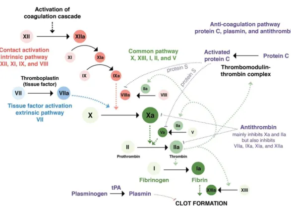
Figure 2: Coagulation cascade with intrinsic, extrinsic and common pathways. The first one it is activated by collagen exposure due to endothelial damage, which will activate XII. The extrinsic pathway is activated due to vessel injury leading to the TF:VIIa complex formation. Representation of the three parts of the coagulation cascade. Both main pathways (intrinsic and extrinsic) join each other by catalysing the activation of factor X to Xa. The common pathway will activate the Xa factor at the beginning to the end lead to fibrin formation. The anti-coagulation pathway is assured by APC, plasmin and antithrombin. In: https://step2.medbullets.com/heme/120239/von-willebrand-disease
Extracellular Vesicles and ECMO
Extracorporeal membrane oxygenation (ECMO) is a medical procedure that allows blood oxygenation and CO2 removal through an external circulation. 27–29 This technique is especially
useful not only for cardiac and respiratory failure but also for premature neonatal support. In the first case, the cardiac failure can be due to post-cardiotomy, post-heart transplantation or severe cardiac failure due to myocarditis or profound cardiac depression. Respiratory failure by adult respiratory distress syndrome, pneumonia, trauma and primary failure graftcan lead to ECMO intervention.28,29 In spite of being a fundamental method to treat adults and children, it is important to be aware of the possible complications of this procedure such as haemolysis, air-embolism, infection and sepsis.28,29 However, the most common complications are bleeding and coagulopathy. 27–30 Bleeding can occur either by systemic anticoagulation administration or massive consumption of platelets and coagulation factors, through DIC. Coagulation cascade can be activated at foreign surfaces due to adhesion of von Willebrand factor (vWF) on the circuit. Platelets will bind to vWF and be activated, realising TF, that will initiate coagulation cascade.30 Besides platelet activation, EV derived from platelets are also reported in ECMO patients. Shear forces from oxygenation membranes are also responsible for platelet-derived.30,31 Studies correlating EVs and ECMO are scarce, therefore the information about the
15
impact that EVs can have on this patients are insufficient. ECMO patients can be a good target population to study how an external circuit can affect cellular activation and vesiculation and the impact of that on homeostasis.
Technical Challenge
The next step in research on EVs is the development, characterization and uniformization of analytical methodologies. The procedures ranging from selection of the samples to EV measurement are far from being standardized.32–34 Simple aspects such as the source of the
EVs, how the samples are manipulated and stored, and the experimental conditions themselves, can largely affect the, number of EV, their stability, aggregation capacity and function.1 Without
uniformization of the above aspects, the comparison of the different studies is difficult.32–34
Therefore, the importance of guidelines to assist the researchers designing their studies is obvious.35
Blood and blood derivates, such as plasma, are one of the most studied biological fluids in the EV field. To guarantee that a study can be correctly compared with others, the source should be characterized. The aspects related with the donor that must be taken in consideration are: a) donor age; b) biological sex; c) current or previous pregnancy; d) menopause; e) pre or postprandial status; f) time of collection; g) physical exercise and the time of the last practice; h) diet and body mass index; i) infections; and j) medication. Besides those, technical factors should be also reported, as: a) fluid collection volume; b) first tube discard; c) type of container; d) time to processing the sample; e) anticoagulant choice; f) temperature during the process; g) conditions of centrifugation and filtration; h) haemolysis’ degree; i) transport conditions; and j)freeze-thaw cycles.1,10,35–37
One of the most studied factors is the anticoagulant choice. When the biological fluid is blood or plasma, it is necessary to add an anticoagulant to the sample. The anticoagulants that are more used in clinical analysis are: heparin, sodium citrate and ethylenediamine tetraacetic acid (EDTA). Several studies show a significant impact on EVs according to anticoagulant choice.35–38 Authors have been reporting EDTA as a inductor of myeloid-derived EVs,
specifically the platelets type, via P-selectin-dependent activation.36–38 Regarding heparin,
studies reveal a higher count and larger EVs in comparison with sodium citrate and EDTA.35,36,38 Sodium citrate is the anticoagulant that gathers the most consensus and is
generally advised to be used in EVs sampling/analysis.36,38 It has been showed that sodium
citrate has the lowest EVs’ concentration, in all sizes, which can be due to the inhibition of vesiculation.36,38
After collection and pre-processing of the samples, EV fraction should be purified. For that, the protocols should include all separation and concentration steps. The separation process can have two purposes, one is to segregate the EVs from non-EVs components, and the other is to separate the different subtypes of EVs. Concentration allows the increase of EVs’ number per volume unit.1 It is important to acknowledge that complete purification is an unrealistic goal
for the currently available technology.1,32,39 To achieve EVs' purification several methods can
be used such as centrifugation, density gradient, chromatography, filtration and immune-isolation.1,33,40
16
The most commonly used method to separate blood cellular fraction from the EVs is centrifugation.32,33,36,40 After this procedure, a platelet-rich plasma (PRP) is obtained but to
achieve a platelet-free plasma (PFP) a differential centrifugation must be done. First, from PRP at 15 000 x g for 20 min, a platelet-poor plasma (PPP) is separated, then again at 15 000 x g for 20 min PFP is recovered.40 As a result, by each centrifugation step, the specificity of EV
recovery increases but the ability of a high yield in recovery is considerably diminished.1,10
Besides protein aggregates and extracellular RNAs, non EV-lipidic vesicles such as very low density lipoproteins (VLDL) and low density lipoproteins (LDL), are examples of particles co-isolated in the plasma that can interfere with the measurement.1,36,39 Therefore, collecting the
blood during a fasting period is advised.36,38
Another critical step is the storage. Given the everyday life in a lab, becomes impossible to analyse all the samples at once. Consequently, it is crucial to develop preservation methods to conserve EVs for later processing. The most commonly used method is freezing; conditions of freezing and thawing can highly affect the final result of a EV measurement given that this process can generate cell fragments that can be identified as EVs.35,38 Therefore, to minimize
the artefacts obtained from the cellular debris, the PFP method is recommended.33,38,40,41 The
snap-freezing should be used to minimize cell damage. Some investigators used liquid nitrogen, but -80ºC snap-freezing is also acceptable.38,41 The temperature of thawing is rarely
reported and the studies in this matter are controversial. The most recent studies showed that thawing should be done at room temperature or 37ºC. 38,41
EVs can be analysed through multiple methods depending on the research goals. To analyse and characterize single particles investigators can use high- or low-resolution imaging. High resolution can provide information about EV structure and composition through electron microscopy, scanning electronic microscopy or transmission electron microscopy. Low-resolution image has the advantage of an increased statistical power given that the particle number analysed are exponentially higher in comparison with the high-resolution methods.1,42 Some of the methods that can also be employed are: a) nanoparticle tracking analysis, b) light scattering (with or without fluorescence detection) and c) Raman tweezers microscopy. Fluorescence-activated cell sorting (FACS), a type of flow cytometry, is the most widely used. FACS measurement is based on light scatter and can be completed with fluorescence detection.2,6,42 The main benefit of this technology is the possibility to analyse individually a high number of particles in a short period of time, and provide information about size and cell origin.6,42 As a consequence, it allows the identification of rare EV subtypes, given an
heterogenous population.6 Despite these advantages, there are also some cons in FACS measurement that should be improved in order to become the gold standard method. The more obviously limitation is the sensitivity threshold. As it was earlier pointed out, EVs are a huge heterogenous population in which can be found vesicles from 30 to 3000 nm. Researchers believe that the predominant subpopulation is small EVs, which means under 300 nm. However, the detection limit of light scatter is around 200nm, which means that a larger fraction is excluded from the analysis.34 Thereby, the complementation with fluorescence based EV detection is quite important since the background noise is usually lower. Nonetheless, since EVs markers are not well stablished, the non-specificity of the fluorophore can lead to false
17
positive signals. This phenomenon can happen due to probe aggregation or as the result of the bounding to non-EV components. On the other hand, there are also EVs, that by not expressing the proteins being recognised by fluorescence, are not detected, leading to false negatives.34,42 Because of this, it is of the utmost importance to add fluorophores as and negative controls. Lastly, FACS units are arbitrary because flow cytometers' sensitivity varies from one another. Due to this fact it is even more urgent to establish a universal calibration method. In 2018, experts in this field gathered at a workshop where they aimed to create a calibration method to reduce interlaboratory variability.43
In conclusion, to reliably analyse EVs, several control steps should be taken into consideration. Beginning with the pre-analytical conditions, it is known that aspects such as gender, prandial status and diseases can influence the measurements. Therefore, it is fundamental to establish precise and transparent inclusion and exclusion criteria. Analytical procedures such as centrifugation settings and anticoagulant used (in case of blood derivates) are also important to be reported; the same reporting quality applies to the storage method used. Finally, the chosen method needs to be compatible with the study goal/hypothesis. In case of FACS use, calibration procedure is essential, due to the interlaboratory variation. The MISEV2018 Checklist listing all regulatory key points is attached to this monography.
18
Objective
In this study, the primary goal was to characterize EVs in two critical disease states (i.e. ECMO and trauma patients) and comparing them to healthy controls. EVs from different cellular sources were analysed: erythrocytes, monocytes, platelets and endothelium. The secondary goal was to understand the influence storage temperature on EV measurements.
19
Methods and Materials
Blood Collection
The trauma patients’ plasma was obtained by venous puncture at a Vienna trauma hospital. The selected patients were over 18 years old and had an Injury Severity Score over 15. The exclusion criteria were: a) pregnancy, b) cancer, c) coronary disease, d) trauma injury with more than 3 hours prior to admission and e) other severe illnesses. Sodium citrate was used as anticoagulant, and the time between collection and centrifugation was less than 2 hours. The settings applied to obtain PPP centrifugation were 2.500 x g for 15 min. The plasma was then aliquoted and stored at -80ºC. Afterwards, the samples were sent to the Ludwig Boltzmann Institute. All participants signed an ethical approval.
Regarding to ECMO patients, patients under 18 years old with acute respiratory failure, post lung transplantation, platelets counts lower than 30.000/mm3 or suffering from heparin-induced thrombocytopenia were not considered. All the remaining conditions were similar with the trauma’s patients collection.
The healthy (control) plasma was obtained from healthy donors at the Ludwig Boltzmann Institute. As with the trauma samples, the time between blood collection and centrifugation was less than 2 hours and the anticoagulant used was sodium citrate. Then the centrifugation was made at 2.500 x g for 15 min. The aliquots were stored at -80ºC.
Flow Cytometer
We proceeded equally for all samples– trauma, healthy and ECMO. The first step was thawing the aliquots in an ice bath. Afterwards, to isolate MVs 250µL were recovered and submitted to centrifugation at 16.100 x g for 30 min at 4ºC. After centrifugation, the supernatant was discarded, and the pellet was resuspended with 200µL of sterile filtered PBS-BSA 1%. The 200 µL were then divided in two. In each tube, 5µL of each of the five antibodies (InvitrogenTM–
eBioscienceTM) were added. Anti-Human CD235a-APC to identify Erythrocyte-derived EV,
CD14-APC eFluor780 to identify Monocyte-derived EVs, CD142-PE to identify TF-positivity, and CD31- PerCP-eFluor710 and CD42b-FITC to distinguish between platelet- and endothelial-derived EVs. After the incubation with the antibodies, they were centrifuged at 16.100 x g for 5 min at 4ºC. Both tubes were then incubated, on ice and in the dark, for 30 min. Then, 1mL of sterile filtered PBS-BSA 1% was added to both tubes. The tubes were centrifuged at 16.100 x g for 30 min at 4ºC. The supernatant of the two tubes was discarded completely and the pellet was resuspended with 300µL of Annexin Binding Buffer 10X (InvitrogenTM).
Then 5µL of Annexin V PE-Cyanine7 (InvitrogenTM) was added to the two tubes and they were
incubated at 0ºC, in the dark, for 15 min.
The samples were analysed with a CytoFLEX – Flow Cytometer (Beckman Coulter®). The
start-up, daily cleaning and quality control were performed every day according to the attached protocol. Extracellular vesicles were identified by size (events <1000 nm) and by the positivity
20
for Annexin V. Cellular origin was then determined by the positivity and/or negativity for specific antigens. Size-associated gating was performed with reference to silica beads of defined dimensions. The samples were vortexed before they were injected in the Flow Cytometer; a clean-up was made between samples using purified water, in order to have independent readings from each sample.
Statistical analysis
All statistical analyses were executed on Prism 8.1.1 for Windows (GraphPad Software). To compare healthy, ECMO and trauma we first submitted our samples to a normality test with Shapiro-Wilk and Kolmogorov-Smirnov. Next, One-way ANOVA followed by Dunnett’s multiple comparisons test was performed. For comparison between the freezing temperatures (-25ºC vs -80ºC) a non-parametric Mann-Whitney was used. All data are presented as mean ± 95% CI and the p-value < 0.05 was considered statistically significant.
21
Results
With the goal to analyse the injury effects on subpopulation of EVs, plasma from healthy donors was compared with trauma and ECMO patients. Moreover, we also investigated the impact of the freezing temperature storage on different subpopulations of EVs. The chosen analytical method was FACS with fluorescent Annexin V staining.
The studied populations were EVs derived from a) erythrocytes, b) monocytes, c) platelets and d) endothelium and e) expressing TF (any cell origin). To specify the EV’s population, antibodies anti-human CD235a, CD14, CD142, CD42b and CD31 were used. CD235a, CD14 and CD142 antibodies identified erythrocytes, monocytes and TF, respectively. The anti-CD42b and CD31 were used to discriminate between platelets and endothelial-derived EVs. The CD 31, also known as PECAM-1, is a molecule present on the surface of granulocytes, monocytes, platelets and endothelial cells. 44 In addition, CD42b or Glycoprotein Ib (GP Ib) can be found at megakaryocytes and platelets.45 Therefore, a combination of signals is necessary to distinguish those two populations. A particle that has a positive sign to CD31 and negative to CD42b is considered an endothelial-derived EV. In contrast, to recognize a platelet-derived EV, a positive and negative signal should be detected for CD42b and CD31, respectively.
Characterization of EVs in ECMO, trauma and healthy subjects
Our results are expressed on a relative and absolute scale. The first is a percentage of positive signal of annexin V and the correspondent antibody for each population, for example, annexin V [+] and CD235a [+]. The absolute scale represents the number of annexin V with the population marker per microliter. The relative scale illustrates the impact of a certain EV population on the study cases (healthy, ECMO and trauma) over the remaining populations. An absolute scale allowed us to examine the trending of those subject populations.
Regarding signal percentage [Figure 3], in plasma from ECMO and trauma patients, there was an increase of particles expressing simultaneously annexin V and CD235a (i.e. erythrocyte-derived EVs). For endothelial-erythrocyte-derived EVs, (annexin V and CD31 positive; CD42b negative), an increased at ECMO and Trauma was also observed. For monocytes, (annexin V and CD14), only an increase in ECMO but not trauma patients’ was detected.
Concerning absolute counts [Figure 4], there was a decrease of detection of annexin V plus CD142 (i.e. EVs expressing TF) only in plasma from ECMO patients. For platelets (CD42b positive; CD31 negative), a decrease in both ECMO and trauma patients was observed in comparison with healthy subjects.
22
Figure 4: Number of EVs per microliter and patient co-detected with annexin V and the specific marker (CD142 – Tissue Factor; CD3-CD42+ - Platelets) at heathy donors (n=5), patients undergoing ECMO (n=16) and trauma patients (n=9).
Data is presented as mean ± 95% CI. p-values < 0.05 was considered statistically significant.
Figure 3: Percentage of vesicles per patient which were co-detected with annexin V and the specific marker (CD235a – erythrocytes; CD14 – monocytes; CD31+CD42- - Endothelium) in heathy donors (n=5), patients undergoing ECMO
(n=16) and trauma patients (n=9). Data is presented as mean ± 95% CI. p-values < 0.05 was considered statistically significant.
23
Characterization of EVs in different storage temperatures
To understand the impact of freezing temperatures on EVs’ populations, heathy donor’s plasma was submitted to two different freezing temperatures, -80ºC and -25ºC. The acquired results [Figure 5] show that at -80ºC, the percentage of EVs is lower for all populations (on both scales) with exception of EVs derived from monocytes.
CD235a (Erythrocytes) -80ºC -25ºC 0 50 100 150 200 A n n e x in v p lu s m a rk e r/ m ic ro L * CD14 (Monocytes) -80ºC -25ºC 0 10 20 30 40 A n n e x in v p lu s m a rk e r/ m ic ro L CD142 (Factor Tissue) -80ºC -25ºC 0 20 40 60 A n n e x in v p lu s m a rk e r/ m ic ro L * CD31+ CD42b- (Endothelium) -80ºC -25ºC 0 10 20 30 40 A n n e x in v p lu s m a rk e r/ m ic ro L * CD31- CD42b+ (Platelets) -80ºC -25ºC 0 200 400 600 800 A n n e x in v p lu s m a rk e r/ m ic ro L *
Figure 5 Percentage of vesicles per sample which were co-detected with annexin V and the specific marker (CD235a – erythrocytes; CD14 – monocytes; CD31+CD42- - Endothelium; CD31- CD42+ - Platelets; CD142 – Tissue Factor) from a
heathy donor where samples were frozen at -80ºC (n=4) or -25ºC (n=4). Data is presented as mean ± 95% CI. p-values < 0.05 was considered statistically significant.
24
Discussion
Both ECMO and trauma are two complex critical care conditions with several activated/ongoing biological processes. Besides the complexity of these two patients’ groups, EVs protocols need to be carefully designed to allow comparable and conclusive studies. ECMO results [Figure 3] showed an increase of erythrocytes, monocytes and endothelial-derived EVs in comparison with healthy donors, relatively to the remain EVs’ populations. EVs derived from erythrocytes could have been elevated either a) due to the hemolysis that red-blood cells suffer in the ECMO circuit or b) due to the influence of various analytical conditions such as storage time and/or temperatures.28,31,46 Underlying comorbidities and general status of ECMO patients justifies an increase in EVs derived from monocytes given that they play an important role in cardiovascular diseases.47 Regarding the endothelium-derived EVs, they can be released due to alterations of the blood-flow caused by the ECMO intervention.31
When looking at absolute EV values per microliter [Figure 4], ECMO patients showed a decrease of platelets and tissue factor-containing EVs. Two explanations can be suggested to justify such an effect. First explanation is related to the ECMO method itself given that platelets and TF can be retained at the ECMO circuit decreasing the EV count in the blood.31 However, it is also possible that both, TF and platelets can be decreased due to consumption of coagulation factors by continuous coagulation activation that is typically observed in ECMO.30,31
Trauma patients, in comparison with the healthy group, have increased populations of EVs derived erythrocytes and endothelial cells [Figure 3]. Hemolysis stemming from analytical conditions is also a possibility. However, severe injuries can justify high prevalence of both EV populations, erythrocytes and endothelial, since endothelium injury and bleeding are common occurrence in trauma’s patients.12
The low number of platelet-derived EVs at trauma’s samples can, once again, be explained by consumption of platelets in DIC [Figure 4].22
An additional goal of this study was to understand the freezing temperature impact on EVs’ populations. A reduction in all populations of EVs were observed at -80ºC in comparison with -25ºC, with exception of EVs derived from monocytes, where no significant difference was observed [Figure 5]. It has previously been reported that low temperatures avoid freezing crystals formations and therefore a smaller amount of cellular debris. Without proper controls, cellular fragments can create false positives signs. Monocytes does not demonstrate a significant difference between -80ºC and -25ºC possibly due to their high sensitivity at both temperatures. Literature recommends cryopreservation of monocytes at -180ºC.48
25
Conclusion
The study of the correlation of EVs with distinct pathophysiological conditions can contribute to better understanding of EV-dependent signalling, interactions and subsequently to the development of new therapeutic approaches. Trauma and ECMO patients are typical intensive care conditions and are characterized by rapid changes and lack of clinical stability. Therefore, research aiming to develop strategies to predict and monitor the clinical status of those patients is greatly desired. Even though trauma and ECMO patients have different characteristics, they also share common features. In both cases, we could observe a decrease of the EVs expressing TF and derived from platelets. Those results underline the importance of DIC syndrome in the studied populations and are a starting point to further research about the influence of EVs in DIC.
As mentioned above, technical procedures to analyse EVs need to be carefully designed, executed and reported in order to allow reliable comparisons of data among laboratories and subsequent conclusions. In our study, we observed that freezing of samples projected for EV analysis should be done at least at -80ºC. Following the existing recommendations and to improve our investigative methodology, we plan include negative controls to our samples in subsequent studies; MISEV 2018 checklist [see attachment] offers guidelines and suggestions. EVs, due to their signalling capabilities are widely implicated in several biological processes and are therefore under investigation by many laboratories worldwide. The exact role of EVs and their interactions with the hosts, its cells and/or pathogens (in case of an ongoing infections) are currently unclear. This wide knowledge gap calls for further in-depth investigations to elucidate the roles of EVs and find potential applications in diagnostics and treatments.
26
Bibliographic References
1. Théry, C. et al. Minimal information for studies of extracellular vesicles 2018 (MISEV2018): a position statement of the International Society for Extracellular Vesicles and update of the MISEV2014 guidelines. J. Extracell. Vesicles 7, (2018). 2. Raeven, P., Zipperle, J. & Drechsler, S. Extracellular vesicles as markers and
mediators in sepsis. Theranostics 8, 3348–3365 (2018).
3. Maas, S. L. N., Breakefield, X. O. & Weaver, A. M. Extracellular vesicles: unique intercellular delivery vehicles. Trends Cell Biol. 27, 172–188 (2018).
4. Karttunen, J., Heiskanen, M., Lipponen, A., Poulsen, D. & Pitkänen, A. Extracellular Vesicles as Diagnostics and Therapeutics for Structural Epilepsies. Int. J. Mol. Sci. 1– 22 (2019). doi:10.3390/ijms20061259
5. Van Niel, G., D’Angelo, G. & Raposo, G. Shedding light on the cell biology of extracellular vesicles. Nat. Rev. Mol. Cell Biol. 19, 213–228 (2018).
6. Victoria C. Ridger; Chantal M. Boulanger; Anne Angelillo-Scherrer; Lina Badimon; Olivier Blanc-Brude; Marie-Luce Bochaton-Piallat8; Eric Boilard; Edit I. Buzas; Andreas Caporali; Francoise Dignat-George, ; Paul C. Evans; Romar; Imo E. Hoefer. Microvesicles in vascular homeostasis and diseases. Thromb. Haemost. 117, 1296– 1316 (2017).
7. Akers, J. C., Gonda, D., Kim, R., Carter, B. S. & Chen, C. C. Biogenesis of extracellular vesicles (EV): Exosomes, microvesicles, retrovirus-like vesicles, and apoptotic bodies. J. Neurooncol. 113, 1–11 (2013).
8. Hurley, J. H. ESCRTs are everywhere. EMBO J. 34, 2398–2407 (2015).
9. Eppensteiner, J., Davis, R. P., Barbas, A. S., Kwun, J. & Lee, J. Immunothrombotic Activity of Damage-Associated Molecular Patterns and Extracellular Vesicles in Secondary Organ Failure Induced by Trauma and Sterile Insults. 9, 1–14 (2018). 10. Fischer, M. B., Weber, V., Fendl, B., Weiss, R. & Spittler, A. Characterization of
extracellular vesicles in whole blood: Influence of pre-analytical parameters and visualization of vesicle-cell interactions using imaging flow cytometry. Biochem. Biophys. Res. Commun. 478, 168–173 (2016).
11. Kastelowitz, N. & Yin, H. Exosomes and microvesicles: Identification and targeting by particle size and lipid chemical probes. ChemBioChem 15, 923–928 (2014). 12. Kuravi, S. J. et al. Changes in the pattern of plasma extracellular vesicles after severe
trauma. PLOSONE 1–17 (2017). doi:10.1371/journal.pone.0183640
13. Tripisciano, C. et al. Different Potential of Extracellular Vesicles to Support Thrombin Generation : Contributions of Phosphatidylserine , Tissue Factor , and Cellular Origin. Sci. Rep. 1–11 (2017). doi:10.1038/s41598-017-03262-2
14. Eppensteiner, J., Davis, R. P., Barbas, A. S., Kwun, J. & Lee, J. Immunothrombotic Activity of Damage-Associated Molecular Patterns and Extracellular Vesicles in Secondary Organ Failure Induced by Trauma and Sterile Insults. Front. Immunol. 9, 1–
27 14 (2018).
15. Potter, D. R. et al. Mesenchymal stem cell-derived extracellular vesicles attenuate pulmonary vascular permeability and lung injury induced by hemorrhagic shock and trauma. J. Trauma Acute Care Surg 84, 245–256 (2018).
16. Andrews, A. M., Lutton, E. M., Merkel, S. F. & Razmpour, R. Mechanical Injury Induces Brain Endothelial-Derived Microvesicle Release : Implications for Cerebral Vascular Injury during Traumatic Brain Injury. Front. Immunol. 10, 1–13 (2016). 17. Matijevic, N. et al. Microvesicle phenotypes are associated with transfusion
requirements and mortality in subjects with severe injuries. J. Extracell. Vesicles 4, 29338 (2015).
18. Azoidis, I., Cox, S. C. & Davies, O. G. The role of extracellular vesicles in
biomineralisation : current perspective and application in regenerative medicine. J. Tissue Eng. 9, 1–11 (2018).
19. Rajendran, L. et al. Emerging Roles of Extracellular Vesicles in the Nervous System. J. Neurosci. 34, 15482–15489 (2014).
20. Relja, B. & Horstmann, J. Traumatic Injury. in Inflammasomes: Clinical and Therapeutic Implications 85–110 (2018). doi:10.1007/978-3-319-89390-7
21. Kerr, N. et al. Inflammasome Proteins in Serum and Serum-Derived Extracellular Vesicles as Biomarkers of Stroke. Front. Immunol. 11, 1–10 (2018).
22. Gando, S., Levi, M. & Toh, C.-H. Disseminated intravascular coagulation. Nat. Rev. Dis. Prim. 2, 16037 (2016).
23. Rumbaut, R. & Thiagarajan, P. Platelet-Vessel Wall Interactions in Hemostasis and Thrombosis. in Platelet-Vessel Wall Interactions in Hemostasis and Thrombosis (Morgan & Claypool Life Sciences, San Rafael (CA), 2010).
24. Heisenberg, W. Origin of Typical Disease Sequelae. (2015). doi:10.1016/B978-0-12-803321-0.00005-7
25. Gil, M. R. Overview of the Coagulation System. Transfusion Medicine and Hemostasis (Elsevier Inc., 2019). doi:10.1016/B978-0-12-813726-0.00091-X
26. Iii, A. P. O. & Mackman, N. Microparticles in Hemostasis and Thrombosis. Circ. Res.
108, 1284–1297 (2012).
27. Nekic, P. & SWSLHD, C. N. E. L. I. C. U. Extra Corporeal Oxygenation (Ecmo) Learning Package. (2016).
28. Marasco, S. F., Lukas, G., McDonald, M., McMillan, J. & Ihle, B. Review of ECMO (Extra Corporeal Membrane Oxygenation) Support in Critically Ill Adult Patients. Hear. Lung Circ. 17, S41–S47 (2008).
29. Makdisi, G. & Wang, I. Extra Corporeal Membrane Oxygenation (ECMO) review of a lifesaving technology. J. Thorac. Dis. Vol 7, No 7 (July 2015) J. Thorac. Dis. (2015). 30. Mulder, M. M. G., Hassan, I. & Lancé, M. ECMO and anticoagulation: A
comprehensive review. Netherlands J. Crit. Care 26, 6–13 (2018).
28
vascular function during extracorporeal circulation. Oncotarget 9, 37229–37251 (2018).
32. Livshits, M. A. et al. Isolation of exosomes by differential centrifugation: Theoretical analysis of a commonly used protocol. Sci. Rep. 5, 17319 (2015).
33. Mateescu, B. et al. Obstacles and opportunities in the functional analysis of
extracellular vesicle RNA – an ISEV position paper. J. Extracell. Vesicles 6, 1286095 (2017).
34. Arraud, N., Gounou, C., Turpin, D. & Brisson, A. R. Fluorescence triggering: A general strategy for enumerating and phenotyping extracellular vesicles by flow cytometry. Cytom. Part A 89, 184–195 (2016).
35. György, B. et al. Improved circulating microparticle analysis in acid-citrate dextrose (ACD) anticoagulant tube. Thromb. Res. 133, 285–292 (2014).
36. Jamaly, S. et al. Impact of preanalytical conditions on plasma concentration and size distribution of extracellular vesicles using Nanoparticle Tracking Analysis. Sci. Rep. 8, 17216 (2018).
37. Wisgrill, L. et al. Peripheral blood microvesicles secretion is influenced by storage time, temperature, and anticoagulants. Cytom. Part A 89, 663–672 (2016).
38. Chatelain, B., Mullier, F., Dogné, J.-M., Bailly, N. & Chatelain, C. Pre-analytical issues in the measurement of circulating microparticles: current recommendations and pending questions. J. Thromb. Haemost. 11, 693–696 (2013).
39. Lötvall, J. et al. Minimal experimental requirements for definition of extracellular vesicles and their functions: a position statement from the International Society for Extracellular Vesicles. J. Extracell. Vesicles 3, 26913 (2014).
40. Chandler, W. L. Microparticle counts in platelet-rich and platelet-free plasma, effect of centrifugation and sample-processing protocols. Blood Coagul. Fibrinolysis 24, 19–21 (2013).
41. Trummer, A., De Rop, C., Tiede, A., Ganser, A. & Eisert, R. Recovery and
composition of microparticles after snap-freezing depends on thawing temperature. Blood Coagul. Fibrinolysis 20, 52–56 (2009).
42. Coumans, F. A. W. et al. Methodological guidelines to study extracellular vesicles. Circ. Res. 120, 1632–1648 (2017).
43. van der Pol, E. et al. Standardization of extracellular vesicle measurements by flow cytometry through vesicle diameter approximation. J. Thromb. Haemost. 16, 1236– 1245 (2018).
44. Lertkiatmongkol, P., Liao, D., Mei, H., Hu, Y. & Newman, P. J. Endothelial functions of platelet/endothelial cell adhesion molecule-1 (CD31). Curr. Opin. Hematol. 23, 253–259 (2016).
45. Sim, X., Poncz, M., Gadue, P. & French, D. L. Understanding platelet generation from megakaryocytes: implications for in vitro-derived platelets. Blood 127, 1227–1233 (2016).
29
Extracellular Vesicles BT - Extracellular Vesicles: Methods and Protocols. in (eds. Kuo, W. P. & Jia, S.) 15–22 (Springer New York, 2017). doi:10.1007/978-1-4939-7253-1_2
47. Pardali, E. et al. Cryopreservation of primary human monocytes does not negatively affect their functionality or their ability to be labelled with radionuclides: basis for molecular imaging and cell therapy. EJNMMI Res. 6, 77 (2016).
48. Utsugi, T., Brown, D., Nii, A. & Fidler, I. J. Frozen-thawed human blood monocytes respond reproducibly to activation stimuli: Implications for screening of BRMs. Biotherapy 5, 301–308 (1992).
30
34 FACS start-up procedure and daily maintenance:
The following Flow Cytometer start-up procedure was performed according to the protocol “FACS STARTUP PROCEDURE” once a day pre to FACS analysis:
• Make sure sheath fluid container is full and waste container is empty (if not, follow instructions below: fill up/empty containers
• Turn on CytoFLEX with black switch on the backside of the device. • Start-up computer
• Select user, type password • Launch Cytexpert software
• Menu on top: Cytometer > System Startup Program
• Follow instructions: make sure tube with sufficient water is inserted. Hit Start (or Initialize first in case device is in standby)
• Always run daily clean program: Cytometer > Daily Clean, type in 2 mins cleaning liquid (FlowClean Cleaning Agent – Beckman Coulter) and 2 mins for water (Aqua bidest.– Fresenius Kabi, “Lösungsmittel für Parenteralia”). Hit Start (or Initialize first in case device is in standby)
• Prepare Quality check (QC) tube (perform once daily):
• Obtain QC from fridge, vortex extensively and place in measurement holder. Press QC, > Start QC, Select lot Nr. In the drop down menu (always select most recent one). Hit Start (or Initialize first in case device is in standby).
• QC proceeds and will pass* (if not, follow trouble shooting instructions: QC failed). Do not forget to return tube to fridge! File > Close QC
• Device is ready to measure.
• Once finished, run daily clean program: Cytometer > Daily Clean, type in 4 mins cleaning liquid and 4 mins for water. Then hit Start (or Initialize first in case device is in standby).
• Close Cytexpert software and turn off computer
• Fill up/empty containers: Carefully unscrew sheath container cap and put on stand (beware of electrical switch and protect FACS slits). Take sheath fluid container and water box, go to sink and fill up to max. level. If box is empty, obtain new box.
• Empty waste container into sink and rinse with tap water. Fill in around 200mL of bleach (DanKlorix).
• Fill water tube in tube holder with “Fresenius” sterile water and cleaning liquid tube with blue Beckman cleansing agent.
• Turn off Cytometer with black switch on the backside of the device.
TROUBLE SHOOTING: QC failed: Prepare fresh QC tube by pipetting 600µL of Fresenius water into new tube. Obtain QC bottle from fridge, vortex extensively and drop in 2 drops of QC liquid into tube, vortex. Repeat QC, adjust events per second count to 200 events per second (add drop of QC liquid or water). If still failing, run Cytometer > Deep Clean, press Start. Once finished, go to Cytometer > Prime, press ok.