This Accepted Author Manuscript is copyrighted and published by Elsevier. It is posted here by agreement between Elsevier and University of Brasilia. Changes resulting from the publishing process - such as editing, corrections, structural formatting, and other quality control
mechanisms - may not be reflected in this version of the text. The definitive version of the text was subsequently published in [Small Ruminant Research, Volume 43, Issue 3, March 2002, Pages 203–209, Pages 1467–1483, doi:10.1016/S0921-4488(02)00017-2].You may download, copy and otherwise use the AAM for non-commercial purposes provided that your license is limited by the following restrictions:
(1) You may use this AAM for non-commercial purposes only under the terms of the CC-BY-NC-ND license.
(2) The integrity of the work and identification of the author, copyright owner, and publisher must be preserved in any copy.
(3) You must attribute this AAM in the following format: [agreed attribution language, including link to CC BY-NC-ND license + Digital Object Identifier link to the published journal article on Elsevier’s ScienceDirect® platform].
________________________________________________________________________
Este Manuscrito do Autor Aceito para Publicação (AAM) é protegido por direitos autorais e publicado pela Elsevier. Ele esta disponível neste Repositório, por acordo entre a Elsevier e a Universidade de Brasília. As alterações decorrentes do processo de publicação - como a edição, correção, formatação estrutural, e outros mecanismos de controle de qualidade - não estão refletidas nesta versão do texto. A versão definitiva do texto foi posteriormente publicado em [Small Ruminant Research, Volume 43, Número 3, Março de 2002, Páginas 203–209,
doi:10.1016/S0921-4488(02)00017-2]. Você pode baixar, copiar e utilizar de outra forma o AAM para fins não comerciais , desde que sua licença seja limitada pelas seguintes restrições: (1) Você pode usar este AAM para fins não comerciais apenas sob os termos da licença CC- BY- NC-ND.
(2) A integridade do trabalho e identificação do autor, detentor dos direitos autorais e editor deve ser preservado em qualquer cópia.
(3) Tem de atribuir este AAM no seguinte formato: [acordo na linguagem atribuída, incluindo o link para CC BY-NC-ND licença Digital + DOI do artigo publicado na revista Elsevier ScienceDirect ® da plataforma].
Degeneration rate of preantral follicles in the ovaries of goats
J.R.V Silva M.A.L Ferreira S.H.F Costa R.R Santos F.C.A Carvalho A.P.R Rodrigues C.M Lucci S.N Báo J.R Figueiredo AbstractThe degeneration rate of ovarian preantral follicles in goats, and the distribution in the follicular classes (primordial, primary or secondary) was assessed. Ovaries from adult goats were collected at a local slaughterhouse. To evaluate the morphology of the caprine preantral follicles in situ, one fragment from each ovary was fixed individually in Carnoy for 12 h, sectioned serially at a thickness of 7 μm and stained with Periodic Acid Shiff-hematoxylin. Preantral follicles were then classified according to the stage of development. Preantral follicles were classified individually either as morphologically normal; as Type 1 degenerated follicles (only the oocyte was degenerated); or as Type 2 degenerated follicles (when degeneration occurred at both oocyte and granulosa cells). The total examined was 235 primordial, 195 primary and 101 secondary follicles. The distribution of degenerated follicles as primordial, primary and secondary follicles was 8.5, 14.3 and 16.8%, respectively. When Types 1 and 2 degenerated follicles were pooled, secondary follicles were significantly more degenerated than primordial and primary follicles. When degeneration Types 1 and 2 was compared in each follicular class, a higher (P<0.05) percentage of Type 1 degeneration was observed in primordial and primary follicles. Conversely, secondary follicles were significantly more affected by Type 2 degeneration. When the follicular classes were taken together, a significantly higher percentage of Type 1 degenerated preantral follicles was observed when compared with Type 2 degenerated follicles (P<0.05). In conclusion, a low percentage of degenerated preantral follicles was observed and secondary follicles were more affected by degeneration than primordial follicles. Thus, primordial follicles constitute a large and potentially valuable source of oocytes for reproductive programs after in vitro growth and maturation.
Keywords: Goat; Preantral follicle; Degeneration; Morphology
1. Introduction
The ovarian preantral follicles represent 90% of the follicular population (Saumande, 1991). The mammalian ovary contains thousands of follicles at birth, but the vast majority (99.9%) become atretic during their growth and maturation (Carroll et al., 1990). Some investigators (Byskov, 1974, Sadrkhanloo et al., 1987 and Huges and Gorospe, 1991) maintain that atresia can occur at any stage of development, suggesting that those follicles which ultimately ovulate have escaped atresia many times during the course of development. Others
maintain that the phenomenon of atresia is limited to a particular stage of development (Grimes et al., 1987 and Hirshfield, 1988), and that there is a single period of vulnerability during the development of antral follicles. Quantitative data pertinent to this is scarce. Of the few studies, most reports relate only on atresia in antral follicles (Osman, 1985 and Hirshfield, 1986). Others fail to define atretic follicles into classes, indicating only the overall incidence of atresia (Byskov, 1974). Only a few studies provide detailed data (Hirshfield, 1988) on the size distribution of atretic follicles in laboratory species. Some of these studies indicate that atresia is restricted to a discrete developmental period that occurs near the final stages of follicular growth (Hirshfield, 1988). Conversely, in goats, Bezerra et al. (1998) and Lucci et al. (1999) showed that atresia also occurs in preantral follicles, but to our knowledge there is no information available about what class of preantral follicles (primordial, primary or secondary follicles) is more sensitive to atretic changes. The knowledge of the quality of preantral follicles in each class is of importance to understand follicular atresia at the preantral stage as well as to provide healthy oocytes for in vitro studies. The in vitro growth of preantral follicles in combination with in vitro maturation promises to be a powerful technology for use in assisted reproductive programs (Telfer et al., 2000). The aim of the present study was to investigate the degeneration rate in goat preantral follicles and to determine what class of preantral follicles (primordial, primary and secondary follicles) is more sensitive to degenerative changes in vivo.
2. Materials and methods
Ovaries (n=45) from adult (1–3 years) cross-bred goats were collected at a local slaughterhouse. The animals were cycling and in good body condition. The ovaries were stripped of surrounding fatty tissue and ligaments, washed in a 70% alcohol solution for approximately 10 s, and then twice in a 0.9% saline solution and immediately processed as described below.
To evaluate the morphology of the caprine preantral follicles in situ, one fragment from each ovary was fixed individually in a Carnoy solution for 12 h. The fragments were then dehydrated in a graded series of ethanol dilutions, treated with xylol and embedded in paraffin wax. The ovarian tissue was then sectioned serially (at a thickness of 7 μm) and stained with Periodic Acid Shiff-hematoxylin (Lucci et al., 1999). Each section was deparaffinized with xylol and rehydrated in graded alcohol. Sections were examined by light microscopy (Zeiss, Germany). The nucleus of the oocyte was used as a marker for analyzing the follicles.
Preantral follicles were classified in three classes according to the stage of development as primordial (one layer of flattened or flattened-cuboidal granulosa cells around the oocyte); primary (a single layer of cuboidal granulosa cells around the oocyte); or, secondary (oocyte surrounded by two or more layers of cuboidal granulosa cells) (Hulshof et al., 1994). Follicular morphology was evaluated based on the integrity of the basement membrane, cellular density, presence or absence of pyknotic bodies and integrity of the oocyte. Based on these parameters, primordial, primary and secondary follicles were classified individually as morphologically normal; as Type 1 degenerated follicles (only the oocyte was degenerated) or as Type 2 degenerated follicles (when degeneration occurred at both oocyte and granulosa cells).
Ultrastructural analysis was performed using normal and degenerated follicles. Briefly, small pieces of ovarian cortex from four ovaries were fixed in a solution containing 2% paraformaldehyde and 2.5% glutaraldehyde in a 0.1 M sodium cacodylate buffer (pH 7.2). After fixation, specimens were rinsed in the buffer and post-fixed in 1% osmium tetroxide, 0.8% potassium ferricyanide and 5 mM CaCl2 in a 0.1 M sodium cacodylate buffer (Silva et al., 2001). Subsequently, the samples were dehydrated in acetone and embedded in Spurr’s epoxy resin. Thin sections (60 nm) were prepared when the oocyte nucleus was present in the semi-thin sections. Semi-semi-thin sections (3 μm) were stained with toluidine blue while semi-thin sections were then contrasted with uranyl acetate and lead citrate, and examined using Jeol 100 C or Zeiss 912 transmission electron microscope.
Data of degenerated preantral follicles from ovarian fragments were pooled. The percentage of degenerated primordial, primary and secondary follicles was compared using a χ2-test (Instat for Macintosh). Values were considered statistically significant at 0.05 or less.
3. Results
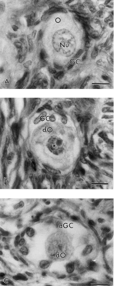
Normal preantral follicles exhibited a healthy spherical or oval oocyte with a large central or eccentrically located nucleus. Granulosa cells, well organized in layers, without pyknotic nucleus were observed surrounding the oocyte (Fig. 1a). Type 1 degenerated follicles exhibited an oocyte with a pyknotic nucleus and well-organized granulosa cells without pyknotic nuclei (Fig. 1b). A retracted oocyte and swollen granulosa cells detached from basement membrane were observed in the Type 2 degenerated follicles (Fig. 1c). It is important to note that neither pyknotic bodies in the granulosa cells nor rupture of the basement membrane were observed in degenerated follicles.
Fig. 1. Histological section of ovarian fragment after staining with Periodic Acid Shiff-hematoxylin, showing (a) normal preantral follicles; (b) Type 1 degenerated follicles; (c) Type 2 degenerated follicles. O: oocyte; Nu: oocyte nucleus; GC: granulosa cells; dO: degenerated oocyte (∗ mark denotes pyknotic nucleus) and dGC: degenerated granulosa cells; bars=10 μm.
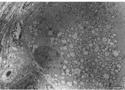
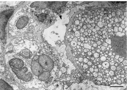
Fig. 2 shows the ultrastructural aspects of a normal preantral follicle. The oocyte cytoplasm of normal follicles present mitochondria with peripherical cristae and continuous mitochondrial membranes. Both smooth and rough endoplasmic reticulum were observed (Fig. 2). Type 1 degenerated follicles had a large number of vacuoles spread throughout the cytoplasm in all oocytes and a shrunken oocyte nucleus. Granulosa cells were normal, and had irregularly-shaped nuclei, with high nuclear-to-cytoplasm ratio. The cytoplasm of granulosa cells contained a great number of elongated mitochondria with lamellar cristae and well-developed rough endoplasmic reticulum (Fig. 3). Type 2 degenerated follicles showed a retracted oocyte, a shrunken oocyte nucleus and great irregularity in the follicular, oocyte and nuclear outlines. At this stage, the oocyte cytoplasm was extremely vacuolated and generally, organelles were no longer recognizable. Granulosa cells became disorganized and many times, the connection between the oocyte and granulosa cells was lost (Fig. 4).
Fig. 2. Electron micrograph of a normal preantral follicle. O: oocyte; Nu: oocyte nucleus; GC: granulosa cells; m: mitochondria; ser: smooth endoplasmic reticulum; bars=2 μm.
Fig. 3. Electron micrograph showing a Type 1 degenerated follicle with vacuolated cytoplasm oocyte, shrunken oocyte nucleus and normal granulosa cells. O: oocyte; Nu: oocyte nucleus; V: vacuoles; GC: granulosa cells; bars=5 μm.
Fig. 4. Electron micrograph of a Type 2 degenerated follicle. Note the retracted oocyte (arrow-head marks abnormal spaces between the oocyte and granulosa cells) and the low density of granulosa cells. O: oocyte; Nu: oocyte nucleus; GC: granulosa cells; V: vacuoles; bars=5 μm.
In total 235, 195 and 101 primordial, primary and secondary follicles were examined, respectively. The occurrence and distribution of degenerated follicles as primordial, primary and secondary follicles was 8.5, 14.3 and 16.8%, respectively (Table 1). The percentage of primary follicles showing Type 1 degeneration was significantly higher than secondary follicles. In contrast, the percentage of Type 2 degeneration in secondary follicles was higher (P<0.05) compared to primordial and primary follicles. When pooling Types 1 and 2 degenerated follicles, secondary follicles were significantly more affected by degeneration than primordial and primary follicles. When Types 1 and 2 degeneration were compared in each follicular class, higher (P<0.05) percentage of Type 1 degeneration was observed in primordial and primary follicles, compared to Type 2 degeneration ( Table 1). Conversely, secondary follicles were significantly more prone to Type 2 degeneration. When the follicular classes were taking together, a significantly higher percentage of Type 1 degenerated follicles was observed, compared to Type 2 degenerated follicles (P<0.05).
Table 1.
Percentage of degenerated preantral follicles in goat ovariesa
a,b
Values within the column not followed by the same letter differ (P<0.05).
4. Discussion
This study demonstrates the occurrence of degeneration of primordial, primary and secondary follicles in the ovaries of cross-bred type goats. Histological and ultrastructural analysis of degenerated follicles showed that a somewhat shrunken oocyte with a pyknotic nucleus and numerous vacuoles in the ooplasm are the main sign of Type 1 degenerated follicles. These features are commonly observed in degenerated preantral follicles of other animals (ewe: Jorio et al., 1991; cow: Erickson et al., 1976; cat: Wood et al., 1997; and rat: Hirshfield, 1988). van den Hurk et al. (1998) and Devine et al. (2000) demonstrated that oocytes with atretic signs contain numerous vacuoles in the ooplasm. These vacuoles represent endoplasmic reticulum swelling (Hay et al., 1976), or altered mitochondria (Fuku et al., 1995). Silva et al. (2001) reported that mitochondria exhibiting extensive swelling and disappearance of most of cristae as well as endoplasmic reticulum enlarged in volume are the first sign of degeneration in preantral follicles. The low density of granulosa cells recorded in Type 2 degenerated follicles may be due to their enlarged volume. Jennings et al. (1975) demonstrated that changes in the cellular membrane permeability cause changes the levels of intracellular Na+ K+ and Cl−, which are associated with changes in volume and increase of intracellular water.
When the follicular classes and degeneration types were taken together, approximately 12% of goat preantral follicles were degenerated. In caprine and ovine studies of estimation of follicular population in ovaries, 6 and 0.1% of degenerated preantral follicles were observed, respectively (Lucci et al., 1999 and Amorim et al., 2000). In contrast, the degeneration rate of preantral follicles was quoted as being 65% in feline ovary (Wood et al., 1997). The difference of these results compared to those of other authors may be due to species differences, variation in the follicular analysis and number of follicles included in the final analysis. Ingram (1962) showed that factors such as age, reproductive cycle, pregnancy and lactation, hormones and nutrition may affect the follicular atresia rate. For bovine (Erickson et al., 1976) and ovine (Jorio et al., 1991) species, an increase in atretic preantral follicles with an increment in age was recorded. Figueiredo et al. (1994) showed that the quality of bovine preantral follicles may be affected by the physiological state of the ovarian donor. Britt (1991) also suggested that negative energy balance may cause atresia in preantral follicles. However, in this trial, the effects of these factors on the preantral follicles were unknown due to the fact that working with slaughterhouse material makes it impossible to know the exact management of the animal before slaughter.
The oocyte of primordial and primary follicles were more sensitive to degeneration than granulosa cells. The little morphological evidence of biosynthetic activity in the granulosa cells of primordial follicles can make these cells less sensitive to degeneration (Hirshfield, 1991). The higher incidence of Type 1 degeneration in primordial and primary follicles could be a consequence of improper growth activation (Mhawi et al., 1991). After the oocyte activation, organelle multiplication and an increase of the uptake of nutrient occurs. Some of these oocytes die after activation due to an inadequate environment to continue their normal development (Rüsse, 1983). In addition, in vitro studies have shown that in certain primordial and primary follicles the oocytes degenerates or completely disappear, while granulosa cells appear healthy and continue to proliferate, showing that the granulosa cells are more resistant to degenerative events than the oocytes (Braw-Tal and Yossefi, 1997). In contrast, the oocyte and granulosa cells of the secondary follicles are equally affected by the degenerative events. Current results demonstrate an increase of the number of granulosa cells in secondary follicles which could make these cells more sensitive to atretic processes. The first sign of degeneration is observed in granulosa cells in tertiary follicles with a large number of granulosa cells (Hirshfield, 1986 and Hirshfield, 1988). In contrast to tertiary follicles, no preantral follicles showing only degeneration of granulosa were observed. It is important to note that the rupture of the basement membrane was not observed in any of the degenerated follicles studied. Other researches also reported primordial and tertiary follicles at initial and advanced stages of atresia to contain an intact basement membrane (Hay et al., 1976 and Tassel and Kennedy, 1980).
When Types 1 and 2 degenerated follicular data was pooled, it was observed that secondary follicles were more affected by the degeneration than primordial and primary follicles. In vitro studies have shown that growing follicles are more sensitive to degenerative events than primordial follicles (Wandji et al., 1996). In addition, Hirshfield (1983) showed that with the increase of follicular diameter, secondary follicles become more sensitive to degeneration. The higher sensitivity of secondary follicles to degeneration may be due to the fact that these follicles are in a stage of growth, showing higher morphological evidence of biosynthetic activity and nutrient uptake.
In conclusion, the study confirms for the first time that goat secondary follicles are more sensitive to degenerative events than primordial and primary follicles. Granulosa cells of primordial and primary follicles are more resistant to degeneration than oocyte. However, in secondary follicles, the oocyte and granulosa cells were equally affected by degenerative events. Thus, with follicular growth, granulosa cells became more sensitive to degeneration. As primordial follicles are the most abundant follicular stage in ovary and less susceptible to
degeneration, these follicles constitute a large and potentially valuable source of health oocytes that could be used in assisted reproductive programs after in vitro growth and maturation.
Acknowledgements
This study was supported by the Laboratório de Histopatologia of Federal University of Pará and the Laboratório de Microscopia Eletrônica of University of Brasilia, Brazil. The authors thank Dr. V.J.F. Freitas for technical support.
Reference
Amorim et al., 2000 C.A Amorim, C.M Lucci, A.P.R Rodrigues, F.C.A Carvalho, J.R Figueiredo, D Rondina, R Cecchi, A Giorgetti, A Martini, P.B.D Gonçalves Quantitative and qualitative analysis of the efficiency of the mechanical method for the isolation of preantral follicles from ovine ovaries Theriogenology, 53 (2000), pp. 1251–1262
Bezerra et al., 1998 M.B Bezerra, D Rondina, A.K.F Lima, L.C Oliveira, R Cecchi, C.M Lucci, A Giorgetti, J.R Figueiredo Quantitative and qualitative aspects of pre-natal folliculogenesis in caprine species Ciência Anim., 8 (1998), pp. 47–56
Braw-Tal and Yossefi, 1997 R Braw-Tal, S Yossefi Studies in vivo and in vitro on the initiation of follicle growth in the bovine ovary J. Reprod. Fert., 109 (1997), pp. 165–171
Britt, 1991 J.H Britt Impact of early pospartum metabolism on follicular development and fertility Bovine Pract., 24 (1991), pp. 39–43
Byskov, 1974 A.G.S Byskov Cell kinetics studies of follicular atresia in the mouse ovary J. Reprod. Fert., 37 (1974), pp. 277–285
Carroll et al., 1990 J Carroll, D.G Whittingham, M.J Wood, E Telfer, R.G Gosden Extra-ovarian production of mature viable mouse oocytes from frozen primary follicles J. Reprod. Fert., 90 (1990), pp. 321–327
Devine et al., 2000 P.J Devine, C.M Payne, M.K McCuskey, P.B Hoyer Ultrastructural evaluation of oocytes during atresia in rat ovarian follicles Biol. Reprod., 63 (2000), pp. 1245–1252 rickson et al., 1976 B.H Erickson, R.A Reynolds, R.L Murphree Ovarian characteristics and reproductive performance of the aged cow Biol. Reprod., 15 (1976), pp. 555–560
F igueiredo et al., 1994 J.R Figueiredo, S.C.J Hulshof, R van den Hurk, M.M Bevers, M Thiry, B Nusgens, J.F Beckers The physiological status of the ovarian donor affects in vitro development of isolated bovine preantral follicles Theriogenology, 42 (1994), pp. 1303–1310
Fuku et al., 1995 E Fuku, L Xia, B.R Downey Ultrastructural changes in bovine oocytes cryopreserved by vitrification Cryobiology, 32 (1995), pp. 139–156
G rimes et al., 1987 R.W Grimes, P Matton, J.J Ireland A comparison of histological and non-histological indices of atresia and follicular function Biol. Reprod., 37 (1987), pp. 82–88 Hay et al., 1976 M.F Hay, D.G Cran, R.M Moor Structural changes occurring during atresia in sheep ovarian follicles Cell Tiss. Res., 169 (1976), pp. 515–529
Hirshfield, 1983 A.N Hirshfield Compensatory ovarian hypertrophy in the long term
hemicastrate rat: size distribution of growing and atretic follicles Biol. Reprod., 28 (1983), pp. 271–278
Hirshfield, 1986 A.N Hirshfield Effect of low dose of pregnant mare’s serum gonadotropin on follicular recruitment and atresia in cycling rats Biol. Reprod., 35 (1986), pp. 113–118
Hirshfield, 1988 A.N Hirshfield Size–frequency analysis of atresia in cycling rats Biol. Reprod., 38 (1988), pp. 1181–1188
Hirshfield, 1991 A.N Hirshfield Development of follicles in mammalian ovary Int. Rev. Cytol., 124 (1991), pp. 43–101
Huges and Gorospe, 1991 M.H Huges, W.C Gorospe Biochemical identification of apoptosis (programed cell death) in granulosa cells: evidence for a potential mechanism underlying follicular atresia Endocrinology, 129 (1991), pp. 2415–2422
Hulshof et al., 1994 S.C.J Hulshof, J.R Figueiredo, J.F Beckers, M.M Bevers, R van den Hurk Isolation and characterization of preantral follicles from foetal bovine ovaries Vet. Quart., 2 (1994), pp. 78–80
Ingram, 1962 Ingram, D.L., 1962. Atresia. In: Zuckerman, S. (Ed.), The Ovary. Academic Press, New York, pp. 247–273.
J ennings et al., 1975 R.B Jennings, C.E Ganote, K.R Reimer Ischemic tissue injury Am. J. Pathol., 81 (1975), pp. 179–197
Jorio et al., 1991 A Jorio, J.C Mariana, A Lahlou-Kassi Development of the population of ovarian follicles during the prepubertal period in D’man and Timahdite sheep Anim. Reprod. Sci., 26 (1991), pp. 239–250
Lucci et al., 1999 C.M Lucci, C.A Amorim, A.P.R Rodrigues, J.R Figueiredo, S.N Báo, J.R.V Silva, P.B.D Gonçalves Study of preantral follicles population in situ and after mechanical isolation from caprine ovaries at different reproductive stages Anim. Reprod. Sci., 56 (1999), pp. 223– 236
Mhawi et al., 1991 A.J Mhawi, J Kanka, J Motlik Follicle and oocyte growth in early posnatal calves: cytochemical, autoradiographical and electron microscopical studies Reprod. Nutr. Dev., 31 (1991), pp. 115–126
Osman, 1985 P Osman Rate and course of atresia during follicular development in the adult cyclic rat J. Reprod. Fert., 73 (1985), pp. 261–270
Sadrkhanloo et al., 1987 R Sadrkhanloo, C Hofeditz, G.F Erickson Evidence of widespread atresia in the hypophysectomized estrogen-treated rat Endocrinology, 120 (1987), pp. 146– 155
Saumande, 1991 J Saumande Folliculogenesis in the ruminants Rec. Méd. Vét., 167 (1991), pp. 205–218]
Silva et al., 2001 J.R.V Silva, S.N Báo, C.M Lucci, F.C.A Carvalho, E.R Andrade, M.A.L Ferreira, J.R Figueiredo Morphological and ultrastructural changes occurring during degeneration of goat preantral follicles preserved in vitro Anim. Reprod. Sci., 66 (2001), pp. 209–223
Tassel and Kennedy, 1980 R Tassel, J.P Kennedy Early follicular development and atretic changes in ovary of the lamb—fine structure and histochemistry Aust. J. Biol. Sci., 33 (1980), pp. 675–687
Telfer et al., 2000 E.E Telfer, J.P Binnie, F.H McCaffery, B.K Campbell In vitro development of oocytes from porcine and bovine primary follicles Mol. Cel. Endocrinol., 163 (2000), pp. 117– 123
van den Hurk et al., 1998 R van den Hurk, E.R Spek, W.J Hage, T Fair, J.H Ralph, K Schotanus Ultrastructure and viability of isolated bovine preantral follicles Human Reprod. Update, 4 (1998), pp. 833–841
Wood et al., 1997 T.C Wood, R.J Montali, D.E Wildt Follicle-oocyte atresia and temporal taphonomy in cold-stored domestic cat ovaries Mol. Reprod. Dev., 46 (1997), pp. 190–200