Antisense Oligonucleotide technologies for neuronal cell
regeneration after spinal cord injury
Artur Guilherme Machado de Lima Costa
Dissertação de Mestrado em Bioquímica
Universidade do Porto
Faculdade de Ciências
Instituto de Ciências Biomédicas Abel Salazar
Artur Guilherme Machado de Lima Costa
Antisense Oligonucleotide technologies for neuronal cell regeneration
after spinal cord injury
Dissertação de Candidatura ao grau de Mestre em Bioquímica da Universidade do Porto
Orientador – Doutor Pedro Moreno Categoria – Investigador Assistente Afiliação – Instituto de Engenharia Biomédica, Porto
Co-orientador – Doutora Ana Paula Pêgo Categoria – Investigador Principal Afiliação – Instituto de Engenharia Biomédica, Porto
FACULDADE DE CIÊNCIAS DA UNIVERSIDADE DO PORTO
ANTISENSE OLIGONUCLEOTIDE TECHNOLOGIES FOR
NEURONAL CELL REGENERATION AFTER SPINAL CORD
INJURY
Artur Guilherme Machado de Lima Costa
BSc in Biochemistry
from Faculdade de Ciências da Universidade do Porto
Master’s thesis in Biochemistry
Done under the supervision of
Pedro Moreno, PhD, and Ana Paula Pêgo, PhD
Abstract
Spinal cord injury is a serious clinical problem, since it does not spontaneously heal and frequently leads to paralysis, leading to severe health, economic and social consequences to both patients and their families. Neurons have low potential for regeneration and the lesion site
environment severely hinders the formation of neurite extensions, essential for the reestablishment of neuronal connections. Among the reported events, a pathway featuring several myelin inhibitors as well as a common downstream regulator was identified as having a major role limiting neurite formation. A gene therapy approach is likely able to target the inhibitory genes, at lower dosage and dose frequency than conventional pharmacotherapy, and with fewer side effects. However, low stability on bodily fluids and low cellular internalization of nucleic acids raise the need for a vector.
Antisense oligonucleotides (AON) were used to induce RNase H-mediated degradation of target mRNA sequences. These codify for myelin inhibitors-activated proteins, namely RhoA from the Rho family of small GTPases and Glycogen Synthase Kinase 3β (GSK3β). AON sequences were defined based on target sequence availability, binding affinity and specificity, then tested in vitro in cell cultures. A commercial transfection reagent was used to ensure transfection efficiency during activity studies, although safety and specificity issues discourage its use for in vivo applications. Gene downregulation was determined by reverse transcription-polymerase chain reaction.
Aiming for in vivo delivery of AONs, a chitosan-derived biomaterial was developed in order to bind AON and deliver them to target cells. Trimethylchitosan is not dependent on pH for solubility and binding stability since the trimethyl substituent ensures permanent positive charge at most pH values. Theoretically, electrostatic interactions are sustained at physiological conditions.
Additionally, hydrophobic moieties, which were expected to improve AON binding properties, cell membrane interaction and lysosomal escape, were also tested. Particle size, chitosan-AON binding properties, cellular binding and uptake were analyzed.
The tested AON sequences were successful in downregulating the target mRNA, at optimum transfection conditions with the commercial reagent. Unmodified TMC was not able to mediate efficient transfection and required high N/P ratios to produce stable TMC-AON interaction; Stearic acid-modified TMC, on the other hand, showed improved AON binding properties and was able to transfect cells at high N/P ratios.
The work was developed in the context of a larger project, and included the cooperation of teammates, e.g. in the biomaterial synthesis, target protein characterization. This study was conducted in the context of the project “Characterization of Cell-intrinsic axonal regeneration determinants and their use to promote repair after CNS injury”, funded by grant HMSP-ICT/0020/2010 from FCT (Fundação para a Ciência e Tecnologia).
Table of contents
Abstract ____________________________________________________________________________ III Table of contents ____________________________________________________________________ IV List of Abbreviations_________________________________________________________________ VI List of Figures______________________________________________________________________ VII Introduction __________________________________________________________________________ 1 Spinal Cord Injury __________________________________________________________________ 1 RhoA ______________________________________________________________________________ 1 Glycogen Synthase Kinase 3β _______________________________________________________ 2 Therapeutic Strategies ______________________________________________________________ 3 The Genetic Approach ______________________________________________________________ 5 Vector-aided Delivery ______________________________________________________________ 11 Methods ____________________________________________________________________________ 15 AON design _______________________________________________________________________ 15 Cell Culture _______________________________________________________________________ 16 Cellular transfection _______________________________________________________________ 16 RhoA- and GSK3β-AON sequence screening ________________________________________ 16 Complex formation ________________________________________________________________ 17 Complex characterization __________________________________________________________ 17 Gel retention electrophoresis assay ______________________________________________ 17 Nucleic acid binding dye accessibility assay ______________________________________ 18 Dynamic Light Scattering analysis _______________________________________________ 18 Transmission Electron Microscopy _______________________________________________ 18 Transfection studies _______________________________________________________________ 18 Luciferase assay ________________________________________________________________ 18 Flow cytometry _________________________________________________________________ 19 Fluorescence microscopy _______________________________________________________ 19 Results _____________________________________________________________________________ 20 PART I – Nucleic acid drug _________________________________________________________ 20
Analysis of antisense oligonucleotide activity _____________________________________ 22 PART II – Polymer vector___________________________________________________________ 25
Physical and chemical properties of TMC _________________________________________ 25 In vitro transfection properties of TMC ____________________________________________ 33 Discussion and Future Directions ____________________________________________________ 39 Acknowledgments ___________________________________________________________________ 40 Bibliography ________________________________________________________________________ 41
List of Abbreviations
AON Antisense oligonucleotide cAMP Cyclic Adenosine monophosphate CNS Central Nervous System
CQ Chloroquine
CLASP2 Cytoplasmic linker-associated protein 2 DMEM Dulbecco’s Modified Eagle Medium EDTA Ethylenediaminetetraacetic acid GSK3β Glycogen Synthase Kinase 3β GDI Guanine Dissociation Inhibitor GPI Glycosylphosphatidylinositol
HEPES 4-(2-Hydroxyethyl)piperazine-1-ethanesulfonic acid HKR HEPES-Krebs-Ringer solution
LNA Locked Nucleic Acid
MAG Myelin-associated glycoprotein MAI Myelin-associated inhibitors MT Microtubules
NgR Nogo Receptor NP Nanoparticle
N/P ratio Ratio of polymer amines to nucleic acid phosphates OMgp Oligodendrocyte-myelin Glycoprotein
SCI Spinal Cord Injury PBS Phosphate buffered saline PCR Polymerase Chain Reaction PEG Polyethylene glycol coating
PS Phosphoriothioate nucleotide modification
RT-PCR Reverse Transcription Polymerase Chain Reaction TAE Tris-Acetic acid-EDTA solution
TBE Tris-Boric acid-EDTA solution
Tm Melting temperature (nucleic acid interaction) TMC Trimethylchitosan
List of Figures
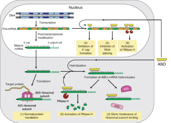
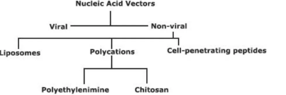
Figure 1 – GSK3 activity influence on CLASP-MT association22. Moderate GSK3 activity promotes axon growth, since it secures CLASP2 MT stabilizing effects and does not trigger CLASP2-induced MT looping. ___________________________________________________________ 3 Figure 2 – Most frequently used gene therapy approaches. ______________________________ 6 Figure 3 – RNase H cleavage mechanism. RNase H cleaves the target strand of a RNA/DNA heteroduplex 7 nucleotide residues away from the recognized sequence43. ____________________ 7 Figure 4 – Antisense mechanisms42. Not only are antisense oligonucleotides able to activate
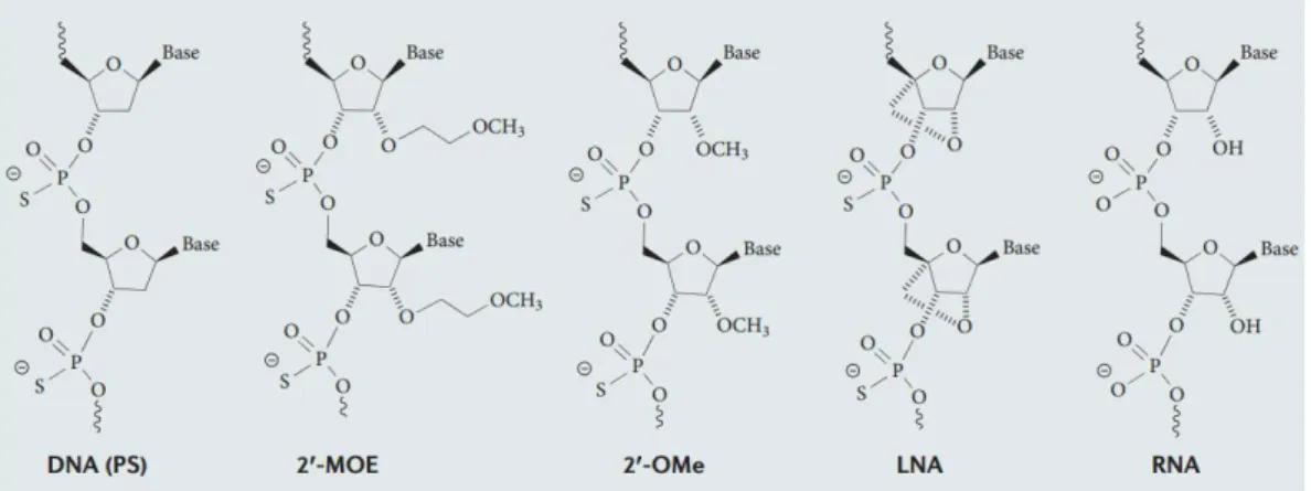
RNase H, they may also interfere with RNA processing and translation mechanisms, lowering protein expression levels all the same. Antisense oligonucleotides may be referred as AON, but also as ASO. __________________________________________________________________________ 8 Figure 5 – AON structural modifications47. Newer and more extensive structural modifications
(e.g. Locked Nucleic Acids) are more effective in downregulating mRNA levels and resisting degradation, but also involve more complex synthesis. _____________________________________ 9 Figure 6 – Sequence structure of a gapmer. Modified nucleotides with improved stability and binding properties are included in the flanks. In the center there are nucleotides that enable RNase H activity. ____________________________________________________________________________ 10 Figure 7 – Commonly used nucleic acid vectors. _______________________________________ 11 Figure 8 – Mean fluorescence of flow cytometry studies at 0.1µM AON. Several concentrations of TransIT-Oligo (Mirus), a commercial transfection reagent were tested, alongside a free oligo control. ______________________________________________________________________________ 20 Figure 9 – Fluoresnce microscopy analysis of different transfection reagent concentrations. Free oligo at 0.1 µM (top), 1.3% (v/v) TransIT-Oligo and 0.1 µM AON (middle) and 1.3% (v/v) TransIT-Oligo and 0.3 µM AON (bottom). The pictures were taken at 40x zoom, and at identical exposures times. _____________________________________________________________________ 21 Figure 10 – Antisense activity analysis of GSK3 AON-treated, PCR-amplified samples. There is a clear downregulation of the target gene (**p < 0.01, samples vs non-treated), although high variability hinders an effective comparison between the different sequences (not statistically significant). __________________________________________________________________________ 23 Figure 11 - Antisense activity analysis of RhoA-AON treated, PCR-amplified samples. There is a clear downregulation of the target gene (**p < 0.01; samples vs non-treated), although high variability hinders an effective comparison between the different sequences (not statistically significant). __________________________________________________________________________ 23 Figure 12 – Agarose gel retention assay of unmodified TMC (top) and 5% stearic
acid-substituted TMC (bottom). N/P ratios are indicated above each column, 0 indicates free oligo (no
acid-modified TMC. The former has partial retention at N/P 30 and complete retention at N/P 100, while the latter has partial retention at N/P 1 and complete retention at N/P 2. _________________ 26 Figure 13 – Polyacrylamide gel retention assay of unmodified (top), 2.5% stearic
acid-modified (mid) and 5% stearic acid acid-modified (bottom) TMC. Polyacrylamide gels enable nucleic
acid staining after having been run, contrary to agarose ones. The difference in the results supports that SybrGold has a destabilizing effect on TMC-AON interaction, especially on unmodified TMC samples. In other words, stearic acid-modified TMC produces more robust interactions with AON. _________________________________________________________________ 27 Figure 14 – Relative fluorescence levels, in comparison to the free oligonucleotide control,
of the SybrGold analysis of unmodified TMC and TMC-SA 5%. N/P 100 of TMC-SA 5% was not
tested. Most values are higher than expected, given the results from retention studies. A loose, disorganized structure is a viable hypothesis for the reduction of SybrGold exclusion from nucleic acid dye binding. The low fluorescence values at TMC N/P 1 and 2 and TMC-SA 5% N/P 1 may be due to extensive aggregation, from mutual neutralization of opposite charges. The hydrophobic properties of the stearic acid substituents support the formation of an uncompacted micelle-like particle structure of TMC-SA 5%-AON complexes that allows accessibility of TMC-bound AON to SybrGold binding. TMC N/P 10 is statistically significant from TMC-SA 5%, *p<0.05. ___________ 29 Figure 15 – Dynamic Light Scattering analysis of unmodified, 2.5% and 5% stearic
acid-modified TMC. The high polydispersity index refers to a broad particle diameter distribution. The
tendency for aggregation is lower in hydrophically modified-TMC than in the original biomaterial. 31 Figure 16 – Typycal Dynamic Light Scattering analysis profile for TMC-SA 5% at N/P 80. Although the example of 5% SA-TMC at N/P80 was used, similar profiles were observed for other N/P ratios. Not all of the high PdI reported values are associated to broad nanoparticle size curves; in some cases, a bimodal distribution is responsible for it. __________________________________ 31 Figure 17 – Transmission electron microscopy images of 5%-SA TMC at N/P 2 (left) and N/P
10 (right). The regular spherical shape of the nanoparticles and the slight variance in diameter are
according to expected. ________________________________________________________________ 32 Figure 18 – Histograms of particle size distribution of 5%-SA TMC at N/P 2 (left) and N/P 10
(right) produced from manual annotations made with the Fiji software97. Nanoparticle
diameter was manually annotated from TEM pictures in order to assess its distribution. The X axis represents particle diameter in nm. Each column has a width of 3.774 nm or 2.664 nm respectively for N/P 2 and N/P 10. _________________________________________________________________ 33 Figure 19 – -Luminiscence levels, relative to the untreated control, of the Luciferase
transfection assay. Lipofectamine 2000, a commercial transfection reagent, was used as a
control both with the same AON concentration as the remaining samples and with 1/3 of that concentration (0.3 and 0.1 µM, respectively). The difference between TMC-SA 2.5% at N/P 40 treated with CQ and TMC-SA 2.5% at N/P 40 is statistically significant (***p<0.001), although the
equivalent comparisons for the untreated TMC and the TMC-SA 5% are not statistically significant. ____________________________________________________________________________________ 34 Figure 20 – Geometric mean of the fluorescence levels of TMC-treated samples, determined
by flow cytometry analysis. HeLa cells were treated with Cy5-labeled AON and TMC in different
concentrations, in triplicate. The low fluorescence levels in the unmodified TMC-treated samples are likely to derive from the aggregation properties of the biomaterial. The higher levels associated to the hydrophobically-modified TMC are indicative that the stearic acid modification is effective in stimulating cellular binding. The differences in fluorescence from 2.5% SA TMC at N/P 80 to both the free oligo control and 5% SA-TMC at N/P 80 are statistically significant (**p<0.01), as well as the difference between 5% SA TMC at N/P 80 and the free oligo control (***p<0.001). _________ 36 Figure 21 – Fluorescence microscopy image of HeLa cells treated with TMC at N/P 80,
TMC-SA 2.5% at N/P 40 and TMC-TMC-SA 5% at N/P 40(from top to bottom). Fluorescence image (left);
fluorescence and brightfield merge image (right). With TMC, the Cy5-labeled is mainly localized in vesicle-like structures (possibly endosomes/lysosomes), pictured by an abundance of granules close to the nucleus. A similar effect is observed in TMC-SA samples, although at higher
Introduction
Spinal Cord Injury
Acute traumatic spinal cord injury (SCI) occurs worldwide with an estimated annual incidence of 130’000 and is associated with severe physical, psychological, social and economic burdens on patients and their families1, 2. As the central nervous system (CNS) does not spontaneously regenerate, paralysis is a common occurrence after SCI. Regeneration may be achieved by re-establishing the neuronal connections via neurite outgrowth; however, it is conditioned by several factors, such as remaining regeneration substrate, length of the lesion site, glial scar formation, inflammatory response, lack of neurotrophic support, and most importantly, an abundance of glial (ephrins, semaphorins3), myelin (MAG, OMgp, Nogo4) and extracellular matrix inhibitors
(chondroitin sulfate proteoglycans5). The abundance of inhibitory molecules that limits neuronal plasticity is related to circuit maturation during development of higher vertebrates, evidenced by lower vertebrates like the gecko or the newt that are able of some spontaneous neuronal regeneration6, 7.
Nogo, oligodendrocyte-myelin glycoprotein (OMgp) and myelin-associated glycoprotein (MAG) are myelin-associated inhibitors (MAI) present in myelin sheaths and have major
importance in the inhibition of neuronal regeneration. Mechanical injury to myelinated fibers leads to the release of these molecules. Nogo66, a Nogo domain, as well as OMgp and MAG, have been shown to bind to Nogo receptor (NgR)8. NgR is a glycosylphosphatidylinositol (GPI)-anchored protein present on the outer leaflet of the plasma membrane. Cleavage of that anchor by
phospholipase C, NgR antibody targeting9 or NgR antisense downregulation10 have been shown to lead to the abolition of growth cone collapse induced by myelin inhibitors. The same effect may be achieved by a small fragment of the Nogo66 domain, a highly efficient peptide antagonist named NEP1-4011. The receptor complex formed after ligand binding also includes p75NTR and Lingo -1. The former was identified as responsible for signal transduction, but Lingo-1 was only recently discovered and is still yet to be characterized. p75NTR acts as a transducer for NgR signaling by releasing RhoA from its interaction with RhoA-GDI (Rho guanine dissociation inhibitor) and enabling its activation by GDP to GTP substitution8. Similarly, chondroitin sulfate proteoglycans and chemorepulsive guidance molecules also activate RhoA, thus inhibiting neurite outgrowth12.
RhoA
cytoskeleton dynamics. Rho kinases alternate between an active GTP-bound state and an inactive GDP-bound state. GDP to GTP substitution is catalyzed by guanine exchange factors (GEF), whereas GTP degradation to GDP is facilitated by GTPase activating proteins (GAP)3. Association with GDP dissociation inhibitor (GDI) maintains Rho in its inactive GDP-bound form. While the two other major Rho kinases, Rac and Cdc42, respond to attractive guidance cues and promote actin polymerization, RhoA activation by negative cues leads to actin depolymerization 13.
RhoA is involved in MAI-induced inhibition of axon regeneration14. Higher GTP-RhoA levels, associated with activated RhoA, have been reported in the presence of inhibitory molecules and siRNA-mediated silencing of RhoA was shown to promote neurite outgrowth10, 15. RhoA is inactivated when cAMP levels are raised, e.g. in the presence of growth factors; inactivation is suggested to occur by dissociation of the NgR signaling complex, mediated by cAMP-activated Protein Kinase A8.
Rho-associated coiled-coil-containing protein kinase (ROCK) is a well characterized Rho downstream effector. It is activated when Rho binds to the Rho binding domain (RBD), interfering with the interaction between the catalytic domain and autoinhibitory region. Inhibiting ROCK leads to enhanced branching and axon elongation16, and also inhibits its activator17.
Glycogen Synthase Kinase 3β
Previous studies in the project “Characterization of Cell-intrinsic axonal regeneration determinants and their use to promote repair after CNS injury”, in which the work developed in this master thesis was integrated, have identified another key component of inhibitory signaling pathways that limit neuronal regeneration after SCI, Glycogen Synthase Kinase 3 β (GSK3β).
Glycogen Synthase Kinase 3 (GSK3) was first discovered to regulate glycogen metabolism regulation, and many other roles have been identified since. There are more than 60 reported substrates18, i.e. this kinase is involved in several cellular processes, like cell cycle, apoptosis and cytoskeleton dynamics. It is usually active under normal resting conditions and is subject to many regulation pathways in order to selectively target its substrates. One of the best characterized mechanisms is phosphorylation at Ser9 by the phosphatidylinositol 3-kinase (PI3K)/Protein kinase B signaling pathway: PI3K activates PKB in response to insulin, which phosphorylates and inhibits GSK3 at Ser9 19, 20. Conversely, Tyr216 phosphorylation results in activation.
There are two mammalian isoforms of Glycogen Synthase Kinase 3, of which β (57 kDa) is more abundant in the CNS and relevant for this work than α (52 kDa). Among GSK3β functions,
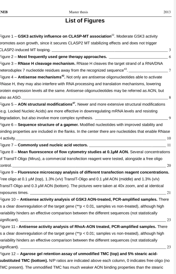
stabilizing microtubules (MT) by phosphorylating MT-associated proteins21 is especially pertinent, they are necessary for neurite outgrowth. Cytoplasmic linker associated protein 2 (CLASP2), one of GSK3β substrates, binds to the plus end of MT or to MT lattices, and regulates MT stabilization 22. At low activity levels, CLASP2 is incapable of stabilizing MT polymerization, and both binding modes are compromised. At intermediate activity levels, CLASP2 selectively binds to the MT plus ends and promotes their extension. At high activity levels, CLASP2 binds to both the plus ends and MT lattices, enabling polymerization but also looping (fig. 1).
Figure 1 – GSK3 activity influence on CLASP-MT association22
. Moderate GSK3 activity promotes axon growth, since it secures CLASP2 MT stabilizing effects and does not trigger CLASP2-induced MT looping.
The induction of GSK3β activity after SCI is linked to lower MT stability. As explored below, several drugs have been used to downregulate GSK3β and have been successful in
remyelinization and neurite extension 23, 24, although there are contradictory perspectives that propose that MAI inhibits GSK3β 25
. Nonetheless, low inhibition seems the best approach towards neuronal regeneration.
Therapeutic Strategies
Currently used therapeutic approaches to SCI have limited success 12, but intensive research on the matter has begun to show practical results. New approaches, under development or on clinical trials, are identified bellow. The following summary focuses on inhibiting negative regulators of neuronal regeneration.
With the identification of Nogo as an important component of MAI-dependent inhibitory pathways of neuronal regeneration, targeting Nogo neutralization became clinically relevant. IN-1, an anti-Nogo antibody, has been shown to promote axon regeneration in many regions of the CNS. Nonetheless, Nogo is just one of the NgR ligands, and compensatory upregulation of the remaining may hinder therapeutic application of Nogo inhibition. On the other hand, NgR may be targeted by NEP1-40, and promising results on regeneration after spinal cord injury and functional recovery have been reported26, 27. p75NTR, although it is also part of the receptor complex, is not a valid target, since it participates in a vast range of transmembrane signaling pathways, including those of neurotrophins such as nerve growth factor (NGF) and brain-derived neurotrophic factor (BDNF).
Rho and Rho-associated kinase (ROCK) are popular targets in neuronal regeneration therapies, as this pathway is crucial to NgR downstream signaling. Y27632 is a specific inhibitor of ROCK, abolishing its negative effects on growth cones, and has been shown to sustain nerve regeneration. However, it is dependent on cAMP levels, related to the intrinsic neuronal growth ability and/or the presence of growth factors 14. The C3 transferase exoenzyme from Clostridium botulinum is another Rho inhibitor, and also has neuronal regenerative effects 8. The major obstacle to the use of C3 in a clinical context is lack of membrane permeability13, which has been tentatively addressed by conjugation with cell-penetrating peptides (explored below)28. However, discrepancies between in vitro and in vivo results are significant, probably due to technical issues, e.g. improper doses, pharmacokinetics and insufficient drug uptake by relevant cells29. Fasudil is one of the few clinically available Rho inhibitors to date3, 30, and has shown much better results than Y27632 and C3; it may have a mechanism other than targeting RhoA 31, 32.Conjugation approaches , e.g. drugs and growth factor-producing bone marrow stromal cells (BMSC), have shown synergistic effects33. Nonetheless, cell therapy is technically demanding, and may pose obstacles to clinical application.
GSK3β participates in the pathophysiology of many disorders, such as Alzheimer’s Disease, schizophrenia and mood disorders34, besides mediating MAI-induced growth cone collapse24, 35. Several inhibitors have been developed, but the available options are far from optimal. Despite a low selectivity, low clearance and high IC50, lithium is frequently used as a mood regulator, targeting GSK3 among other proteins34, 36. The low selectivity and high IC
50 are associated to its binding mechanism of competition for the Mg2+ binding site19. SB216763 and SB415286, which are also among the most used drugs against GSK3 and are more potent and specific than lithium, have off-target effects due to an ATP-binding site-competing binding mechanism. Additionally, they may cause ablation of the anti-tumorigenic role of GSK3, by inhibiting GSK3-mediated degradation of proto-oncogenes, e.g adenomatous polyposis coli (APC), β-catenin, cyclin D1, c-Myc, snail, Bcl-3. The discovery of new pharmacologic drugs is hindered by structural homology to other signaling
Field Code Changed Field Code Changed
proteins, and a new drug platform with increased target specificity would be most advantageous.
As stated above, RhoA inhibition has a positive effect on the blockade of MAI-inhibitory pathways, and GSK3β inhibition provides a solution for improving MT dynamics and growth cone stabilization. Thus, a combinatorial approach inhibiting both RhoA and GSK3β is apt to provide a satisfactory synergistic effect. This study will focus on targeting both proteins, as a new therapeutic approach after SCI.
The Genetic Approach
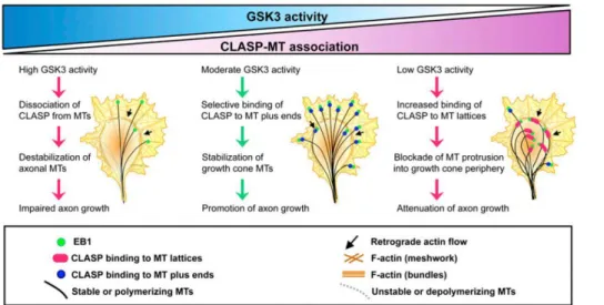
Gene therapy has been subject of great interest since it was introduced in the scientific community, since the concept of correcting genetic pathologies by introducing therapeutic genes or replacing/deleting aberrant ones is a very promising idea. Meanwhile, the definition of gene therapy has expanded and it now broadly includes the use of gene-encoding DNA plasmids, RNA molecules or oligonucleotides to correct genetic information or regulate gene expression (fig. 2). Specifically, regulation of gene expression through oligonucleotide gene therapy is believed to have several potential advantages over conventional pharmacotherapy, such as fewer side effects (derived from higher specificity), a broader set of available targets and easier design. In the context of antisense oligonucleotide therapies, explored below, an analogy to traditional pharmacology can be made: target mRNA may be considered as the receptor, and the antisense sequence as the drug. The specific approach of antisense therapy yields another advantage, since recycling of the degradation complex sustains activity for a much longer period than traditional drugs, and may be further extended by structural modifications that minimize degradation.
RNA interference37 is an endogenous regulatory system of gene expression inhibition, and
may be divided in micro RNA (miRNA) and short interfering RNA (siRNA) mechanisms. In RNAi mechanisms, a double stranded RNA molecule is cleaved by Dicer, a class III RNAse. The resulting fragments, about 20 nucleotides long, are inserted in the RISC-loading complex, forming the pre-RISC38. Upon degradation of the sense strand by Argonaute-2 (Ago2), the catalytic component of the complex, the RNA-induced silencing complex (RISC) is complete. RISC degrades target mRNA complementary to the antisense strand, and as the structure remains theoretically intact at the end of the reaction, multiple catalytic cycles are possible. siRNA and miRNA differ in the complementary sequence specificity required for target RNA degradation – while siRNA is highly specific, and only regulates transcripts levels that undergo Watson-Crick base pairing on all of the sequence length, miRNA is more promiscuous, as it targets sequences that pair with a short “seed” sequence. So, the latter is involved in the regulation of a much broader set of transcripts and its manipulation for therapeutic purposes is more complex. siRNA has drawn
attention to the potential of antisense degradation of target transcripts, which has high specificity and low toxicity from off-target effects. Nonetheless, off-target effects may arise from RISC incorporation of the antisense strand, and intrinsic regulatory mechanisms may be affected by excessive recruitment of RISC.
Figure 2 – Most frequently used gene therapy approaches.
In a similar mechanism, single stranded antisense oligonucleotides (AON) associate with RNAse H to promote degradation of target mRNA, with the possibility of a recycling mechanism and long-lasting effects.Oligonucleotides are designed to have complementary sequences to the target mRNA, which supports high specificity. That may be done with the aid of in silico techniques, which take into consideration secondary structures from mRNA folding and protein occupancy that often reduce the available binding regions39, nevertheless experimental validation is still. Compared to siRNA, AON are not as effective, but as they are easier to manipulate (as seen further below), they have been subject to intensive study for either research or clinical applications. Also, the difference in activity has been tackled by AON structural modifications (explored below). In contrast to siRNA, AON can also work through a number of other mechanisms, namely splicing modulation, RNA processing inhibition, translational arrest, miRNA antagonism and telomerase inhibition.
RNAse H shares with Ago2 a common catalytic mechanism, i.e. cleavage of the sense
strand from a DNA-RNA heteroduplexes complex, formed between target mRNA and
oligonucleotide, and a homolog domain, the catalytic P-element wimpy testis (PIWI) domain40, 41. RNAse H is a highly conserved endonuclease, being RNAse H1 is the major isoform in humans, while RNAse H2 is less abundant and requires binding of two proteins (RNAse H2B and RNAse H2C) for activation. The latter is believed to have a role different from RNA degradation, due to its capacity of recognizing and cleaving a single ribonucleotide in a deoxyrribonucleotide strand, e.g. a
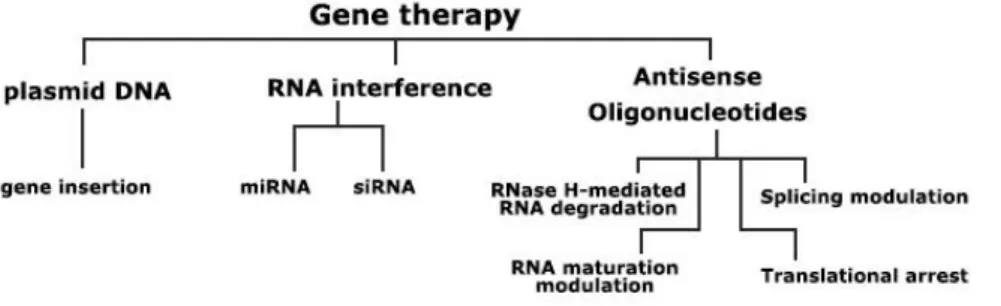
result of a faulty incorporation during DNA synthesis. RNAse H1, usually referred simply as RNAse H (since RNAse H2 has little importance in the context of AON), was discovered to be influenced by the GC content of the nucleic acid drug. AON with 11 or more guanine or cytosine residues have high efficiency, whereas those with 9 or fewer displayed poor inhibition42. Recognition is done by the RNA binding domain (RBD), while cleavage is attributed to the catalytic domain, separated from the former by a spacer sequence. This leads to a shift between the sites where the duplex is recognized and where it is cleaved, of about 7 nucleotide residues (fig. 3)43. As cleavage may be hampered by nucleotide structural modifications, gapmer strategies are often employed, as explored later on.
Figure 3 – RNase H cleavage mechanism. RNase H cleaves the target strand of a RNA/DNA heteroduplex 7 nucleotide residues away from the recognized sequence43.
RNAse H is a well characterized and ubiquitous enzyme in mammalian cells, and has been extensively used in antisense therapy, to the point of the number of clinical trials using this mechanism having exceeded the combined number of the antisense oligonucleotides trials that use other mechanisms43. This approach benefits from the AON ability to activate RNAse H at low concentrations and rapidly deplete theoretically any target mRNA39, 43, 44. Also, it is present in the nucleus, cytoplasm and mitochondria, virtually sustaining AON activity in any intracellular site. However, RNAse H mediated antisense therapies have not yet been used for nerve regeneration38.
Among other antisense oligonucleotide mechanisms of activity (fig. 4), there is splicing
modulation, which involves masking splice sequences by AON binding, thus redirecting the
pre-mRNA splicing reaction with the production of an altered protein phenotypic profile. Such alterations may have relevant effects, e.g. the expression regulation of transcript variants Bcl-XL and Bcl-XS regulates apoptosis45, 46. Exon skipping, a variant of the splicing modulation approach,
has shown to be a valuable tool for correcting specific pathologies such as Duchenne Muscular Dystrophy, which result from different mutations with the ability to disrupt the reading frame47-51. AON inhibition of 3’-polyadenylation or 5’-capping may also be used to target mRNA, as it leads to immature molecules, which are subsequently degraded, since this is a crucial step for functional and stability purposes52.
Figure 4 – Antisense mechanisms42
. Not only are antisense oligonucleotides able to activate RNase H, they may also
interfere with RNA processing and translation mechanisms, lowering protein expression levels all the same. Antisense oligonucleotides may be referred as AON, but also as ASO.
Besides targeting mRNA levels, AON may also be used to inhibit protein synthesis, by a mechanism known as translational arrest. It is achieved by AON binding to translational initiation or adjacent regions, thus blocking scanning of the transcript by the 40S ribosome subunit, assembly of the 40S and 60S subunits, or movement of the ribosome down the transcript after assembly. It has been shown to lead to protein downregulation in vitro, but confirmation of in vivo effects is supported by limited evidence38. Since there is no shift in mRNA levels, quantification faces technical issues. miRNA may also be targeted by miRNA antagonists that bind and prevent miRNA of participating in RNAi regulation pathways. Although both in vitro and in vivo experiments
showed promising results, the mechanism is not clearly characterized yet. miRNA degradation is hypothesized, but on the other hand, hybridization with ASO hinders its detection and quantification. Telomerase is a ribonucleoprotein that ensures sufficient telomere length in dividing, e.g. cancer, cells. The RNA segment used for binding to telomeres is apt to be targeted by ASO and thus preventing elongation. Telomerase targeting has shown promising results for cancer therapy in several studies53-55.
Although AON are not as efficient as siRNA, structural modifications have been developed to minimize and possibly invert that difference (fig. 5). Pharmacokinetic properties have also been greatly improved due to AON resisting nuclease degradation and evading unspecific interactions that enhance clearance and toxic side effects from off-target delivery56. AON modifications are arranged into generations, a classification that relates to their efficiency, extension of modification and relative appearance date. The first includes phosphorotioate (PS), a single atom (oxygen to sulfur) substitution on one of the non-bridging atoms of the phosphate group57. It improves resistance to nucleases 100-300 fold by a chirality-dependent mechanism (Sp phosphorotioate is nuclease resistant, but Rp is as sensitive as phosphodiester bonds58) and increases binding to plasma proteins, which reduces clearance. Thus, circulation time is prolonged and target cells are more easily reached. Neither RNA binding nor RNAse H-activating properties are compromised by this modification59.
Figure 5 – AON structural modifications47
. Newer and more extensive structural modifications (e.g. Locked Nucleic Acids) are more effective in downregulating mRNA levels and resisting degradation, but also involve more complex
synthesis.
Second generation AON have substituents at the 2’ position of the ribose sugar, usually 2’-O-methoxyethyl or 2’-O-methyl60. Potency (i.e. binding affinity, assessed by the melting
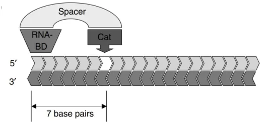
temperature, Tm) and stability are higher, but unfortunately RNAse H activation properties are lost. That, however, may be recovered using a gapmer strategy (fig. 6), i.e. a center region of the
sequence that allows RNAse H binding, flanked by 2’-modified bases. These modifications lead to longer-lasting effects which in an in vivo systemic administration setting of c-raf-1 downregulation in humans61, support a relatively infrequent dosing, e.g. weekly or biweekly dosing38.
The third generation is characterized by extensive alterations to the ribose ring, e.g. substitution by a furanose (morpholino) or by a peptidic backbone (peptide nucleic acid), or conformation constriction (locked nucleic acid). Peptide nucleic acids (PNA) are an example of AON with successful nuclease (and peptidase) evasion, and great biostability. PNA not only selectively bind to target mRNA, but also have anti-gene effects by hybridizing with double stranded DNA in four possible configurations - triplex, triplex invasion, duplex invasion and double duplex invasion42. Locked nucleic acids (LNA) have a conformation restriction derived from the 2’-O, 4’-C methylene bridge in the ribose ring, and a greatly increased hybridization affinity.
Morpholino nucleotides have a substituted sugar ring, and a high resistance to degradation. Although third generation AON have high Tm and biostability, RNAse H recruitment characteristics are lost. As with second generation AON, gapmer design may solve that problem.
Figure 6 – Sequence structure of a gapmer. Modified nucleotides with improved stability and binding properties are included in the flanks. In the center there are nucleotides that enable RNase H activity.
Fomivirsen, an oligonucleotide drug against cytomegalovirus (CMV) retinitis, is the first and,
so far, only clinically approved therapeutic oligonucleotide by FDA, in 199842, 47. However, it was discontinued due to low market demand. Fomivirsen is a 21 nucleotide long phosphoriotioate (first generation) oligodeoxynucleotide, and has excellent pharmacokinetic properties. Despite a rapid diffusion to the retinal epithelium after intraocular injection, systemic distribution is remarkably low60. The low necessary dose (330 µg per 50 µL ), conjugated with the limited systemic distribution, avoided any potential side effects47. Other examples of topical or local application yielded good results regarding tissue distribution, but CNS targeting still requires injection to the cerebrospinal fluid, since oligonucleotide do not cross the blood-brain barrier (BBB).
This successful case of PS modifications encouraged its use on most of the drugs currently on clinical trials. Although it has been reported an increase of non-specific interactions with cell surface and intracellular proteins38, 42, PS remains one of the most successful AON structural modification62. A reduction of production costs has been observed, which promises a commercially competitive alternative in the future to currently used drugs43.
Vector-aided Delivery
Antisense oligonucleotide-based therapies are often required to target a limited cell population, e.g. injured neurons. Furthermore, AON are not easily internalized by most cell types. Hence, systemic administration of naked oligonucleotides is not a valid approach. Conjugation with vectors (fig. 7) enables specific, localized delivery and may further improve resistance to
degradation, surpass anatomical obstacles and enhance cell uptake. Due to their incomparable ability to deliver nucleic acids into the cytoplasm, modified viruses were among the first vectors to be experimented with. However, issues concerning toxicity, immune response and safety diverted attention to alternatives63, 64. Non-viral vector development has presented some valid options so far, but a better understanding of cell entry mechanisms and tissue distribution would greatly benefit the presentation of new, more efficient and safer vectors.
Figure 7 – Commonly used nucleic acid vectors.
Non-viral vectors are easier to manipulate and are generally supported by an electrostatic interaction for the formation of complexes with nucleic acids. Liposome-derived vectors were among the first vectors studied as an alternative to viral vectors. The cationic lipids in their composition allow the encapsulation of bound DNA in an aqueous inner chamber. A tightly packed structure is formed due to the abundance of electrostatic interactions between the negatively charged phosphate groups of the DNA and the positively vector charged groups. A controlled zeta potential (surface charge) supports interaction with negatively charged membranes and facilitates internalization. As with other vectors, cationic liposomes may be incremented with targeting moieties for a more selective delivery43. However, DNA-liposome complexes are associated with several technical obstacles, e.g. reducing particle size for a more efficient internalization, toxicity from the cationic lipids, activity decrease in the presence of serum or antibiotics, low encapsulation efficiency, poor storage stability and rapid clearance from the blood, which impair their potential for the delivery of therapeutic nucleic acids in a clinical context63, 65.
Antennapedia homeodomain and HIV protein Tat were the first described cell-penetrating
peptides (CPP), resulting from the observation of translocation through biological membranes66. The 82 amino acid-long Tat peptide has neurotoxic properties, associated to amino acid residues 31-61, but membrane translocation ability is associated with residues 48-8567. Other CPP have been described, usually less than 30 amino acids long, polybasic or amphipatic, and able to carry molecules as they translocate across cell membranes68. Cargo molecules are varied, including oligonucleotides that may be covalently or non-covalently bound. The internalization mechanism is not yet characterized, but hypotheses focus on either the endocytic pathway or direct translocation through the plasma membrane, and it may be influenced by cargo size67, type of CPP69 and CPP concentration68. Endosomal escape mechanisms are not elucidated as well, although suspicions lie on membrane disruption70.
Although cytotoxicity was not observed in short exposures, long exposures caused extensive cell necrosis67. Nonetheless, CPP toxicity is lower than other nucleic acid vectors, since its
internalization is fast enough to support short exposures. Transfection efficiency is higher than PEI, the standard vector for nucleic acid delivery. A potential drawback arises from the induction of a humoral immune response against the peptide cargo67, which may be useful in DNA vaccination approaches71. In order to use CPP in a clinical context, several issues should be addressed, namely characterizing mechanisms of internalization, endosomal escape, cargo size influence, as well as affinity and specificity. There is also the matter of production costs, which may be reduced once CPP enter larger-scale applications. Additionally, CPP, as other peptides, have not been used in oral administration so far68.
Polyethylenimine (PEI) is one of the most studied and promising polymeric vectors. After
cellular internalization, its high buffering capacity leads to a rapid endosomal escape by a process referred as proton-sponge effect. As with lipoplexes, PEI polyplexes are victim to extensive aggregation from non-specific interactions with plasma proteins. Blood circulation may be prolonged by polyethylene glycol (PEG) coating, i.e. PEGylation, a commonly used protective technique66, 72. Cytotoxicity rises with the augment of the proportion between PEI and the nucleic acid drug. Complex size, surface charge and transfection efficiency are also influenced, i.e. the charge ratio between the polymer amino groups and the phosphate groups of the nucleic acids (N/P ratio) has a profound influence as a formulation determinant. Efficiency is described as optimal at N/P 8 for plasmid transfection, but it is associated to significant toxicity73.
Chitosan is a deacetylated derivative from chitin, a β1→4 N-acetylglucosamine anionic polyssacharide and the major component of crustaceans and insects exoskeleton and fungi cell
wall. It is a biocompatible and biodegradable material with low toxicity, high cationic potential and has functional groups that allow simple coupling of extracellular and intracellular targeting ligands63. It has been extensively used in gene therapy studies74-77. However, before regarding the possibility for its use as a gene carrier in clinical trials, low specificity and low transfection issues must be solved. The latter is highly dependent on formulation, especially on molecular weight (MW), degree of deacetylation (DD), pH (solubility), and charge ratio of chitosan to DNA. High DD ensures vector positive charge by exposing amino groups with a pKa around 6.564, 78. The electrostatic binding between the positively charged chitosan amines (N) and the negatively charged DNA phosphates (P) is also dependent on the charge ratio, i.e. N/P ratio. An adequate ratio must provide complex stability in a physiological milieu, but also enable disassembly after entering the target cell. High ratios grant complex integrity, but may limit drug release and lead to some cytotoxicity. MW influences DNA entrapment, a decisive factor in nuclease evasion, and delivery upon cell entry. These major formulation factors affect each other, .e.g. increasing N:P ratio for better stability may be compensated by lowering MW in order to maintain release capacity and high transfection efficiency79.
pH affects solubility and electrostatic interaction since it may determine whether the amino groups are protonated or not, i.e. positive charge is ensured only at acidic pH, but not at
physiological conditions80. That issue has been addressed with the use of chitosan salts, e.g. with chitosan hydrochloride (CHy), chitosan lactate (CLa), chitosan acetate (CAc), chitosan aspartate (CAs), chitosan glutamate (CGl), that increase transfection efficiency, but also raise optimum N:P ratios65. The use of trimethylated chitosan (TMC) is another solution, since it sustains a constant positive protonated state on the amino groups by covalently binding three methyl substituents. TMC supports higher solubility and DNA binding, and also increases the complex zeta potential, which enhances interactions with the negatively charged cell membranes. An increase in membrane permeability for small hydrophilic compounds has also been observed, nonetheless maintaining a level of cytotoxicity lower than PEI81.
Additional structural modifications may also be used. Examples include the introduction of the non-toxic fragment of the tetanus toxin for targeting nanoparticles to neuronal cell populations82, or hydrophobic moieties that increase hydrophobic interactions with single stranded AON, cell membranes, and endosomal membranes, which enhance complex formation, internalization and endosomal escape, respectively83. There are other strategies, like the use of fibrin scaffolds that enable controled complex release84, or of double stranded oligonucleotides that are more stable and bind more easily to polycations85 (due to increased charged density and lack of exposure of hydrophobic bases to the solvent) than single stranded oligonucleotides. Furthermore, endosomal escape may be addressed by chemical modification with lysine-hystidine dendrons that improve
buffering properties and stimulate the proton sponge effect83, 86. All in all, there are several approaches for increasing the efficiency of polycation-mediated nucleic acid delivery.
This project was part of a study aimed to develop an approach for the induction of nerve regeneration after SCI by downregulating RhoA and GSK3β. A battery of antisense
oligonucleotide sequences were designed and tested for their capacity to down-regulate the target molecules. Based on the results they are expected to be later further developed into more efficient oligos by introduction of 3rd generation nucleotide modifications. A newly developed nucleic-acid delivery platform consisting of trimethylated chitosan conjugated with a hydrophobic group was assessed for the capacity to deliver antisense oligonucleotides in vitro. If successful, this would warrant further development of the platform for future in vivo delivery targeting neuronal cells in the context of SCI nerve regeneration.
Methods
AON design
AON sequences (table 1 and 2) were previously determined based on binding properties (optimized Tm) to available sections of the target mRNA taking into consideration RNA secondary structure (using the web-based software “oligo analyzer” from IDT and “SFold”87). Other putative targets were not found using a BLASTN screening for unspecific binding. 22 nucleotides-long gapmers were produced and purified by GE Health Care Uppsala, Sweden, in which the 10 central nucleotides were phosphorothioate-modified and the six nucleotides in each end were
phosphorothioate- and 2’-O-methyl-modified, as seen on tables 1 and 2. This gapmer structure is believed to sustain RNase H activity. The 7th sequence of both the RhoA and the GSK3β AON sets differs in structure by having no phosphoriothioate bonds between the 2-O-methyl substituted nucleotides; that change was done in order to assess the potential redundancy of having both structural modifications in the same segment. These sequences were designed for rat gene targeting, since testing has been planned to proceed in murine animal models; they may be substituted by other sequences if another species is to be targeted.
Table 1 – RhoA AON sequences. “*” refers to a phosphorothioate bond, that substitutes the typical phosphodiester one, and nucleotides preceded by “m” have 2-O’-methyl modifications. RhoA AON 6b has a similar nucleotide sequence to RhoA AON 6 but differs from all other AON by not having phosphorothioate bonds between the 2-O-methyl-substituted
nucleotides.
RhoA AON 1 mU*mA*mC*mC*mU*mG*C*T*T*C*C*C*G*T*C*C*mA*mC*mU*mU*mC*mA RhoA AON 2 mA*mU*mC*mU*mU*mC*C*T*G*T*C*C*A*G*C*T*mG*mU*mG*mU*mC*mC RhoA AON 3 mC*mU*mC*mC*mC*mG*C*C*T*T*G*T*G*T*G*C*mU*mC*mA*mU*mC*mA RhoA AON 4 mA*mC*mC*mU*mC*mU*C*T*C*A*C*T*C*C*G*T*mC*mU*mU*mU*mG*mG RhoA AON 5 mC*mC*mG*mA*mC*mU*T*T*T*T*C*T*T*C*C*C*mG*mC*mG*mU*mC*mU RhoA AON 6 mA*mU*mC*mU*mC*mU*G*C*C*T*T*C*T*T*C*A*mG*mG*mU*mU*mU*mU
RhoA AON 6b mAmUmCmUmCmUG*C*C*T*T*C*T*T*C*A*mGmGmUmUmUmU
Table 2 – GSK3β AON sequences. “*” refers to a phosphorothioate bond, that substitutes the typical phosphodiester one, and nucleotides preceded by “m” have 2-O’-methyl modifications. GSK3β AON 6b has a similar nucleotide sequence to GSK3β AON 6 but differs from all other AON by not having phosphorothioate bonds between the
2-O-methyl-substituted nucleotides. GSK3b AON 1 mA*mA*mA*mG*mG*mA*G*G*T*G*G*T*T*C*T*C*mG*mG*mU*mC*mG*mC GSK3b AON 2 mC*mC*mU*mC*mA*mU*C*T*T*T*C*T*T*C*T*C*mG*mC*mC*mA*mC*mU GSK3b AON 3 mG*mG*mU*mU*mC*mU*G*T*G*G*T*T*T*A*A*T*mG*mU*mC*mU*mC*mG GSK3b AON 4 mC*mA*mG*mU*mU*mC*T*T*G*A*G*T*G*G*T*A*mA*mA*mG*mU*mU*mG GSK3b AON 5 mG*mA*mG*mG*mA*mG*G*G*A*T*A*A*G*G*A*T*mG*mG*mU*mG*mG*mC GSK3b AON 6 mU*mU*mC*mU*mC*mA*T*G*A*T*C*T*G*G*A*G*mC*mU*mC*mU*mC*mG GSK3b AON 6b mUmUmCmUmCmAT*G*A*T*C*T*G*G*A*G*mCmUmCmUmCmG
Cell Culture
The adherent RN22 (Rat schwanomma), and HeLa/Luc705 (Human cervical
adenocarcinoma) cell lines were used. The latter expressed non-functional Luciferase due to a mutated intron; this cell line is used as reporter system for efficient transfection of a specific, splice-correction AON, since it can trigger RNA expression of functional Luciferase, quantifiable by a functional assay88. The cells were cultured at 37ºC, 5% CO2, in T75 culture flasks (Thermo Scientific) in approximately 10 mL of Dulbecco’s Modified Eagle Medium (DMEM) supplemented with 10% fetal bovine serum (FBS) and 50 µg/mL gentamicin, henceforth referred as “complete medium”. Subcultures were done routinely to prevent overconfluency, by washing the cells with 5 mL of phosphate buffer saline (PBS) and addition of 1 mL 0.25% (w/v) trypsin. Cells were then suspended in an appropriate volume of complete medium, thus inactivating trypsin, and re-seeded at the desired cellular density.
Cellular transfection
500 µL of cell suspension was used for seeding 24-well plates (BD Falcon) at adequate density so that 50-60% confluency was reached after 24h, i.e. at the moment of transfection, and overconfluency was not reached before the experiment ended. GSK3β and RhoA AON were transfected with TransIT-Oligo (Mirus). Although AON and transfection reagent concentrations varied, the transfections were done by adding 50 µL of the transfection mix, containing Opti-MEM I Reduced Serum Medium (Invitrogen), Transit, and the AON, to 250 uL medium. TMC transfections were done by adding 50 µL of the prepared complex solution to 450 µL medium. In both cases, the medium was substituted immediately before the transfection for antibiotic-free medium, and was only re-added 4h after. Chloroquine at 100 µM (final concentration) was added to selected samples at 24h after transfection; medium was replaced by complete medium 4h later.
RhoA- and GSK3β-AON sequence screening
Total RNA of RN22 was harvested at 24h after transfection with TransIT-Oligo, with the Direct-zol RNA MiniPrep kit (Zymo Research). NanoDrop 1000 (Thermo Scientific)
spectrophotometric analysis was used for determining RNA concentration of the extracts and accessing purity levels by the 260/280 ratio (should be equal or higher than 2). Equal amounts of total RNA were amplified by OneStep RT-PCR kit (Qiagen), or by SuperScript II reverse
independent replicates were done for each condition.
Every sample was submitted to amplification of both the target (GSK3β or RhoA) and a reference (rRNA) gene. 15 µL PCR products with 3 µL loading buffer were submitted to 1.2% agarose gel electrophoresis at 100V for 70 minutes and semi-quantitative analyzes performed using the Volume tool of the Image Lab (Bio Rad) software for band intensity. Relative transcript levels are defined by dividing the ratio of target to reference gene of each sample by the equivalent ratio of the untreated, negative control sample.
Complex formation
The biomaterials under study were previously prepared and characterized in the lab. The unmodified TMC had 43.3 kDa, 11% acetylated monomers and 30% quaternized monomers. The modified materials had similar characteristics with an additional degree of stearic acid substitution which is indicated below. All TMC solutions were done in 10 mM HEPES and 5% (w/v) glucose, in the pH range of 7.2-7.4, as determined by an 827 pH Lab Meter (Metrohm).
TMC-AON or SA TMC-AON complexes were prepared immediately before any experiment, following a common protocol with a usually fixed oligonucleotide concentration at 1 µM and a variable concentration of TMC. The AON used in the TMC analyses was 20 nucleotides long with phosphoriothioate modifications (C*C*U*C*U*U*A*C*C*U*C*A*G*U*U*A*C*A), in other words, similar to the AON under screening for downregulation activity.
The complexes were prepared by separately heating the AON and TMC solutions at 60ºC for 5 minutes, then adding the AON solution to the TMC solution, and heating the mixture at the same temperature (60ºC) for 15 minutes under agitation, followed by a cooling down period at room temperature (approximately 25ºC) for 30 minutes. Some samples, i.e. those submitted to gel retention, DLS, TEM and nucleic acid binding dye accessibility analyzes, were also further incubated in PBS for 30 minutes at room temperature (approximately 25ºC), which aimed to simulate physiologic salt and pH conditions.
Complex characterization
Gel retention electrophoresis assay
electrophoresis assay. For agarose gels, 2% agarose (w/v) was dissolved in TAE buffer (Tris base, acetic acid and EDTA), added SybrGold for antisense oligonucleotide staining using the
recommendations of the manufacturer, and run under 120V for 30 minutes. Samples contained 25 µL complexes, 2 µL MilliQ water, 3µL 10x PBS and 6 µL loading buffer. Gradient polyacrylamide gels (4-20%) were also used and ran at 80V for 45 minutes. PAGE gels were stained after the run by incubation in 1x SybrGold in TBE solution for 15 minutes. Gel imaging was done using a ChemiDoc (Bio Rad) camera equipment.
Nucleic acid binding dye accessibility assay
The samples, previously incubated in PBS, were further incubated with 1x SybrGold for 10 minutes at room temperature (approximately 25ºC). Fluorescence emission was determined by exciting the samples at 495 nm and detecting at 540 nm. Blank solutions were prepared with the same TMC concentrations as the samples, but with no AON, and a free oligonucleotide sample was also prepared, with no TMC. Three independent replicates were prepared for each condition, each analyzed in duplicate.
Dynamic Light Scattering analysis
Dynamic Light Scattering (DLS) analysis was done using a Zetasizer Nano (Malvern), with three independent replicates observed three times each. Between each separate analysis, the cuvette was washed with MilliQ water.
Transmission Electron Microscopy
Transmission Electron Microscopy (TEM) analysis was prepared by placing the samples over a formvar-coated carbon grid, dehydrating them and performing negative staining with 1% uranyl acetate.
Transfection studies
Luciferase assay
An additional method was used for assessing the transfection efficiency, a reporter system in which the used HeLa cell line expresses a non-functional Luciferase gene. Upon efficient
expression of functional Luciferase. In order to evaluate relative Luciferase expression levels, cells were washed with 500 µL PBS, lysed with 300 µL HEPES-Krebs-Ringer solution (containing 10mM glucose, 1.2 mM CaCl2, 5 mM KCl, 1.2 mM MgSO4, 1.2 NaHPO4, 130 mM NaCl and 0.15% Tryton X100), stored at -80 ºC for 10 minutes, and then left to thaw at room temperature (approximately 25 ºC) for about 30 minutes. Then, the cell lysates were analyzed with both the Luciferase Assay System Kit (Promega) and the Micro BCA Protein Assay Kit (Thermo Scientific). The former determines luciferase levels through emitted luminescence and the latter determines total protein levels through absorvance at 562 nm. Relative luciferase levels were calculated by establishing the ratio of luciferase to total protein levels, and then the fold increase between the samples and the non treated control.
Flow cytometry
Transfected cells were analyzed in a FACS Calibur (BD), 24h after transfection. The used AON were labeled with Cy5. Cells were suspended by trypsin treatment, washed with 500 µL cold PBS, and filtered through a 100 µm pore filter before the analysis.
Fluorescence microscopy
Samples were visualized at 24h after transfection with an AxioVert Inverted Fluorescence Microscope (Carl Zeiss).
Results
PART I – Nucleic acid drug
Selection of transfection conditions
During optimization of the transfection conditions, an approach with fluorescence
chromophore (Cy5)-labeled antisense oligonucleotides was used. The optimum conditions were selected for antisense activity screening of oligonucleotide sequences with a commercial reagent specific for in vitro transfection of these nucleic acids, TransIT-Oligo (Mirus). Gentamycin was removed from the cell culture medium during the initial 4h of transfection, since it is reported that the presence of antibiotics during transfection may lead to substantial cytotoxicity59. Flow cytometry was used as an initial approach to assess AON association/internalization to cells. This technique does not entirely differentiate between membrane-bound or internalized AON; furthermore, those that enter the cell may be either contained in endocytic vesicles or free in the cytoplasm and nucleus, where they are able to exert their function. In other words, conclusions on transfection efficiency may not be gathered solely from flow cytometry analysis, however a higher degree of relative fluorescence normally relates to increased possibilities of achieving higher transfection efficiency. It is thus useful for a simple and quick screening, which is complemented later by functionality studies.
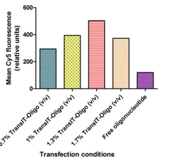
Figure 8 – Mean fluorescence of flow cytometry studies at 0.1µM AON. Several concentrations of TransIT-Oligo (Mirus), a commercial transfection reagent were tested, alongside a free oligo control.
From the analysis of the mean fluorescence of each sample, 1.3% TransIT-Oligo (v/v) can be expected to support efficient AON transfection in the selected cell line (RN22) since it supported the highest fluorescence levels (fig. 8). Some cytotoxicity (verified by observation of cell
morphology under the optical microscope) was seen for the sample with the highest transfection reagent concentration, which may explain the difference of fluorescence to the previous sample. At 0.1 µM, fluorescence level is increased more than 3 times by the presence of 1.3% transit; however, as stated before, no straight-lined conclusions may be gathered from flow cytometry analysis alone.
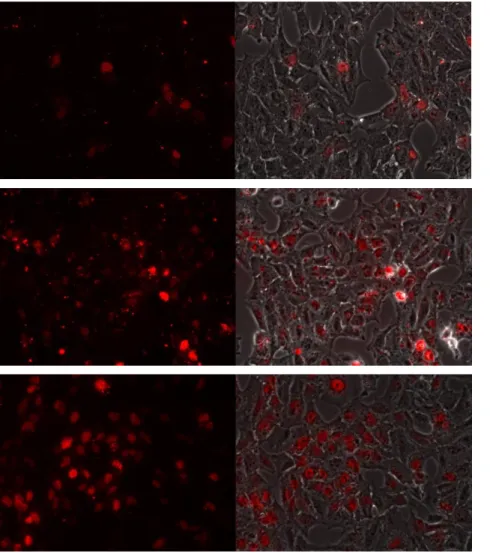
Figure 9 – Fluoresnce microscopy analysis of different transfection reagent concentrations. Free oligo at 0.1 µM (top), 1.3% (v/v) TransIT-Oligo and 0.1 µM AON (middle) and 1.3% (v/v) TransIT-Oligo and 0.3 µM AON (bottom). The
Following the flow cytometry studies, fluorescence microscopy was used to confirm whether the previously selected conditions were efficient in transfecting the selected cell line. In addition to free oligo and 1.3% (v/v) TransIT-Oligo at 0.1 uM AON, 1.3% (v/v) TransIT-Oligo at 0.3 µM AON was also tested, having had their microscopy pictures taken at identical exposure times. The latter provided substantially higher transfection efficiency with no significant signs of cytotoxicity (fig. 9). Efficient transfection was determined by diffuse nuclear staining, referred in previous studies as a characteristic of phosphorothioate-modified AON89, 90. In opposition, cytoplasmatic abundance of small granules is signal of extensive retention of AON at vesicles, most probably endosomes, i.e. there is low endosomal escape.
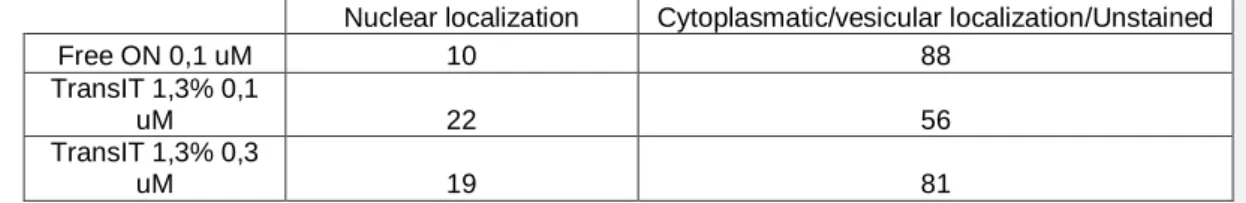
A manual counting of nuclear- or cytoplasmic/vesicular-stained cells was done from
fluorescence microscopy images, and used in a contingency table (table 3). For analysis purposes, cells binned into the first class were excluded of the second, although in reality they are stained in the cytoplasm as well. The chi-square test returned p < 0.001 that the data is statistically significant, i.e. the statistical analysis supports that the tested conditions have different transfection properties.
Table 3 – Contingency table used in the analysis of the fluorescence images. Cell count was determined manually. P<0.001 statistical significance analysed by chi-square test of the 2x3 contingency table.
Nuclear localization Cytoplasmatic/vesicular localization/Unstained
Free ON 0,1 uM 10 88
TransIT 1,3% 0,1
uM 22 56
TransIT 1,3% 0,3
uM 19 81
Since these results from the optimization studies support 1.3% (v/v) TransIT-Oligo and 0.3 µM AON as efficient transfection conditions with about 80% transfected cells, these were selected for further testing of AON downregulation activity.
Analysis of antisense oligonucleotide activity
Rn22 a rat schwannoma cell line expressing RhoA and GSK3beta was then used as a model for testing the downregulation efficiency of a series of antisense oligonucleotides. These were selected previously based on thermodynamic analysis of binding affinities of the oligos to the target mRNAs (having in consideration melting temperatures and RNA secondary structure). After transfection with conditions determined from the results presented above, reverse transcription PCR with semi-quantitative analysis was selected as an initial method to screen for the different oligonucleotide efficiencies.
Figure 10 – Antisense activity analysis of GSK3 AON-treated, PCR-amplified samples. There is a clear downregulation of the target gene (**p < 0.01, samples vs non-treated), although high variability hinders an effective
comparison between the different sequences (not statistically significant).
Figure 11 - Antisense activity analysis of RhoA-AON treated, PCR-amplified samples. There is a clear downregulation of the target gene (**p < 0.01; samples vs non-treated), although high variability hinders an effective
This semi-quantitative means of analysis yielded a very high standard deviation, which did not enable sequence discrimination (fig. 10 and 11). Nonetheless, all of them were effective in downregulating the target gene. The variability observed in the antisense studies could reflect some toxicity issues observed during the transfection procedure with some of the AONs (as seen by cell morphology changes visualized under the light microscope – data not shown). Despite the variability and the absence of statistical significance between every pair of treated samples, all AON sequences tested yielded gene downregulation with p < 0.01. Based on these results quantitative, real-time RT-PCR will be used in future to confirm and better discriminate between some of the AON sequences.
PART II – Polymer vector
In order for antisense oligonucleotides to have effects in a therapeutic context, they need to be delivered to target cells in an efficient way. Due to their chemical nature (negatively charged hydrophilic molecule), these are not taken up efficiently by cells; furthermore, pharmacokinetics and bioavailability are an issue in in vivo conditions. Thus, safe and non-toxic delivery vectors are needed for therapeutic applications of antisense oligonucleotides. In that context, chitosan, a natural polymer with inherent biodegradability properties, is a promising vector.
A new chitosan derivative was tested for efficient complexation (formation of nanoparticles) and for transfection efficiency of AONs. Trimethyl modification of the deacetylated amines of chitosan produced a biomaterial with improved electrostatic interaction and solubility properties, trimethylchitosan (TMC). An additional modification with stearic acid was tested for AON binding properties, nanoparticle diameter, cellular binding and transfection efficiency properties, which were expected to improve over the unmodified TMC.
Physical and chemical properties of TMC
Unmodified TMC and stearic acid conjugated TMC (TMC-SA) were tested for the ability to bind to AONs. Initially, a TMC-SA with 5% SA (TMC-SA 5%) modification was tested. Later in the project a TMC-SA with 2.5% SA (TMC-SA 2.5%) modification became available, which was included in some of the analyses. An initial approach to study the AON binding properties of both the unmodified and TMC-SA 5% was done by using agarose gel electrophoresis and SybrGold staining. Since the nucleic acid dye should only bind free oligonucleotides, this was perceived as a simple and effective analysis. Using a free oligo control as a reference for migration length, several TMC N/P ratios could be tested for their ability to strongly interact and bind AON thereby
preventing its migration in the gel. AON weakly interacting or not interacting at all with the TMCs after complexation will migrate. The fraction of migrating AON thus directly reflects the extension of interaction between AON and TMCs.
The smearing pattern observed is indication that the oligonucleotide is also being released from the interaction with the TMC over time (during the agarose run). This could indicate a destabilizing effect of the nucleic acid binding dye (SybrGOLD which was present in the gel during the electrophoresis) and/or the electrophoresis voltage conditions. Presence of a smear but absence of free oligo band suggests complete AON binding at the beginning of the run, with posterior release during the run.
Figure 12 – Agarose gel retention assay of unmodified TMC (top) and 5% stearic acid-substituted TMC (bottom). N/P ratios are indicated above each column, 0 indicates free oligo (no TMC present). The unmodified TMC has much
weaker AON binding properties than the stearic acid-modified TMC. The former has partial retention at N/P 30 and complete retention at N/P 100, while the latter has partial retention at N/P 1 and complete retention at N/P 2.
As expected, the hydrophobic substitution was effective in improving the AON binding properties of TMC, since at N/P 2 no free oligo was present at the beginning of the run (fig. 12); however, the intense smear indicates potential low interaction stability. Higher N/P ratios have reduced smear, and at N/P 10 and 30, there is none at all. Although it lacks confirmation in vitro, excessive N/P ratios may pose an obstacle to oligonucleotide release after entering the cell, in the case of the interaction being too stable and the complex failing to dissociate. Cytotoxicity could be another valid reason to avoid those N/P ratios. Although the hydrophobically modified TMC (with 5% SA modification) showed complete retention of the oligo at low N/P ratios, the unmodified
trymethylchitosan was only effective in slowing the AON migration at N/P 100 (although some degree of destabilization is still observed – presence of smear).
A different approach was used at a later stage – considering that the presence of nucleic acid binding dyes during the electrophoresis could interfere with AON migration, posterior staining was suggested. Polyacrylamide gels were selected because they are more efficient in post staining than agarose gels. For this analysis, unmodified TMC, TMC-SA 5% and TMC-SA 2.5% were available to use.