João Luís Cruz Prates
Effect of fetal bovine serum on
Candida glabrata cultures
João Luís Cruz Pr
at es Ef fect of fe tal bo vine serum on Candida glabrata cultures 15
João Luís Cruz Prates
Effect of fetal bovine serum on
Candida glabrata cultures
Trabalho efetuado sob orientação da
Professora Doutora Mariana Contente Rangel Henriques
Universidade do Minho
e do
Professor Doutor Patrick Van Dijck
KU Leuven
Dissertação de Mestrado
Mestrado Integrado em Engenharia Biomédica
Ramo de Engenharia Clínica
Nome: João Luís Cruz Prates
Endereço eletrónico: [email protected] Telefone: 910357794/259939562
Número do Cartão de Cidadão: 13461633 Título da dissertação:
Effect of fetal bovine serum on Candida glabrata cultures Orientador/a/es:
Professora Doutora Mariana Contente Rangel Henriques Professor Doutor Patrick Van Dijck
Ano de conclusão: 2015
Mestrado Integrado em Engenharia Biomédica Ramo de Engenharia Clínica
DE ACORDO COM A LEGISLAÇÃO EM VIGOR, NÃO É PERMITIDA A REPRODUÇÃO DE QUALQUER PARTE DESTA TESE/TRABALHO.
Universidade do Minho, _____/_____/_________
A
CKNOWLEDGMENTSMy first words are to my supervisors, Dr. Patrick Van Dijck and Dr.ª Mariana Henriques. I would like to thank both for all the support. To Dr. Patrick Van Dijck, my words of thankfulness for the opportunity to integrate a so respectful institution like the Laboratory of Molecular Cell Biology in KU Leuven. His advice helped me during my research and the conditions of the lab were so great, I can only assure that it was one of the most important periods of my life academically and personally. To Dr.ª Mariana Henriques, my words of admiration for all the time and patience she spent helping me and for all the motivation and guidance that I needed to pull this project through.
Another person that had an incredible role of support and guidance during my time in the Laboratory of Molecular Cell Biology in Leuven was Bea Timmermans. She always helped me with my doubts and questions and she always made an incredible effort to motivate me and to teach me everything she could to make of me a better worker on the lab. For all her availability, friendly words, advices and knowledge I have to thank. I have no doubt she will have success in Science. I wish her the best. My words of appreciation to the other persons working in the Laboratory of Molecular Cell Biology in KU Leuven.
To my Erasmus mate Cláudia Rodrigues for all the friendship, patience and help during our time in Leuven. Her importance during the time we lived in a different and far away country is undeniable and I have to thank her a lot for all the support. I have to refer too to the rest of my portuguese friends and to all the friends I made during my Erasmus program from all over the world. They were very important motivating me and giving me great memories that I will never forget.
Another person that has been there for me in some of the best and worst moments is my girlfriend, to whom I have to thank every day, for all the patience, friendship and love.
My last words are to all my family, especially to my parents and my brother, for their love, confidence, encouragement, understanding, help and patience. They gave me everything I have and that is a debt I will maintain for the rest of my life. Thank you very much for your magic powers that have been making things possible.
To my maternal grandfather and my paternal grandmother, This one I really need to share with both of them, they would be proud of me.
A
BSTRACTCandida glabrata, is a yeast that takes part of the human’s microbial flora as an opportunistic pathogen and has been emerging, becoming an important and dangerous nosocomial pathogen.
The main evidence that raised the development of this project was the observation that there was a secretion of unknown composition identified on C. glabrata cultures grown in the presence of fetal bovine serum (FBS). However, the results showed that all the strains tested from C. glabrata, Saccharomyces cerevisiae and Candida albicans produced secretion only in the presence of FBS, what was not expected. The goals were to evaluate the role of FBS in C. glabrata secretion production, to characterize its composition and to evaluate its effect in C. glabrata’s virulence factors.
It was confirmed by SDS-PAGE that the secretion only occurred in the presence of FBS. In addition, it was shown that the proteins on the secretion were not part of the proteic composition of the FBS but came from the cells. The way the cells were affected by the presence of the FBS is still unclear. By mass spectrometry, five genes were found on the secretion that had homology on S. cerevisiae cell wall glucanases.
As the origin of the secretion and the composition of that secretion were already investigated, next a deletion collection was screened to try to determine the gene(s) responsible for the secretion. However, there were no correlation between the mutants and the production of secretion.
A susceptibility test was performed for fluconazole and it was possible to verify a morphological difference between the cells grown in FBS and without it and a MIC value higher in the presence of FBS. The way the FBS affects the MIC is not elucidated. Congo red and calcofluor white were also tested in the presence of FBS, but, the results were inconclusive as the FBS did not have any remarkable effect on the two cell wall stress dyes tested.
Regarding adhesion, it was possible to verify that with increasing concentration of FBS, the cell adhesion decreased.
In conclusion, the secretion in the presence of FBS is not exclusive for C. glabrata cultures however, this study can bring new insights on C. glabrata virulence.
R
ESUMOA Candida glabrata é um fungo que faz parte da flora microbiana do organismo humano, sendo um agente patogénico oportunista, que se tem vindo a tornar um importante e perigoso patogeno nosocomial.
A evidência para este projeto surgiu da observação de que existia uma secreção de composição desconhecida nas culturas de C. glabrata com soro fetal bovino (FBS). No entanto, os resultados mostram que todas as estirpes de C. glabrata, Saccharomyces cerevisiae e Candida albicans que foram usadas produziram secreção na presença de FBS, o que não era esperado.
Os objetivos foram avaliar o papel do FBS na produção dessa secreção em culturas de C. glabrata, caracterizar a sua composição e avaliar o seu efeito nos fatores de virulência da C. glabrata.
Foi confirmado através de SDS-PAGE que a secreção apenas ocorria na presença de FBS. Para além disso, foi mostrado que as proteínas da secreção não faziam parte da composição do FBS mas tinham origem nas células. A forma como as células são afetadas pela presença do FBS ainda não é clara. Através de espectrometria de massa foram encontrados cinco genes na secreção com homologia nas glucanases da parede celular da S. cerevisiae.
Assim que a origem da secreção e a sua composição foram investigadas, uma “deletion collection” de C. glabrata foi cultivada na presença de FBS com o objetivo de determiner o(s) gene(s) responsáveis pela secreção. No entanto, não foi possível estabelecer uma correlação entre os mutantes e a produção da secreção.
Um teste de susceptibilidade ao fluconazole foi realizado e foi possível verificar a existência de diferenças morfológicas entre as células suplementadas por FBS e sem FBS e, para além disso, o valor do MIC aumentou na presença do FBS. A forma como o FBS afeta o MIC não está ainda determinada. O Congo red e o calcofluor white foram também testados na presença de FBS mas os resultados foram inconclusivos uma vez que o FBS não teve efeito em nenhum dos corantes testados.
Em relação à adesão, foi possível verificar que com o aumento da concentração de FBS, a adesão celular diminuiu.
Para concluir, a secreção na presença de FBS não é exclusiva para culturas de C. glabrata, no entanto este estudo pode trazer novos desenvolvimentos ao conhecimento da virulência da C. glabrata.
T
ABLE OFC
ONTENTS Acknowledgments ... iii Abstract ... v Resumo ... vii Table of Contents ... ix List of Figures ... xiList of Tables ...xiii
Abbreviations ... xv
1. Chapter I – General Introduction ... 1
1.1. Motivation and Goals ... 3
1.2. State of Art ... 4
1.2.1 Nosocomial bloodstream infections ... 4
1.2.2 Candida glabrata ... 5
1.2.3 Virulence factors ... 6
Response to macrophages ... 6
Enzyme production ... 7
Adherence to host cells, non-biological surfaces, and surface hydrophobicity 10 Biofilm formation ... 11
Tolerance to azole antifungals ... 13
Melanin-like pigment ... 13
1.2.4 Antifungal classification and resistance ... 14
Polyenes ... 15
Azoles ... 16
Echinocandins ... 18
Other antifungals ... 19
2. Chapter II – Materials and Methods ... 21
2.1. Strains ... 23
2.2. Secretion Analysis by Strain Spotting ... 23
2.3.1 Sample preparation ... 23
2.3.2 SDS-PAGE ... 24
2.3.3 Gel staining ... 24
2.4. Extraction of Proteins via SDS for LC MSMS ... 25
2.4.1 SDS removal and digest ... 25
2.4.2 Salt removal ... 25
2.5. Screening for Defective Secretion ... 26
2.6. Minimal Inhibitory Concentration ... 26
2.7. Spot Assay – Congo Red and Calcofluor White ... 27
2.8. Adhesion Assay ... 27
3. Chapter III - Results and Discussion ... 29
3.1. Evaluation of the Secretion Produced by C. albicans and S. cerevisiae ... 31
3.2. Determination of Culture Secretion Origin ... 35
3.3. Mass Spectrometry (MS) ... 38
3.4. Screening of the C. glabrata Deletion Collection ... 39
3.5. Determination of the Minimal Inhibitory Concentration (MIC) ... 40
3.6. Susceptibility Testing with Congo Red and Calcofluor White ... 42
3.7. Candida glabrata Adhesion ... 43
4. Chapter IV - Conclusion and Future Work ... 45
L
IST OF FIGURESFigure 1 - The left picture illustrates several colonies of C. glabrata cultured in minimal medium with agar and supplemented with glucose and FBS; the right picture represents two falcons with cultures of C. glabrata in minimal medium with glucose (A) and C. glabrata in minimal medium supplemented with glucose and FBS (B).
Figure 2 - Representation of different classes and antifungal drugs in use nowadays (Roemer &
Krysan, 2014).
Figure 3 - SDS-PAGE of the pelleted fraction resultant from the supernatant obtained from C. glabrata wild type cells grown in SCD plus FBS (lane 2) and of the remainant supernatant from the same culture (lane 3); samples constituted of the supernantant obtained from a C. glabrata wild type culture in SCD (lane 4) and of the remainant supernatant from the same culture (lane 5); Control samples with only SCD (lane 6) and with SCD plus FBS (lane 7); Pelleted fraction resultant from the supernatant obtained from S. cerevisiae Σ1278b cells grown in SCD (lane 8) and of the remainant supernatant from the same culture (lane 9); samples constituted of the supernantant obtained from a S. cerevisiae Σ1278b culture in SCD plus FBS (lane 10) and of the remainant supernatant from the same culture (lane 11); Secretion on C. glabrata wild type cells cultured on SCD with agar and FBS (lane 12); Lane 1 - SeeBlue® Plus2 Pre-stained Protein Standard.
Figure 4 - The left picture illustrates several colonies of C. glabrata cultured in minimal medium with agar and supplemented with glucose and FBS; the right picture represents two falcons with cultures of C. glabrata in minimal medium with glucose (A) and C. glabrata in minimal medium supplemented with glucose and FBS (B).
Figure 5 - SDS-PAGE of the pelleted fraction resultant from the supernatants of C. glabrata grown in SCD (minimal medium with glucose) (lane 2) and in the presence of FBS (lane 5). Controls were included for SCD + FBS (lane 3) and SCD + FBS (3x diluted) (lane 4). Lane 1 - SeeBlue® Plus2 Pre-stained Protein Standard.
Figure 6 - SDS-PAGE of the pelleted fraction resultant from the supernatants obtained from the
Amicon subfraction <10 kDa for cells grown in SCD (lane 2) and with FBS (lane 3); from the Amicon subfraction >10 kDa for cells grown in SCD (lane 4) and with FBS (lane 5); control samples, without Amicon centrifugation, supertantant from cells grown in SCD with FBS (lanes 6 and 7) and without it
(lanes 8 and 9). Samples on lanes 7 and 9 are 2x dilutions from samples 6 and 8, respectively. Lane 1 - SeeBlue® Plus2 Pre-stained Protein Standard.
Figure 7 - SDS-PAGE of different washes of the pelleted fraction resultant from the supernatants
resulting from C. glabrata grown in SCD and FBS; Lane 2 – pelleted supernatant; Lane 3 – first wash of the pelleted supernatant; Lanes 4 to 9 – second to seventh washes of the pelleted supernantant, respectively; Lane 10 – pelleted supertantant after washes. Lane 1 - SeeBlue® Plus2 Pre-stained Protein Standard.
Figure 8 - Image of the 96-well plate containing the MIC assay (24h) to test fluconazole. The first row
of the plate is composed of empty wells. In the second to fourth rows there are cultures of C. glabrata wild type cells in SCD. In the fifth to seventh rows there are C. glabrata wild type cells in SCD plus FBS. The concentration of antifungal in both this groups of rows decreases from well to well till 0 (as according to the information on the first row). The last row of the plate contains the controls (no antifungal), constituted only by minimal medium with glucose (SCD Medium), C. glabrata cultured in minimal medium with glucose (SCD cells), minimal medium with glucose supplemented with FBS (FBS+SCD Medium) and C. glabrata cultured in minimal medium with glucose supplemented with FBS (FBS+SCD cells), respectively.
Figure 9 - In vitro adhesion of C. glabrata ATCC2001 analyzed by growing cells supplemented with different percentages of FBS in 96-wells plate. Nonadherent cells were washed away with PBS, and remaining adherent cells were stained with XTT menadione solution.
L
IST OF TABLESTable 1 - Temporal variation of bloodstream infections for different Candida spp. in North America (M. a Pfaller & Diekema, 2010)
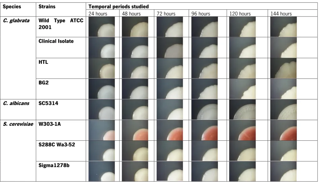
Table 2 - Representation of the evolution of the secretion for different species and strains each 24
hours till 144 hours, cultured on minimal medium with glucose (SCD)
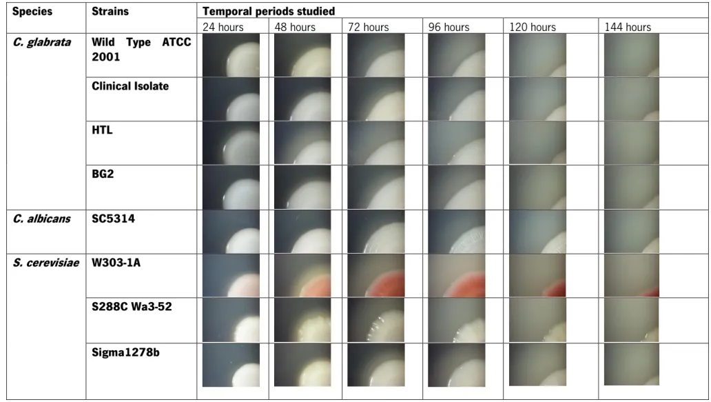
Table 3 - Representation of the evolution of the secretion for different species and strains each 24
hours till 144 hours, cultured on minimal medium with glucose (SCD) and FBS
Table 4 - Significant genes obtained from the mass spectrometry results and secreted proteins by C. glabrata
Table 5 - Example of two strains (HTL and BG2) from the C. glabrata deletion collection used, screened on agar plates with minimal medium and glucose (SCD) and minimal medium and glucose and FBS (SCD+FBS), at 48 hours incubation. It is possible to observe that both strains secreted in the presence of FBS
Table 6 - Result of spot assay (24 and 48 h) in minimal culture with glucose (SCD) (A) or SCD
supplemented with 5% FBS (SCD+FBS) (B) for CFW and CR
Table 7 - Summarization of the various factors influencing each quantification protocol; (-)
A
BBREVIATIONSABC: ammonium bicarbonate ACN: acetonitrile
CR: Congo red
CSM: complete supplement mixture CW: calcofluor white
DTT: dithiothreitol
ECM: extracellular matrix FBS: Fetal bovine serum
GPI: glycosylphosphatidylinositol HTL: his3Δ::FRT leu2Δ::FRT trp1Δ::FRT IAA: iodoacetamide
MIC: minimal inhibitory concentration MS: mass spectrometry
PBS: phosphate-buffered saline Rpm: rounds per minute
SC: synthetic complete medium SCD: minimal medium with glucose SDS: sodium dodecyl sulfate TE: tris-EDTA buffer
TFA: trifluoroacetic acid UA: ureabuffer
XTT: 2,3-Bis-(2-Methoxy-4-Nitro-5-Sulfophenyl)-2H-Tetrazolium-5-Carboxanilide) YPD: yeast extract peptone dextrose
1. C
HAPTERI
1.1 Motivation and Goals
Candida species are yeasts that are part of the human’s microbial flora as opportunistic pathogens. After Candida albicans, Candida glabrata is the most prevalent yeast pathogen, with a high mortality rate in hospitals all around the world. One of the biggest problems associated with this increasing prevalence is the delay till the initiation of the right therapy (because C. glabrata is tolerant to the azole antifungals that are used to treat Candida infections).
The main evidence for this project was the observation that there was a secretion of unknown composition identified on Candida glabrata cultures in minimal medium with glucose and fetal bovine serum (FBS) (Figure 1) on solid medium and liquid medium.
Figure 1 - The left picture illustrates several colonies of C. glabrata cultured in minimal medium with agar and supplemented with glucose and FBS; the right picture represents two falcons with cultures of C. glabrata cultured in minimal medium with glucose (A) and C. glabrata in minimal medium supplemented with glucose and FBS (B).
From the observation of the Figure 1 it is possible to identify a clear region around the cultures of C. glabrata spotted in solid minimal medium with FBS. That clear region can be observed also on the liquid cultures and with more evidence. Comparing with the culture of C. glabrata without FBS, the falcon with the culture of C. glabrata supplemented with FBS presents a layer of white secretion up to the pellet without the cells and a less clear supernatant layer.
So, the main purpose of this project was to determine if the secretion only occurred for C. glabrata and to evaluate the role of FBS in the secretion production. Moreover, it was also a goal to characterize the composition of that secretion. This analysis might be important for a better understanding of C. glabrata colonization and virulence mechanisms, and probably will give new important insights on this species that could be useful for the development in the future of more effective drugs against this pathogen.
1.2 State of Art
1.2.1 Nosocomial bloodstream infections
Nowadays, Candida is the fourth most common cause of nosocomial bloodstream infections (BSI) in the USA. Candida albicans is the most common Candida species causing BSI accounting for 54% of the cases. Secondly ranked is Candida glabrata, a species that is responsible for 19% of BSI infections in this country (Wisplinghoff et al., 2004). Candida is obviously an important cause of candidemia being nowadays an important and dangerous nosocomial pathogen (Pfaller et al., 2003; Trick et al., 2002). In the last three decades there was a significant increase in the cases of fungal infections in humans (Lass-Flörl, 2009), usually due to the use of invasive surgical procedures, immunosuppressive therapies and broad-spectrum antibiotics (Williams et al., 2011). These last two factors seem to increase in proportion with the number of infections caused by C. glabrata (Hajjeh et al., 2004). Over the last 15 years, the number of C. glabrata infections associated to BSI increased in North America (from only 12% of the Candida species, between 1992 and 1993, to 26% of the Candida species, between 2004 and 2008), while the number of C. albicans and other Candida (Candida parapsilosis, Candida tropicalis and Candida krusei) BSI cases stabilize (Table 1), allowing a better understanding of C. glabrata importance.
Table 1 - Temporal variation of bloodstream infections for different Candida spp. in North America (Pfaller & Diekema, 2010)
% Total by Candida spp.*
Location Study
period Reference isolates No. of Candida albicans Candida glabrata Candida Other
United States 1992-1993 Kao et al. (1999) 837 52 12 35 United States 1993-1995 Pfaller et al. (2002a) 79 56 15 25 United States 1995-1997 Pappas et al. (2003) 1,593 46 20 28 United States 1995-1998 Pfaller et al. (2002a) 934 53 20 25 United States 1998-2000 Hajjeh et al. (2004) 935 45 24 27 Canada 1999-2004 Laupland et al. (2005) 209 51 22 17 North America 2001-2004 Pfaller et al. (2007a) 2,773 51 22 23 North America 2001-2006 Pfaller et al. (2008a) 1,489 50 24 24 North America 2001-2007 Pfaller et al. (2009b) 11,682 49 21 24 North America 2004-2006 Diekema et al. (2009a) 1,657 52 23 22 North America 2004-2008 Horn et al. (2009) 2,019 46 26 27
Moreover, results from Silva et al. (2011)indicate that infections with both C. albicans and C. glabrata are of major importance, causing increased tissue damage, since the presence of C. albicans
seems to increase the invasiveness of C. glabrata. In this way both species are able to occupy similar niches what makes it possible for them to co-exist (Silva et al., 2011).
1.2.2 Candida glabrata
Candida glabrata, together with other Candida species, belongs to the class Fungi Imperfecti, the order Moniliales, and the family Cryptococcaceae.
C. glabrata is closely related to the non-pathogenic Saccharomyces cerevisiae. It shares a recent common ancestor with several Saccharomyces species, and clearly belongs to a clade different from that of other Candida species which display particular features such as the recoding of the CUG codon to serine (Roetzer et al., 2011).
It has an optimal growth temperature of 37 °C and, being a nicotinamide adenine dinucleotide (NAD+) auxotroph, its growth is dependent on exogenous supply of NAD+ precursors (Roetzer et al., 2008).
The skeletal component of the cell wall of the majority of fungal pathogens, including Candida albicans, and S. cerevisiae is based on a core structure of β-(1,3)-glucan covalently linked to β-(1,6)-glucan and chitin (a β-(1,4)-linked polymer of N-acetylglucosamine (GlcNAc)). These polymers form hydrogen bonds between adjacent polysaccharide chains to create a tough three-dimensional network of microfibrils. In addition to the glucan and chitin skeleton, C. albicans cell wall contains a matrix that mainly comprises glycosylated proteins. In C. albicans, the major class of cell wall proteins are glycosylphosphatidylinositol (GPI)-anchor-dependent cell wall proteins (GPI-CWPs), which are attached through a GPI remnant to β-(1,3)-glucan or chitin by a highly branched β-(1,6)-glucan linker (Netea et al., 2008).
The cell wall organization of C. glabrata appears similar to that of the closely related nonpathogenic yeast S. cerevisiae. However, C. glabrata cell wall contains more mannoprotein. These proteins, on the outer layer of the wall of C. glabrata, are expected to play key roles in adhesion and biofilm formation and other host-pathogen interactions that mediate fungal virulence (Groot et al., 2008). C. glabrata contains covalently bound cell wall proteins (CWPs) that can be divided into two groups, the largest group being glycosylphosphatidylinositol (GPI)-modified proteins that are covalently bound to the wall 1,6-β-glucan whereas the second group is directly bound to 1,3-β-glucan via an ASL (Groot et al., 2008).
1.2.3 Virulence factors
Analyzing Candida albicans and Candida glabrata virulence factors it is possible to identify some differences and resemblances, what increases the scientific knowledge and consequently makes it possible to evolve clinically, finding efficient therapies and saving more and more infected patients. Several virulence factors were already identified as part of Candida species pathogenicity as the morphological behavior on the presence of macrophages, the enzyme production and secretion, the mechanisms of adherence to host cells and non-biological surfaces, the surface cell wall hydrophobicity, the biofilm formation and the resistance to antifungal drugs.
Response to macrophages
Candida albicans resides as a harmless lifelong commensal organism (Mayer et al., 2013), however it is possible to identify some factors that turn it in a dangerous pathogen. One of those virulence factors is its capacity to morphologically switch from yeast cells to pseudohyphae or true hyphae (Whiteway & Oberholzer, 2004). The production of germ tubes results in conversion to a filamentous growth phase or hyphae, also called the mycelial form (Biswas et al., 2007). When on the presence of macrophages, C. albicans wild type strain, which is able to make the switch, can kill the macrophages being more effective causing disease. On the other hand, some C. albicans mutants (like cph1/cph1 efg1/efg1 mutant) when interacting with macrophages fail to form filaments and finish suffering phagocytosis and dying (Lo et al., 1997).
In contrast, Candida glabrata is not polymorphic, growing only as a blastoconidia (yeast) (Silva et al., 2012). When C. glabrata cells are engulfed by host macrophages they have to adapt to the acidic phagosomal environment in order to survive. Inside the macrophages they suffer oxidative stress and they are starved for carbon, so they induce genes involved in gluconeogenesis, β-oxidation, glyoxylate cycle, and transporters for amino acids and acetate (Kaur et al., 2007; Roetzer et al., 2010). C. glabrata induces peroxisomes transiently in the process of recycling of internal resources, what plays an important protective role in this condition because the proliferation of peroxisomes in fungal cells contributes to the adjustment of carbon metabolism (Roetzer et al., 2010).
Li et al. (2007), showed that C. glabrata even at the lowest infectivity ratio triggers a higher granulocyte macrophage colony stimulating factor response than C. albicans, inducing a proinflammatory cytokine response in oral epithelial cells. However, its tissue/cell damaging ability,
epithelial cytokines and chemokines as a potent activator of leukocytes and lymphocytes to the site of mucosal infection. So, as C. glabrata cells are able to survive and replicate inside macrophages, this makes it tempting to speculate that luring more macrophages to the site of infection may even be beneficial for the fungus and may constitute part of its immune evasion strategy (Seider et al., 2011). The capability of C. glabrata cells to survive, function, and replicate inside the macrophage is due to chromatin remodeling process, that allows energy homeostasis and genes required for its virulence (Rai et al., 2012).
Enzyme production
Another important virulence factor of Candida albicans is the secretion of hydrolases. The three most significant extracellular hydrolytic enzymes produced by C. albicans are the secreted aspartyl proteinases (Sap), phospholipase B and lipases (Naglik et al., 2003).
SAPs, encoded by a family of ten genes (SAP1-SAP10), can be divided into subfamilies based on amino acid sequence homology alignments [SAP1-SAP3, SAP4-SAP6, SAP9-SAP10 (SAP7 and SAP8 are divergent and are not represented as subfamily members)] (Naglik et al., 2008).
From the three families, it is possible to identify 2 main subfamilies (SAP1-SAP3 and SAP4-SAP6) that are expressed during infections and are believed to be a major virulence factor in C. albicans at least for superficial infections (Naglik et al., 2004). Taking in consideration Naglik et al. (2003), there is solid evidence suggesting that Sap9 and Sap10 may not be secreted from the cell and seem to be regulatory proteinases that may play a role in the cell surface integrity, function that differs from the functions of the other Sap. An in vivo study addressing the expression and evaluation of SAP genes of C. albicans was conducted in 137 human subjects with oral and vaginal candidosis, recurring to reverse-transcriptase polymerase chain reaction. According to the results, SAP2 and SAP5 are predominant proteinase genes that may be required by C. albicans to fulfill basic functions in relation to survival and proliferation in the human host. The other members of the first subfamily presented, SAP1 and SAP3, are more frequently found in association with vaginal infections. The universal expression of SAP2 during colonization and infection and the preferential expression of SAP1, in particular, during active human disease supports the current evidence that the SAP1-SAP3 subfamily contributes to the pathogenesis and virulence of C. albicans at mucosal surfaces (Naglik et al., 2003). Regarding the other SAP gene subfamily (SAP4-SAP6), the results indicate that SAP4 and SAP6 are more frequently expressed during oral and vaginal infections, what indicates that this subfamily is involved in human mucosal disease (Naglik et al., 2003). Sap6, in particular, plays a dominant role during systemic
infections by aiding penetration of tissue and survival of the fungus in phagocytes (Felk et al., 2002). SAP7 function is completely unknown, however, its expression in both oral and vaginal infections may support a role during human mucosal infections. SAP8 is expressed in C. albicans isolates from active oral and vaginal infections, however the way it contributes to human mucosal infections is still unclear too (Naglik et al., 2003).
An in vitro proteinase production assay was performed on several types of Candida, including Candida glabrata. The results showed that C. glabrata produces proteinases (Chakrabarti et al., 1991), however the role of these proteinases is not specified. Some species that are not involved in Sap proteinases secretion may produce enzymes of aspartyl proteinases activity, closely related to S. cerevisiae yapsins (Sikora et al., 2011). That’s the case for C. glabrata as it is closely related to S. cerevisiae (Roetzer et al., 2011).
The work of Sikora et al. (2011), showed that all C. glabrata strains examined secreted proteinases, due to the detected Yapsin-related aspartyl proteinase genes – YPS2, YPS4, YPS6. They play an important role in the maintenance of cell wall integrity, adherence to host cells, and survival of fungal pathogens in macrophages and in virulence.
Regarding phospholipase production, it has been reported that C. albicans has, in total, seven phospholipase genes (PLA, PLB1, PLB2, PLC1, PLC2, PLC3 and PLD1), but so far only PLB1, PLB2, PLC1 and PLD1 are well characterized, extensively investigated, and also correlated with C. albicans pathogenicity. Concerning PLA, limited studies were conducted (Samaranayake et al., 2006). The results of the reintroduction of PLB1 into a C. albicans strain to restore and evaluate its virulence in vivo, showed that this gene may be responsible for host invasion, degrading the phospholipid constituents of the host cell membranes and allowing fungal hyphae to have access to the cytoplasm (Mukherjee et al., 2001). Regarding PLB2, and despite its homology with PLB1, any major functional role during colonization and infection in humans can be attributed (Ghannoum, 2000). According to Kunze et al. (2005), the role of PLC1 is not entirely known but it seems that this is an essential gene, involved in multiple cellular processes and that may in fact act in the nucleus. PLC2 and PLC3 are upregulated under filament-inducing conditions but dispensable for filamentation in C. albicans, being also not required for escape from macrophages (Knechtle et al., 2005). The function of both CaPLC2 and CaPLC3 must be clearly different from CaPLC1, as neither of them can compensate for CaPLC1 loss. The fact that these two genes are unique for C. albicans and do not exist in S. cerevisiae may suggest a role in adaptation to the human host (Kunze et al., 2005). Hube et al. (2001) performed in
vivo and in vitro expression studies using Northern blots and reverse transcriptase-PCR and determined that PLD1 exists in C. albicans and expresses higher levels during the yeast to hyphae transition.
A phospholipase assay was performed, on Candida glabrata isolates, obtained from blood samples from patients on intensive care units, dialysis units and oncology units, and few of the strains of this species obtained were phospholipase producers (Mohan & Ballal, 2008). This indicates that C. glabrata is a phospholipase producer as C. albicans.
The LIP genes constitute a large gene family that may have evolved to adapt to the permanent association of Candida albicans with the human or animal host and, therefore, they may also have important functions during persistence and infection processes (Hube et al., 2000). Fu et al. (1997) transformed Saccharomyces cerevisiae with C. albicans genomic library and screened for lipolytic activity on egg-yolk agar plates (a traditional screen for phospholipase activity), and two identical clones were obtained and exhibited lipolytic activity. The study results suggested that C. albicans, in addition to phospholipases, also had lipases, and by Northern blot analysis, it was shown that expression of the LIP1 transcript was detected only when C. albicans was grown in media containing Tween 80, other Tweens or triglycerides as the sole carbon source (Fu et al., 1997). Strong structural similarities of LIP2-LIP10 (except LIP7) to LIP1 suggest that like Lip1, they may be as well secreted. In addition, the existence of a family of genes that encode secreted lipases of C. albicans may be an indication that different lipase genes are needed during different stages or types of infection (Hube et al., 2000).
The hydrolysis of lipids by the lipases allows C. albicans to use fatty acids and/or glycerol as growing substrates transporting hydrolytic products into its cells. The high number of LIP genes may provide an advantage to the fungus, allowing it to survive even in the absence of carbohydrates and assisting C. albicans in the competition with other microorganisms. Moreover, release of fatty acids may change the pH in the microenvironment (observed decrease from 5.5 to 2.8 during growth in lipid media) allowing the optimal conditions for other fungal proteins like the secreted proteinases (Hube et al., 2000).
In addition, pathogenic microorganisms as C. albicans and C. glabrata can grow in the host using haemin or haemoglobin as sources of iron. Haemolysins are used by Candida species to degrade haemoglobin and facilitate recovery of the elemental iron from host cells. This enables pathogen survival and persistence in the host (Silva et al., 2012).
Adherence to host cells, non-biological surfaces, and surface hydrophobicity
Other important virulence factor is Candida adherence to host cells, non-biological surfaces, and its surface hydrophobicity (Luo & Samaranayake, 2002).
As with other pathogenic Candida species, the cell wall of Candida glabrata is the point of contact between host and fungus. In addition it protects the fungal cell from hostile environments, enabling its adherence to host surfaces and maintaining cell shape (West et al., 2013). Therefore, they thus are excellent targets for development of antifungal drugs and diagnostic tools, being crucial to get more insights into C. glabrata’s pathogenesis (Groot et al., 2008).
Cell wall proteins called adhesins are critical to mediate adhesion of C. albicans to various substrates, like abiotic surfaces, and other yeast cells, microorganisms or host cells. Among many adhesins, members of the agglutinin-like sequence (Als) family of glycoproteins are particularly active in cell aggregation, adhesion to endothelia and epithelia, adherence to a broad variety of host substrates and consequent infection of those host surfaces, formation of biofilms and pathogenesis in mouse models (Garcia et al., 2011; Phan et al., 2007). However, Als3 and Als1 are the only members of this family that have a significant invasive function being capable of inducing epithelial cell endocytosis by itself. Als6, Als7 and Als9 do not appear to mediate significant endothelial cell endocytosis, and Als2 and Als4 have not yet been tested (Phan et al., 2007). The study of the proteins that may have a function in adherence led to the identification of a hypha-specific surface protein (Hwp1). According to the results, the presence of Hwp1 might enable C. albicans to form tight attachments to the oral mucosa through TGase-catalyzed cross-linking between Hwp1 and epithelial cell proteins, which is important in the pathogenesis of candidiasis (Staab et al.,1999). Morphology-independent proteins can also contribute to adhesion. These include GPI-linked proteins (Eap1, Iff4 and Ecm33), non-covalent wall-associated proteins (Mp65, a putative β - glucanase, and Phr1, a β -1,3 glucanosyl transferase), cell-surface associated proteases (Sap9 and Sap10) and the integrin-like surface protein Int1 (Mayer et al., 2013).
On C. glabrata cell wall, there is one group called the EPA (epithelial adhesin) genes that are not present in the Saccharomyces cerevisiae genome. This subtelomeric gene family encodes adhesion proteins. Approximately 67 genes encoding adhesion-like glycosylphosphatidylinositol (GPI) - anchored proteins reside within the C. glabrata genome, and at least 17 or 23 (depending on the background) of these proteins can be allocated to the Epa family (Groot et al., 2008; Kaur et al., 2005). According to Cormack et al. (1999), Epa1 is responsible for 95% of the adherence to human epithelial cells in vitro.
to mention. C. glabrata Ace2 is a homologue of the S. cerevisiae transcription factor Ace2 (Stead et al., 2009). According to data from a murine model of candidiasis, inactivation of CgAce2 results in a hypervirulent strain whose infection results in a fatal systemic disease on 100% of the infected mice (Kamran et al., 2004).
Adherence to non-biological surfaces is essentially important in a clinical environment, where it is important to make sure that all indwelling devices used on patients are not infected by C. albicans or C. glabrata and do not contribute to increase patients’ illness. These concern is due to the fact that these devices represent a potential niche for adhesion and further formation of biofilm-associated infections (Kucharíková et al., 2014).
The surface hydrophobicity is another virulence factor for Candida albicans. According to the results of an ex vivo assay in mouse tissue, hydrophobic yeast cells are able to bind to host tissues diffusely and abundantly (Hazen et al., 1991). These results of Hazen et al. (1990) indicated in that C. albicans hydrophobicity was due to changes in the external surface protein exposure and that protein mannosylation would influence exposure of hydrophobic surface proteins. Hydrophobic cells are more resistant than hydrophilic cells to phagocytic killing (Antley & Hazen, 1988). Also for C. glabrata, Epa6 mediates strong hydrophobic interactions, as a basis for biofilm formation on abiotic surfaces (El-Kirat-Chatel et al., 2015).
Biofilm formation
A biofilm can be defined as an aggregate of microbial cells adherent to a living or nonliving surface, embedded within a matrix of extracellular polymeric substances (EPS) of microbial origin (Hall-Stoodley et al., 2012). It confers an advantage for all microorganisms being important for their survival as commensals and pathogens of humans. It allows them to evade host immune mechanisms and to resist both treatment with antifungals and competitive pressure from other microorganisms. In addition, biofilm formation may allow the species to be better adapted to colonization of tissues and indwelling devices (Silva et al., 2009).
Candida albicans cell wall proteins called adhesins are critical for biofilm formation. Among many adhesins, the members of the agglutinin-like sequence (Als) family of glycoproteins are particularly active in formation of biofilms and pathogenesis in mouse models (Garcia et al., 2011). Moreover, phospholipase B genes have temporal differences in gene expression levels in biofilms, what was observed to be model-dependent. PLB1 was downregulated in biofilm growth in microtiter plates (MTP) and in the in vivo and reconstituted human epithelium (RHE) models, however not in those
grown on silicone disks in a continuous flow system (CDC reactor). PLB2, was also underexpressed in biofilms grown in the MTP and in the in vivo and RHE models (up to 12 hours), and was upregulated in biofilms grown in the CDC reactor and in the RHE model (after 24 hours and 48 hours) (Nailis et al., 2010).
Using a clinically relevant model for subcutaneous catheter-related infection on rat, in vivo biofilms were compared with 6-day-old in vitro biofilms formed on the same devices placed in a 24-well plate and submerged in RPMI 1640 medium. A subset of genes – EPA1, EPA3, EPA6 and AWP1-AWP7 was chosen. Comparing the in vivo model system results with planktonic cells incubated in RPMI 1640 medium for 6 days with continuous shaking, it is possible to see that the studied genes are upregulated on the in vivo conditions. In addition, comparing the results from 6-day-old in vitro biofilms with planktonically grown cells, the genes of the EPA gene family chosen to study (EPA1, EPA3 and EPA6) were not significantly differentially expressed, whereas non-EPA adhesion genes, such as AWP3, AWP6 and AWP7, were up-regulated. Comparing the expression levels in old in vivo biofilm with 6-day-old in vitro biofilm, it was verified the up-regulation of EPA3 and EPA6 for the in vivo assay, whereas the expression of EPA1 was not different from that under in vitro conditions. These different results point to the fact that the infection process strongly depends on the environment. EPA3 expression results (at least 4-fold higher in vivo than in vitro) might be an indication that it is one of the most important genes responsible for in vivo biofilm development in a subcutaneous model. Regarding the AWP gene family, all genes shown to be expressed however increased expression of AWP2, AWP3 and AWP5 was observed in mature in vivo Candida glabrata biofilms (Kucharíková et al., 2014). Epa6, as already referred, mediates adherence to epithelial cells (Castaño et al., 2005), being an adhesin required for biofilm formation in C. glabrata.EPA6 and its close paralogue EPA7 are located in subtelomeric regions and their transcription is regulated by Sir4p and Rif1p, two proteins involved in subtelomeric silencing. Biofilm growth conditions induce the transcription of EPA6 and EPA7: this is dependent on the presence of an intact subtelomeric silencing machinery and is independent of the Mpk1p signalling pathway. Finally, the kinase Yak1p is required for expression of both adhesin genes and acts through a subtelomeric silencing machinery-dependent pathway (Iraqui et al., 2005).
The ability of Candida to form biofilm is an important feature that promotes both infection and persistence in the host. Biofilm activity is significant since high activity might be associated with enhanced expression of putative virulence factors. For C. glabrata, it was shown that their biofilms had lowest metabolic activity but higher number of cultivable cells per unit area when compared with other
biofilm matrices from C. glabrata have higher amounts of both proteins and carbohydrates than those species (Silva et al., 2009). Moreover, C. glabrata was unable to generate filamentous forms unlike the other Candida species tested (Silva et al., 2010).Thein et al. (2007) reported that after 48 hours, the biofilms of aerobically grown C. glabrata generally revealed a multilayer biofilm structure packed with blastospores devoid of either pseudohyphae or hyphae. Under anaerobic/static conditions, C. glabrata forms non-contiguous microcolonies of blastospores (Thein et al., 2007).
Tolerance to azole antifungals
One important virulence factor of Candida glabrata is its tolerance to azole antifungal drugs. It has been demonstrated that resistance to fluconazole is associated with an increased expression of the ATP binding cassette (ABC) transporters CgCDR1 and CgPDH1, which appeared to occur after patient exposure to this drug (Bennett et al., 2004). Another study demonstrates that some C. glabrata isolates can acquire decreased susceptibility to azole drugs, during exposure to azole compounds (Panackal et al., 2006).
For more information about other antifungal drugs see the section Antifungal classification and resistance.
Melanin-like pigment
Another Candida glabrata virulence factor is the production of a melanin-like pigment, since C. glabrata cells have a high rate of survival when treated with hydrogen peroxide (Brunke et al., 2010). A decreased neutrophil-mediated damage was seen in the presence of this melanin-like pigment and increased ability to cause epithelial damage. However C. glabrata pigment is formed as a by-product of tryptophan catabolism via the Ehrlich pathway. The gene ARO8 is transcriptionally induced in the medium containing tryptophan as the sole nitrogen source, and its product Aro8 can generate indole pyruvate from tryptophan. This intermediate is then either catabolized further to indole acetaldehyde by ARO10, or is secreted into the supernatant. Once outside the yeast cell, the intermediate spontaneously reacts to form the pigment only if oxygen is present. As the pigmented cell cause increased damage in vitro, expression of the pigment in vivo would explain in part the pathogenic potential of C. glabrata. However, the pigment production by C. glabrata is limited to very specific conditions so it is a rather unlikely event in any relevant in vivo scenario of C. glabrata infections (Brunke et al., 2010).
1.2.4 Antifungal classification and resistance
Nowadays, the therapeutic options for invasive fungal infections are limited to three most important classes of antifungals: polyenes, azoles and echinocandins (Figure 2).
Figure 2 - Representation of different classes and antifungal drugs in use nowadays (Roemer & Krysan, 2014).
The limited options are firstly due the fact that, unlike antimicrobials targeting bacteria, fungal pathogens are more closely related to the host. As many fundamental biochemical and cell biological processes are conserved from fungi to humans, many small molecules that are toxic to yeast are also toxic to humans. For this reason the antifungal drug has to target structures unique to fungi as the three antifungals classes do (Roemer & Krysan, 2014). Secondly, the development of a new antifungal agent requires many economic resources as much as time for finding the new compound and to test its security and efficacy. Then, years are required to proceed to clinical trials and to turn the new antifungal into an approved drug.
Antifungal resistance can be divided into in vitro and clinical resistance. The first, in vitro resistance is a laboratory measurement incorporating clinical experience and set against the range of susceptibility results for particular drugs and fungal species. It can be divided into primary/intrinsic resistance in which the fungus is resistant prior to drug exposure, or secondary/acquired resistance
persistence or progression of the fungal infection and it occurs despite the intervention of the adequate antifungal therapy, in the appropriate dose and by the most efficient administration route (Rogers, 2006). Antifungal resistance results of the combination of both in vitro and clinical resistance. This means that resistance is present if isolates are not inhibited by an antifungal drug administrated at normal doses and normal dosage schedules. In addition to this definition, MIC values can be a sign of antifungal resistance (Pfaller, 2012).
Polyenes
All the polyenes interact with sterols, change the membrane permeability of eukaryotic cells and lead to cell lysis. Taking in consideration the classification of polyene antibiotics according to its chemical structure and biological effects by Kotler-Brajtburg et al. (1979), it is possible to distinguish between two groups. The first group includes those which caused little or no K+ leakage or growth
inhibition without cell killing or lysis and the second group includes those producing significant K+
leakage or growth inhibition at much lower concentrations than concentrations required for cell lysis or death(Kotler-Brajtburg et al., 1979).
Amphotericin B, the most important polyene, acts by binding to sterols in the fungal cell wall and altering membrane permeability, thereby allowing the leakage of cytoplasmic components (Khoo et al., 1994) as K+ and Mg2+. These losses, along with a subsequent influx of protons into the fungal cell,
cause acidification of the fungal interior with precipitation of the cytoplasm and ultimate cell death (Hamill, 2013).
This molecule is delivered by intravenous infusion and is the agent with the broadest antifungal spectrum. However, it has some important side effects as fever and chills, rigors, arthralgias, nausea vomiting and headaches. In addition, it presents a substantial incidence of renal toxicity. For this reason, amphotericin B lipid formulations were developed to reduce toxicity and provide a safer alternative than conventional amphotericin B (Hamill, 2013). The disadvantage of such formulations is the quite expensive price and the fact that are available only in some regions (Roemer & Krysan, 2014). The targets of polyenes are plasma membrane sterols. However, the mechanisms of resistance to amphotericin B are poorly understood, but a decrease or lack of ergosterol content in the fungal cell membrane has been associated with resistance without affecting cell viability. Defects in the ERG3 gene have been reported to lead to an accumulation of other sterols instead of ergosterol (Pemán et al., 2009).
It was observed a decrease in Candida glabrata susceptibility to amphotericin B in some regions like Europe where 4.4% of the isolates had MIC values in excess of 2 µg/ml (Pfaller et al., 2004). This means an increase of resistance so higher doses of amphotericin B may be required for optimal treatment of infections with this particular pathogen (Nguyen et al., 1998).
Azoles
Azole antifungal drugs consist of a five-membered ring containing two or three nitrogens, classified as imidazoles (as ketoconazole, miconazole and clotrimazole) or triazoles (as itraconazole and fluconazole), respectively. The use of imidazoles is limited to treatment of superficial mycoses (with the exception of ketoconazole), whereas triazole drugs have a broader range of applications in the treatment of both superficial and systemic fungal infections (Sheehan et al., 1999). This class of antifungal agents blocks ergosterol biosynthesis by inhibition of lanosterol 14α-demethylase, leading to a diminished ergosterol production. Therefore cells lack the ability to build and renew sterols in cell membranes, changing the membrane fluidity and function of vital processes such as: signaling, transport, exocytosis and endocytosis (Rodrigues et al., 2013).
Azoles are extremely well tolerated, although they interfere with the metabolism of a number of other drugs owing to their ability to inhibit cytochrome P450. In general, fluconazole has broad activity against clinically relevant yeast including Candida species and Cryptococcus. Because amphotericin B and 5-flucytosine are not available in many resource-limited regions, fluconazole is widely used to treat cryptococcal meningitis despite the fact that it is less effective. Moreover, many isolates of Candida glabrata and Candida krusei, however, are intrinsically less susceptible (Roemer & Krysan, 2014).
Four major mechanisms of resistance to azoles have been described in Candida species: (i) decrease of intracellular drug concentrations, (ii) target site alteration, (iii) overexpression of the target enzyme and (iv) development of a bypass pathway. Candida cells can develop a decreased drug concentration by upregulation of active efflux pumps. According to a multiple efflux mechanism study (Albertson et al., 1996), resistance to fluconazole can be acquired by mutations causing an increased expression of CDR1, while other mutations causing increased CDR1 expression are associated with multiazole resistance. In a different study, a new ABC transporter closely related to Cdr1 that was named Cdr2, was cloned. The protein Cdr2p could confer resistance to azole antifungal agents, other antifungals (terbinafine, amorolfine) and to a variety of metabolic inhibitors (Sanglard et al., 1997). Another family of transporters is the Mdr family where Mdr1 appears to be the major facilitator (White,
1997). For Candida albicans, the expression of increased mRNA levels of MDR1 and CDR are associated with increased resistance (White, 1997).
A second mechanism of resistance in Candida species is the acquisition of point mutations in the gene encoding for the target of the azoles (ERG11). This results in an altered target that has a reduced affinity or is incapable to bind to the azole drugs (Pfaller, 2012).
Candida can also overexpress or upregulate the Erg11 target enzyme, leading to a deficient binding of targets with azole drugs, or to an increased quantity of those drugs. However, minimal upregulation of target enzymes has been observed to date, and this mechanism does not appear to be a major cause of azole resistance in Candida at this time (Pfaller, 2012). The antifungal agent is, therefore, overwhelmed and routine therapeutic concentrations can no longer effectively inhibit ergosterol synthesis. Target enzyme up-regulation can be achieved through gene amplification, increased transcription rate, or decreased degradation of the gene product (Kanafani & Perfect, 2008).
The last mechanism of azole resistance in Candida species is associated with the development of bypass pathways, which difficult the disruptive action on the membrane by the azole drugs. This has been linked with mutations in ERG3 in certain resistant strains of Candida (Pfaller, 2012). After fluconazole treatment, the predominant ergosterol in the fungal membrane is 14α-methyl-3,6-diol. Mutations in the ERG3 gene preventing the accumulation of 14α-methyl-3,6-diol, allow the formation of the precursor 14α-methylfecosterol, which allows Candida to grow even in the presence of the drug (Kelly et al., 1997). Therefore, replacement of ergosterol with the latter product leads to functional membranes and abolishes the action of azoles on the ergosterol biosynthetic pathway (Kanafani & Perfect, 2008).
According to Artemis Disk Global Antifungal Surveillance Study data, C. glabrata strains from North America (21.1% from all the Candida spp. in this region against 11.3% in Europe), exhibit higher rates of azole resistance than strains from Europe (19.5% for fluconazole in North America compared to 16.3% in Europe) (Pfaller et al., 2010). In general, health care unit patients infected with this pathogen have a higher risk of receiving an inadequate therapy, in comparison with those infected by fungal pathogens susceptible to azoles (Klevay et al., 2009).
For this reasons, the common use of fluconazole as initial therapy to treat suspected candidemia, may not be advisable due to C. glabrata’s reduced susceptibility to azoles (Klevay et al., 2009). However it is better to treat C. glabrata infections with an amphotericin B based drug, rather than fluconazole (Nguyen et al., 1996).
Echinocandins
The echinocandins are a class of drugs that target the cell wall by inhibiting β-1,3-D-glucan synthesis. The drug induces the formation of a defective cell wall, leading to cell rupture in yeasts and aberrant hyphal growth in molds (Kanafani & Perfect, 2008).
They have broad fungicidal activity against Candida species and have emerged as an important therapeutic option for candidiasis. For many of the most common invasive fungal infections, the better tolerated azoles and echinocandins have emerged as first-line agents (Roemer & Krysan, 2014). However, in comparison with fluconazole, echinocandins proved to be very powerful drugs, eliminating Candida glabrata biofilm-associated infections. Micafungin, caspofungin and anidulafungin significantly decreased mature C. glabrata biofilms developed subcutaneously after 7 days of treatment. The high efficiency of echinocandins against biofilms might result from their fungicidal nature, although increasing use of echinocandins in the therapy of patients leads to rising numbers of C. glabrata resistant strains (Kucharíková et al., 2014).
The mechanisms of echinocandin resistance are still being investigated (Kanafani & Perfect, 2008) however, an assay with Candida albicans strains allowed confident detection of mutations in FKS1 gene conferring reduced susceptibility to caspofungin. The mutations are associated with alterations of serine 645 of Fks1p (Balashov et al., 2006).
It was demonstrated for a minority of C. albicans strains and specifically for caspofungin that growth occurs at concentrations well above the MIC. This paradoxical effect appears to be less common with other Candida species (Stevens et al., 2004). It was shown that caspofungin has an effect on cell wall hexoses, including glucans and chitin. The glucan content is depressed by the drug and the chitin content is upregulated. The paradoxical effect seems to be due to an increase in the chitin content on the cell wall of Candida isolates that allow them to survive in the presence of high concentrations of caspofungin. According to the same data, caspofungin also inhibits synthesis of β-1,6-glucan and β-1,3-glucan (Stevens et al., 2006).
There are several studies published about cases of susceptibility reduction of C. glabrata isolates to echinocandins like caspofungin. Concerning C. glabrata isolates from a patient in an intensive care unit, recovered from gall drainages and urine, resistance to caspofungin was found (Krogh-Madsen et al., 2006). Another study of caspofungin effect on C. glabrata clinical isolates confirmed the role of C. glabrata FKS2 mutation on the reduction of echinocandin susceptibility (Katiyar
Other antifungals
In another antifungals class it is possible to identify flucytosine, a base pyrimidine analogue that inhibits cellular DNA and RNA synthesis. However, organisms have already developed acquired resistance to this antifungal drug that resulted from defects in its metabolism through enzymatic mutations (Pemán et al., 2009).
According to Pfaller & Diekema (2007), it is apparent that no class of antifungal agent is immune to the development of resistance. In addition, mortality attributable to invasive candidiasis remains high largely due to delays in the administration of appropriate antifungal therapy (Pfaller & Diekema, 2007). Because of the increasing failure of treatment, new targets for novel therapeutic approaches are required (Kucharíková et al., 2014) to assure the following generations to have a safe and efficient therapy capable of assuring humans health.
2. C
HAPTERII
2.1 Strains
The Candida glabrata strains used in this work were: C. glabrata (ATCC 2001); a his3∆ trp1∆ leu1∆ strain (ATCC 2001 background); the BG2 strain (that contains Epa1p, which is a member of the GPI protein family) (Schwarzmüller et al., 2014); a clinical isolate from the Leuven hospital and 619 strains from the recently published deletion collection (Schwarzmüller et al., 2014).
The Saccharomyces cerevisiae strains used were W303-1A (possesses a ybp1-1 mutation (I7L, F328V, K343E, N571D) which abolishes Ybp1p function, increasing sensitivity to oxidative stress); S288C (strain used in the systematic sequencing project and that does not form pseudohyphae) and Σ1278b (genome is closely related to S288c, and shares some other genomic regions with W303).
The Candida albicans strain used was SC5314 (wild type strain).
2.2 Secretion Analysis by Strain Spotting
To study the secretion of cells of different species (Candida glabrata, Candida albicans and Saccharomyces cerevisiae), several strains were spotted on two different medium plates: SC with 2% agar plus 5% glucose and SC with 2% agar plus 5% glucose plus 5% fetal bovine serum (FBS) (Gibco®) (5 % FBS had the most significant effect on a C. glabrata growth curve with different concentrations of serum). C. glabrata strains were spotted (including the wild type, a clinical isolate, a his3∆ trp1∆ leu1∆ strain (ATCC 2001 background) and BG2). This way, wild type ATCC 2001 was used as a control and, as several other strains were spotted, it was possible to verify if there was a strain that produced a bigger, lesser or no secretion. This assay was based on the comparison between the secretion that different strains would produce on the presence and absence of FBS. Therefore, S. cerevisiae, as a close related microorganism of C. glabrata, and C. albicans, the most common and pathogenic Candida sp., were included. The C. glabrata plates were incubated at 37 ºC and the S. cerevisiae and C. albicans plates were incubated at 30 ºC and checked each 24 h, during 6 days.
2.3 SDS-PAGE
2.3.1 Sample preparation
Cultures of 24h of ATCC 2001 in synthetic complete (SC) medium (0.77 g/l complete supplement mixture (CSM) (formedium); 1.7 g/l yeast nitrogen base without amino acids (Remel) and
5 g/l ammonium sulfate (Sigma Aldrich); pH 6.5) plus 2% glucose and in SC plus 2% glucose and 5% FBS were centrifuged at 500 rpm during 10 min at 4 °C. Then the supernatant of each culture was kept, removing the pelleted cells. This step was repeated, with increased rounds per minute (rpm). After the last centrifugation step, each culture was checked for the absence of cells by observation under the microscope.
Then, as cells were absent, a centrifugation at 14000 rpm (high speed centrifugation) at 4 °C, during 3 min was made to pellet the proteins left.
Afterwards, the pellets were washed with Tris-EDTA (TE) buffer (100 mM/L Tris(pH7.4) + 10 mM/L EDTA(pH8.0)). Therefore 1 ml of TE buffer was added in the samples incubated on ice for 5 min. A high speed centrifugation at 4 °C, during 3 min was made and the supernatant was discarded in the end. Again 1 ml of TE buffer was added to assure that the wash was efficient. After another centrifugation step, the pellet was resuspended in 500 µl buffer solution (4% SDS, 1% DTT and 100 mM Tris (MW=121.12 g/mol); HCl till pH = 7.6).
2.3.2 SDS-PAGE
A solution with 40 µl of each sample and 10 µl loading dye (30% v/v β-mercapto-ethanol, 250 mM Tris pH=8, 10% SDS, 0.5% bromofenol blue, 50% glycerol) was performed. Sodium dodecyl sulfate (SDS) dissolves cells membranes and denaturates the proteins covering them with negative charges (because of the sulfate group). This way, when on an electric field, the proteins are able to migrate towards the positive pole. Glycerol has a high density which assures the loading of the samples in the gel. Bromophenol blue is a dye to see the progression of the electrophoresis. Next, the samples were heated at 65 °C during 5 min and this was centrifugated at 3000 rpm during 4 min. Then, 10 µl of the samples and a molecular weight ladder (SeeBlue® Plus2 Pre-stained Protein Standard) were loaded in the gel. The gel was run by an electric current of 100 V for 10 min and subsequently 150 V for 75 min.
2.3.3 Gel staining
The gel was removed from the cassette and rinsed 3 times with 100 ml Milli-Q water during 5 min. To stain the gel, 20 ml Simply BlueTM Safe Stain (LifeTechnologies) was added to cover the gel and
this was incubated for at least 1 h at room temperature while gently shaking. Subsequently, 100 ml Milli-Q water was added to destain the gel, during at least one hour.
2.4 Extraction of Proteins via SDS for LC MSMS
After carrying out the section SDS Page – Sample Preparation, protein concentrations were determined using the nanodrop 2000.
2.4.1 SDS removal and digest
To remove the remaining SDS from the protein samples, 20 µg protein from each sample was mixed with 400 µl Ureabuffer (UA) (8 M urea in 0.1 M Tris/HCl (pH 8.5)). The samples were added on Microcon YM-30 filters (Millipore, Billerica, MA, USA) and centrifuged (13 000 rpm; 20 min). The flow-through was discarded and the samples were washed twice with UA-buffer (Kraft-Terry & Gendelman, 2012). Then, 200 µl UA-buffer containing 20 mM 1% dithiothreitol (DTT) was added to the filter. After 15 min incubation at room temperature, the samples were centrifuged again (14000 rpm; 20 min). 100 µl UA-buffer containing 0.05 M of iodoacetamide (IAA) was added to the filters, incubated for 30 min and centrifuged again. Then, 200 µl UB-buffer (8 M urea in 0.1 M Tris/HCl (pH 8.0)) was added to the filters, followed by centrifugation at 14 000 rpm during 20 min. This step was repeated twice. Again 40 µl UB-buffer was added, containing Lys-C (enzyme-protein ratio of 1:50) and this was incubated overnight at room temperature. Afterwards, 120 µl of 40 mM ammonium bicarbonate (ABC) containing trypsin (enzyme-protein ratio 1:100) was added to the filters, followed by incubation during 4 h at 37 ºC (Antunes et al., 2014).
After digestion, the filters were centrifuged at 14 000 rpm during 20 min but this time the flow-through was kept. In addition, 50 µl of 0.5 M NaCl was added to the filters following centrifugation. The flow-through was collected (Antunes et al., 2014).
2.4.2 Salt removal
Pierce C18 Spin Columns (Thermo Scientific) were used to remove the salts from the samples. The digested samples were acidified with a buffer containing 2% trifluoroacetic acid (TFA) and 20% acetonitrile (ACN) (sample: buffer ratio 3:1) resulting in a final concentration of 0.5% TFA and 5% ACN. The C18 columns were activated and equilibrated by adding 200 µl of 50% ACN and 200 µl 0.5% TFA in 5% ACN respectively, followed by centrifugation at 1,500 g for 1 min after each step. The samples were added to columns and centrifuged at 1,500 g for 1 min. This step was repeated once to maximize the sample binding to the column. The columns were washed with 200 µl of 0.5% TFA in 5% ACN, and centrifuged at 1,500 x g for 1 min. This step was repeated once to remove the maximum amount of salt contaminants. The samples were eluted from the column by applying twice 25 µl of 70% ACN and
centrifuged at 1,500 g for 1 min. Samples were dried in a vacuum operator until they were dry and the peptides were dissolved in 10 µl of 0.1% FA and 5% CAN (Antunes et al., 2014).
Then the stock TFA solution was diluted 10 times with the protein sample. The sample was stored in the freezer and then transfer to be analysed by mass spectrometry at SyBioMa (Katholieke Universiteit Leuven Association, Belgium).
2.5 Screening for Defective Secretion
A large-scale Candida glabrata deletion library consisting of 619 strains was used to screen for defective secretion (Schwarzmüller et al., 2014). The strains were grown in 200 µl of SC medium with 2 % agar in a 96-well format overnight at 37 °C while shaking. Subsequently 96-well plates were filled with 200 µl SC agar medium plus 2% D-glucose and 5% FBS. The overnight culture was pinned on the agar 96-well plates, incubated for two days at 37 °C and checked for secretion.
2.6 Minimal Inhibitory Concentration
The minimal inhibitory concentration (MIC) value corresponds to the lowest concentration of an antimicrobial agent that causes a specified visible reduction in the growth in a broth dilution susceptibility test. The CSLI standard method used allows setting levels of microbial resistance to an antimicrobial agent after a specific incubation time (Rex et al., 2008).
The MIC was determined in 96-well plates. An amount of 80 µl of SC media plus 2%glucose or SC media plus 2% glucose plus 5% FBS was added to each well together with 20 µl of a 10 times concentrated antifungal solution. The fluconazole concentrations used, as documented on MIC guidelines (Rex et al., 2008), was a serial twofold dilutions indexed to the base 2 (final concentrations: 64, 32, 16, 8, 4, 2, 1, 0.5, 0.25, 0.125 and 0.0625 µg/ml). Those values already include the MIC value previously obtained for Candida glabrata cells treated with fluconazole: 8 µg/mL (Rex et al., 2008). The highest and lowest concentrations were chosen due to the unpredictable MIC value for C. glabrata cells cultured on FBS when treated with fluconazole and because of the drug concentration ranges used previously (Rex et al., 2008). The wild type C. glabrata (ATCC 2001) was counted in a Neubauer counting chamber and diluted in H2O to 5000 cells/ml. Then, 100 µl of cell suspension (in