ii Nome: Patrícia Dias Carvalho
Endereço eletrónico: [email protected] Telefone:967751409
Número do Bilhete de Identidade: 13926854
Título da dissertação:
Exploring galectin-3/KRASmut/p16INK4a interplay in colorectal cancer Orientadores:
Prof.ª Doutora Ana Arminda Lopes Preto Almeida Prof.ª Doutora Maria José Cardoso Oliveira
Ano de conclusão: 2014
Designação do Mestrado: Mestrado em Genética Molecular
DE ACORDO COM A LEGISLAÇÃO EM VIGOR, NÃO É PERMITIDA A REPRODUÇÃO DE QUALQUER PARTE DESTA TESE
Universidade do Minho, 2 de Dezembro de 2014
iii
Terminada esta etapa não posso deixar de transmitir os meus sinceros agradecimentos a todos os que me apoiaram nesta caminhada e que, direta ou indiretamente, contribuíram para a realização deste trabalho.
Começo por agradecer à minha orientadora Dra. Ana Preto pela oportunidade de realização deste trabalho e pela confiança que depositou em mim ao longo deste ano. Não posso também deixar de mostrar a minha gratidão por todo o empenho e dedicação, por todos os conhecimentos que me transmitiu, pela presença e apoio incondicionais e acima de tudo pela amizade. Agradeço também o facto de ter posto no meu caminho a minha coorientadora, Dra. Maria José Oliveira. A ela agradeço tudo o que me ensinou e todo o tempo que dispensou para o fazer, todo o apoio, força e confiança que me transmitiu, todo o profissionalismo com que sempre me recebeu e por toda a amizade e carinho, um grande obrigada! Foi sem dúvida uma honra trabalhar com as duas e só posso agradecer toda a paciência que tiveram comigo e quanto me ajudaram a crescer!
Às pessoas que mais me aturaram durante estes anos de mestrado, todas as páginas desta tese não seriam suficientes para mostrar o quão grata estou por fazerem parte da minha vida. À Cátia, à Rita e à Tatiana, obrigada por todos os momentos que partilhamos durante estes anos, pela amizade que nutrimos, pelos sucessos que celebramos e pelas derrotas em que nos amparamos, por toda a preocupação, por estarem sempre dispostas para me ajudar, por independentemente de tudo estarem sempre lá com uma palavra de apoio e um abraço quentinho… obrigada por serem as melhores amigas do mundo!
Ao João e à Suellen por tudo o que ensinaram, pela ajuda sempre preciosa e apoio incondicional durante este ano, muito obrigada! À restante “grande família” LBA/LCCA, principalmente à Carla, à Ana Rita, à Cristina, à Cidália, à Dalila, à Cátia e à Lisandra (que continua a ter uma costela LBA), obrigada por terem tornado a minha integração tão simples, pelo companheirismo e amizade, pelo apoio, pelos momentos e confidências que partilhamos dentro e fora do laboratório, obrigada por tornarem todas as horas de trabalho mais leves e animadas! Um agradecimento também à Sara Alves pelos conhecimentos que me transmitiu.
Às meninas do laboratório do INEB no IPATIMUP, Ana, Cátia e Marta, obrigada por me terem recebido de braços de abertos, por tudo o que me ensinaram e por estarem sempre disponíveis para me ajudar. Um obrigada muito especial à María Lázaro por toda a paciência que
iv
Por último, mas porque “os últimos são sempre os primeiros”, a eles devo tudo e por isso lhes dedico todo o meu esforço e trabalho: o maior agradecimento vai para os meus pais, o meu grande suporte. Obrigada por me darem a oportunidade de concretizar mais esta etapa, por estarem sempre presentes nos bons e maus momentos, por acreditarem em mim, por vezes mais do que eu própria, e por me darem sempre coragem para continuar. Um agradecimento especial também aos meus irmãos, à minha avó e restante família, por terem sempre uma palavra de incentivo.
Um agradecimento também ao projecto European Marie Curie Initial Training Networks (ITN): FP7-PEOPLE-2012-ITN: “GLYCOPHARM: The sugar code: from (bio)chemical concept to clinics” pelo financiamento e pela colaboração nas pessoas da Prof. Dra. Cândida Lucas e do Prof. Dr. Hans-Joachim Gabius; ao Centro de Biologia Molecular e Ambiental (CBMA) e ao Instituto Nacional de Engenharia Biomédica (INEB), ao programa FEDER através do POFC-COMPETE e COMPETE FCOMP-01-0124-FEDER-010915 e à Fundação para a Ciência e Tecnologia (FCT) através dos projectos PEst-OE/BIA/UI4050/2014 e PTDC-SAU-ONC/112511/2009.
v ABSTRACT
KRAS is the most frequently mutated RAS isoform in many cancers, including colorectal cancer (CRC; 30-50% of the cases), which is a leading cause of death worldwide. Hotspot mutations on this oncogene on codons 12, 13 and 61, mainly resulting in G12V, G12D or G13D substitutions, have an important impact on therapy-related decisions. KRAS signalling nanoclusters are stabilized by the scaffold protein galectin-3 (Gal-3). Gal-3, the only chimaera-type galectin within the galectin family, has numerous intra and extracellular ligands, playing central roles in many cellular functions. Alterations in Gal-3 expression profile are correlated to a variety of cancers, including CRC, being involved in cancer cell growth, transformation, apoptosis, angiogenesis, adhesion, invasion and metastasis. Gal-3 mediated transformation is partially attributed to its specific interaction with KRAS and consequent activation of its downstream signalling effectors. The phenotypic outcomes of this interaction are not well established and thus constitute an important subject of research, with possible therapeutic implications. On its turn, p16 is a well-known tumour suppressor, which plays several additional roles, including in the regulation of angiogenesis, apoptosis, anoikis, immortalization and senescence. p16 seems to be related with KRAS and Gal-3, exerting its tumour suppressor function by downregulating both proteins to achieve cancer cell anoikis resistance reversion.
It is possible to infer that KRAS and Gal-3 have a mutual and dual relationship, and that p16 downregulates both Gal-3 and KRAS, nevertheless, there is very limited knowledge about the Gal-3/KRAS/p16 interplay. In this project we aimed to understand if there is a direct interaction between these three proteins in CRC cell lines and to explore the effect of KRAS and/or Gal-3 silencing in their interplay and in cancer-associated phenotypic characteristics. For that purpose we used SW480 and HCT116 CRC-derived cell lines, the normal colon cell line NCM460 and four others NCM460-derived cell lines previously transfected with Flag-KRASwt and FLAG-KRAS harbouring hotspot mutations: Flag-KRASG12V, Flag-KRASG12D and Flag-KRASG13D.
In conclusion, this work provides, for the first time, evidences that support the existence of a biochemical interaction between Gal-3, KRAS and p16 in the CRC model, establishing a new research field that requires further exploration. Our data also suggest that KRAS can constitute a good target for new therapeutic strategies in CRC, as it impairs various cancer hallmarks.
vi RESUMO
O KRAS é a isoforma RAS mais frequentemente mutada em vários cancros, incluindo no cancro colorretal (CCR; 30-50% dos casos), que é uma importante causa de morte em todo o mundo. As mutações mais frequentes neste oncogene, mutações pontuais nos codões 12, 13 e 61, que maioritariamente resultam em substituições G12V, G12D, ou G13D, têm um impacto importante nas decisões terapêuticas. As plataformas de sinalização KRAS na membrana são estabilizadas pela proteína galectina-3 (Gal-3). Gal-3, a única galectina tipo “quimera” dentro da família das galectinas, tem inúmeros ligandos intra e extracelulares, desempenhando papéis centrais em várias funções celulares. Alterações no perfil de expressão da Gal-3 estão correlacionadas com uma variedade de tumores, incluindo CCR, estando envolvidas no crescimento tumoral, transformação, apoptose, angiogénese, adesão, invasão e metastização. A transformação mediada pela Gal-3 é parcialmente atribuída à sua interação específica com KRAS e consequente ativação das suas vias de sinalização. Os resultados fenotípicos dessa interação não estão bem estabelecidos, constituindo assim um importante tema de investigação com possíveis implicações terapêuticas. Por sua vez, p16 é um conhecido supressor tumoral que desempenha vários papéis adicionais, inclusivamente na regulação da angiogénese, apoptose, anoikis, imortalização e senescência. A p16 parece estar relacionada com KRAS e Gal-3, exercendo a sua função de supressão tumoral por regulação negativa de ambas as proteínas para atingir reversão de resistência a anoikis.
Assim, é possível inferir que a KRAS e a Gal-3 têm uma relação mútua e dupla, e que a p16 regula negativamente tanto Gal-3 como KRAS, no entanto, o conhecimento sobre a interação p16/KRAS/Gal-3 é muito limitado. Neste projeto pretendíamos compreender se existe uma interação direta entre estas três proteínas em linhas celulares de CCR e explorar o resultado do silenciamento de KRAS e/ou Gal-3 nessa interação e em características fenotípicas tumorais. Com esse objetivo usamos as linhas celulares derivadas de CCR SW480 e HCT116, uma linha celular de cólon normal NCM460 e quatro outras linhas celulares derivadas da NCM460 anteriormente transfectadas com Flag-KRASwt e FLAG-KRAS com as mutações mais frequentes: Flag-KRASG12V, Flag-KRASG12D and Flag-KRASG13D.
Este trabalho fornece pela primeira vez, evidências a favor da existência de uma interação bioquímica entre Gal-3, KRAS e p16 no modelo CRC, estabelecendo um novo conceito que definitivamente deve constituir objeto de investigação futura. Sugerimos também que o
vii
viii
Agradecimentos ... iii
Abstract ... v
Resumo ... vi
List of contents ... viii
List of figures ... ix
List of tables ... xi
List of abbreviations ... xi
I. Introduction ... 15
1.1. Cancer: the global burden ... 17
1.1.1. Colorectal carcinogenesis ... 18
1.2. The RAS family: small proteins, high significance ... 20
1.2.1. RAS signalling output: localization matters ... 22
1.2.2. RAS mutations and cancer ... 24
1.2.2.1. KRAS mutations in colorectal cancer: role and significance ... 25
1.3. The galectin family... 26
1.3.1. Galectin-3: a pleiotropic lectin ... 27
1.3.1.1. Galectin-3 in cancer: roles and KRAS interaction ... 30
1.3.1.1.1. Galectin-3 in colorectal cancer ... 33
1.4. p16INK4a: more than a tumour suppressor gene ... 34
1.4.1. p16 connection with galectin-3 and KRAS ... 36
1.5. Rationale and aims ... 38
II. Materials and Methods ... 41
2.1. Cell lines and culture conditions ... 43
2.2. Western Blotting analysis ... 43
2.2.1. Total protein extraction ... 43
2.2.2. Protein Quantification ... 44
2.2.3. Western Blotting ... 44
2.3. Immunofluorescence Assay ... 46
2.3.1. Cells fixation in methanol ... 46
ix
2.6. Gelatin Zymography ... 50
2.7. Trypan Blue exclusion assay ... 51
2.8. Time-lapse microscopy ... 51
2.9. Statistical analysis ... 52
III. Results ... 53
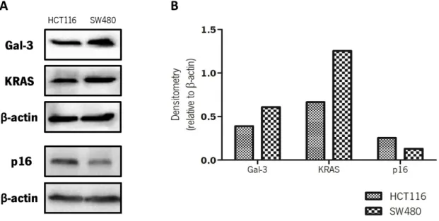
3.1. Basal expression levels of galectin-3, p16 and KRAS in colorectal cancer cells ... 55
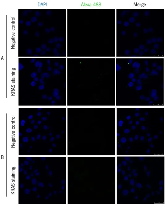
3.2. Galectin-3, KRAS and p16 localization patterns in normal colon and colorectal cancer cells …….………..56
3.2.1. Galectin-3, KRAS and p16 immunostaining in colorectal cancer cells ... 56
3.2.2. Galectin-3 and p16 double immunostaining in colorectal cancer cells ... 61
3.2.3. Galectin-3, KRAS and p16 immunostaining in NCM460-transfected cells ... 66
3.3. Study of galectin-3, KRAS and p16 interaction by co-immunoprecipitation in SW480 cells………71
3.4. Optimization of galectin-3 silencing conditions by RNA interference ... 73
3.5. Phenotypic alterations induced by galectin-3 and/or KRAS silencing in SW480 cells………73
3.5.1. Effect of galectin-3 and/or KRAS silencing on cell morphology ... 74
3.5.2. Effect of galectin-3 and/or KRAS silencing on cell viability ... 76
3.5.3. Effect of galectin-3 and/or KRAS silencing on galectin-3, KRAS and p16 expression levels…….……….77
3.5.4. Effect of galectin-3 and/or KRAS silencing on MMPs production/activity ... 78
3.5.5. Effect of galectin-3 and/or KRAS silencing on cell migration ... 79
IV. Discussion ... 83
V. Concluding remarks and Future perspectives ... 93
VI. References ... 99
VII. Supplementary data ... 111
LIST OF FIGURES Fig. 1.1. The hallmarks and emerging characteristics of cancer………..18
Fig. 1.2. A genetic model for colorectal carcinogenesis………...19
x
Fig. 1.6. Intracellular ligands and functions of Gal-3………..29 Fig. 1.7. Gal-3 contributes to tumourigenesis and tumour progression through several different mechanisms………32 Fig. 1.8. Cell cycle regulation by p16………35 Fig. 1.9. Rationale of the project………39 Fig.3.1. Expression pattern of Gal-3, KRAS and p16 levels in CRC-derived cells HCT116 and SW480………...55 Fig. 3.2. Confocal fluorescence microscopy analysis of KRAS localization shows no positive staining………..…57 Fig. 3.3. Confocal fluorescence microscopy analysis of Gal-3 staining in SW480 cells evidences differences between Gal-3 localization in low and high confluent cells………….……….58 Fig. 3.4. Confocal fluorescence microscopy analysis of Gal-3 staining in HCT116 cells evidences a similar Gal-3 localization pattern in low and high confluent cells.……….59 Fig. 3.5. Confocal fluorescence microscopy analysis of p16 staining in SW480 cells evidences differences between Gal-3 localization in low and high confluent cells……….……..60 Fig. 3.6. Confocal fluorescence microscopy analysis of p16 staining in HCT116 cells evidences a similar p16 localization pattern in low and high confluent cells………..…..61 Fig. 3.7. Confocal fluorescence microscopy analysis of Gal3+p16 double immunostaining secondary antibodies control conditions in SW480 (A) and HCT116 cells (B)………63 Fig. 3.8. Confocal fluorescence microscopy analysis of Gal-3+p16 double immunostaining in low and high confluent SW480 cells reveals the presence of some discreet yellow dots indicative of close proximity and co-localization of these proteins ………64 Fig. 3.9. Confocal fluorescence microscopy analysis of Gal-3+p16 double immunostaining in low and high confluent HCT116 cells reveals the presence of some discreet yellow dots mainly localized at the cells nuclei and suggestive of co-localization……….65 Fig. 3.10. Representative western blot analysis of Gal-3, endogenous KRAS, Flag-KRAS and p16 basal expression levels in NCM460 parental (P) cell line and NCM460 transfected with Flag-KRASwt, Flag-KRASG12V, Flag-KRASG12D and Flag-KRASG13D……….…66 Fig. 3.11. Confocal fluorescence microscopy analysis of Flag-KRAS and Gal-3 double immunostaining control conditions in NCM460 Flag-KRAS wt (A) and staining positive conditions in NCM460 Flag-KRAS wt, G12V, G12D and G13D (B)……….67 Fig. 3.12. Confocal fluorescence microscopy analysis of Gal-3 and p16 double immunostaining control conditions in NCM460 KRAS wt (A) and staining positive conditions in NCM460 Flag-KRAS wt, G12V, G12D and G13D (B)………..….68 Fig. 3.13. Confocal fluorescence microscopy analysis of Flag-KRAS and p16 double immunostaining control conditions in NCM460 Flag-KRAS wt (A) and staining positive conditions in NCM460 Flag-KRAS wt, G12V, G12D and G13D (B)……….69 Fig. 3.14. Gal-3, KRAS and p16 co-immunoprecipitate ………...72
xi
Fig. 3.16. Gal-3 and/or KRAS silencing induce morphology chances in SW480 cells………75 Fig. 3.17. Gal-3 and/or KRAS silencing decrease cell viability of SW480 cells………...76 Fig. 3.18. Gal-3 silencing induces an increase in KRAS expression levels whereas KRAS silencing slightly decrease Gal-3 expression………..77 Fig. 3.19. KRAS silencing and Gal-3/KRAS double silencing led to a decrease in Pro-MMP-9, MMP-9 and Pro-MMP-2 levels ……….79 Fig. 3.20. Gal-3 and/or KRAS silencing have no effect on colon cancer SW480 cells migration.81 Fig. 4.1. Results outline of this project. ……….91 Fig. S1. Confocal fluorescence analysis of Gal-3 and p16 negative staining controls in high confluent SW480 and HCT116 cells………..113 Fig. S2. Representative western blot showing similar Gal-3 expression independently of the cells confluence and nutritional availability ………114 Fig. S3. Analysis of Gal-3 content in conditioned media (CM) from HCT116 and SW480 cells.……….114 LIST OF TABLES
Table. 1.1. Evidences of Gal-3-KRAS interaction in cancer cell models………..33 Table. 2.1. Primary and secondary antibodies used in this study and their respective dilutions……….45 Table. 2.2. Primary and secondary-conjugated antibodies and the respective dilutions used in immunofluorescence, with reference to cell lines and fixation methods……….48 Table. 3.1. Localization patterns of Gal-3 and p16 in SW480 and HCT116 cells and Gal-3, p16 and (Flag-)KRAS in NCM460 cells transfected with KRASwt and the three KRAS hotspot mutations………..71 LIST OF ABBREVIATIONS
AKT V-AKT murine thymoma viral oncogene
APC Adenomatous polyposis coli
BRAF V-RAF murine sarcoma viral oncogene homolog B
BSA Bovine serum albumin
CDK Cyclin dependent kinase
xii
Co-IP Co-immunoprecipitation
COX-2 Cyclooxygenase 2
CRC Colorectal cancer
DCC Deleted in colorectal carcinoma
EGF Epidermal growth factor
EGRF Epidermal growth factor receptor ERK Extracellular regulated MAP kinase
FAP Familial adenomatous polyposis
FBS Fetal bovine serum
Gal-1 Galectin-1
Gal-3 Galectin-3
GAPs GTpase activating proteins
GDP Guanosine diphosphate
GEFs Guanine nucleotide exchange factors
GTP Guanosine triphosphate
GTPase Guanosine triphosphatase HBSS Hank´s balanced salt solution
HNPCC Hereditary non polyposis colorectal cancer HRAS Harvey rat sarcoma virus oncogene
IF Immunofluorescence
IgG Immunoglobulin G
IP Immunoprecipitation
KRAS Kirsten rat sarcoma viral oncogene MAPK Mitogen activated protein kinase
MEK Mitogen associated extracellular signal-regulated kinase MGMT O6-methylguanine-methyltransferase
MLH1 MutL homolog 1
MMP Matrix metalloproteinase
MRR Mismatch repair
MSI Microsatellite unstable
MSS Microsatellite stable
xiii
PBS Phosphate buffered saline
PFA Paraformaldehyde
PI Propidium iodide
pRB Retinoblastoma protein
PVDF Polyvinylidene difluoride
RAF Rapidly accelerated fibrosarcoma kinase
RAS Rat sarcoma
RIPA Buffer Radioimmunoprecipitation assay buffer
RNA Ribonucleic acid
RNAi RNA interference
Rpm Revolutions per minute
RPMI Roswell Park Memorial Institute medium
RT Room temperature
RTK Receptor tyrosine Kinase
SDS Sodium dodecyl sulfate
shRNA Short hairpin RNA
siRNA Small interference RNA
I.
I
NTRODUCTION
17 1.1. Cancer: the global burden
According to the World Health Organization (WHO), cancer is defined as “the uncontrolled growth and spread of cells. It can affect almost any part of the body. The growths often invade surrounding tissue and can metastasize to distant sites” (WHO 2014). In 2012 cancer accounted for 14.1 million new cases and 8.2 million deaths worldwide. Predictions point to an increase of 19.3 million new cancer cases by 2025, due to growth and ageing of the global world population (GLOBOCAN, 2012).
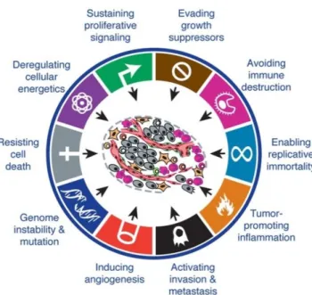
Carcinogenesis is a multistep process with each step displaying dynamic changes in the genome. Such changes are translated by mutations in oncogenes with dominant gain of function, and tumour suppressor genes with recessive loss of function, which progressively transform normal cells into malignant derivatives. Through those steps, cells acquire a succession of capabilities that were initially referred as the “hallmarks of cancer” and were defined by the authors as “six essential alterations in cell physiology that collectively dictate malignant growth” (Hanahan & Weinberg 2000). Those six hallmarks were proposed as: self-sufficiency in growth signals, insensitivity to growth-inhibitory signals, evasion of programmed cell death, limitless replicative potential, sustained angiogenesis, and tissue invasion and metastasis (Hanahan & Weinberg 2000). Meanwhile, the past decade witnessed remarkable progresses in cancer biology that led to the update of the hallmarks list (Fig.1.1), adding two more: the capability to modify or reprogram the cellular metabolism and the ability of cancer cells to evade the immunological destruction. Moreover, two consequential characteristics that facilitate acquisition of both core and emerging hallmarks were mentioned: the genomic instability and mutability and the inflammation by innate immune cells (Hanahan & Weinberg 2011). Moreover, Floor et al. 2012, referred to another important characteristic - the loss of differentiation.
Deregulation of signalling pathways is very important to acquire these hallmark capabilities, and the interconnections and crosstalk between the individual sub-circuits can make a certain oncogenic event to affect multiple capabilities (Hanahan & Weinberg 2011).
The reductionist view that a tumour is nothing more than a collection of relatively homogeneous cancer cells has been put aside and tumours have increasingly been recognized as complex organs with specialized cell types and a tumour microenvironment constructed during the tumourigenesis process (Hanahan & Weinberg 2011). Therefore, this dynamic, complex and intra/inter-tumour heterogeneity contribute to explain the difficulty of treatment and the need for multi-target therapies. Despite the increasing accumulation of knowledge about
18
carcinogenesis and the successful advances in treatment strategies, scientific research must continue (Duffy 2013).
1.1.1. Colorectal carcinogenesis
Colorectal cancer is the third most common cancer in men and the second in women, being the third most frequently diagnosed worldwide. About 694 000 deaths from CRC are estimated worldwide, making it the fourth most common cause of cancer mortality. In Portugal, this is the second most common type of cancer in men and women, being responsible for the highest number of cancer mortality (GLOBOCAN, 2012). Hereditary predisposition, age and lifestyle factors, namely fat and alcohol regular consumption, physical inactivity, obesity and tobacco smoking represent risk factors for CRC development (Haggar & Boushey 2009).
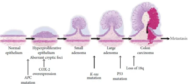
The first model of colorectal carcinogenesis was suggested in 1990 by Fearon and Vogelstein (Fearon & Vogelstein 1990) as a sequential pathway in which normal epithelium becomes hyperproliferative evolving to adenoma and ultimately to carcinoma. This pathway essentially requires mutational events in oncogenes, such as KRAS, and in tumour suppressor genes, such as APC, DCC and p53. Additional events may eventually lead to metastasis (Fig.1.2).
19
CRC can be subdivided in sporadic, which results from a stepwise accumulation of genetic and epigenetic alterations, or hereditary/familial, which arises from germline mutations in genes involved in colorectal carcinogenesis. Hereditary CRC accounts for less than 5% of all cases and among all the identified syndromes the most common are Familial Adenomatous Polyposis (FAP) and Lynch syndrome (also called Hereditary Non Polyposis Colorectal Cancer- HNPCC) (Al-Sohaily et al. 2012; Armaghany et al. 2012).
Currently, genomic instability is known to play an important role in colorectal carcinogenesis. Accordingly, three not mutually exclusive events are considered to cause this instability: (1) chromosomal instability (CIN), (2) microsatellite instability (MSI) and (3) CpG island methylator phenotype (CIMP) (Al-Sohaily et al. 2012; Armaghany et al. 2012). CIN, also called adenoma-carcinoma sequence, is the most common cause of genomic instability. It is characterized by karyotypic abnormalities and the accumulation of mutations that led to the activation of proto-oncogenes (e.g.: KRAS) and inactivation of tumour suppressor genes (e.g.: APC and p53). MSI results from a defective mismatch repair (MMR) system that leaves microsatellites either longer or shorter than usual. According to defined markers, MSI is classified as microsatellite high (MSI-H), microsatellite low (MSI-L) or microsatellite stable (MSS). Inactivation of MMR enzymes can result either from abnormal methylation of promoter CpG islands or from point mutations in MMR family members. Germline mutations in MMR genes are in the origin of HNPCC (Al-Sohaily et al. 2012; Armaghany et al. 2012). Aberrant epigenetic regulation via inappropriate methylation of gene promoter regions is common in CRC and is as
Fig. 1.2. A genetic model for colorectal carcinogenesis. Mutations in APC, activation of KRAS and inactivation of p53 are essential events in this carcinogenic process (Adapted from Sandouk et al. 2013).
20
significant as DNA mutation in the inactivation of tumour suppressor genes. CIMP is a result of aberrant hypermethylation that takes place in repetitive CG dinucleotides or CpG-rich stretches of DNA in a given gene, such as MLH1, promoter region. As well as MSI, CIMP has also been classified in levels (e.g.: CIMP-low, CIMP-high) (Armaghany et al. 2012).
In addition to the traditional carcinogenesis adenoma-carcinoma sequence, some CRCs are believed to arise from lesions called serrated polyps, establishing the serrated pathway. Therefore, based on the molecular and pathological profile, Jass (2007) categorized 5 subtypes of CRC:
(1) CIMP-high, methylation of MLH1, BRAF mutation, chromosomally stable, MSI-H, origin in serrated polyps, known generally as sporadic MSI-H (accounts for 12% of CRCs);
(2) CIMP-high, partial methylation of MLH1, BRAF mutation, chromosomally stable, MSS or MSI-L, origin in serrated polyps (accounts for 8% of CRCs);
(3) CIMP-low, KRAS mutation, MGMT methylation, CIN, MSS or MSI-L, origin in adenomas or serrated polyps (accounts for 20% of CRCs);
(4) CIMP-negative, CIN, mainly MSS, origin in adenomas (sporadic or hereditary) (accounts for 57%).
(5) Lynch syndrome, CIMP-negative, BRAF mutation negative, chromosomally stable, MSI-H, origin in adenomas (accounts for 3%) (also referred as familial MSI-H CRC).
1.2. The RAS family: small proteins, high significance
RAS proteins belong to the RAS superfamily of small GTPases, which is divided into at least 5 subfamilies: the RAS, Rho/Rac, Rab, Arf and Ran (Castellano & Santos 2011). Mammalian RAS proteins have a molecular weight of approximately 21 kDa and are codified by 3 similar genes Harvey-RAS (HRAS), Neuroblastome-RAS (NRAS) and Kirsten-RAS (KRAS). KRAS gene originates two alternative spliced isoforms, A and B, being the latter an ubiquitously expressed form (Vögler et al. 2008) and hereafter designated as KRAS. As a result of their oncogenic role, these genes started to be identified more than 40 years ago and since then information has been accumulating, proving their relevance on signalling transduction and on molecular oncology (Malumbres & Barbacid 2003).
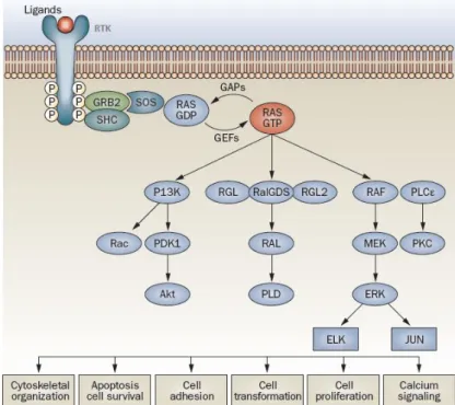
RAS proteins are regulated by guanine nucleotide exchange factors (GEFs) which activate the system by inducing GDP dissociation and GTP binding; and by GTPase activating proteins (GAPs) that inversely regulate the system (Fig.1.3). This binary behaviour enables RAS proteins to
21
function as molecular switches in a broad range of signalling transduction processes, from extracellular signals, for example soluble epidermal growth factor (EGF), to the interior of the cells. Importantly, the high number of RAS activators and effectors identified in mammalian cells, places these proteins at the crossroads of various cellular signalling networks, such us RAF/MEK/ERK, PI3K/AKT and RalGDS pathways. Consequently, they affect a wide range of cellular responses that influence cell fate, including proliferation, differentiation and apoptosis (Fig.1.3) (Normanno et al. 2009; Castellano & Santos 2011).
The structure of these small monomeric G proteins can be divided into three domains (Vögler et al. 2008):
(1) The G domain that is approximately 95% conserved among the different isoforms. This region of the protein binds guanine nucleotides, contains the switch 1 and 2 loops, which undergo major conformational changes on GTP–GDP exchange and are also the binding surfaces for effectors, exchange factors, and GAPs;
Fig. 1.3. RAS mediated intracellular signal transduction pathways. Ligand binding to the extracellular
domain of receptor tyrosine kinases (RTKs) result in activation and initiation of signalling cascades. Activated receptor complexes contain adaptors such as SHC and GRB2 that recruit proteins like SOS1, increasing RAS–GTP levels by catalyzing nucleotide exchange on RAS. GTP-bound RAS activates different signalling pathways including: RAF/MEK/ERK cascade, that regulate cell proliferation and motility via the transcription factors JUN and ELK; PI3K, which via AKT controls a number of downstream effectors responsible for anti-apoptotic responses and via Rac, regulates the actin cytoskeleton dynamics, being important in RAS-mediated transformation; RalGDS, RGL and RGL2, which activate PLD, resulting in control of cell-cycle progression and in RAS-dependent transformation; and PLCε, which regulates calcium release and PKC activation. To terminate this signal transduction, GAPs (eg. neurofibromin- NF1) bind to RAS–GTP and accelerate its conversion to RAS–GDP (Adapted from Normanno et al. 2009).
22
(2) The poorly conserved C-terminal HVR (hypervariable region; less than 15% of sequence identity) domain that undergoes direct post-translational processing, being essential for plasma membrane anchoring as well as for the trafficking of newly synthesized and processed RAS, from the cytosolic surface of the endoplasmic reticulum to the inner surface of the plasma membrane. It also comprises the linker region that connects the anchor sequence with the N-terminal G-domain;
(3) The C-terminal CAAX (C=cysteine, A=aliphatic, X=amino acid) motif that is post-translationally processed to generate an S-farnesyl cysteine carboxymethyl ester. The membrane anchor is completed by one (NRAS) or two (HRAS) proximal S-palmitoylable cysteine residues or a polybasic domain of six lysine residues in KRAS.
1.2.1. RAS signalling output: localization matters
Besides being mainly found at the inner surface of the plasma membrane, these proteins can also be found in other intracellular membranes such as endosomes, endoplasmic reticulum/Golgi and mitochondria, where they can generate signal output as well (Omerovic & Prior 2009). RAS interaction with the plasma membrane is highly dynamic and is located in specific microdomains (sites within the plasma membrane that have a distinct lipid and/or protein composition) (Hancock 2003). One of the best characterized microdomains are the lipid rafts, which consist of dynamic assemblies of cholesterol and sphingolipids at the exoplasmic leaflet of the bilayer. Additionally, caveolae is one subset of lipid rafts found in cell surface invaginations, formed by polymerization of caveolins- hairpin-like palmitoylated integral membrane proteins- that tightly bind cholesterol (Simons & Toomre 2000). As there are different C-terminal lipid anchors on N, H and KRAS isoforms - a farnesyl group with one, two palmitate groups or a polybasic domain, respectively - it would therefore be expected that they sort or target RAS proteins to different membrane microdomains (Hancock 2003). In fact, the HRAS membrane anchor targets this protein to lipid rafts and caveolae but, in contrast, the membrane anchor of KRAS predominantly (85% of the proteins) targets it to non-raft plasma membrane. Notably, HRAS is in dynamic equilibrium between lipid rafts and non-rafts sites of the plasma membrane: when GDP-loaded, a percentage of HRAS localizes at lipid rafts, however, GTP-loading redistributes HRAS from rafts to non-raft sites. Although localization in the rafts is necessary for signalling, this release from rafts is essential for HRAS efficient activation of RAF (Prior et al. 2001). Additionally, many studies have demonstrated that activated HRAS and KRAS
23
operate in non-overlapping, non-raft microdomains of the plasma membrane (Hancock 2003). This differential lateral segregation of RAS proteins within the plasma membrane can be a plausible explanation for distinct signal outputs generated by these highly homologous proteins. Furthermore, the C-terminal membrane anchor was shown to regulate RAS signal output (Hancock 2003).
Additionally, at the plasma membrane, approximately 40% of activated RAS proteins form nanoclusters, whereas the remainder is distributed as monomers. These nanoclusters display some specific features: KRAS nanoclusters are actin-dependent and cholesterol-independent, and are stabilized by the scaffold protein galectin-3 (Gal-3), whereas HRAS-GDP nanoclusters are cholesterol and dependent but, when activated (GTP loaded), are cholesterol and actin-independent and are stabilized by galectin-1 (Gal-1) (Abankwa et al. 2007; Omerovic & Prior 2009). Importantly, using KRASG12V it was shown that only nanoclustered proteins can recruit downstream signalling effectors and transduce signals with high-fidelity (Tian et al. 2007). In addition, RAF is recruited to and retained in KRAS nanoclusters but, in turn, it is not retained in HRAS nanoclusters. Similarly, upon epidermal growth factor receptor (EGFR) activation, RAF is preferentially recruited to KRAS and not to HRAS nanoclusters (Plowman et al. 2008).
RAS interactions with Gal-1 and Gal-3 act as regulators of signal duration and effector usage. Gal-1 binds active HRAS-GTP and active KRAS-GTP (preferentially and in a higher extent with the former) and Gal-3 only binds KRAS-GTP (Elad-Sfadia et al. 2002; Elad-Sfadia et al. 2004), all in a RAS farnesyl moiety-dependent manner (Paz et al. 2001; Elad-Sfadia et al. 2004). These interactions between RAS and galectins stabilize RAS in the active state (reducing the efficiency of GAP-mediated GTP hydrolysis) and promote its association with distinctive effectors (Elad-Sfadia et al. 2002; Elad-Sfadia et al. 2004). Thus, when HRAS-GTP interacts with Gal-1, it promotes activation of RAF but not of PI3K (Elad-Sfadia et al. 2002). In contrast, when KRAS-GTP interacts with Gal-3, it promotes activation of RAF and PI3K (Elad-Sfadia et al. 2004).
In summary, despite the similarity of RAS isoforms, they display some functional and biological specificity. This is most likely a result of their intrinsically different biological potency and of their different spatiotemporal and subcellular compartmentalization. The combination of these features with the different cellular contexts in which each isoform is expressed, dictates their distinct signalling and consequent output (Omerovic & Prior 2009; Castellano & Santos 2011).
24
Fig.1.4. RAS isoform-specific codon mutations. (A) RAS oncogenes are frequently mutated at codons 12 (in purple), 13 (in blue) and 61 (in green) and the incidence of mutation differs between the isoforms. (B) Isoforms – specific mutation patterns are associated with particular cancer types (Adapted from Prior et al. 2012).
1.2.2. RAS mutations and cancer
The central role of RAS gene products in normal cell signalling is consistent with the high frequency of oncogenic activation of RAS genes in human cancers (Castellano & Santos 2011). RAS genes exhibit a pattern of isoform-specific codon and point mutations, which display varying incidences in different cancers and presents a specific association with particular cancers types (Fig.1.4). Typically, single mutations occur at codons 12, 13 or 61 with different frequencies in each isoform (Prior et al. 2012). These mutations favour GTP binding, leading to constitutive activation of RAS, meaning that the protein no longer requires ligand for activation. This constitutive activation deregulates RAS target signalling pathways leading to malignant transformation (Mccubrey et al. 2007).
KRAS is the most frequently mutated isoform in most cancers, having a higher occurrence in pancreatic cancer, where 90% of the tumours harbour KRAS mutations. Its patterns of mutation are dominated by G→A transitions at the second base of codons 12 or 13, resulting in G12D or G13D mutations; G→T transversions at the second base of codon 12, producing G12V, and a special case in lung cancer where G→T transversion at the first base of codon 12 produce G12C mutations (Prior et al. 2012).
Conclusively, one might speculate that this high frequency of RAS proto-oncogenes activation in human cancers makes RAS and their signalling pathways attractive targets for cancer therapy.
25
1.2.2.1. KRAS mutations in colorectal cancer: role and significance
Back in 1990, the significance of KRAS mutations in CRC had already been recognized as an initial event in colorectal carcinogenesis (Fearon & Vogelstein 1990). Indeed, KRAS mutations are very frequent in CRC, occurring in approximately 30-50% of the cases, being considered an important factor to be taken into account in therapy-related decisions (Arrington et al. 2012).
The RASCAL (Kirsten-RAS in colorectal cancer collaborative group) carried out the biggest studies about the impact of KRAS in CRC, reaching some important conclusions: KRAS mutations are associated with CRC development and progression, and their presence increase the risk of death; followed by codon 13, codon 12 is the most frequently mutated, being the change of glycine (G) to aspartate (D) the more frequent alteration. Additionally, also in codon 12, the mutation that results in the change of glycine (G) to valine (V) is associated with more aggressive and advanced cancers and represents an increased risk of tumour relapse and death (Andreyev et al. 1998; Andreyev et al. 2001). In sporadic MSS CRC, KRAS mutations have been related to more aggressive tumours and it has been suggested as a critical gene in the metastization process, increasing the ability to invade (Oliveira et al. 2007). In fact, primary CRC harbouring KRAS mutations is frequently associated with distant metastasis in liver and lungs, which are the main cause of death in CRC patients (Cejas et al. 2009; Nash et al. 2010). In MSI CRC, KRAS is less mutated than in MSS and mutations in this gene are inversely associated and mutually exclusive to the oncogenic BRAFV600E mutation, more frequent in this type of cancer (Rajagopalan et al. 2002; Oliveira et al. 2003).
Additionally, the epidermal growth factor receptor (EGFR) is frequently overexpressed in CRC and its simultaneous presence with mutated KRAS, a downstream effector of EGFR-induced signalling cascade, has been identified as a potent predictor of anti-EGFR therapies resistance, namely to cetuximab and panitumumab (Vectibix) (Heinemann et al. 2009; Normanno et al. 2009). Recently, meta-analysis studies have suggested that among the patients with KRAS mutations, the ones that harbour the mutation on codon 13 are more sensitive and may benefit more from the anti-EGFR therapy with cetuximab (Chen et al. 2013a ; Mao et al. 2013). Hence, KRAS and EGFR mutation status should be evaluated before the therapeutic decisions are considered (Heinemann et al. 2009; Normanno et al. 2009).
In conclusion, the presence of KRAS mutations in CRC seems to be a bad prognostic biomarker with a relevant impact on therapy.
26
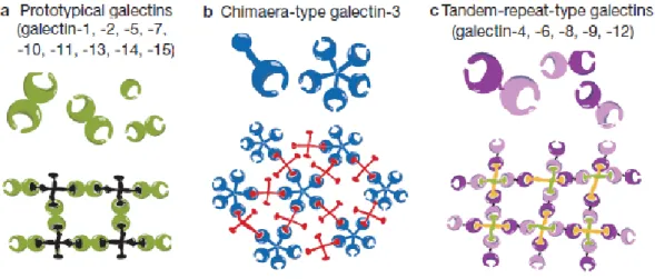
Fig. 1.5. Galectin family division into 3 groups: (a) prototypical galectins, (b) chimaera-type Gal-3 and (c)
tandem-repeat-type galectins. As galectins are bivalent or multivalent, they can form lattices with multivalent glycoconjugates (Adapted from Yang et al. 2008).
1.3. The galectin family
Galectins are members of a larger family of β-galactoside-binding lectins that before being named as galectins in 1994, were referred as S-type or S-lac lectins. The author that suggested the terminology defined that membership to galectin family requires the fulfilment of two essential criteria: (1) affinity for β-galactosides and (2) a significant sequence similarity in the carbohydrate-binding site (Barondes et al. 1994). These lectins are evolutionary ancient being expressed and conserved from vertebrates to invertebrates and protists. Their presence in so many species through evolution suggests that they may have evolved to play fundamental roles in cell biology (Cooper 2002).
The 15 identified mammalian galectins all contain conserved carbohydrate-recognition domains (CRDs) of about 130 amino acids, which served as a criteria for their classification into: prototypical galectins with one CRD (galectin-1, -2, -5, -7, -10, -11, -13, -14 and -15); the chimaera-type Gal-3 that contains a non-lectin N-terminal region (of about 120 amino acids) connected to a CRD; and the tandem-repeat-type galectins that contain two homologous CRDs in a single polypeptide chain, separated by a linker of up to 70 amino acids (galectin-4, -6, -8, -9 and -12) (Fig.1.5). Galectins have sugar-binding specificity and with regard to their binding activity, they are bivalent or multivalent having the ability to form lattice-like structures (Yang et al. 2008).
In the extracellular space, galectins can modulate the interactions between matrix components and cell-surface integrins, regulating cell-cell and cell-matrix adhesion. These
27
interactions are extremely important in cellular motility, polarity and tissue formation, and their loss is associated with some diseases, such as inflammation and tumour progression (Hughes 2001). Also, due to their multivalency, they can crosslink cell-surface glycoconjugates and trigger signalling events that can modulate cell behaviour (Liu & Rabinovich 2005). Intracellularly, by interacting with some specific ligands, galectins play a role in the regulation of essential cell processes like growth, apoptosis and cell cycle regulation (Liu et al. 2002).
1.3.1. Galectin-3: a pleiotropic lectin
Firstly identified in 1982 as a 32kDa antigen (Muc-2) on the surface of mouse thioglycollate-elicited macrophages (Ho & Springer 1982), over the years Gal-3 was isolated from many other sources and received many designations, until the introduction of the “galectin” nomenclature. Among the galectins family, Gal-3 is the most studied member (Dumic et al. 2006).
Structurally Gal-3 is composed by two distinct conserved domains: the N-terminal domain (ND), responsible for multimerization, consists of a 110-130 amino acids sequence with 7-14 tandem repeats of Pro-Gly-Ala-Tyr-Pro-Gly followed by three additional amino acids, and the 130 amino acids C-terminal CDR, that accommodates the carbohydrate-binding site (Dumic et al. 2006). These two domains are necessary for Gal-3 functionality: the ND contributes to carbohydrate-binding (Barboni et al. 2000) and the C-terminal is involved in Gal-3 self-association (depending on whether it is bound to saccharides or not) (Yang et al. 1998). The ND is also necessary for Gal-3 subcellular localization (Gong et al. 1999) and is susceptible to cleavage by metalloproteinases (MMPs; particularly MMP-2 and MMP-9), which play an important function in several physiological processes such as extracellular matrix degradation and tissue remodeling (Ochieng et al. 1998). Its Ser6 residue phosphorylation was demonstrated to play key roles in Gal-3 ligand binding, acting like the “on/off” switch of some downstream biological effects, including its anti-apoptotic and anti-anoikis (apoptosis upon loss of anchorage) activities (Mazurek et al. 2000; Yoshii et al. 2002). The former has been attributed to the C-terminal NWGR (Asn-Trp-Gly-Arg) motif, which is similar to the BH1 domain of the Bcl-2 family proteins (Akahani et al. 1997). Gal-3 is one of the members of the family that exhibits a dual localization, intra and extracellular, but lacks a transport signal sequence, thus its mechanism of externalization does not involve the classical endoplasmic reticulum-Golgi complex pathway (Hughes 1999). Meanwhile, Gal-3 was found in dendritic cells’ secreted exosomes (Théry et al. 2001), and has
28
been shown to interact with membrane phospholipids and cholesterol, having the propensity to rapidly penetrate and traverse the lipid bilayer in either direction (Lukyanov et al. 2005). In the intracellular space, Gal-3 shuttles between the nucleus and the cytoplasm, being exclusively/mainly in one compartment or distributed by both, depending for example on cell type or specific in vitro experimental conditions, and many studies have been carried out in search for import and export signals (Haudek et al. 2010). Gal-3 can migrate into the nucleus by distinct pathways: passive diffusion, ND-dependent active transport system (Nakahara et al. 2006a), or it can be imported by the importin (karyopherin) α/β complex, as it has a nuclear localization signal (NLS)-like sequence in the C-terminal (Nakahara et al. 2006b). Recently, it was elucidated that nucleoporin Nup98 mediates Gal-3 nuclear export by interacting with its C-terminal domain (Funasaka et al. 2013).
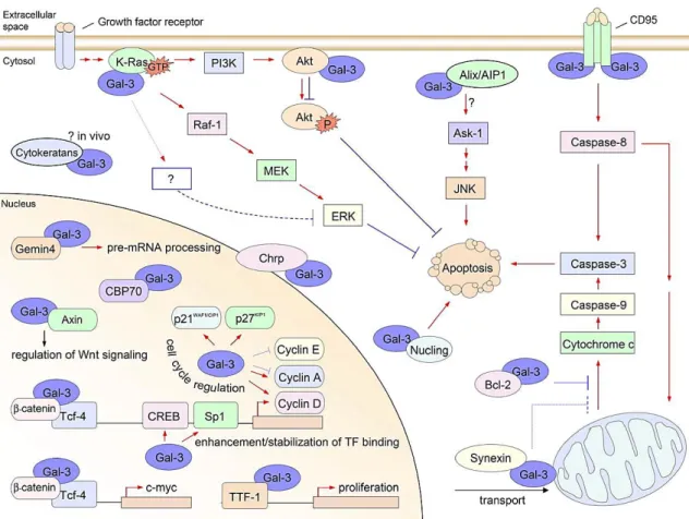
Numerous intracellular molecules have been identified as Gal-3 ligands (Fig.1.6) and the interaction with most of them is established through protein-protein interactions instead of protein-carbohydrate recognitions (Haudek et al. 2010). In the nucleus, Gal-3 plays a part in spliceossome assembly by interacting with Gemin4 (Park et al. 2001), participates in the regulation of transcription by interacting with transcription factors like the thyroid transcription factor 1 (TTF-1) (Paron et al. 2003), or by enhancing/stabilizing transcription factor binding to CRE and possibly SP1 sites, which are often found at the promoter region of many genes, including cyclin D1 (Lin et al. 2002). In the cytoplasm Gal-3 interacts, among others, with Bcl-2, functioning in its pathway (Yang et al. 1996); with synexin, which mediates its translocation to the perinuclear mitochondrial membranes, where it regulates mitochondrial integrity (Yu et al. 2002); and with nucling, which downregulates Gal-3 and consequently mediates apoptosis (Liu et al. 2004). Furthermore, Gal-3 also can exert its anti-apoptotic activity by modulating mitochondrial homeostasis, in particular by reducing intracellular ROS generation and influencing the mitochondrial membrane potential (Matarrese et al. 2000). Hence, intracellular Gal-3 is an active participant and regulator of essential cell processes namely proliferation/cell cycle regulation, survival and death.
Gal-3 biological roles are also regulated by its subcellular localization, for example: in contrast with the intracellular anti-apoptotic activity, when extracellular, Gal-3 has an opposite effect, acting as a pro-apoptotic factor, what has been demonstrated in human thymocytes and T cells (Stillman et al. 2006). Extracellularly, Gal-3 is found on cell surfaces, in the extracellular
29
Fig. 1.6. Intracellular ligands and functions of Gal-3. Gal-3 plays important roles in cell biology, such as in:
pre-mRNA splicing, transcription regulation, proliferation and regulation of cell cycle and apoptosis. (Red arrows indicate positive effects, blue lines indicate negative effects) (Adapted from Dumic et al. 2006).
matrix, in biological fluids and sera, as well as in culture media of certain cell lines, exhibiting several autocrine and paracrine effects (Dumic et al. 2006).
As a result of its multivalent properties and the ability to bind cell surface glycoproteins and glycosylatedcomponents, like laminin, fibronectin, hensin, elastin, collagen IV and tenascin-C and -R, some integrins and lipopolysaccharides, Gal-3 participates in cell-cell, cell-matrix and also
cell-pathogen interactions. These interactions translate into the modulation of cell adhesion, cell activation, receptor turnover and endocytosis, interfering with important biological processes, namely maintenance of cellular homeostasis, immune reactions, organogenesis and angiogenesis, tumour cell invasion and metastasis (Ochieng et al. 2004; Rabinovich et al. 2007). Regarding its membrane localization, in migrating dendritic cells, this lectin was found localized in membrane lipid rafts (in a lectin-carbohydrate interaction independent way), arising the suggestion that it is necessary for the formation of more complex ruffle structures, and possibly regulates cell migration at least in part by regulating the cell membrane architecture (Hsu et al. 2009).
30
As far as the immunoregulatory properties of this lectin are concerned, it acts as a chemoattractant for immune cells, namely monocytes, macrophages as well as neutrophils (Sano et al. 2000), participates in many T-cell mediated inflammatory processes, infections and autoimmune diseases (e.g.: diabetes mellitus and multiple sclerosis), acting in general as pro-inflammatory, and in innate anti-tumour immunity favouring tumour progression (Radosavljevic et al. 2012).
In summary, Gal-3 is a multifaceted protein: it displays pleiotropic effects that are dependent on its localization and interaction with its extensive list of ligands, being involved in normal cell function but also in pathological processes.
1.3.1.1. Galectin-3 in cancer: roles and KRAS interaction
Gal-3 altered expression has been connected to the progression of a variety of cancers, such as prostate (Knapp et al. 2013), thyroid (Chiu et al. 2010), breast (Zhang et al. 2014), colorectal (Endo et al. 2005; Povegliano et al. 2011), ovarian (Kim et al. 2011), hepatocellular (Jiang et al. 2014) and melanoma (Brown et al. 2012), being in some cases considered a potent diagnostic and prognostic biomarker and therefore a good therapeutic target.
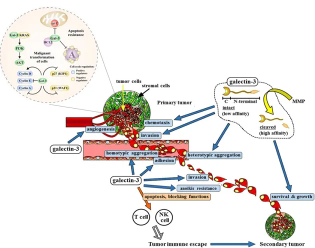
Notably, Gal-3 is an active player in the carcinogenesis process from transformation to metastization (Fig. 1.7). Direct evidence has shown that Gal-3 plays a pivotal role in malignant transformation: inhibition of Gal-3 in breast cancer cells led to the reversion of the transformed phenotype both in vivo and in vitro (Honjo et al. 2001), and the introduction of Gal-3 cDNA into normal thyroid follicular cells conferred them a malignant phenotype (Takenaka et al. 2003). Additionally, Gal-3 participates in every step of the metastatic cascade including in angiogenesis, dissemination through the blood flow, tumour embolism formation and extravasation (Funasaka et al. 2014).
A list of evidences has been highlighting Gal-3 angiogenic role through different mechanisms (Funasaka et al. 2014). For example, Gal-3 seems to be connected to vascular endothelial factor (VEGF) and basic fibroblast growth factor-mediated angiogenesis by its carbohydrate-mediated interaction with αvβ3 integrin, which leads to subsequent activation of signalling pathways that promote the formation of new blood vessels (Markowska et al. 2010). Likewise, increased circulating Gal-3 is capable of induce secretion of metastasis promoting cytokines such as interleukin-6 and colony-stimulating factor from the blood vascular endothelial cells (in vitro and in vivo) that will enhance the expression of endothelial cell surface adhesion
31
molecules resulting in increased association between cancer and endothelial cells leading to increased migration and tubule formation (Chen et al. 2013b). Moreover, it has been recently demonstrated that Gal-3 produced by the tumour can induce M2-like macrophages migration and thereby, indirectly promoting angiogenesis and tumour growth (Jia et al. 2013). In addition, this lectin is required for stabilization of the epithelial-endothelial interactions, with proteolytically cleaved Gal-3 displaying a 20-fold higher affinity for endothelial cells when compared to the full-length protein (Shekhar et al. 2004). This ND-cleaved form occurs in vivo and has been demonstrated to play an important role in angiogenesis: tumour cells expressing non-cleavable Gal-3, showed reduced growth and angiogenesis (Nangia-Makker et al. 2007). In the same way, cells expressing an MMP-2/-9 cleavable-Gal-3 form showed increased endothelial cells chemotaxis, invasion and interaction with endothelial cells resulting in increased angiogenesis and 3D morphogenesis (Nangia-Makker et al. 2010).
Gal-3 subcellular localization also appears to be important and determinant for its roles in cancer. In prostate cancer, Gal-3 expression is generally decreased, but when detected it is only present in the cytoplasm, localization that is related to disease progression (Van Den Brûle et al. 2000). The same group proved that, in human prostate cancer, Gal-3 exerts opposite biological activities according to its localization: nuclear Gal-3 has anti-tumour functions whereas cytoplasmic Gal-3 promotes tumour progression (Califice et al. 2004). Also, in tongue cancer a translocation of Gal-3 from the nucleus to the cytoplasm during neoplastic progression was observed and was suggested to act as a prognostic factor (Honjo et al. 2000). On the other hand, in clear cell renal cell carcinoma, Gal-3 seems to be overexpressed and is significantly translocated into the nucleus (Straube et al. 2011).
Furthermore, by suppressing cancer cell drug-induced apoptosis, Gal-3 has been frequently implicated in drug resistance and its targeting could improve the efficacy of anticancer drug chemotherapy in several types of cancer (Fukumori et al. 2007). Fortunately, the investment on the design of therapeutic Gal-3 inhibitors is becoming a reality (Blanchard et al. 2014).
32
KRAS is one of the Gal-3 ligands (Fig. 1.6): they have been shown to interact in various cellular models and Gal-3 mediated transformation is partially attributed to this interaction (Fig. 1.7) (Elad-Sfadia et al. 2004; Shalom-Feuerstein et al. 2005; Levy et al. 2010; Song et al. 2012; Wu et al. 2013). Gal-3 specific interaction with KRAS-GTP promotes strong and prolonged KRAS activation of PI3K and RAF-1, but on the other hand, promotes attenuation of ERK activation by an unknown mechanism (Elad-Sfadia et al. 2004). Importantly, some results indicate that tumours can acquire KRAS-transforming characteristics without a KRAS activating mutation but by elevated expression of Gal-3 (Shalom-Feuerstein et al. 2005). Gal-3 is capable of directly bind KRAS and mediate the activation of its signalling pathways (Shalom-Feuerstein et al. 2005; Shalom-feuerstein et al. 2008; Song et al. 2012), but also indirectly increases KRAS expression
Fig. 1.7. Gal-3 contributes to tumourigenesis and tumour progression through several different mechanisms. Galectin-3 has an important role in the initiation of tumourigenesis especially through its interaction
with KRAS triggering its downstream signalling pathways (not only PI3K/AKT pathway, represented in the image, but also the RAF/MEK/ERK pathway). Additionally, it has anti-apoptotic activity and maintains cell survival. Cell surface Gal-3 acts as an adhesion molecule in homotypic (cell–cell) and heterotypic (cell–matrix) interactions and is involved in the formation of tumour emboli and attachment of tumour cells to endothelium during metastasis. It is also able to protect cancer cells against anoikis allowing them to survive and spread through the blood flow, and to contribute to tumour immune escape by inducing apoptosis of immune cells. Gal-3 can also regulate tumour cell migration and invasion and sustains secondary tumour survival and growth. Particularly the ND-cleaved Gal-3 form has angiogenic activity promoting new capillaries formation (Adapted from Radosavljevic et al. 2012; Funasaka et al. 2014).
33
and improves its membrane stability and activity, by downregulating miRNA let-7, which is known for negatively regulating KRAS transcription (Levy et al. 2011). Data about this interaction using cancer cell models has been accumulating and proving its relevance in malignancy (Table 1.1).
Table. 1.1. Evidences of Gal-3-KRAS interaction in cancer cell models
Model Gal-3-KRAS connection Reference Breast cancer
cells
- Overexpression of Gal-3 coincide with a significant increase in KRASwt-GTP
coupled with loss of NRASwt-GTP;
- Gal-3 acquires several oncogenic properties by binding to KRASwt with the
consequent activation of RAF-MEK-ERK signalling.
Shalom-Feuerstein et
al. 2005
- Gal-3 is an integrant structural component of KRAS-GTP membrane nanoclusters and is determinant for nanoclusters formation and signal output.
Shalom-Feuerstein et
al. 2008
Thyroid
cancer cells - Gal-3 overexpression positively correlates with high levels of KRAS
wt -GTP,
contributing to malignant phenotype. Levy et al. 2010
Pancreatic cancers and cancer cells
- Gal-3 binds RAS, is responsible for its attachment to the membrane and
mediates its downstream signalling activation. Song et al. 2012
Colon cancer
cells - Through the activation KRAS/RAF/MEK/ERK pathway, Gal-3 mediates cancer cell migration and potential distal localization. Wu et al. 2013 1.3.1.1.1. Galectin-3 in colorectal cancer
Gal-3 is expressed in the normal colonic mucosa both in the nucleus and cytoplasm, but more strongly in the nucleus (Lotz et al. 1993; Sanjuán et al. 1997). Despite some studies demonstrated a decreased Gal-3 expression in CRC (Lotz et al. 1993; Sanjuán et al. 1997) and that Gal-3 decrease is related with the malignant invasive behaviour (Tsuboi et al., 2007), the majority of the studies have confirmed an increased Gal-3 expression and a correlation with the degree of dysplasia and metastatic potential (Lotan et al. 1991; Irimura et al. 1991; Schoeppner et al. 1995; Legendre et al. 2003; Hittelet et al. 2003; Endo et al. 2005; Povegliano et al. 2011).
Emphasizing the relevance of Gal-3 in CRC, a CRC rat model was designed to study its expression and its ligands along carcinogenesis. The expression of Gal-3 demonstrated to be increased in primary lesions and diminished in tumours and metastasis (Hill et al. 2010), which is partially in agreement with the studies aforementioned. In one aspect seems to be consensus: along tumour progression, Gal-3 intracellular localization is cytoplasmic rather than nuclear. This localization seems to be related to a higher risk of recurrence and thereby can be considered a poor prognosis factor (Lotz et al. 1993; Sanjuán et al. 1997; Lotan et al. 1991; Irimura et al. 1991; Hittelet et al. 2003; Povegliano et al. 2011).
34
Overexpression of this lectin was also found at the cell surface of CRC cells (Lee et al. 1991; Greco et al. 2004) where it is suggested to facilitate metastization (Lee et al. 1991). In agreement with this author and the ones that described increased expression of Gal-3 correlated with metastatic potential, is the finding that reduction of Gal-3 mRNA and protein levels (both on cytoplasm and cell surface), through antisense inhibition, significantly reduced cells’ ability to colonize the liver and conversely, elevation of the lectin levels resulted in a derivative cell line with a high liver-colonizing potential (Bresalier et al. 1998). This relation between Gal-3 and metastasis seems to be explained by its interaction with cancer-associated MUC1, via the oncofetal Thomsen-Friedenreich (TF) antigen, which induces polarization of MUC1 cell surface localization and thereby allows exposure of some underlying adhesion molecules, enhancing cancer-endothelial adhesion and promoting metastasis (Zhao et al. 2009). Gal-3 role in CRC cells adhesion, is also supported by its range of ligands that are involved in cell adhesion and metastasis, such as carcinoembyonic antigen (CEA; to which Gal-3 co-localizes on the cell membrane) and other members of the immunoglobulin superfamily, laminin, lamp-1 and lamp-2 (Ohannesian et al. 1995). Meanwhile, in the rat model, it was observed that Gal-3 ligands at the extracellular matrix are expressed in late phases of tumourigenesis (Hill et al. 2010), which is in agreement with the proposed role for Gal-3–ligand interactions in cancer invasion and metastasis.
Furthermore, serum levels of Gal-3 are greatly elevated in CRC patients, particularly those with metastasis (Barrow et al. 2011). Simultaneous detection of serum Gal-3/-4 levels can distinguish metastatic from non-metastatic patients with high specificity and sensitivity and can be a useful tool to detect CRC metastasis (Barrow et al. 2013).
Importantly, using human colon cancer cell lines (DLD-1 and Caco-2), it has been demonstrated that overexpression of Gal-3 is related to increased cell migration rate and to lamellipodia formation and distal lung localization in SCID mice, whereas its knockdown decreased the migration. Importantly, this Gal-3 effect on migration is mediated by KRAS and its subsequent activation of RAF/MEK/ERK pathway (Wu et al. 2013).
1.4. p16INK4a: more than a tumour suppressor gene
p16INK4a (abbreviated as p16) is part of the INK4 family of cyclin-dependent kinase (CDK) inhibitors, which includes three more members: p15INK4b, p18INK4c, p19INK4d (Vermeulen et al. 2003). This protein was isolated in a yeast two-hybrid screening searching for proteins that interact with
35
the human CDK4 and it was suggested to be a negative regulator of proliferation in normal cells (Serrano et al. 1993). After its isolation, some authors established a direct connection between p16 inactivation and tumour susceptibility and it was confirmed as a tumour suppressor gene (Liggett & Sidransky 1998).
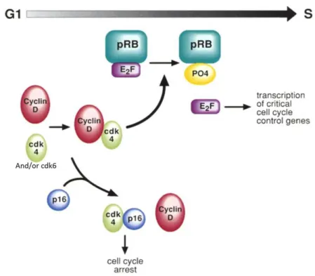
The formation of CDK4/6-cyclin D active complexes is essential for entry in G1 phase, and during early G1 CDK4/6-cyclin D active complexes are responsible for retinoblastoma protein (pRb) phosphorylation. This, in turn, leads to the disruption of pRB complexes with the histone deacetylase protein and release of the transcription factors E2F-1 and DP-1, which positively regulate the transcription of genes whose products are required for S phase progression. As the CDK inhibitor (CKI) p16 inhibits the association of CDK4/6-cyclin D complexes, pRb is not phosphorylated and thereby the genes necessary for the progression to S phase are not transcribed, resulting in cell cycle arrest in G1 (Fig. 1.8). Therefore, cells that lose p16 will indiscriminately progress through the cell cycle (Liggett & Sidransky 1998; Vermeulen et al. 2003).
Fig. 1.8. Cell cycle regulation by p16. Cyclin D1 regulate the function of CDK4 and/or CDK6, which
phosphorylates pRB. Phosporylation inactives pRB and allows E2F release followed by the transcription of critical cell-cycle proteins for the progression through the restriction point. p16 has a negative effect in this regulation and acting as a CKI, prevents cyclin D-CDK4 and/or CDK6 complex formation rendering cells to cell cycle arrest (Adapted from Liggett & Sidransky 1998)
36
The mechanisms behind p16 inactivation involve point mutations, promoter methylation or deletions (Rocco & Sidransky 2001). The ubiquitous and frequent inactivation of p16 in human tumours led to the hypothesis that loss of p16 must provide a selective cellular growth advantage to many tumours (Liggett & Sidransky 1998). Importantly, in CRC cell lines, inactivation of p16 seems to be common and promoter methylation-dependent (Herman et al. 1995; Tominaga et al. 1997; Kim et al. 2005). Conversely, in CRC tissues, the methylation status had been demonstrated to be variable and p16 expression has been repeatedly detected, overexpressed in some cases (Ohhara et al. 1996; Dai et al. 2000; Palmqvist et al. 2000; Norrie et al. 2003; Kim et al. 2005). Decreased or no p16 expression as a result of promoter methylation is associated with a more aggressive phenotype and poor prognosis (Liang et al. 1999; Yi et al. 2001; Kim et al. 2005; Bihl et al. 2012). Moreover, the variable density of methylation (high, low or non-methylated) appears to be important since it correlates with p16 mRNA expression (Kim et al. 2005) and yet it might not result in transcriptional silencing (Norrie et al. 2003; Kim et al. 2005). Aside from its tumour suppressor CKI status, p16 has been proving to exert a function in other cell biology aspects, including in the regulation of angiogenesis (Alhaja et al. 2004; Lu et al. 2012), apoptosis (Al-Mohanna et al. 2004; Lu et al. 2012), anoikis (Plath et al. 2000; André et al. 2007; Sanchez-Ruderisch et al. 2010; Amano et al. 2012) and, immortalization and senescence (Hara et al. 1996; Serrano et al. 1997; Lin et al. 1998; Jacobs & Lange 2004; Lu et al. 2012).
1.4.1. p16 connection with galectin-3 and KRAS
From the point of view of p16 connection with Gal-3 and KRAS, some of p16 roles in anoikis and senescence need to be addressed.
In a variety of cell lines, reconstitution of p16 expression confers the lost capability to undergo anoikis, what seems to be related to a p16-reconstitution induced upregulation of the fibronectin receptor (α5β1 integrin) (Plath et al. 2000). Beyond the transcriptional upregulation of the α5β1 integrin, p16 might have significant biological influences in other aspects. In fact, in Capan-1 pancreatic carcinoma p16 positive cells, a concerned expression of β1,4-galactosyltransferases as well as a decreased sialylation of O-/N-glycans, were observed. These aspects correlate with increased β1-integrin maturation, subunit assembly and increased binding activity of the α5β1-integrin. Besides, as a result of the reduced sialylation, that normally masks terminal galactoside residues for lectins, the presentation of glycan binding sites is
37
increased along with the cells capacity to bind Gal-1 (a galectin that favours anoikis), that was also increased at the transcription level (André et al. 2007). Moreover, in addition to altering α2,6-sialyltransferase expression, p16 acts through the regulation of enzymes on the pathway of sialic acid biosynthesis (Amano et al. 2012).
The concerned effects of p16 to restore tumour cells anoikis susceptibility also affect Gal-3.As already mentioned, Gal-3 is important to protect cells from anoikis induction, effect that is exerted intracellularly and at the cell surface by antagonizing Gal-1 pro-anoikis effects. Likewise, forced expression of Gal-3 in p16-positive cells reduces their susceptibility to anoikis but the presence of p16 decreases Gal-3 mRNA levels and cell surface presentation (Sanchez-Ruderisch et al. 2010). In addition, it was shown that this tumour suppressor controls oncogenic KRAS function in human pancreatic cancer cells: restitution of p16 downregulates KRASG12V activity, decreases its stability and expression, event that is necessary for p16-mediated restoration of anoikis resistance and suppression of cells clonogenicity (phenomena that was also verified in CRC cells- SW480 cell line) (Rabien et al. 2012). In this model, reduced levels of Gal-3 were found (Rabien et al. 2012), and in the light of the exposed information one might speculate that those could be a result of p16 expression and might also have contributed for the observed effects on KRAS. In summary, p16 coordinately orchestrates important changes to suppress the transformed phenotype with relevant impact in the galectin network and on the oncogenic KRAS.
As far as the p16 role in senescence is concerned, besides being involved in other senescence mechanisms, namely the ones induced by telomere erosion or DNA damage, p16 is involved in prematurely oncogenic stimuli-induced senescence (OIS). This mechanism was firstly referred by Serrano et al. 1997 and has been suggested to have a tumour suppressive function in a variety of different tumours (Collado & Serrano 2013). p16 levels increase in response to prolonged oncogenic HRASG12V expression/activation and provoke permanent cell cycle arrest in G1 phase. The mechanism underlying this requires RAS-mediated MAPK activation, which can produce two precisely opposite effects: cell-cycle arrest or forced mitogenesis, depending on the integrity of the senescence cooperative program controlled by p53 and p16, thereby reinforcing the tumour suppressor status of these genes (Serrano et al. 1997; Lin et al. 1998). This duality of effects seems to depend on the level of RAS activation: low levels led to hyperplasia, whereas high levels induce irreversible cellular senescence. This oncogene-induced senescence is followed by RAS downregulation and subsequent apoptosis, which could be explained by the so called “RAS addiction” hypothesis: once established, oncogenic RAS activation may be required