Ânia Robim Costa e Sousa
Licenciada em Análises Clínicas e Saúde Pública
Efficacy and toxicity evaluation of
celastrol in adjuvant-induced arthritis rat model
Dissertação para obtenção do Grau de Mestre em
Genética Molecular e Biomedicina
Orientador: D
outora Rita Cascão, JEFonseca’s Lab
, Instituto de Medicina
Molecular, Faculdade de Medicina da Universidade de Lisboa
Setembro, 2016
Júri:
III
Efficacy and toxicity evaluation of celastrol in adjuvant-induced arthritis rat model
Copyright © Ânia Robim Costa e Sousa, Faculdade de Ciências e Tecnologia, Universidade
Nova de Lisboa.
V
“Ah, not in knowledge is happiness, but in the acquisition of knowledge! In forever
knowing, we are forever blessed; but to know all, were the curse of a fiend.”
VII
Acknowledgments
First of all, I would like to thank my advisor, Rita Cascão, for having accepted me in her work, and for her patience and dedication to the team. Secondly I would also like to thank Professor Doctor João Eurico Fonseca for receiving me in his lab, in which the work was developed in.
I would also like to thank Bruno Vidal and Inês Lopes, for their unconditional help and patience in the experimental procedures. I must also thank my co-worker Ana Raquel Maia, for her help and company in this project.
IX
Abstract
Rheumatoid Arthritis (RA) is a chronic systemic autoimmune inflammatory disease that mainly affects the joints, and is characterized by active inflammation as well as bone and cartilage destruction. Since structural joint damage is irreversible, early recognition and treatment are currently being emphasized, with the goal of inducing remission of the disease. Current RA therapies fail or produce only partial responses in most patients and have adverse toxicological effects, so there is still an unmet need for a drug that can offer an effective and safe treatment of RA.
Celastrol, is a compound extracted from an herb used in Chinese medicine, which was previously identified by our work group as a potential candidate for the development of a new therapeutical drug for inflammatory diseases, such as RA. Therefore, the main goal of this project was to evaluate the efficacy and toxicity of the oral administration of a range of Celastrol dosages, using an adjuvant-induced arthritis (AIA) rat model. In order to achieve this, we treated AIA rats with dosages of Celastrol of 1 μg/g, 2.5μg/g, 12.5μg/g and 25μg/g, from day 8 post disease induction until day 22, when rats where sacrificed. Blood and paw samples were collected for quantification of bone turnover and degradation serum markers, histological and immunohistochemical evaluation, as well as for quantification of toxicological blood parameters.
Our work showed that an orally administered dosage of 2.5 μg/g of celastrol in the rat AIA model effectively reduces inflammation, infiltration and proliferation of synovial cells, suppresses bone erosion, reduces the number of osteoclasts and osteoblasts and reduces the number of synovial CD68+ cells, thus suggesting this treatment as effective. Moreover, we also showed that this treatment has no adverse toxicological effects at dosages of 1 μg/g and 2.5 μg/g, and that dosages of 25 μg/g and 12.5 μg/g can be considered lethal dose (LD) and LD50, respectively.
XI
Resumo
A Artrite Reumatoide (AR) é uma doença inflamatória autoimune sistémica crónica que afeta principalmente as articulações, e é caraterizada por inflamação ativa assim como por destruição do osso e cartilagem. Dado que os danos estruturais da articulação são irreversíveis, o tratamento e reconhecimento precoce são a ênfase atual, com o objetivo de induzir a remissão da doença. As terapias atuais para a AR falham ou produzem respostas somente parciais na maioria dos doentes e têm efeitos tóxicos adversos, existindo assim ainda uma grande necessidade de uma terapêutica que possa oferecer um tratamento eficaz e seguro para a AR.
O celastrol é um composto extraído de uma planta utilizada na medicina Chinesa, e foi previamente identificado pelo nosso grupo como um candidato potencial para o desenvolvimento de uma nova terapêutica para doenças inflamatórias, como a AR. Assim, o principal objetivo deste projeto foi testar a eficácia e toxicidade da administração oral de diferentes dosagens de celastrol, utilizando um modelo de rato de artrite induzida por adjuvante (AIA). Para isso, tratámos ratos AIA com dosagens de celastrol de 1 μg/g, 2.5 μg/g, 12.5 μg/g e 25 μg/g, desde o dia 8 após a indução da doença e até ao dia 22, quando os ratos foram sacrificados. Foram recolhidas amostras de sangue e da pata para quantificação de marcadores séricos de turnover e degradação óssea, avaliação histológica e imunohistoquímica, assim como para quantificação de parâmetros toxicológicos do sangue.
O nosso trabalho demonstrou que a administração oral de uma dosagem de celastrol de 2.5 μg/g no modelo de rato AIA reduz eficazmente a inflamação, a infiltração e a proliferação das células da sinóvia, suprime a erosão óssea, reduz do número de osteoclastos e osteoblastos e reduz o número de células sinoviais CD68+, sugerindo que este tratamento é eficaz. Além disso, demonstrámos que este tratamento não tem efeitos tóxicos adversos nas dosagens de 1 μg/g e 2.5 μg/g, e que as dosagens de 25 μg/g e 12.5 μg/g podem ser consideradas dose letal (DL) e DL50, respetivamente.
XIII
Table of Contents
LIST OF FIGURES ...XV LIST OF TABLES ... XVII ABBREVIATIONS ... XIX
1. INTRODUCTION ... 1
1.1.RHEUMATOID ARTHRITIS ... 1
1.1.1. Definition ... 1
1.1.2. Etiology ... 2
1.1.3. Pathophysiology ... 2
1.2.SKELETAL BONE AND RHEUMATOID ARTHRITIS... 4
1.2.1. Bone Homeostasis ... 4
1.2.2. Rheumatoid Arthritis and Bone ... 5
1.3.TREATMENT OPTIONS ... 6
1.4.CELASTROL ... 7
1.5.GOALS ... 9
2. MATERIALS AND METHODS ... 11
2.1.ANIMAL MODEL AND EXPERIMENTAL DESIGN ... 11
2.2.BLOOD TOXICOLOGICAL PARAMETERS MEASUREMENT ... 12
2.3.BONE TURNOVER AND DEGRADATION MARKERS MEASUREMENT ... 12
2.4.HISTOLOGICAL AND IMMUNOHISTOCHEMICAL EVALUATION ... 12
2.5.STATISTICAL ANALYSIS... 13
3. RESULTS ... 15
3.1.CELASTROL’S EFFECTS ON INFLAMMATORY SCORE AND ANKLE PERIMETER ... 15
3.2.CELASTROL’S EFFECTS ON BONE TURNOVER AND BONE DEGRADATION MARKERS ... 16
3.3.HISTOLOGICAL EVALUATION ... 18
3.4.IMMUNOHISTOCHEMICAL EVALUATION ... 20
3.5.CELASTROL’S TOXICOLOGICAL EFFECTS ... 21
4. DISCUSSION ... 23
XV
List of Figures
FIGURE 1.1-SCHEMATIC REPRESENTATION OF A) A NORMAL SYNOVIAL JOINT AND B) A SYNOVIAL JOINT WITH RA. ... 3
FIGURE 3.1–GRAPHICS SHOWING A) THE EVOLUTION OF THE INFLAMMATORY SCORE IN ALL GROUPS THROUGHOUT THE EXPERIMENT,
B) THE INFLAMMATORY SCORE AT DAY 22 AND C) THE ANKLE PERIMETER AT DAY 22. ... 16
FIGURE 3.2-BLOOD LEVELS OF BONE TURNOVER MARKERS A)CTX-I AND B)P1NP AND BONE DEGRADATION MARKER C)TRACP5B,
MEASURED IN RAT SERUM, AT DAY 22. ... 17
FIGURE 3.3–SCORES FROM THE HISTOLOGICAL OBSERVATION OF LEFT HIND PAW SECTIONS AT DAY 22, STAINED WITH HEMATOXYLIN
AND EOSIN. ... 19
FIGURE 3.4–IMMUNOHISTOCHEMICAL EVALUATION SCORES OF LEFT HIND PAW SECTIONS AT DAY 22 USING THE PRIMARY ANTIBODIES
A)KI-67, B)CATHEPSIN K, C)OSTEOCALCIN AND D)CD68. ... 21
FIGURE 3.5–CELASTROL’S TOXICOLOGICAL EFFECTS ON A)RATS’ BODY WEIGHT AND BLOOD TOXICOLOGICAL PARAMETERS OF B)
CREATINE KINASE (CK), C)UREA, D)LACTATE DEHYDROGENASE (LDH), E)ALANINE TRANSAMINASE (ALT) AND F) PRO-ATRIAL
XVII
List of tables
XIX
Abbreviations
ACPA Anti-Citrullinated Protein Antibody ACR American College of Rheumatology AIA Adjuvant-Induced Arthritis
ALT Alanine Transaminase APC Antigen Presenting Cell bDMARD Biological DMARD CCP Cyclic Citrullinated Peptide CIA Collagen-Induced Arthritis
CK Creatine Kinase
CPR C-Reactive Protein
CTX-I Cross Linked Collagen type I
DMARD Disease-Modifying Anti-Rheumatic Drug ELISA Enzyme Linked Immunosorbent Assay ESR Erythrocyte Sedimentation Rate EULAR European League Against Rheumatism FCA Freunds Complete Adjuvant
HLA Human Leukocyte Antigen
HLA-DRB1 HLA – antigen D Related Beta Chain 1
IFN Interferon
IL Interleukin
LD Lethal Dose
LDH Lactate Dehydrogenase MMP Matrix Metalloproteinases
NSAID Nonsteroidal Anti-Inflammatory Drug
Osx Osterix
P1NP Pro-Collagen Type I N-Terminal Propeptide pro-ANP pro-Atrial Natriuretic Peptide
XX
RF Rheumatoid Factor
Runx2 Runt-Related Transcription Factor 2 sDMARD Synthetic DMARD
SE Shared Epitope
SPF Specific Pathogen Free TNF Tumor Necrosis Factor
1
1.
Introduction
1.1.
Rheumatoid Arthritis
1.1.1.
Definition
Rheumatoid Arthritis (RA) is a chronic systemic autoimmune inflammatory disease that affects small joints, with articular damage and periarticular bone loss (Alamanos & Drosos, 2005; Firestein, 2003; Haugeberg et al., 2004). Besides generalized bone loss, which may lead to an elevated fracture risk, RA also causes substantial co-morbidity and is associated with a significant loss of physical, emotional and social quality of life, and decreased life-span (Alamanos & Drosos, 2005). In fact, this disease is commonly associated with other conditions, such as cardiovascular, pulmonary, psychological and skeletal disorders, and with some cancers and infections (Michaud & Wolfe, 2007).
Several prevalence and incidence studies of RA estimate a prevalence of 0.5-1% in the adult population worldwide (Alamanos & Drosos, 2005; Firestein, 2003) with a mean annual incidence of 0.02-0.05% in North American and North European countries (Aho, Kaipiainen-Seppänen, Heliövaara, & Klaukka, 1998; Alamanos & Drosos, 2005; Gabriel, Crowson, & O’Fallon, 1999; Riise, Jacobsen, & Gran, 2000), and the expected survival of patients is likely to decrease 3-10 years (Alamanos & Drosos, 2005; Gabriel et al., 2003; Wolfe et al., 1994). The incidence of this disease is higher in women than in men, with a sex ratio between 2:1 and 3:1 (Kourilovitch, Galarza-Maldonado, & Ortiz-Prado, 2014; Kvien, Uhlig, Ødegård, & Heiberg, 2006; van Vollenhoven, 2009a), suggesting an influence of reproductive and hormonal factors in the occurrence of the disease. The age of disease onset is around 50 years (Alamanos & Drosos, 2005).
The main symptom that characterizes this disease is symmetrical inflammation of small articulations (hands and feet), followed by chronic pain, swelling, stiffness and joint destruction that usually progresses from distal and small to more proximal and large joints (Aletaha et al., 2010; Kourilovitch et al., 2014). Since structural joint damage is irreversible, early recognition and treatment are currently being emphasized, with the goal of halting progression of the disease and induce remission.
2 RA diagnosis is commonly based on a set of clinical, serological and radiological criteria. RA’s classification is done accordingly to the American College of Rheumatology / European League Against Rheumatism (ACR/EULAR) criteria established in 2010 (Aletaha et al., 2010). These criteria classify a disease as ‘definiteRA’, based on the confirmed presence of synovitis in at least one joint, absence of an alternative diagnosis better explaining the synovitis, and achievement of a total score of 6 or greater (of a possible 10) from the individual scores in four domains: number and site of involved joints (range 0–5); serological abnormality (range 0–3), including at least a serological test for RF or ACPAs; elevated acute-phase response (range 0–1), including at least a measure of Erythrocyte Sedimentation Rate (ESR) or C-reactive protein (CPR); and symptom duration (range 0–1) (Aletaha et al., 2010).
1.1.2.
Etiology
RA is a multifactorial disease, resulting from the interaction of both genetic and environmental factors, which contribute to its occurrence and expression. The etiology of the disease is incompletely understood but is thought to be an interaction between genetic susceptibility, sex and age, smoking, infectious agents, and hormonal, dietary, socioeconomic and ethnic factors. Most of these factors are likely associated with both disease occurrence and severity (Alamanos & Drosos, 2005; McInnes & Schett, 2011).
The most important genetic factor associated with RA is the Human Leukocyte Antigen – antigen D Related Beta chain 1 (HLA-DRB1) complex, which is present in all individuals (McInnes & Schett, 2011; Ollier & MacGregor, 1995). This gene encodes an HLA class II protein, expressed in antigen presenting cells (APCs), which play a central role in the immune system by presenting antigens to T helper cells. The HLA-DRB1 locus has been shown to be linked to and associated with RA, with an especially high risk in individuals with shared epitope (SE) genes (Jawaheer & Gregersen, 2002; Ollier & MacGregor, 1995). A known interaction between genetic susceptibility and environmental factors is the fact that tobacco smoke exposure increases the risk factor for anti-Cyclic Citrullinated Peptide (CCP) antibodies production in HLA-DRB1 SE positive patients with RA (Klareskog et al., 2006; Liao, Alfredsson, & Karlson, 2009).
1.1.3.
Pathophysiology
3 Figure 1.1 - Schematic representation of a) a normal synovial joint and b) a synovial joint with RA. The synovial joint is composed of two bony ends covered with cartilage and separated by a synovial space, involving the synovial fluid and membrane. Adapted from (Choy, 2012).
role in RA include interferon (IFN)- and IL-17, which are used by T cells in the activation process of monocytes, macrophages and synovial fibroblasts (van Lent & van den Berg, 2007).
When T and B cells become activated, they produce cytokines and chemokines, which lead to more interactions between T cells, B cells, and macrophages, and consequentially to more cytokines and chemokines being produced, potentiating a feedback mechanism which perpetuates an autoimmune response. This autoimmune response becomes organized near the perivascular areas of the synovial membrane (Figure 1.1), leading to the formation of new blood vessels, or angiogenesis, to facilitate the delivery of nutrients to proliferating cells, and to the migration and accumulation of neutrophils in the synovial fluid (Harris, 1990). The process of blood vessels formation in the synovial membrane, is essential to the evolution of rheumatoid synovitis, that is, the inflammation of the synovial membrane (Harris, 1990).
4
1.2.
Skeletal Bone and Rheumatoid Arthritis
1.2.1.
Bone Homeostasis
Bone is a type of connective tissue that is composed of minerals (65%), primarily carbonated apatite, but also of organic components (20-25%), such as type I collagen, lipids and other noncollagenous proteins. The remainder is composed of water (10%) bound to the collagen-mineral composite and free water (Burr & Akkus, 2014). This composition, together with the general organization of the bone matrix, gives this tissue special mechanical properties such as, stiffness, rigidity, ductility and tensile strength (Del Fattore, Teti, & Rucci, 2012).
Although bone appears to be metabolically inert, it is in fact a dynamic organ that is controlled by the action of two main types of cells: osteoblasts and osteoclasts (Nakashima, Hayashi, & Takayanagi, 2012; Nakashima & Takayanagi, 2009). These cells work in collaboration to resorb damaged bone and to resynthesize new bone (Manolagas & Jilka, 1995). This continuous process of shaping and repairing the bone is called remodeling. This process is complex, and is tightly regulated by osteoblasts and osteoclasts (Kular, Tickner, Chim, & Xu, 2012). The coordinated balance between the activities of these cells, the bone-forming osteoblasts and the bone-resorbing osteoclasts, is crucial to maintain the homeostasis of the bone (Karsenty & Wagner, 2002). An imbalance in the bone remodeling process, favoring either osteoclast or osteoblast activity, has severe consequences for the organism, leading to serious bone pathologies, including Osteoporosis and Osteopetrosis, and other diseases that may involve the immune system, including RA (Kular et al., 2012; Rodan & Martin, 2000).
Bone remodeling, which is responsible for normal bone turnover, begins with an initiation phase that includes the recruitment and migration of partially differentiated mononucleated osteoclast precursors to the bone surface, their differentiation into mature osteoclasts, and the activation and maintenance of bone resorption, that occurs in the resorption lacunae or “pits” (Kular et al., 2012; Teitelbaum & Ross, 2003). In the next step, the reversal phase, occurs a transition from osteoclastic to osteoblastic activity, where osteoclastic bone resorption is inhibited and osteoclasts undergo apoptosis whilst osteoblasts and their constituent progenitor cells migrate to the newly resorbed surface where they produce an osteoid matrix and mineralize the osteoclast-orchestrated cavities. The final phase then follows, with the osteoblasts laying down bone until the resorbed bone is completely replaced (Hadjidakis & Androulakis, 2006; Kular et al., 2012).
5 the rest of the cell. The ruffled membrane border releases several hydrolytic lysosomal enzymes, such as Cathepsin K, matrix metalloproteinase (MMP)-9 and tartrate-resistant acid phosphatase type 5b (TRAcP5b), which attack the exposed collagen matrix, cleaving collagen fibers, and effectively remove small quantities of bone (Hadjidakis & Androulakis, 2006; Kular et al., 2012).
Osteoblasts are specialized cuboid bone forming cells that are responsible for the synthesis of bone matrix, regulation of mineralization and also differentiate into osteocytes or bone lining cells. These cells are found in clusters, lining on the layer of bone matrix they are producing. Osteoblasts originate from multipotent mesenchymal stem cells which have the potential to differentiate into mature osteoblasts (Hadjidakis & Androulakis, 2006; Kular et al., 2012). Numerous secreted factors of paracrine, autocrine and endocrine origin influence osteoblast development and maturation, such as the parathyroid hormone (PTH), the Runt-related transcription factor 2 (Runx2) and Osterix (Osx). Osteoblasts secrete type I collagen, the basic building block of bone, and several noncollagenous proteins including osteocalcin and alkaline phosphatase, which are essential for mineral deposition. Mature osteoblasts have one of three fates: they undergo apoptosis, differentiate further into osteocytes or become quiescent lining cells (Kular et al., 2012).
1.2.2.
Rheumatoid Arthritis and Bone
Structural damage that occurs as a result of RA is a direct consequence of a complex process that involves bone erosion, cartilage degradation and joint inflammation. In the case of this disease, the normal bone homeostasis, or the osteoblast-osteoclast axis, is severely disrupted, resulting in an enhanced osteoclast function and lack of bone repair activities after the formation of the erosions (Schett, 2007). In fact, as a typical feature of inflammatory tissue, the synovial membrane in RA contains many monocytes/macrophages that have the potential to differentiate into osteoclast, upon contact with appropriate signals (Schett, 2007).
In RA, monocytes migrate into the inflamed joint space, and differentiate into osteoclasts. Synovial fibroblast-like cells and activate T cells produce the necessary signals for this process to occur. In addition, the synovial fibroblast-like cells are found as part of the invasive pannus tissue, contributing to a localized differentiation into osteoclasts, and consequent bone erosion (Gravallese et al., 2000). Moreover, activated T cells produce IL-17, that together with other proinflammatory cytokines present in the synovial membrane of patients with RA, such as TNF, IL-1 and IL-6, enhances osteoclast differentiation and activity, leading to an accelerated process of structural damage (Lam et al., 2000; Sato et al., 2006; Schett, 2007; Wei, Kitaura, Zhou, Ross, & Teitelbaum, 2005).
pro-6 collagen type I N-terminal propeptide (P1NP), a propeptide of type I collagen that is found in circulation and directly reflects the rate of synthesis of type I collagen (Pollmann et al., 2007; Seibel, 2000).
1.3.
Treatment Options
Due to the inflammatory condition of RA, first-line therapy has traditionally included medications that suppress inflammation, and act rapidly to improve pain and swelling, such as nonsteroidal anti-inflammatory drugs (NSAIDs) and glucocorticoids (Gaffo, Saag, & Curtis, 2006). With time came the dramatic realization that RA is a serious and potentially devastating disease that requires aggressive management, including the use of Disease-modifying anti-rheumatic drugs (DMARDs) in the early onset of the disease, and the active pursuit of optimum results by frequent changes in therapy and the use of combination therapies. This new approach to treatment spawned a large number of new therapeutic agents, both pharmacological and biologic that changed the treatment of RA (Sardar & Andersson, 2016; van Vollenhoven, 2009b). Current therapy strategies include NSAIDs, glucocorticoids, synthetic DMARDs (sDMARDs) and biological DMARDs (bDMARDs), that can either be used in monotherapy or in combination therapy (Table 1.1) (Koenders & van den Berg, 2015; Venkatesha, Dudics, Acharya, & Moudgil, 2014).
DMARDs are slow-acting compounds that not only improve symptoms, but also slow clinical and radiographic progression of the disease, unlike NSAIDs. sDMARDs, such as methotrexate, are the most used drugs in therapy, are considered acceptably safe, and have a slow onset of action, ranging from several weeks to months (American College of Rheumatology AD Hoc Committee on Clinical Guidelines, 1996; O’Dell, 2004). The use of NSAIDs, such as aspirin and ibuprofen, is usually well tolerated by patients for a short period of time, however their chronic use may sometimes lead to gastrointestinal complications and even renal insufficiency. As for glucocorticoids, these present toxicity risks even at low dosages (American College of Rheumatology AD Hoc Committee on Clinical Guidelines, 1996; Chiba et al., 2005).
7 Table 1.1 – Main categories of RA treatment compounds and examples. Adapted from (Gaffo et al., 2006; Venkatesha et al., 2014)
CATEGORY EXAMPLE(S)
NSAIDs Aspirin, Ibuprofen
Glucocortidoids Prednisone, Methylprednisolone
sDMARDs Methotrexate, Hydroxychloroquine, Sulfasalazine, Leflunomide bDMARDs
Anti-TNF Infliximab, Etanercept, Adalimumab
IL-1 Inhibitors Anakinra
IL-6 Inhibitors Tocilizumab
Costimulation Blockers Abatacept
B-cell Targeted Therapies Rituximab
NSAID – Nonsteroidal anti-inflammatory drugs; sDMARD – Synthetic Disease-modifying anti-rheumatic drug; bDMARD – biological DMARD; TNF - Tumor necrosis factor; IL – Interleukin.
1.4.
Celastrol
Our group (Cascão, 2012) observed that both IL-1 and TNF are two cytokines which play an important role in RA. Taken this into account, they performed an in vitro library drug screening, searching for
drugs that downregulated the production of IL-1 and TNF, followed by an in vivo drug therapy study
using a Wistar rat model of adjuvant-induced arthritis (AIA). The results from this study identified celastrol, a compound extracted from the root bark of Tripterygium wilfordii Hook f (TwHf), as a
potential candidate for the development of a new therapy targeting RA.
RA, like many other chronic conditions, is associated with a high level of complementary and alternative medicine use, in particular, herbal treatments (Setty & Sigal, 2005). One of the main herbs used in Chinese medicine is the TwHf (Tao & Lipsky, 2000), also known as thunder god vine, an herb whose extracts have proven immunomodulatory (Yu, Venkatesha, & Moudgil, 2012) and anti-inflammatory effects (Sassa, Takaishi, & Terada, 1990) in vitro and in vivo, in various animal models, including RA
(Cascão et al., 2012; Nanjundaiah et al., 2012; Venkatesha, Yu, Rajaiah, Tong, & Moudgil, 2011), atherosclerosis (Gu et al., 2013), Alzheimer’s disease (Allison, Cacabelos, Lombardi, Alvarez, & Vigo, 2001; Paris et al., 2010), asthma (Kim, Park, Jeoung, & Ro, 2009) and systemic lupus erythematosus (Xu, Wu, Xu, Ren, & Ge, 2003).
8 Idris et al., 2010 used several in vitro cell cultures, such as osteoblast, osteoclast precursors, and
osteoclast plus macrophage cultures. These cells were cultured in the presence of celastrol and the results showed that this bioactive compound inhibits osteoclast formation, bone resorption and macrophage viability, while also stimulating osteoclast apoptosis.
Venkatesha et al., 2011, used the rat AIA model, treating male Lewis rats with a dose of celastrol of 1 μg/g/day, diluted in PBS and administered intraperitoneally. The treated group showed suppression of key proinflammatory cytokines (IL-6, IL-17 and IFN-). A follow up study by Yu et al., 2012, also used the AIA rat model, treating male Lewis rats with the same dosage of celastrol as the above mentioned study, and with the same administration process. This study used gene expression profiling and pathway analysis to show that celastrol actively modulated the immune responses rather than inducing global immunosuppression. Nanjundaiah et al., 2012, used the AIA rat model and the same administration process as the above mentioned studies. The results showed that celastrol reduced the number of osteoclasts, and the inflammation-induced bone damage by favoring anti-osteoclastic activity.
Cascão et al., 2012, also used the AIA rat model and administered celastrol intraperitoneally. Their results demonstrated the effective treatment of AIA through the use of celastrol, a downregulator of the production of IL-1 and TNF, supporting the in vivo anti-inflammatory and anti-proliferative effects of
this compound. Cascão et al., 2015, further tested the effects of celastrol in AIA rats, reporting a significant decrease in the number of sublining CD68 macrophages and in the overall number of inflammatory cells in the synovium, following treatment with celastrol, thus suggesting that this compound halted joint destruction without side effects, and validating celastrol as a potential candidate for a treatment drug targeting RA.
9
1.5.
Goals
There is still a large need for further development of drugs that target RA, to fulfill the inadequate response that is being observed on a large number of patients, where therapies sometimes fail or produce only partial responses, and also produce adverse toxicological effects. Therefore, there is still an unmet need for a drug that can offer an effective and safe treatment.
The main goal of this work is to evaluate the efficacy and toxicity of the oral administration of a range of celastrol dosages, by studying its anti-inflammatory, anti-proliferative and bone protective properties, using an AIA rat model.
In order to accomplish this, we assessed the in vivo efficacy, in the AIA rat model, of the oral
administration of celastrol by evaluating the dose-dependent effects in:
Joint inflammation and ankle perimeter;
Bone turnover and bone degradation markers in the serum;
Articular joint tissues;
Cell proliferation markers, osteoblastic and osteoclastic cells markers, and in CD68+ cells, in tissue sections of the paw.
Moreover, we also assessed the in vivo dose-dependent toxicological profile of celastrol by evaluating
the effects of the oral administration of celastrol in:
Animal body weight;
11
2.
Materials and Methods
2.1.
Animal Model and Experimental Design
In this work it was used the Wistar AIA rat model to assess the efficacy and toxicity of orally administrated celastrol. The advantages for using this model include the presence of: a robust, easily measurable, polyarticular and systemic inflammation, with a reliable onset and progression; marked bone resorption; and marked periosteal bone proliferation (Bendele, 2001). Eight weeks old female Wistar AIA rats, weighing 250g, were purchased from Charles River Laboratories International (Massachusetts, USA) and maintained under specific pathogen free (SPF) conditions. All experiments were approved by the Animal User and Ethical Committees at the Instituto de Medicina Molecular, according to the Portuguese law and the European recommendations.
Induction of adjuvant disease was done with Freunds Complete Adjuvant (FCA) supplemented with mycobacterium, injected subcutaneously at the right hind paw. Paw swelling is monitored from day 4 to day 22, the end point of the experiment. Clinical evidence of arthritis occurs on day 8-9 post injection of adjuvant (Bendele, 2001). Treatments were initiated on day 8 after disease induction, at the onset of the disease, which is considered a therapeutic model (Bendele, 2001), and finished on day 22, when the disease as already reached the peak in chronic stage (Stolina et al., 2009).
Celastrol (Sigma, Missouri, USA) was administrated at doses of 1 μg/g, 2.5 μg/g, 12.5 μg/g and 25 μg/g of body weight per day. Celastrol was dissolved in ethanol, due to its solvent properties, and PEG400 was also added for the intragastric administration. AIA rats were separated into five groups: the four treatment groups that received distinct dosages of celastrol, each group with five rats; and the arthritic group, with ten rats, that received ethanol in PEG400, and served as a positive control. An additional group composed of eight healthy rats received water, and served as a negative control. All rats were fed the corresponding solutions through gavage.
12
2.2.
Blood Toxicological Parameters Measurement
The levels of blood toxicological parameters of creatine kinase (CK), urea, lactate dehydrogenase (LDH), alanine transaminase (ALT) (BioAssay Systems, California, USA), and pro-atrial natriuretic peptide (pro-ANP) (Biomedica Immunoassays, Vienna, Austria), were measured in rat serum by commercially available enzyme linked immunosorbent assay (ELISA), according to the manufacturer’s instructions. Standard curves for each parameter were created using reference concentrations supplied by the manufacturer. Samples were analyzed using plate reader Tecan Infinite 200 PRO (Tecan, Männedorf, Switzerland).
2.3.
Bone Turnover and Degradation Markers Measurement
Bone turnover markers CTX-I and P1NP, and bone degradation marker TRAcP5b (Immunodiagnostic System, Boldon, UK) were measured in rat serum by commercially available ELISA, according to the manufacturer’s instructions. Standard curves for each marker were created using reference concentrations supplied by the manufacturer. Samples were analyzed using plate reader Tecan Infinite 200 PRO (Tecan, Männedorf, Switzerland).
2.4.
Histological and Immunohistochemical Evaluation
13 microscope equipped with a color camera. Immunohistochemical evaluation of the sections was done according to a semi-quantitative score from 0–4: 0– no staining; 1– less than 25% staining; 2– 25 to 50% staining; 3– 50 to 75% staining; 4– more than 75% staining (Cascão et al., 2012).
2.5.
Statistical Analysis
15
3.
Results
3.1.
Celastrol’s
Effects on Inflammatory Score and Ankle Perimeter
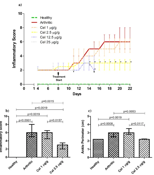
Inflammatory Score was measured from day 4 to day 22, when the rats were euthanized (Figure 3.1a). It is worth noting that three AIA rats from the Cel 12.5 μg/g group and all five AIA rats from the Cel 25 μg/g group were euthanized before day 22 due to excessive body weight loss (more than 20% of the initial body weight) and to the presence of dyspnea and diarrhea. This suggests that celastrol may be toxic at these concentrations.
16
a)
b) c)
Figure 3.1 – Graphics showing a) the evolution of the Inflammatory Score in all groups throughout the experiment, b) the Inflammatory Score at day 22 and c) the Ankle Perimeter at day 22. a) Treatment with Celastrol in the Celastrol-treated groups began at day 8. Asterisks (*) denote a significant difference between the groups treated with Celastrol and the Arthritic group. †Last day of data from the Cel 25 μg/g group. ¥ Last day of data
from the Cel 12.5 μg/g group. All data is represented as median with interquartile range and differences were
considered statistically significant for p<0.05. Healthy n=8, Arthritic n=10, Cel 1 μg/g n=5, Cel 2.5 μg/g n=5,
Cel 12.5 μg/g n=5¥ and Cel 25 μg/g n=5†.
3.2.
Celastrol’s Effects
on Bone Turnover and Bone Degradation Markers
17 Figure 3.2 - Blood levels of bone turnover markers a) CTX-I and b) P1NP and bone degradation marker c) TRAcP5b, measured in rat serum, at day 22. All data is represented as median with interquartile range and differences were considered statistically significant for p<0.05. Healthy n=8, Arthritic n=10, Cel 1 μg/g n=5 and Cel 2.5 μg/g n=5.
b) a)
c)
By the end of the study (Figure 3.2a), arthritic rats showed increased levels of osteoclastic activity (p=0.0186 in arthritic rats vs healthy rats), with no differences between arthritic and celastrol-treated animals. However, celastrol-treated rats also showed no differences in the levels of CTX-I, when compared to the healthy rats group.
Regarding serum levels of P1NP (Figure 3.2b), both arthritic rats and celastrol-treated rats at the lower dose showed increased levels of osteoblastic activity (p=0.0074 and p=0.0177 in arthritic rats and celastrol-treated rats at the dose of 1 μg/g vs healthy rats, respectively), with no differences between arthritic and both celastrol-treated rat groups. There was also no difference between the Cel 2.5 μg/g group and the healthy animals.
18
3.3.
Histological Evaluation
For histological observation, we used sections of the left hind paw, stained with hematoxylin and eosin. The histological evaluation of the stained sections (Figure 3.3) was done according to four semi-quantitative scores: the sublining layer infiltration score, the lining layer cell number score, the bone erosion score, and the global severity of the disease score.
The sublining layer infiltration score, evaluated the extent of the cell infiltration in the joint. As can be observed in Figure 3.3a, both arthritic rats and celastrol-treated rats at the lower dose showed an increased infiltration of the sublining layer (p=0.0001 and p=0.0019 in arthritic rats and celastrol-treated rats at the dose of 1 μg/g vs healthy rats, respectively), with no differences between arthritic and Cel 1 μg/g animals. The Cel 2.5 μg/g group showed a significant reduction in infiltration in comparison with arthritic rats (p=0.0010), and also with Cel 1 μg/g animals (p=0.0097), and showed no significant differences to the healthy rats.
In regards to the proliferation of the synovial lining layer, assessed by the lining layer cell number score (Figure 3.3b), both arthritic rats and celastrol-treated rats at the lower dose showed an increased proliferation (p=0.0019 in arthritic rats and celastrol-treated rats at the dose of 1 μg/g vs healthy rats), with a higher level of proliferation present in Cel 1 μg/g animals, when compared to the arthritic group (p=0.0365). The Cel 2.5 μg/g group showed a significant reduction in proliferation in comparison with arthritic rats (p=0.0110), and also with Cel 1 μg/g animals (p=0.0109), and showed no significant differences to the healthy rats.
As for the bone erosion score (Figure 3.3c), both arthritic rats and celastrol-treated rats at the lower dose showed an increased bone erosion (p=0.0008 and p=0.0019 in arthritic rats and celastrol-treated rats at the dose of 1 μg/g vs healthy rats, respectively), with no differences between arthritic and Cel 1 μg/g animals. The Cel 2.5 μg/g group showed a significant reduction in bone erosion in comparison with arthritic rats (p=0.0053), and also with Cel 1 μg/g animals (p=0.0117), and showed no significant differences to the healthy rats.
19 Figure 3.3 – Scores from the histological observation of left hind paw sections at day 22, stained with hematoxylin and eosin. a) Sublining layer infiltration, b) Lining layer cell number, c) Bone erosion and d) Global severity of the disease. All data is represented as median with interquartile range and differences were considered statistically significant for p<0.05. Healthy n=8, Arthritic n=10, Cel 1 μg/g n=5 and Cel 2.5 μg/g n=5.
b) a)
20
3.4.
Immunohistochemical Evaluation
Sections of the left hind paw were also incubated with primary antibodies Ki-67, Cathepsin K, osteocalcin and CD68 (Figure 3.4). Immunohistochemical evaluation of the sections was done according to a semi-quantitative score of the staining.
Ki-67 is a marker of immune cell proliferation and its presence reflects the levels of synovial cells proliferation. We observed (Figure 3.4a) that the immunohistochemical scores of this marker showed that the arthritic rats and the rats treated with a dosage of 1 μg/g of celastrol had a higher level of proliferation than that of the healthy rats (p=0.0001 and p=0.0019, respectively) and there was no difference between the levels of synovial cells proliferation in the arthritic rats and in the Cel 1 μg/g rat group. We also observed that the rats treated with a dosage of 2.5 μg/g of celastrol had a level of proliferation lower than both the arthritic rats and the rats treated with a dosage of 1 μg/g of celastrol (p=0.0020 and p=0.0219, respectively), and that Cel 2.5 μg/g rats showed no significant difference in the immune cell proliferation level to the healthy rat group.
Cathepsin K is expressed in osteoclasts and its presence reflects the number of osteoclasts and their precursors in the tissue that is being analyzed. The immunohistochemical scores of this marker (Figure 3.4b) showed that both the arthritic group and the group of rats treated with a celastrol dosage of 1 μg/g have an increased number of osteoclasts (p=0.0019 in arthritic rats and Cel 1 μg/g group vs healthy rats), with no difference between them. The Cel 2.5 μg/g group showed a significant reduction in the number of osteoclasts in comparison with arthritic rats (p=0.0449), and with Cel 1 μg/g rats (p=0.0442). There was no significant difference between the number of osteoclasts in the rat group treated with a dosage of 2.5 μg/g of celastrol and the healthy rat group.
Osteocalcin is a marker of osteoblasts, and its presence reflects the number of these cells in the tissue. The immunohistochemical scores of this marker (Figure 3.4c) showed that both arthritic rats and rats treated with a dosage of 1 μg/g of celastrol had a higher number of osteoblasts (p=0.0010 and p=0.0019 in arthritic rats and celastrol-treated rats at the dose of 1 μg/g vs healthy rats, respectively), with no differences between arthritic and Cel 1 μg/g animals. The Cel 2.5μg/g group showed a significant reduction in the number of osteoblasts in comparison with arthritic rats (p=0.0086), and also with Cel 1 μg/g animals (p=0.0097). The rats treated with a dosage of 2.5 μg/g of celastrol had no difference to the healthy animals.
21 Figure 3.4 – Immunohistochemical evaluation scores of left hind paw sections at day 22 using the primary antibodies a) Ki-67, b) Cathepsin K, c) Osteocalcin and d) CD68. Evaluation of the sections was done according to a semi-quantitative score. All data is represented as median with interquartile range and differences were considered statistically significant for p<0.05. Healthy n=8, Arthritic n=10, Cel 1 μg/g n=5 and Cel 2.5 μg/g n=5.
b) a)
c) d)
and p=0.0097 in arthritic and celastrol-treated rats at the dose of 1 μg/g vs Cel 2.5 μg/g, respectively), and showed no differences to the healthy animals.
3.5.
Celastrol’s Toxicological Effects
22 Celastrol-treated rats did not suffer any body weight loss (Figure 3.5a), at the end of the study, when compared to the arthritic rats’ body weight. However, both arthritic rats and celastrol-treated rat groups showed a significant loss in body weight, when compared to healthy rats (p=0.0003 and p=0.0016 in arthritic rats and both celastrol-treated rat groups vs healthy rats, respectively).
In regards to blood toxicological parameters (Figure 3.5b-f), both arthritic and celastrol-treated rats showed no signs of toxicological effects (arthritic rats and celastrol-treated rats vs healthy rats), with no differences between arthritic and celastrol-treated animals.
Figure 3.5 – Celastrol’s toxicological effects on a) Rats’ body weight and blood toxicological parameters of b)
Creatine Kinase (CK), c) Urea, d) Lactate Dehydrogenase (LDH), e) Alanine Transaminase (ALT) and f) pro-Atrial Natriuretic Peptide (pro-ANP), measured in the rats’ serum, at day 22. All data is represented as median with interquartile range and differences were considered statistically significant for p<0.05. Healthy n=8, Arthritic
n=10, Cel 1 μg/g n=5 and Cel 2.5 μg/g n=5.
b)
d) c)
f) e)
23
4.
Discussion
RA is a disease characterized by inflammation of small joints of hands and feet, leading to articular erosion and periarticular bone loss (Alamanos & Drosos, 2005; Firestein, 2003; Haugeberg et al., 2004). Current RA therapies lack in providing an adequate response in a large number of patients and also produce adverse toxicological effects, so there is still an unmet need for a drug that can offer an effective and safe therapeutical option for the treatment of RA.
The main aim of this work is to evaluate the efficacy and toxicity of the oral administration of a range of celastrol dosages, using an AIA rat model. To accomplish this, we assessed the in vivo efficacy of the
oral administration of celastrol in treating inflammation, inhibiting proliferation and preventing bone damage, by evaluating the dose-dependent effects in: joint inflammation and ankle perimeter; bone turnover and bone degradation markers in the serum; articular joint tissues; and cell proliferation markers, osteoblastic and osteoclastic cells markers, and in the number of CD68+ cells, a marker of the efficacy of the treatment, in tissue sections of the paw. Moreover, we also assessed the in vivo
dose-dependent toxicological profile of celastrol by evaluating the effects of the oral administration of celastrol in animal body weight and in markers for systemic tissue damage, nephrotic damage, hepatic damage and cardiac damage.
The inflammatory score measured in our work, together with the ankle perimeter measure, served as indicators of joint inflammation levels in the animals. The ankle perimeter measure in particular also gives information of the articular swelling, that is linked to the degree of inflammation of the joints. Arthritic rats started to show escalating worsening of the disease symptoms from day 10, which is in agreement with literature (Bendele, 2001; Stolina et al., 2009).
24 To assess the efficacy of an oral dosage of celastrol in preventing bone degradation, we measured its effects in serum levels of CTX-I and P1NP, bone turnover markers, and TRAcP5b, a marker of bone resorption and osteoclast number (Halleen, Tiitinen, Ylipahkala, Fagerlund, & Väänänen, 2006). CTX-I is released upon degradation of bone matrix (Garnero et al., 2003) and is elevated in animal models of RA (Vidal et al., 2015). We measured this marker on the serum of all rat groups, and observed that the osteoclastic activity in arthritic rats was higher than that of healthy rats. Still, the osteoclastic activity in both rat groups treated with celastrol showed no statistically significant difference to any of the other two groups. These findings suggest that an orally administered dosage of 2.5 μg/g of celastrol might not be effective in reducing the osteoclastic activity, and a higher dose may be required.
We also measured the serum levels of P1NP, which directly reflects the rate of synthesis of type I collagen, the main building block of bone matrix, carried out by osteoblasts (Seibel, 2000). We observed that the osteoblastic activity is elevated in arthritic rats and in rats treated with celastrol at a dosage of 1 μg/g when compared to healthy rats. The behavior of this bone formation marker is explained by the natural mechanism of the organism to achieve bone homeostasis, when in presence of excessive bone destruction, as it happens in the AIA rat model of RA. This bone destruction then leads to an increase in bone formation, in an attempt to achieve a state of bone homeostasis, showing a compensatory mechanism in bone turnover (Vidal et al., 2015). The osteoblastic activity of the rat group treated with a dosage of 2.5 μg/g of celastrol showed no difference with the healthy rats. Still, the osteoblastic activity in this group also showed no difference to both the arthritic rat group and to the rat group treated with a dosage of 1 μg/g of celastrol. These findings suggest that, an orally administered dosage of 2.5 μg/g of celastrol has no effect on osteoblast activity. Furthermore, this also suggests that an orally administered dosage of 2.5 μg/g of celastrol might have effects on bone degradation, by reducing osteoclast activity (CTX-I), without interfering in bone forming processes.
25 The histological evaluation of sections of the left hind paw, stained with hematoxylin and eosin, was done according to four distinct scores. The sublining layer infiltration score, the lining layer cell number score, the bone erosion score, and the global severity of the disease score, all showed an increased cell infiltration, lining layer proliferation, bone articular destruction and severity of the disease in both arthritic and Cel 1 μg/g rat groups, when compared to healthy rats. Moreover, the results also showed a reduction in cell infiltration, lining layer proliferation, bone articular destruction and severity of the disease following treatment with a dosage of celastrol of 2.5 μg/g, when compared to both arthritic and Cel 1 μg/g rat groups, and with no difference to healthy rats. These findings suggest that an orally administered dosage of 2.5 μg/g of celastrol is effective in reducing inflammation, the infiltration and proliferation of cells in the synovium, suppressing bone erosion, and ameliorating the global symptoms of the disease. These results are supported by another work developed by our research group, that administered celastrol intraperitoneally (Cascão et al., 2015).
Immunohistochemical evaluation of sections of the left hind paw was done by staining with primary antibodies Ki-67, Cathepsin K, osteocalcin and CD68.
The score of Ki-67, a marker of immune cell proliferation, showed that the arthritic rats and the rats treated with a dosage of 1 μg/g of celastrol had a higher level of proliferation, when compared to healthy rats. This score also showed that the rats treated with a dosage of 2.5 μg/g of celastrol had a level of proliferation lower than both the arthritic rats and the rats treated with a dosage of 1 μg/g of celastrol, and had no differences to the healthy rat group. These findings suggest that there was a reduction on the levels of synovial cells proliferation, following treatment with an orally administered dosage of 2.5 μg/g of celastrol, which might imply an anti-inflammatory effect of celastrol in preventing the formation of the pannus. These results are supported by other works developed by our research group, that administered celastrol intraperitoneally (Cascão, 2012; Cascão et al., 2015), while at the same time suggests that they can also be achieved by administering celastrol orally.
26 synovium and also the relative RNA expression of the Cathepsin K gene, thus further attesting our results. Moreover, our findings, together with the ones from CTX-I show that this local reduction in the number of osteoclasts is not observed in the serum markers, although there is an observed tendency for this marker to return to healthy levels following treatment with celastrol.
We also measured the expression of Osteocalcin in joint tissue, whose immunohistochemical staining reflects the number of osteoblasts in the tissue. In our work we observed that the arthritic rats and the rats treated with a dosage of 1 μg/g of celastrol had a higher number of osteoblasts than those present in healthy rats. We also observed that the rats treated with a dosage of 2.5 μg/g of celastrol had a lower number of osteoblasts than both the arthritic rats and the rats treated with a dosage of 1 μg/g of celastrol, and had no differences to the healthy rat group. Despite these findings suggesting that an orally administered dosage of 2.5 μg/g of celastrol reduces the number of osteoblasts in the articular joint tissues, we need to take into consideration the natural mechanism of the organism to achieve bone homeostasis. Since we already showed that celastrol was effective in reducing the number of osteoclasts, these findings may suggest that there is indeed a reduction in the number of osteoblasts due to the natural mechanism of homeostasis, rather than due to the direct effect of celastrol on osteoblastic cells. Furthermore, these findings, together with the ones from P1NP show that this local reduction in the number of osteoblasts is not observed in the serum markers, although there is an observed tendency for this marker to return to healthy levels following treatment with celastrol.
CD68 is expressed in activated macrophage cells and a significant reduction on the number of synovial CD68+ cells is usually associated with effective experimental drugs in humans and animals (Vieira-Sousa et al., 2011; Wijbrandts et al., 2007). The score of this marker, showed that the arthritic rats and the rats treated with a dosage of 1 μg/g of celastrol had a higher number of CD68+ cells, when compared to healthy rats. This score also showed that the rats treated with a dosage of 2.5 μg/g of celastrol had a reduction on the number of CD68+ cells, when compared to both the arthritic rats and the rats treated with a dosage of 1 μg/g of celastrol, and had no differences to the healthy rat group. These findings suggest that an orally administered dosage of 2.5 μg/g of celastrol is effective in reducing synovial CD68+ cells, thus suggesting this treatment as effective, also supported by other works developed by our research group (Cascão et al., 2015).
27 This raises questions about the safety of the compound and, therefore, we performed a body weight measure and a toxicological screening test. In regards to the body weight, we observed that celastrol-treated rats did not suffer any body weight loss at the end of the study, when compared to the arthritic rats’ body weight, suggesting that an oral administration of celastrol at the dosages of 1 μg/g and 2.5 μg/g has no effect on body weight. Still, we observed that both arthritic rats and celastrol-treated rat groups showed a loss in body weight, when compared to healthy rats, further supporting that the observed body weight loss is caused by the disease itself and not by celastrol treatment.
The toxicological screening test included markers for systemic tissue damage (CK and LDH), nephrotic damage (Urea), hepatic damage (ALT) and cardiac damage (pro-ANP), and also a histological analysis of internal organs performed by a clinical pathologist. Regarding the toxicological tests, all rat groups showed no signs of toxicological effects. Of note, we have noticed a tendency to an increase in ALT and CK levels in the two surviving rats from the group treated with a dosage of 12.5 μg/g of celastrol. However, these findings must be considered with attention since they are only based on data from two animals. The histological analysis was performed on internal organ samples from all rats, by a blinded-clinical pathologist (data not shown), which reported histopathological findings supporting our results.
Altogether, these findings suggest that an orally administered dosage of 2.5 μg/g of celastrol as no adverse toxicological effects, also supported by other works developed by our research group (Cascão et al., 2015).
In conclusion, we were able to demonstrate that an orally administered dosage of 2.5 μg/g of celastrol in an in vivo rat AIA model is effective in reducing inflammation, the infiltration and proliferation of
synovial cells, suppressing bone erosion, ameliorating the global symptoms of the disease, reducing the number of osteoclasts and osteoblasts in paw sections and in reducing the number of synovial CD68+ cells, thus suggesting this treatment as effective. Moreover, we also showed that this treatment, at an oral dosage of 2.5 μg/g of Celastrol, has no adverse toxicological effects.
29
References
Aho, K., Kaipiainen-Seppänen, O., Heliövaara, M., & Klaukka, T. (1998). Epidemiology of rheumatoid arthritis in Finland. Seminars in Arthritis and Rheumatism, 27(5), 325–334.
Alamanos, Y., & Drosos, A. A. (2005). Epidemiology of adult rheumatoid arthritis. Autoimmunity
Reviews, 4(3), 130–136. http://doi.org/10.1016/j.autrev.2004.09.002
Aletaha, D., Neogi, T., Silman, A. J., Funovits, J., Felson, D. T., Bingham, C. O., … Hawker, G. (2010). 2010 rheumatoid arthritis classification criteria: an American College of Rheumatology/European League Against Rheumatism collaborative initiative. Annals of the
Rheumatic Diseases, 69(9), 1580–1588. http://doi.org/10.1136/ard.2010.138461
Allison, A. C., Cacabelos, R., Lombardi, V. R., Alvarez, X. A., & Vigo, C. (2001). Celastrol, a potent antioxidant and anti-inflammatory drug, as a possible treatment for Alzheimer’s disease.
Progress in Neuro-Psychopharmacology & Biological Psychiatry, 25(7), 1341–1357.
American College of Rheumatology AD Hoc Committee on Clinical Guidelines. (1996). Guidelines for monitoring drug therapy in rheumatoid arthritis. American College of Rheumatology Ad Hoc Committee on Clinical Guidelines. Arthritis and Rheumatism, 39(5), 723–731.
Bendele, A. (2001). Animal models of rheumatoid arthritis. Journal of Musculoskeletal & Neuronal
Interactions, 1(4), 377–385.
Burr, D. B., & Akkus, O. (2014). Chapter 1 - Bone Morphology and Organization. In Basic and Applied
Bone Biology (pp. 3–25). San Diego: Academic Press. Retrieved from
http://www.sciencedirect.com/science/article/pii/B9780124160156000010
Cascão, R. (2012). Role of IL-1β in rheumatoid arthritis. PhD Thesis. Faculdade de Medicina,
Universidade de Lisboa. Retrieved from http://repositorio.ul.pt/handle/10451/7058
Cascão, R., Vidal, B., Lopes, I. P., Paisana, E., Rino, J., Moita, L. F., & Fonseca, J. E. (2015). Decrease of CD68 Synovial Macrophages in Celastrol Treated Arthritic Rats. PLOS ONE, 10(12),
30 Cascão, R., Vidal, B., Raquel, H., Neves-Costa, A., Figueiredo, N., Gupta, V., … Moita, L. F. (2012). Effective treatment of rat adjuvant-induced arthritis by celastrol. Autoimmunity Reviews,
11(12), 856–862. http://doi.org/10.1016/j.autrev.2012.02.022
Chiba, T., Sato, K., Endo, M., Ando, T., Inomata, M., Orii, S., & Suzuki, K. (2005). Upper gastrointestinal disorders induced by non-steroidal anti-inflammatory drugs.
Hepato-Gastroenterology, 52(64), 1134–1138.
da Silva, J., Fonseca, J., Graça, L., Moita, L., & Carmo-Fonseca, M. (1995). Reinnervation of post-arthritic joints in the rat. Clinical and Experimental Rheumatology, 14(1), 43–51.
Del Fattore, A., Teti, A., & Rucci, N. (2012). Bone cells and the mechanisms of bone remodelling.
Frontiers in Bioscience (Elite Edition), 4, 2302–2321.
Firestein, G. S. (2003). Evolving concepts of rheumatoid arthritis. Nature, 423(6937), 356–361.
http://doi.org/10.1038/nature01661
Gabriel, S. E., Crowson, C. S., Kremers, H. M., Doran, M. F., Turesson, C., O’Fallon, W. M., & Matteson, E. L. (2003). Survival in rheumatoid arthritis: a population-based analysis of trends over 40 years. Arthritis and Rheumatism, 48(1), 54–58. http://doi.org/10.1002/art.10705
Gabriel, S. E., Crowson, C. S., & O’Fallon, W. M. (1999). The epidemiology of rheumatoid arthritis in Rochester, Minnesota, 1955-1985. Arthritis and Rheumatism, 42(3), 415–420.
http://doi.org/10.1002/1529-0131(199904)42:3<415::AID-ANR4>3.0.CO;2-Z
Gaffo, A., Saag, K. G., & Curtis, J. R. (2006). Treatment of rheumatoid arthritis. American Journal of
Health-System Pharmacy: AJHP: Official Journal of the American Society of Health-System
Pharmacists, 63(24), 2451–2465. http://doi.org/10.2146/ajhp050514
Gan, K., Xu, L., Feng, X., Zhang, Q., Wang, F., Zhang, M., & Tan, W. (2015). Celastrol attenuates bone erosion in collagen-Induced arthritis mice and inhibits osteoclast differentiation and function in RANKL-induced RAW264.7. International Immunopharmacology, 24(2), 239–246.
http://doi.org/10.1016/j.intimp.2014.12.012
Garnero, P., Ferreras, M., Karsdal, M. A., Nicamhlaoibh, R., Risteli, J., Borel, O., … Delaissé, J. M.
31
American Society for Bone and Mineral Research, 18(5), 859–867.
http://doi.org/10.1359/jbmr.2003.18.5.859
Gravallese, E. M., Harada, Y., Wang, J. T., Gorn, A. H., Thornhill, T. S., & Goldring, S. R. (1998). Identification of cell types responsible for bone resorption in rheumatoid arthritis and juvenile rheumatoid arthritis. The American Journal of Pathology, 152(4), 943–951.
Gravallese, E. M., Manning, C., Tsay, A., Naito, A., Pan, C., Amento, E., & Goldring, S. R. (2000). Synovial tissue in rheumatoid arthritis is a source of osteoclast differentiation factor. Arthritis
and Rheumatism, 43(2), 250–258.
http://doi.org/10.1002/1529-0131(200002)43:2<250::AID-ANR3>3.0.CO;2-P
Gu, L., Bai, W., Li, S., Zhang, Y., Han, Y., Gu, Y., … Ji, Y. (2013). Celastrol prevents atherosclerosis via inhibiting LOX-1 and oxidative stress. PloS One, 8(6), e65477.
http://doi.org/10.1371/journal.pone.0065477
Hadjidakis, D. J., & Androulakis, I. I. (2006). Bone Remodeling. Annals of the New York Academy of
Sciences, 1092(1), 385–396. http://doi.org/10.1196/annals.1365.035
Halleen, J. M., Tiitinen, S. L., Ylipahkala, H., Fagerlund, K. M., & Väänänen, H. K. (2006). Tartrate-resistant acid phosphatase 5b (TRACP 5b) as a marker of bone resorption. Clinical Laboratory,
52(9–10), 499–509.
Harre, U., Kittan, N. A., & Schett, G. (2014). Autoantibody-mediated bone loss. Current Osteoporosis
Reports, 12(1), 17–21. http://doi.org/10.1007/s11914-013-0185-9
Harris, E. D. J. (1990). Rheumatoid Arthritis. New England Journal of Medicine, 322(18), 1277–1289.
http://doi.org/10.1056/NEJM199005033221805
Haugeberg, G., Lodder, M. C., Lems, W. F., Uhlig, T., Ørstavik, R. E., Dijkmans, B. a. C., … Woolf,
A. D. (2004). Hand cortical bone mass and its associations with radiographic joint damage and fractures in 50-70 year old female patients with rheumatoid arthritis: cross sectional Oslo-Truro-Amsterdam (OSTRA) collaborative study. Annals of the Rheumatic Diseases, 63(10), 1331–
1334. http://doi.org/10.1136/ard.2003.015065
32 vitro and prevent ovariectomy-induced bone loss in vivo. FASEB Journal: Official Publication
of the Federation of American Societies for Experimental Biology, 24(11), 4545–4555.
http://doi.org/10.1096/fj.10-164095
Jawaheer, D., & Gregersen, P. K. (2002). Rheumatoid arthritis. The genetic components. Rheumatic
Diseases Clinics of North America, 28(1), 1–15, v.
Karsenty, G., & Wagner, E. F. (2002). Reaching a genetic and molecular understanding of skeletal development. Developmental Cell, 2(4), 389–406.
Kim, D. Y., Park, J. W., Jeoung, D., & Ro, J. Y. (2009). Celastrol suppresses allergen-induced airway inflammation in a mouse allergic asthma model. European Journal of Pharmacology, 612(1–
3), 98–105. http://doi.org/10.1016/j.ejphar.2009.03.078
Klareskog, L., Stolt, P., Lundberg, K., Källberg, H., Bengtsson, C., Grunewald, J., … Alfredsson, L.
(2006). A new model for an etiology of rheumatoid arthritis: smoking may trigger HLA-DR (shared epitope)-restricted immune reactions to autoantigens modified by citrullination.
Arthritis and Rheumatism, 54(1), 38–46. http://doi.org/10.1002/art.21575
Koenders, M. I., & van den Berg, W. B. (2015). Novel therapeutic targets in rheumatoid arthritis. Trends
in Pharmacological Sciences, 36(4), 189–195. http://doi.org/10.1016/j.tips.2015.02.001
Kourilovitch, M., Galarza-Maldonado, C., & Ortiz-Prado, E. (2014). Diagnosis and classification of rheumatoid arthritis. Journal of Autoimmunity, 48–49, 26–30.
http://doi.org/10.1016/j.jaut.2014.01.027
Kular, J., Tickner, J., Chim, S. M., & Xu, J. (2012). An overview of the regulation of bone remodelling at the cellular level. Clinical Biochemistry, 45(12), 863–873.
http://doi.org/10.1016/j.clinbiochem.2012.03.021
Kvien, T. K., Uhlig, T., Ødegård, S., & Heiberg, M. S. (2006). Epidemiological aspects of rheumatoid arthritis: the sex ratio. Annals of the New York Academy of Sciences, 1069, 212–222.
http://doi.org/10.1196/annals.1351.019
33 of RANK ligand. The Journal of Clinical Investigation, 106(12), 1481–1488.
http://doi.org/10.1172/JCI11176
Liao, K. P., Alfredsson, L., & Karlson, E. W. (2009). Environmental influences on risk for rheumatoid arthritis. Current Opinion in Rheumatology, 21(3), 279–283.
http://doi.org/10.1097/BOR.0b013e32832a2e16
Manolagas, S. C., & Jilka, R. L. (1995). Bone Marrow, Cytokines, and Bone Remodeling — Emerging Insights into the Pathophysiology of Osteoporosis. New England Journal of Medicine, 332(5),
305–311. http://doi.org/10.1056/NEJM199502023320506
McInnes, I. B., & Schett, G. (2011). The Pathogenesis of Rheumatoid Arthritis. New England Journal
of Medicine, 365(23), 2205–2219. http://doi.org/10.1056/NEJMra1004965
Michaud, K., & Wolfe, F. (2007). Comorbidities in rheumatoid arthritis. Best Practice & Research.
Clinical Rheumatology, 21(5), 885–906. http://doi.org/10.1016/j.berh.2007.06.002
Nakashima, T., Hayashi, M., & Takayanagi, H. (2012). New insights into osteoclastogenic signaling mechanisms. Trends in Endocrinology and Metabolism: TEM, 23(11), 582–590.
http://doi.org/10.1016/j.tem.2012.05.005
Nakashima, T., & Takayanagi, H. (2009). Osteoimmunology: Crosstalk Between the Immune and Bone Systems. Journal of Clinical Immunology, 29(5), 555–567.
http://doi.org/10.1007/s10875-009-9316-6
Nam, J. L., Ramiro, S., Gaujoux-Viala, C., Takase, K., Leon-Garcia, M., Emery, P., … Buch, M. H. (2014). Efficacy of biological disease-modifying antirheumatic drugs: a systematic literature review informing the 2013 update of the EULAR recommendations for the management of rheumatoid arthritis. Annals of the Rheumatic Diseases, 73(3), 516–528.
http://doi.org/10.1136/annrheumdis-2013-204577
Nanjundaiah, S. M., Venkatesha, S. H., Yu, H., Tong, L., Stains, J. P., & Moudgil, K. D. (2012). Celastrus and Its Bioactive Celastrol Protect against Bone Damage in Autoimmune Arthritis by Modulating Osteoimmune Cross-talk. Journal of Biological Chemistry, 287(26), 22216–22226.
34 O’Dell, J. R. (2004). Therapeutic Strategies for Rheumatoid Arthritis. New England Journal of
Medicine, 350(25), 2591–2602. http://doi.org/10.1056/NEJMra040226
O’Dell, J. R., Mikuls, T. R., Taylor, T. H., Ahluwalia, V., Brophy, M., Warren, S. R., … Keystone, E.
(2013). Therapies for Active Rheumatoid Arthritis after Methotrexate Failure. New England
Journal of Medicine, 369(4), 307–318. http://doi.org/10.1056/NEJMoa1303006
Ollier, W. E., & MacGregor, A. (1995). Genetic epidemiology of rheumatoid disease. British Medical
Bulletin, 51(2), 267–285.
Paris, D., Ganey, N. J., Laporte, V., Patel, N. S., Beaulieu-Abdelahad, D., Bachmeier, C., … Mullan, M. J. (2010). Reduction of beta-amyloid pathology by celastrol in a transgenic mouse model of Alzheimer’s disease. Journal of Neuroinflammation, 7, 17.
http://doi.org/10.1186/1742-2094-7-17
Pollmann, D., Siepmann, S., Geppert, R., Wernecke, K. D., Possinger, K., & Lüftner, D. (2007). The amino-terminal propeptide (PINP) of type I collagen is a clinically valid indicator of bone turnover and extent of metastatic spread in osseous metastatic breast cancer. Anticancer
Research, 27(4A), 1853–1862.
Riise, T., Jacobsen, B. K., & Gran, J. T. (2000). Incidence and prevalence of rheumatoid arthritis in the county of Troms, northern Norway. The Journal of Rheumatology, 27(6), 1386–1389.
Rodan, G. A., & Martin, T. J. (2000). Therapeutic approaches to bone diseases. Science (New York,
N.Y.), 289(5484), 1508–1514.
Rodríguez-Pinto, D. (2005). B cells as antigen presenting cells. Cellular Immunology, 238(2), 67–75.
http://doi.org/10.1016/j.cellimm.2006.02.005
Rossi, D., Modena, V., Sciascia, S., & Roccatello, D. (2015). Rheumatoid arthritis: Biological therapy other than anti-TNF. International Immunopharmacology, 27(2), 185–188.
http://doi.org/10.1016/j.intimp.2015.03.019
Sardar, S., & Andersson, Å. (2016). Old and new therapeutics for Rheumatoid Arthritis: in vivo models and drug development. Immunopharmacology and Immunotoxicology, 38(1), 2–13.