MSc
Studies on the
Eucalyptus Leaf Disease Complex
in Portugal
Dissertação para obtenção do Grau de Doutor em Biologia (especialidade em Microbiologia)
Orientador: Doutor Alan John Lander Phillips, FCT/UNL
Co-orientador: Doutora Maria Helena Neves Machado, INIAV
MSc
Studies on the
Eucalyptus Leaf Disease Complex
in Portugal
Dissertação para obtenção do Grau de Doutor em Biologia (especialidade em Microbiologia)
Orientador: Doutor Alan John Lander Phillips, FCT/UNL
Co-orientador: Doutora Maria Helena Neves Machado,
INIAV
V
Para os meus meninos To my little boys
VII
“Copyright”
Márcia Filipa Lopes Rosendo de Castro Silva FCT/UNL e da UNL
IX
XI
gratitude.
I am grateful to my supervisor, Prof. Dr. Alan Phillips, for his support during this thesis. His knowledge, experience and guidance were crucial for the success of the preparation of the manuscripts.
I am thankful to my co-supervisor, Dr. Helena Machado for her support throughout almost a decade, for her critical opinion and experience along with the valuable discussions, all so helpful.
To Prof. Dr. Mike Wingfield and Prof. Dr. Pedro Crous for their advice in some aspects of the thesis.
I also acknowledge the Portuguese Foundation for Science and Technology, which provided financial support through the grant SFRH/BD/40784/2007. As for the financial support for laboratory work and most of the biological material provided by experimental plantations supported by technical assistance protocol of Instituto RAIZ/AltriFlorestal/INRB.
I am also grateful to Eng. Carlos Valente, Dr. Clara Araújo and Dr. Lucinda Neves for their support in the fieldwork and the financial support for part of the fieldwork and laboratory work that was provided by Instituto RAIZ/AltriFlorestal/INIAV Protocol.
To Dr. William Quaedvlieg and Dr. Ewald Groenewald who kindly supplied some sequences.
To PhD Committee, Prof. Dr. Álvaro Fonseca and Prof. Dr. Octávio Paulo who gave some suggestions during the work and to PhD Coordinator, Prof. Dr. Isabel Sá-Nogueira who was always available for me.
To Unit for Research and Services on Agricultural and Forestry Systems and Plant Health of INIA for all facilities directed by Dr. Amélia Lopes and coordinated by Dr. Eugénia de Andrade. To Dr. Teresa David (Coordinator of Forestry Systems) for her encouragement and friendship.
To Dr. Edmundo de Sousa as Coordenador of Unit of Forestry Research for the enthusiasm and support with some of the equipment.
To the Head of Lab of Molecular Genetics, Dr. Filomena Nóbrega for lab equipment and for her friendship.
To the Head of Lab of Seed Lab, Eng. Lourdes Santos for lab equipment, especially for plant climatic chambers.
To the Head of Lab of Mycology, Dr. Helena Bragança, lab staff and Eugénio Diogo, and all
“ex-Forest Protection Department” staff Adérito, Florinda, Joana, Lurdes, Luís, Margarida
Fontes, Margarida Vieira, Maria João, Marina, Pedro, Sofia, Victor and the “outsider” Joaquim,
for their support and friendship.
Rosemary Gale and Vitaliy Kolesov who encouraged me in the worst days.
Ângela, Clara, Eduardo and Inês for the friendship, confidence and help given.
To Augusta for her companionship, encouragement and inspiration during the difficult days.
All of my friends for their important companionship and invaluable support throughout this journey. I would like to express my deepest gratitude to Salomé (I will not forget), and Rute for our conversations.
To my family
My parents for their return in the last difficult two months.
My great-grandmother, avó Maria, my grandparents, avô Mário and avó Inácia that I lost during this work, for their character, unconditional love and support, they were extremely important, even without their physical presence.
Mário, blood of my blood, for his values, character, encouragement that has given me strength when I needed the most, unconditional love and support, which were extremely important.
To my two sons, Dinis and Francisco, for their sweetness smile, who were born during this work and did not have much of my time, at least as they should have and deserve.
XIII
Native from south eastern Australia, Eucalyptus globulus is the main species in eucalypts
plantations in Portugal. The most serious foliar disease in eucalypt plantations is linked to
Mycosphaerella senso lato, which affects young trees in the juvenile phase foliage causing leaf
necrosis. This disease results in reduced growth rate of the host and lower wood volume, thus causing significant productivity losses. The most common name for this disease was Mycosphaerella Leaf Disease that became inappropriate when most of the pathogens on eucalypts were re-distributed into several genera. The term "Eucalyptus Leaf Disease Complex" is now more appropriate. The overall aim of this thesis was to investigate the Eucalyptus Leaf Disease Complex in Portugal, focusing on species diversity, taxonomy and the role played by each species in the disease complex on Eucalyptus globulus. Literature on the Eucalyptus Leaf
Disease Complex was reviewed and the species were distributed into several genera. A survey based on symptomatic leaves collected from several Eucalyptus globulus plantations and
characterized by morphological and molecular tools provided an overview of species incidence and of the most frequent species in the disease complex. The present work reveals additional species of Mycosphaerella senso lato associated with eucalypt plantations in Portugal. Thus,
five new records of Teratosphaeria and phylogenetically related species were added to the
Iberian Peninsula, namely, Neodevriesia hilliana, for the first time on Myrtaceae; Quasiteratosphaeria mexicana, Teratosphaericola pseudoafricana, Teratosphaeria pluritubularis and Teratosphaeria lusitanica, a new species. Furthermore, new anamorphic
structures were found and two new combinations were made. Regarding other genera, some species were observed for the first time, such as Cladosporium cladosporioides, Fusicladium eucalypti, Mycosphaerella madeirae, in the mainland. In addition to leave diseases, Teratosphaeria gauchensis was found causing a severe stem and trunk canker on Eucalyptus globulus. The aggressiveness of several species was compared to evaluate each species
individually in the complex, permitting to distinguish different behaviours, from primary to secondary pathogens. Cladosporiumcladosporioides, M. communis and M. lateralis, appeared
to be more aggressive than Teratosphaeria nubilosa. In fact, contrary to the prevailing views on
this disease complex, Teratosphaeria nubilosa is not the only species responsible for the disease,
which clearly involves a complex of species acting together.
XV
Originário do sudoeste da Austrália, Eucalyptus globulus é a principal espécie de
eucalipto em plantações em Portugal. A doença mais grave nas folhas de eucalipto está relacionada com espécies de Mycosphaerella senso lato as quais afetam árvores jovens com
folhagem na sua fase juvenil, resultando na diminuição da sua taxa de crescimento e do volume de madeira produzido, o que causa perdas significativas de produtividade. O nome mais comum desta doença é Doença das Manchas das Folhas do Eucalipto (Mycosphaerella Leaf Disease) que se tornou inapropriado uma vez que as diversas espécies envolvidas na doença foram reorganizadas em diversos géneros, sendo agora o uso do termo “Complexo da Doença das Folhas do Eucalipto” (Eucalyptus Leaf Disease Complex) o mais adequado. O objetivo geral desta tese é investigar o Complexo da Doença das Folhas do Eucalipto em Portugal, especialmente a diversidade de espécies, a taxonomia e o papel de cada espécie no complexo da doença. O Complexo da Doença das Folhas do Eucalipto foi recentemente revisto e as espécies envolvidas distribuídas em diversos géneros. Efetuou-se a prospeção de folhas sintomáticas em diversas plantações de Eucalyptus globulus e as espécies detetadas foram caracterizadas
morfologicamente e com ferramentas moleculares, permitindo uma análise geral das espécies que se encontram no complexo da doença. O presente trabalho indica um aumento do número de espécies de Mycosphaerella senso lato associadas às plantações de eucalipto em Portugal.
Em relação ao género Teratosphaeria e espécies filogeneticamente próximas, foram adicionadas
cinco novas observações na Península Ibérica, Neodevriesia hilliana, pela primeira vez em Myrtaceae; Quasiteratosphaeria mexicana, Teratosphaericola pseudoafricana, Teratosphaeria pluritubularis e Teratosphaeria lusitanica, uma nova espécie; novas estruturas do estado
assexuado e duas novas reclassificações de géneros foram também acrescentadas. Relativamente a outros géneros, foram observadas algumas espécies pela primeira vez, tal como
Cladosporium cladosporioides, Fusicladium eucalypti, Mycosphaerella madeirae, no
continente. Foi também descrita a primeira observação de Teratosphaeria gauchensis que causa
um cancro severo em Eucalyptus globulus. Foi comparada a agressividade de diversas espécies,
de modo a avaliar o contributo de cada espécie no complexo da doença, permitindo distinguir diferentes comportamentos, desde agente patogénico primário a secundário. As espécies de
Cladosporium cladosporioides, Mycosphaerella communis e Mycosphaerella lateralis,
destacam-se por um comportamento mais agressivo relativamente à Teratosphaeria nubilosa.
De facto, a espécie Teratosphaeria nubilosa não é a única responsável pela doença mas envolve
claramente um complexo de espécies que atuam em conjunto.
XVII Peer reviewed journals
Silva M.R.C., E. Diogo, H. Bragança, H. Machado and A.J.L. Phillips, 2015. Teratosphaeria gauchensis associated with trunk, stem and foliar lesions of Eucalyptus globulus in
Portugal. Forest Pathology 45, 224–234. Doi: 10.1111/efp.12160.
Silva M.R.C., H. Machado, L. Neves, C. Araujo and A.J.L. Phillips, 2012. Mycosphaerella and Teratosphaeria species associated with Mycosphaerella Leaf Disease on Eucalyptus globulus in Portugal. Forest Systems 21, (2) 300-305. ISSN: 2171-5068,
http://dx.doi.org/10.5424/fs/2012212-02211
International Conference posters
Silva M.R.C., H. Machado, L. Neves, C. Valente and A.J.L. Phillips, 2012. Distribution of Mycosphaerella leaf disease on Eucalyptus in Portugal. IUFRO WP 7.02.02 2011 Global
Change and Forest Diseases: New Threats, New Strategies, Montesclaros Monastery in Cantabria (Spain) from 23rd-28th of May 2011. Journal of Agricultural Extension and Rural Development, Conference Proceedings 4, (9) 298.
Silva M.R.C., H. Machado and A.J.L. Phillips, 2010. In vitro pathogenicity tests with Mycosphaerella species isolated from Portuguese Eucalyptus plantations. 9th International
Mycological Congress – The Biology of Fungi, 1-6 August 2010, Edinburgh-UK. Delegate
CD-ROM [P3.264].
National Conference posters
Silva M.R.C., H. Machado and A.J.L. Phillips, 2014. Eucalyptus Leaf Disease Complex in Portugal and its recent taxonomy. 1º Simpósio SCAP "Novos desafios na Proteção das Plantas e 7º Congresso da SPF. P03.13. p.53, 20 e 21 Novembro, Oeiras.
Silva M.R.C., E. Diogo, H. Bragança, H. Machado and A.J.L. Phillips, 2014. New disease linked with trunk and stem canker of Eucalyptus in Portugal. 1º Simpósio SCAP "Novos
desafios na Proteção das Plantas e 7ºCongresso da SPF. P03.12. p.52, 20 e 21 Novembro, Oeiras.
Machado, H.*, H. Bragança, M.R.C. Silva and E. Diogo, 2013. Doenças do eucalipto em Portugal – novas ameaças e desafios. 7º Congresso Florestal Nacional “Florestas –
Conhecimento e Inovação”, Vila Real/Bragança, 5-8 junho. Livro de Resumos p. 197.
Silva M.R.C., H. Machado, B.N. Gonzalez, C. Valente, L. Neves and A.J.L. Phillips, 2010. Survey of Mycosphaerella species complex on Eucalyptus globulus in Portugal. 9th
Complex. Phytopathologia Mediterranea (submitted).
Silva M.R.C., H. Machado and A.J.L. Phillips. New records, species and combinations in
Teratosphaeria on Eucalyptus in Portugal. Forest Pathology (submitted).
Silva M.R.C., H. Machado and A.J.L. Phillips. Pathogenicity of species in the Eucalyptus Leaf Disease complex. European Journal of Plant Pathology (submitted).
Silva M.R.C., H. Machado, L. Neves and A.J.L. Phillips. Species in the Eucalyptus Leaf
XIX Chapter 1 Eucalyptus Leaf Disease Complex
Fig. 1.1 Leaves, lesions and ascospores of Mycosphaerellacea and
Teratosphaeriaceae on Eucalyptus globulus: (a) Austroafricana parva (b) Mycosphaerella communis (c) Mycosphaerella lateralis (d) Paramycosphaerella marksii (e) Teratosphaeria molleriana and (f) Teratosphaeria nubilosa. For each species letter, respectively: (a1, b1, c1, d1, e1 and f1) Adaxial leaf surface(on left) Abaxial leaf surface (on right); (a2, b2, c2, d2, e2 and f2) Lesion from adaxial leaf surface (on left)and abaxial
leaf surface (on right); (a3, b3, c3, d3, e3 and f3) Germinating ascospores on
MEA. Scale bars: (a2-d2) = 10 μm (same bar to all), (e2) and (f2) = 10 μm, (a3-f3) = 10 μm, respectively. …………..……….... 41
Chapter 3 New records, species and combinations in Teratosphaeria on Eucalyptus in
Portugal
Fig. 3.1 Maximum likelihood (ML) tree constructed from ITS sequence data with
RAxML selecting the gamma model of rate heterogeneity for containing all isolates related with Teratosphaeria leaf disease of Eucalyptus. Maximum
Likelihood bootstrap value are given at the nodes. The scale bar shows 0.2 changes. Ex-type strains are indicated with a (*) ... 97
Fig. 3.2 Austroafricana parva: (a) Leaf spot (abaxial surface) isolation site (white
arrow) from Eucalyptus globulus (b) Lesion from abaxial leaf surface (c)
Lesion from adaxial leaf surface (d) Culture on MEA at 25 °C in the dark
after 30 days (e–l) Germinating ascospores on MEA (m–r) Conidiogenous
and conidia, scale bar: (a) = 1 cm, (e–l) and (m–r) = 10μm ………... 99
Fig. 3.3 Neodevriesia hilliana: (a) Leaf spot (abaxial surface) isolation site (white
arrow) from Eucalyptus globulus (b) Lesion from abaxial leaf surface (c)
Lesion from adaxial leaf surface (d) Culture on MEA at 25 °C in the dark after 30 days (e–g) Germinating ascospores on MEA (h–n) Conidiogenous
and conidia, scale bar: (a) = 1 cm, (e–n) = 10μm ………... 101
Fig. 3.4 Quasiteratosphaeria mexicana: (a) Leaf spot (abaxial surface) isolation site
(white arrow) from Eucalyptus globulus(b) Lesion from abaxial leaf surface (c) Lesion from adaxial leaf surface (d) Culture on MEAat 25 °C in the dark
after 30 days (e–j) Germinating ascospores on MEA (h–n) Conidiogenous
and conidia, scale bar: (a) = 1 cm, (e–j) and (k–v) = 10μm ………. 102
Fig. 3.5 Teratosphaeria lusitanica: (a) Leaf spot (abaxial surface) from Eucalyptus globulus (b) Lesion from abaxial leaf surface (c) Lesion from adaxial leaf surface (d) Culture on MEA at 25 °C in the dark after 30 days (e)
Germinating ascospore on MEA (f–u) Conidiogenous and conidia, scale bar:
(a) = 1 cm, (e) and (f–u) = 10μm ……….. 104
Fig. 3.6 Teratosphaeria pluritubularis: (a) Leaf spot (abaxial surface) from Eucalyptus globulus (b) Lesion from abaxial leaf surface (c) Lesion from
adaxial leaf surface (d) Culture on MEA at 25 °C in the dark after 30 days (e) Germinating ascospore on MEA (f) Conidia, scale bar: (a) = 1 cm, (e)
Eucalyptus globulus (b) Lesion from abaxial leaf surface (c) Lesion from
adaxial leaf surface (d) Culture on MEA at 25 °C in the dark after 30 days (e–j) Germinating ascospore on MEA (k–p) Conidia, scale bar: (a) = 1 cm, (e–j) and (k–p) = 10μm ………... 107
Chapter 4 Species in the Eucalyptus Leaf Disease Complex in Portugal
Fig. 4.1 Maximum likelihood (ML) tree constructed from ITS sequence data with
RAxML selecting the gamma model of rate heterogeneity for containing isolates related with Austroafricana parva, Mycosphaerella communis, M. lateralis, M. madeirae, Neodevriesia hilliana, Paramycosphaerella marksii,
Quasiteratosphaeria mexicana, T. molleriana, T. nubilosa, T. pluritubularis
and Teratosphaericola pseudoafricana species associated with the
Eucalyptus Leaf Disease Complex (ELDC). Maximum Likelihood bootstrap values are given at the nodes. The scale bar shows 0.2 changes. Ex-type
strains are indicated with a (*)………. 127
Chapter 5 Teratosphaeria gauchensis on Eucalyptus in Portugal
Fig. 5.1 Symptoms of Teratosphaeria canker disease. (a) Infected leaf. (b) Conidia of Teratosphaeria gauchensis. (c) Infected shoot with necrosis. (d) Cankered
shoot. (e) Canker stem. (f, g) Cankers on trunk. Scale bars: (a) = 1 cm, (b) =
10 µm ………... 150
Fig. 5.2 Characteristics of Portuguese isolates of Teratosphaeria gauchensis. (a)
LISFA PF2 (type A) at 20°C. (b) LISFA PE78 (type B) at 25°C. (c) LISFA
PE19 (type C) at 20°C ………. 151
Fig. 5.3 Maximum-likelihood (ML) tree constructed from ITS sequence data with
RAxML selecting the gamma model of rate heterogeneity for Teratosphaeria
gauchensis and T. zuluensis. Maximum-likelihood bootstrap values are given
at the nodes. The scale bar shows 0.02 changes. Ex-type strains are indicated
with a (T) ………... 154
Fig. 5.4 A Bayesian 50% majority rule consensus tree based on a combined ITS,
EF-1α alignment, containing all isolates associated with Teratosphaeria callophylla, T. corymbiae, T. considenianae, T. foliensis, T. gauchensis, T. majorizuluensis, T. ovata, T. stellenboschiana and T. zuluensis species
available at GenBank with both two genes. Bayesian posterior probabilities support values for the respective nodes are displayed in the tree. The tree was rooted to Pseudoteratosphaeria secundaria (CBS118507). The scale bar
indicates 0.1 expected changes per site. Ex-type strains are indicated with a
(T) ……….. 155
Chapter 6 Pathogenicity of species in the Eucalyptus Leaf Disease Complex
Fig. 6.1 Mean values of lesion diameter (mm) on abaxial surface of Mycosphaerella communis (Cm), Teratosphaeria nubilosa (N) and Austroafricana parva (P)
during 102 days after inoculation on non-wounded (nW) and wounded (W)
XXI Fig. A.1 Pathogenicity test
in vitro on E. globulus at 0, 7, 15 and 35 days, after
inoculation with mycelia of Mycosphaerella communis, M. lateralis, Teratosphaeria molleriana (≡M. vespa), Austroafricana parva (≡M. grandis), A. parva and T. nubilosa ………... 197 Appendix B Plantations, Symptomatic leaves, Pattern of Ascospore Germination and
Cultures
Fig. B.1 Plantations sites of Eucalyptus globulus selected to evaluate ELDC species. (1)
Oural - Ponte de Lima; (2) Serra Cruz Cadeia – Valongo; (3) Recarei – Penafiel; (4) Possarros – Trofa; (5) Fôjodo Vale – Trofa; (6) Alquerubim – Aveiro; (7) Qta
S. Gens – Ourém; (8) Valeira – Santarém; (9) Sta Irene – Rio Maior; (10) Qta do
Pisão – Bombarral; (11) Qta do Pombo – Bombarral; (12) Sta Ana – Cadaval; (13)
Bogalheira – Torres Vedras; (14) Alcácer do Sal; (15) Cercal do Alentejo……… 209
Fig. B.2 Patterns of ascospore germination of several Mycosphaerella s.l. (A) T. cryptica; (B) Pseudocercospora gracilis (≡M. gracilis), Paramycosphaerella marksii; (C) Pallidocercospora heimii (≡M. heimii), Phaeophleospora gregaria (≡M. gregaria), Teratosphaeria molleriana; T. nubilosa, Sonderhenia eucalypticola (≡M. walkeri); (D) Zasmidium parkii (≡M. parkii); (E) Suberoteratosphaeria suberosa (≡M. suberosa); (F) M. juvenis
(≡T. nubilosa); (G) Amycosphaerella africana (≡T. africana); (H) Q. mexicana (≡T. mexicana); (I) Pallidocercospora crystallina (≡M. crystallina), M. ellipsoidea (now≡Am. africana), M. endophytica, M. lateralis, Pallidocercospora irregulariramosa (≡M. irregulariramosa), Parapenidiella (≡M. tasmaniensis);(J) Pallidocercospora colombiensis (≡M. colombiensis, M. keniensis; (K) Pseudoteratosphaeria flexuosa (≡M. flexuosa); (L) T. suttonii (≡M. suttoniae); (M) Pallidocercospora heimioides (≡M. heimioides);(N) Austroafricana (≡T. parva)(adaptado de Crous, 1998) 210 Fig. B.3 Isolations of symptomatic leaves of Eucalyptus Leaf Disease Complex
(ELDC) (Crous, 1998) ……… 210
Fig. B.4 Adult symptomatic leaf with a amplified lesion of Teratosphaeria nubilosa
………...………... 211
Fig. B.5 Isolations of Teratosphaeria nubilosa in juvenile leaves of Eucalyptus globulus. (1) The fruiting bodies (pseudothecias); (2) Ascospore germination;
(3) Juvenil leaf with ELDC ………...………. 211
Fig. B.6 Fungi fruiting bodies (pseudothecias) on Eucalyptus globulus……….……... 212 Fig. B.7 Symptomatic juvenile leaf of ELDC from Eucalyptus globulus with six
lesions (1)M. communis and A. parva;(2) M. lateralis and A. parva; (3) M. communis and A. parva; (4) M. communis and A. parva; (5) M. lateralis, T. nubilosa; (6) A. parva; Bar = 10 mm ………... 212 Fig. B.8 Adult leaf with ELDC with Austroafricana parva, Paramycosphaerella
marksii, Teratosphaeria molleriana, on the right = abaxial leaf surface, on the
on MEA at 25°C in the dark) (a) Austroafricana parva (b) Mycosphaerella communis (c) M. lateralis (d) M. madeirae (e) Neodevriesia hilliana (f) Paramycosphaerella marksii (g) Quasiteratosphaeria mexicana (h) Teratosphaeria lusitanica (i)T. molleriana (j) T. nubilosa (k) T. pluritubularis (l) Teratosphaericola pseudoafricana ……… 213
Fig. B.10 Single spore cultures of others species used in this work (after 1 month on
MEA at 25°C in the dark) (a) Cladosporium sp. (b) Fusicladium eucalyptorum
XXIII Chapter 1 Eucalyptus Leaf Disease Complex
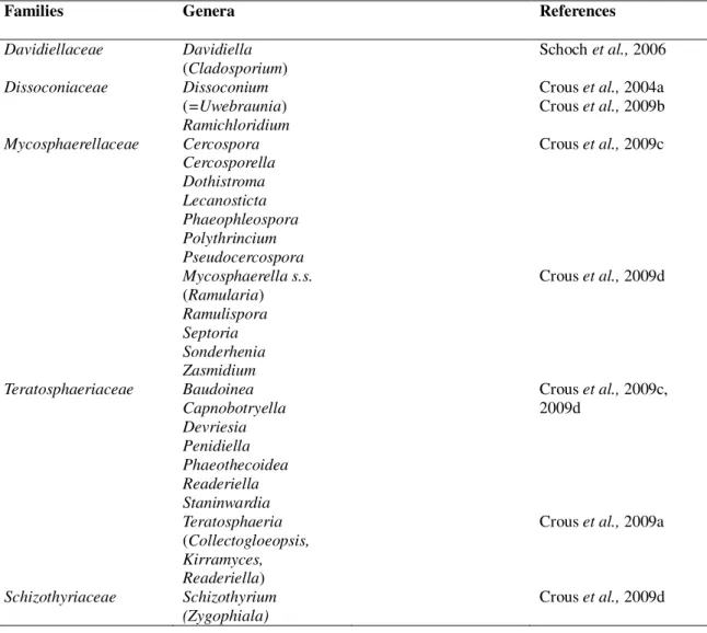
Table 1.1 Morphological characteristics of several genera of Mycosphaerellasensu lato (Crous et al., 2007a) ……… 19 Table 1.2 Phylogenetic studies that have analysed the Mycosphaerella complex
between 2006 and 2009 ………...……... 21
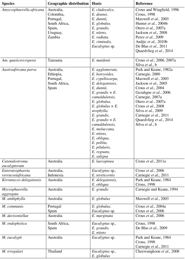
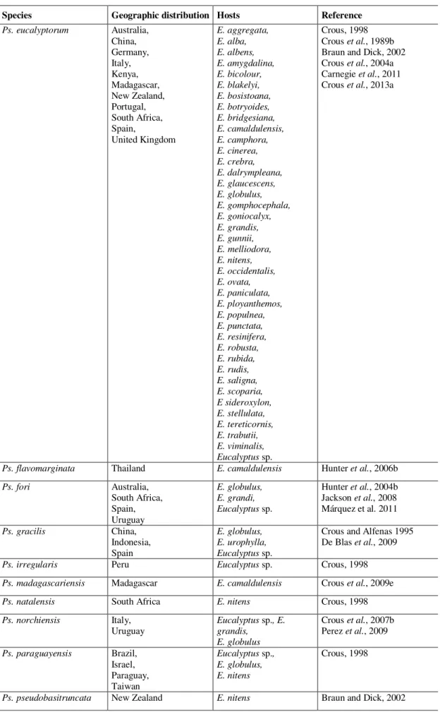
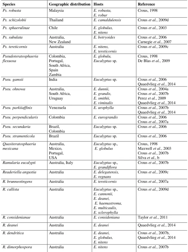
Table 1.3 Genera and species of fungi occurring on Eucalyptus leaves and stems … 23 Table 1.4 Teratosphaeria canker records by year, country and host …... 49
Table 1.5 Recent new families that left Mycosphaerellaceae and Teratosphaeriaceae
and new genera, combinations on Mycosphaerellaceae and
Teratosphaeriaceae and the respective original name ... 50 Chapter 2 Mycosphaerella and Teratosphaeria species associated with Mycosphaerella Leaf
Disease on Eucalyptus globulus in Portugal
Table 2.1 Locations of the ten plantations studied along the coastal border, ordered
from north to south, total number of fungal species identified at each location and its presence (1) and absence (0): (C) M. communis; (G) M. grandis; (L) M. lateralis; (MK) M. marksii; (V) M. vespa; (MLL) T. molleriana; (N) T. nubilosa; (P) T. parva and number of other species
unidentified ………... 77
Table 2.2 Species identified on juvenile leaves of Eucalyptus globulus based in the
percentage of similarity of each isolate to its closest match at the GenBank
nucleotide sequences database ……….. 79
Chapter 3 New records, species and combinations in Teratosphaeria on Eucalyptus in
Portugal
Table 3.1 Isolates included in the phylogenetic analyses ………. 89
Table 3.2 The Devriesia and Neodevriesia species and their hosts ……….. 109 Chapter 4 Species in the Eucalyptus Leaf Disease Complex in Portugal
Table 4.1 Reports of the most commonly found foliar fungi in the field for several
countries………... 120
Table 4.2 Isolates included in the phylogenetic analyses ………...…… 122
Table 4.3 Species identified on juvenile leaves of Eucalyptus globulus based on the
percentage similarity of each isolate to its closest match in GenBank, but not included in the phylogenetic tree..……..…... 126
Table 4.4 Total of occurrences of species and combinations among them in
symptomatic leaf lesions………. 129
Table 4.5 Number of lesions, Severity level (SL), number of species (NSpp) per
Chapter 5 Teratosphaeria gauchensis on Eucalyptus in Portugal
Table 5.1 Collection details and GenBank accession numbers of isolates included in
this study ……… 146
Table 5.2 Comparison with others studies of the culture growth at different
temperatures and conidial dimensions ………... 151
Table 5.3 Mean of culture growth (mm) of Portuguese isolates of Teratosphaeria gauchensis at different temperatures after 4 weeks on MEA. Means
comparison were made with Fishers Least Significant Difference (LSD) test. Within each column means with different letters are significantly
different (p <0.05) ……….. 151
Chapter 6 Pathogenicity of species in the Eucalyptus Leaf Disease complex
Table 6.1 List of taxa and strains isolated from juvenile leaves of Eucalyptus globulus
plantations surveyed in Portugal between 2004 and 2010 and inoculated on
this study ………. 171
Table 6.2 Mean values and standard error of lesion diameter (mm) on abaxial surface
of non-wounded (nW) and wounded (W) leaves, 102 days after inoculation with different species. For each column, values with different letters differ
significantly according with the Duncan’s test. Values are sorted by ∆(nW -W) representing the variation between mean values of lesions on nW and W
leaves ………... 173
Appendix B Plantations, Symptomatic leaves and Pattern of Ascospore Germination and
Cultures
Table B.1 Number of Eucalyptus globulus plantations selected to evaluate ELDC,
collections, trees, symptomatic leaves, lesions, isolates obtained and
XXV
ACT Actin
ANOVA Analysis of variance
ATPase 6 gene mitochondrial adenosine triphosphate 6 gene
BLAST Basic Local Alignment Search Tool
bs bootstrap support
CBS Culture collection of Centraalbureau voor Schimmelcultures
Fungal Biodiversity Centre, Utrecht, The Netherlands
cf. confer–“compare”
CI Consistency index
CMW Culture collection of M.J. Wingfield, housed at Forestry and
Agricultural Biotechnology Institute
comb. nov. combinatio nova–“new combination”
CPC Culture collection of P.W. Crous, housed at CBS
diam. in diameter
DNA Deoxyribonucleic acid
dNTP Deoxyribonucleotide triphosphate
EDTA Ethylenediaminetetra acetic acid
EF-1α Translation elongation factor 1-alpha
EFN culture collection of ex-Estação Florestal Nacional, Instituto
Nacional dos Recursos Biológicos, Oeiras, Portugal, now housed at LISFA
e.g. exempli gratia–“for example”
ELDC Eucalyptus Leaf Disease Complex
et al. et alii –“and co-workers”
gen. nov. genus novum–“new genus”
HI Homoplasy index
INIA National Institute for Agrarian and Veterinary Research
ITS Internal transcribed spacer
ITS1-5.8S-ITS2 cluster Cluster of Internal transcribed spacer 1,
5.8S ribosomal RNA gene, and internal transcribed spacer 2
LISFA Herbarium Code of Unit for Research and Services on
Agricultural and Forestry Systems and Plant Health, INIA I.P., Oeiras, Portugal
LSU Large subunit of 28S rDNA
MEA Malt extract agar
ML Maximum-likelihood
MLD Mycosphaerella leaf disease
MP Maximum parsimony
MUCC MurdochUniversity culture collection, Australia
NCBI National Center for Biotechnology Information
NJ Neighbor joining
PCR Polymerase chain reaction
RAPD randomly amplified polymorphic DNA
RAxML Randomized Axelerated Maximum Likelihood
rDNA Ribosomal DNA
RH relative humidity
RI Retention index
SE Standard error of the mean
s. l. sensu lato–“in the wide or broad sense”
sp. nov. species nova–“new species” sp., spp. (plural) species
SSU small subunit of 18S rDNA
STE-U Culture collection of the Department of Plant Pathology,
Stellenbosch University, South Africa.
TL Tree length
TLD Teratosphaeria Leaf Disease
UV Ultraviolet
vs against (versus)
XXVII
Current name Synonyms Family
Amycosphaerella africana ≡ Mycosphaerella africana (Basionym) ≡ Teratosphaeria africana
≡ Mycosphaerella ellipsoidea ≡ Mycosphaerella aurantia
Mycosphaerellaceae
Amycosphaerella quasicercospora ≡ Mycosphaerella quasicercospora (Basionym) ≡ Teratosphaeria quasicercospora
Mycosphaerellaceae
Austroafricana parva ≡ Mycosphaerella parva (Basionym) ≡ Teratosphaeria parva
≡ Mycosphaerella grandis
Teratosphaeriaceae
Neodevriesia hilliana ≡ Devriesia hilliana (Basionym) Neodevriesiaceae
Pallidocercospora heimii ≡ Mycosphaerella heimii ≡ Pseudocercospora heimii
Mycosphaerellaceae
Paramycosphaerella marksii ≡ Mycosphaerella marksii (Basionym) Mycosphaerellaceae
Quasiteratosphaeria mexicana ≡ Mycosphaerella mexicana (Basionym)
≡ Teratosphaeria mexicana
Teratosphaeriaceae
Teratosphaeria lusitanica Sp. nov. Teratosphaeriaceae
Teratosphaeria molleriana ≡ Sphaerella molleriana (Basionym) ≡ Mycosphaerella molleriana ≡ Colletogloeopsis molleriana ≡ Readeriella molleriana ≡ Mycosphaerella vespa ≡ Mycosphaerella ambiphylla ≡ Teratosphaeria xenocryptica
Teratosphaeriaceae
Teratosphaeria nubilosa ≡ Sphaerella nubilosa (Basionym)
≡ Mycosphaerella nubilosa ≡ Mycosphaerella juvenis
Teratosphaeriaceae
Teratosphaeria pluritubularis ≡ Mycosphaerella pluritubularis (Basionym) Teratosphaeriaceae
Teratosphaericola pseudoafricana ≡ Mycosphaerella pseudoafricana (Basionym)
≡ Teratosphaeria pseudoafricana Teratosphaeriaceae
XXIX Am. Amycosphaerella
A. Austroafricana
M. Mycosphaerella
P. Paramycosphaerella
Pc. Pallidocercospora
Ph. Phaeophleospora
Pth. Phaeothecoidea
Pp. Parapenidiella
Ps. Pseudocercospora
Pseu. Pseudoteratosphaeria
R. Readeriella
S. Septoria
So. Sonderhenia
Su. Suberoteratosphaeria
T. Teratosphaeria
U. Uwebraunia
X. Xenomycosphaerella
XXXI
Acknowledgments ……….. XI
A
BSTRACT………... XIIIR
ESUMO ………...XVPublications arising from the current thesis ………...... XVII
Index of Figures ………. XIX
Index of Tables ………... XXIII List of Abbreviations and Acronyms……….……….……… XXV List of Mycosphaerellaceae, Neodevriesiaceae and
Teratosphaeriaceae Genera and its synonyms used within the current thesis ………… XXVII List of genera abbreviations used in this thesis ……….. XXIX
I. G
ENERALI
NTRODUCTION ………... 01II. LITERATURE
R
EVIEW……….. 09Chapter 1 Eucalyptus Leaf Disease Complex ………... 11
III. S
TATUS OFE
UCALYPTUSL
EAFD
ISEASEC
OMPLEX INP
ORTUGAL …………... 69Chapter 2 Mycosphaerella and Teratosphaeria species associated with
Mycosphaerella Leaf Disease on Eucalyptus globulus in Portugal …………..
……… 71
Chapter 3 New records, species and combinations in Teratosphaeria
on Eucalyptus in Portugal ……...……….83
Chapter 4 Species in the Eucalyptus Leaf Disease Complex in Portugal ………. 115
IV. O
THERT
ERATOSPHAERIA DISEASES ……….. 137Chapter 5 Teratosphaeria gauchensis on Eucalyptus in Portugal...………..…………... 139
V. S
PECIES BEHAVIOUR IN THEC
OMPLEX………..………..………...
163Chapter 6 Pathogenicity of species in the Eucalyptus Leaf Disease Complex ………… 165
VI. G
ENERALC
ONCLUSIONS ………..………..………..…………... 1833 General Introduction
This study is mainly focused on morphological and molecular identification of species associated with the Eucalyptus Leaf Disease Complex (ELDC) and others species involved. Some attention was also centred on understanding the role of each species on the complex.
An overview of the background knowledge on Mycosphaerella s.l. is presented and
developed in the Literature Review (Chapter 1). The general objectives of the study are displayed forward, aiming to unravel some of the prevailing unknown species.
Background knowledge
Approximately 700 species of fungi have been linked to Eucalyptus leaves and stems.
The disease associated with Mycosphaerella s.l. teleomorphs and several anamorph genera
started to be studied in detail at the end of the 1990s (Crous, 1998). Previous names related to this disease complex were Mycosphaerella Leaf Spot, Mycosphaerella Leaf Blotch, Crinkle
Leaf Blight and Mycosphaerella Leaf Disease because many different anamorphic fungi were
collected from affected leaves and many of the fungi were associated with the teleomorph genus
Mycosphaerella (Park et al., 2000). Between 2006 and 2009 there was a taxonomic
reorganization of Mycosphaerella s.l. that resulted in a redistribution of the genera and species
into different families. Thus the name Mycosphaerella Leaf Disease became particularly
unsuitable since most of the important pathogens on eucalypts were transferred from
Mycosphaerella to Teratosphaeria (e.g., T. nubilosa and T. cryptica) and for a short time the
disease was called Teratosphaeria Leaf Disease (TLD) (Schoch et al., 2006; Crous et al., 2007;
Crous et al., 2009a, 2009b, 2009c). Thereafter it became clear that many different genera were
associated with the disease and the application of a fungal genus name to the disease was not suitable. Furthermore, it became clear that this disease was not caused by a single pathogen but a complex of species related to similar symptoms. For all those reasons it was decided that in this thesis the term "Eucalyptus Leaf Disease Complex" (ELDC) will be used, and recommend that this term be used in all future studies.
Earlier studies used 14 different ascospore germination patterns as a character in morphological identification of Mycosphaerella s.l. (Fig. B.2, B.3). However, it is now known
4
addition, many of these species are difficult to grow in culture, while others grow very slowly and do not form asexual spores in culture. Some of the species are host-specific and produce small fruiting structures with very conserved morphology (Crous, 1998; Hunter et al., 2006). As
a result, morphological identification is extremely difficult and it is essential to confirm the identity by molecular means. Sequence data of the ITS region provides enough resolution to differentiate most taxa (Hunter et al., 2006).
In 1829 eucalypts were introduced into Portugal as ornamental trees and began to be exploited commercially for its wood quality for pulp production on the last half part of XXth Century (Potts, 2004). Today these species represent the first continental forestry area with 812 000 ha, about 26% (ICNF 2013) particularly planted in the coastal regions (Valente et al.,
2008).
Eucalypts benefited from the “absence” of pests and diseases before 1970 (Crous and Wingfield, 1997; Valente et al., 2008). The earliest report of Mycosphaerella s. l. outside of
Australia was reported in 1881 (T. molleriana) in Portugal on leaves of Eucalyptus globulus
(Crous and Wingfield, 1997). However this disease was unnoticed in Portugal before 1999, due to the absence of severe defoliation on young trees. Early studies reported about 10 species on eucalypts in Portugal, but this was based on informal collections (Crous and Wingfield, 1997; Crous, 1998; Crous et al., 2006). This disease affects mainly young trees in the juvenile foliage
phase, resulting in reduced growth rate of the trees and lower wood volume, which causes significant productivity losses.
Some studies of epidemiology and pathogenicity have been made on Mycosphaerella s. l.
on Eucalyptus in particular on Austroafricana parva, Teratosphaeria cryptica and T. nubilosa
(e.g. Park & Kean, 1982a, 1982b) the last two were considered the most virulent on eucalypts (Fig. B.4, B.5). However, there have been no such studies on others species belonging to this complex.
All these subjects are described in detail in Chapter 1 – Literature Review.
This work
In Chapter 1 the complex of species of Mycosphaerella s. l. was reviewed, in particular
the latest developments about evaluation of genera and how the species have been re-distributed into several genera, including Teratosphaeria. This provides an update on the
5
geographical distribution of the disease, the taxonomy of the pathogens and new potential pathogens that can also cause diseases on eucalypts.
A survey of species associated with ELDC in Portugal is reported in Chapter 2. Symptomatic leaves were collected from E. globulus plantations, fungi were isolated and
characterized in terms of their morphology (Table B.1; Fig. B.1). DNA sequences of the ITS region were used to give an indication of the species that are associated with the disease and to indicate the most frequent species in the disease complex.
Teratosphaeria species and some phylogenetically closely species are the subject of
Chapter 3. In this chapter five new records for the Iberian Peninsula were added: Neodevriesia hilliana was reported for the first time on Myrtaceae; Quasiteratosphaeria mexicana, Teratosphaericola pseudoafricana and Teratosphaeria pluritubularis, the last one only in
Portugal and Teratosphaeria lusitanica was introduced as a new species in the Iberian
Peninsula. New anamorphic structures were included and described for A. parva, Q. mexicana, T. pluritubularis and T. pseudoafricana. Furthermore, two new combinations were made,
namely Amycosphaerella quasicercospora and Quasicercospora mexicana. Thus an update of Teratosphaeria species and allies on E.globulus in Portuguese plantations is presented.
With the aim of determining the role played by the species present in the complex of the lesions studied, and based on the ascospores germination patterns with similar culture morphology, several isolates were confirmed by molecular characterization and their phylogenetic analysis was accomplished. Furthermore, species present in a single lesion were quantified and the results compared to severity levels in order to evaluate the composition of the complex. Some species were reported for the first time related to the disease symptoms including Cladosporium cladosporioides, Fusicladium eucalypti, Mycosphaerella madeirae
(first report from mainland Portugal) and Venturiaceae sp. All the other species previously
reported were also found (Chapter 4).
During the surveys, attention was made to check for new symptoms on eucalypts leaves and to determine if any cankers are involved (Chapter 5). Regarding the cankers observed, severe damage on E. globulus has been observed for the first time in Portugal. The cause of this
canker was identified from sequence data of the ITS1-5.8S-ITS2 and EF-1α clusters and morphological characteristics and determined to be Teratosphaeria gauchensis.
Leaf spots on Eucalyptus are normally attributed to species like T. nubilosa and T. cryptica, which are considered the most virulent ones, but many other species can inhabit the
6
evaluate the individual behaviour of the several species found in the leaf disease complex and compare their capability to colonize leaf tissues and cause leaf necrosis. Such a study is reported in Chapter 6.
Objectives
The purpose of this work is to explore some of the knowledge gaps related to the "Eucalyptus leaf disease complex" in Portugal aiming to identify the species found during a survey.
The specific objectives of this thesis are:
To compile aliterature review on Mycosphaerellas. l. (Chapter 1).
To conduct a survey of fungi associated with symptomatic leaves collected from
E. globulus plantations, identify them in terms of morphology and DNA
phylogeny and determine the most frequent species in the disease complex (Chapter 2 and 4).
To evaluate morphologically and phylogenetically the existence of new species, make new combinations where necessary (Chapter 3).
To evaluate the relative frequency of species in E. globulus plantations during
several seasons, its combinations in symptomatic lesions and the relation of the complex composition with disease severity (Chapter 4).
To observe collections for new leaf symptoms and for canker development (Chapter 5).
To characterize aggressiveness of the species in the complex (Chapter 6).
7 References
Crous P.W., 1998. Mycosphaerella spp. and their anamorphs associated with leaf spot diseases of Eucalyptus. Mycologia Memoir, 21, 1–170. St. Paul, Minn., USA, APS Press.
Crous P.W., C.L. Schoch, K.D. Hyde, R. Wood, C. Gueidan, G.S. de Hoog and J.Z. Groenewald, 2009a. Phylogenetic lineages in the Capnodiales. Studies in Mycology 64, 17–47.
Crous P.W., B.A. Summerell, A.J. Carnegie, M.J. Wingfield and J.Z. Groenewald, 2009b. Novel species of Mycosphaerellaceae and Teratosphaeriaceae. Persoonia 23, 119–146.
Crous P.W., B.A. Summerell, A.J. Carnegie, M.J. Wingfield, G.C. Hunter, T.I. Burgess, V. Andjic, P.A. Barber and J.Z. Groenewald, 2009c. Unravelling Mycosphaerella: do you believe in genera? Persoonia 23, 99–118.
Crous P.W. and M.J. Wingfield, 1997. Colletogloeopsis, a new coelomycete genus to accommodate anamorphs of two species of Mycosphaerella on Eucalyptus. Can J Bot 75, 667–674.
Hunter G., B.D. Wingfield, P.W. Crous and M.J. Wingfield, 2006. A multi-gene phylogeny for species of Mycosphaerella occurring on Eucalyptus leaves. Studies in Mycology 55, 147–161.
Park R.F. and P.J Keane, 1982a. Three Mycosphaerella species from leaf diseases of Eucalyptus. Transactions of the British Mycological Society 79, 95–100.
Park R.F. and P.J. Keane, 1982b. Leaf diseases of Eucalyptus associated with Mycosphaerella species. Transactions of the British Mycological Society 79, 101–115.
Park R.F., P.J. Keane, M.J. Wingfield and P.W. Crous, 2000. Fungal diseases of eucalypt foliage. In Diseases and pathogens of eucalypts. (Keane, P.J., G.A. Kile, , F.D. Podger and B.N. Brown, Ed.), 153–239, CSIRO publishers, Australia.
Schoch C.L., R.A. Shoemaker, K.A. Seifert, S. Hambleton, J.W. Spatafora and P.W. Crous, 2006. A multigene phylogeny of the Dothideomycetes using four nuclear loci. Mycologia 98, 1043–1054. Valente, C., H. Machado and M. Silva, 2008. Eucalypts pests and diseases. In: Pragas e doenças em
C
H
A
P
TE
R
1
Eucalyptus Leaf Disease Complex
13
Chapter 1
REVIEW PAPER
Eucalyptus Leaf Disease Complex
Márcia R. C. Silva1,3, Helena Machado1, Alan J. L. Phillips2
1Instituto Nacional de Investigação Agrária e Veterinária, I.P., Unidades Estratégicas de Investigação e Serviços, Sistemas Agrários e Florestais e Sanidade Vegetal, Quinta do Marquês, 2780-159 Oeiras, Portugal;
2UCIBIO, Departamento de Ciências da Vida, Faculdade de Ciências e Tecnologia, Universidade Nova de Lisboa, 2829-516 Caparica, Portugal;
14 Summary
Eucalypts are the second most important forest plantation species grown worldwide. A complex of species of Mycosphaerellasensu lato causes leaf disease on Eucalyptus that results
in significant economic losses wherever eucalypts are grown. Although the causal agents were included mainly in Mycosphaerella this genus was recently re-evaluated and the species
distributed into several genera, including Teratosphaeria and others. This review provides an
update on the disease, the damage caused, impact, influence of weather conditions, disease control, and pathogenicity, focussing on selected countries especially Portugal. Also reviewed are the geographical distribution of the disease, taxonomy of the pathogens and includes new and other possible leaf diseases that can also cause cankers on eucalypts.
Key words. Mycosphaerella, Teratosphaeria, Capnodiales, Dothideomycetes, eucalypts.
Introduction
Host
Eucalypts, commonly known as gum trees are a important forest plantation species grown worldwide. The genus Eucalyptus was introduced by L’Héritier de Brutelle (1789) for a single
species, E. obliqua. In the following 200 or more years, many species names have been
published in the genus (Brooker, 2000). Eucalypts are found principally in the southern hemisphere and are native to Australia, but since the late 18th century they have been spread around the world (Potts, 2004).
Eucalypts are well-known for their straight form, fast growth, also on hard habitat, facility in vegetative propagation, adaptation to soils and to a wide range of climates including high rainfall, semi-arid, sea level and alpine tree line zones (Old et al., 2003; Potts, 2004).
Commercially they are important for their special wood properties and for pulp production, these attractive product qualities have induce the extensive installation of eucalypt plantations in many countries (Old et al., 2003).
15
This may cause widespread epidemic fungi and pathogens can be transmitted on infected seeds or planting stocks (Old et al., 2003). Some studies on the Leaf Disease Complex,
(predominantly species of Mycosphaerella and Teratosphaeria) and involving different species
of eucalypts point to E. globulus as the most susceptible species (Carnegie et al., 1994; Dungey et al., 1997; Tejedor, 2004).
Species concepts
Before discussing the taxonomy of Mycosphaerella is necessary to outline the concepts
that are used to circumscribe the species. Several criteria have been used to contribute to taxonomical studies of fungi. First of all it is important to distinguish between species concept and species criteria. Species concept is a description of the kind of entity that constitutes a species, while criteria that delimit a particular species, i.e. the practical standards for the recognizing whether individuals should be considered members of the same species, are called species criteria (Taylor et al., 2000; Cai et al., 2011).
The diverse criteria that allow the delimitation of species can be characterized as morphological, physiological, intersterility, host specificitation, and phylogenetic. These species recognition criteria try to recognize evolutionary independent lineages (Taylor et al., 2000; Cai et al., 2011). These comprise the Morphological Species Concept (MSC); the Biological Species
Concept (BSC); the Ecological Species Concept (ESC); the Phylogenetic Species Concept (PSC) and the Genealogical Concordance Phylogenetic Species Recognition (GCPSR); Polyphasic Method (PM) and finally the Consolidated Species Concept (CSC). However searching for a single species criterion valid for all cases is basically impossible (Giraud et al.,
2008).
16
The BSC was proposed by Ernst Mayr (1942) and considers species as “groups of
actually or potentially interbreeding natural populations, which are reproductively isolated from
other such groups”. The capacity to mate resulting in a fertile progeny is the major aim of this species concept. This concept can potentally be applied to fungi that have a sexual stage in their life cycle. However, mating and production of the sexual state of most fungi in culture has not been possible, and many species are known to be purely asexual. Therefore, the BSC is not a feesible concept for fungi.
The ecological species concept (ESC) is used to describe populations that are adapted to certain ecological niches and due to their adaptations will form discrete morphological clusters. According to the ESC, populations form the discrete phenetic clusters that we identify as species because the ecological and evolutionary processes controlling how resources are divided up tend to produce those clusters (Ridley, 2003). Giraude et al. (2010) emphasizes the
adaptation to a particular ecological niche is also linked to the emergence of novel fungal diseases of plants. Thus, host shift speciation is one of the primordial paths for emergence of new fungal diseases and is a particular case of ecological speciation.
ESC has the advantage of recognizing the role played by the environment in controlling morphological development. On the other hand, it has disadvantages such as potentially ignoring cryptic species (two or more distinct species classified as a single species) and not easily defining ecological niches as a whole. In addition, ESC encompasses the idea of host association and naming species according to the host on which they occur. This can result in an enormous proliferation of names, many of which are synonyms.
Over the last years, the phylogenetics approach became a rapid DNA tool that could resolve fungal taxa that BSC could not resolve (Taylor et al., 2000; Hunter et al., 2006b). The
PSC became well suited for fungi because it relates both sexual and asexual organisms and emphasizes a nucleotide divergence between monophyletic lineages (Taylor et al., 2000). The
problem with this criterion is that it is based on the assumption that the whole genome is represented by a gene that should have the same evolutionary history, which in some cases did not occur.
Taylor et al., (2000) described GCPSR as an extension of the PSC and offered a better
17
During the last decade, the polyphasic approach of combining Biological, Morphological and Phylogenetic Species Concepts has revolutionised the taxonomy of fungi (Lombard et al.,
2010). More recently Quaedvlieg et al., (2014) proposed a formal change in the name of the
polyphasic approach, which they called the Consolidated Species Concept (CSC).
Taxonomy of Mycosphaerella and related genera
Persoon (1794) described Sphaeria corylea (Pers.) on dead leaves of Corylus. Over the
next 3 years he discovered more related species and then (Persoon, 1797) transferred them all to
Sphaeria maculiformis (Pers.) relegating the species epithet to the status of variety.
Subsequently, Saccardo transferred all species of Sphaeria with 1-septate, hyaline ascospores to Sphaerella (Saccardo, 1882). However, the genus name Sphaerella was already occupied by
green algae and all Sphaerella species were placed in Mycosphaerella (Aptroot, 2006). Thus,
the first generic description for Mycosphaerella Johanson (1884) was that of Sphaerella (1882).
Early mycologists frequently described new species based on the host with which they were associated. This lead to a proliferation of species names, many of which were later reduced to synonymy (Von Arx, 1949; Barr, 1972; Tomilin, 1979; Corlett, 1991). Aptroot (2006) re-examined more than 10 000 taxa in Mycosphaerella and recognized about 3000 species.
The first phylogenetic studies, based on ITS sequence data, suggested that
Mycosphaerella is monophyletic (Crous et al., 2000; Goodwind et al., 2001). Later, however,
Maxwell (2004) showed that based on ITS sequence data many of the anamorph genera within
Mycosphaerella are polyphyletic.
Müller and Oehrens (1982) characterized the genus Teratosphaeria as “globose,
perithecium-like ascomata growing inside the living leaf-tissue, with bitunicate asci and
brownish, bicellular ascospores”. Taylor et al., (2003) resolved the relationships among
members of the Mycosphaerellaceae by phylogenetic analysis of ITS sequence data and
concluded that Teratosphaeria was a synonym of Mycosphaerella. Furthermore,
phylogenetically it was monophyletic based on ITS (Crous et al., 2001).
Crous et al., (2007) used combined ITS and LSU sequence data and demonstrated
polyphyly (within Teratosphaeria) and paraphyly (within the Capnodiales) in Mycosphaerella.
18
Crous et al. (2007a) recognized a subset of isolates, representing various species as
morphologically different from Mycosphaerella s. str. (Crous, 2009). That study positioned Teratosphaeria within the Teratosphaeriaceae because of the distinct asexual morphs and DNA
phylogenetic data, also the presence of pseudoparenchymatal remnants in ascomata, ascospores that turn brown and verruculose while still in the asci, and ascospores with a mucoid sheath. Thus, polyphyly was demonstrated in a phylogeny based on combined ITS and partial large subunit (LSU) of the nuclear rRNA operon sequence data. Generic concepts were stabilized when Teratosphaeria was reinstated and several species of Mycosphaerella were transferred to Teratosphaeria. All genera, except Schizothyrium, were characterized by pseudothecial
ascomata (Table 1.1). Furthermore, Colletogloeopsis, Kirramyces and Readeriella anamorphs
were transferred to Teratosphaeria (Crous et al, 2009a, 2009d).
Basically, between 2006 and 2009, phylogenetic studies based on partial LSU gene sequences supported by anamorph and teleomorph morphologies revealed that the
Mycosphaerella complex resides in several different families (Davidiellaceae, Dissoconiaceae, Mycosphaerellaceae, Teratosphaeriaceae and Schizothyriaceae) and are represented by several
genera (Table 1.2) (Schoch et al., 2006; Crous et al., 2009b, 2009c, 2009d).
In 2009, Crous et al. (2009c) considered generic boundaries in the Teratosphaeriaceae and Mycosphaerellaceae. Thus, Mycosphaerellaceae has Cercospora, Cercosporella, Dothistroma, Lecanosticta, Phaeophleospora, Polythrincium, Pseudocercospora, Ramularia
(Mycosphaerella s. s.), Ramulispora, Septoria, Sonderhenia and Zasmidium anamorphs. The
genera Ramichloridium and Dissoconium were excluded from the Mycosphaerellaceae and
shown to represent an undefined family.
In 2013, Hyde et al. (2013) added 10 more families to the Dothideomycetes, 7 new
orders, 38 asexual genera within Mycosphaerellaceae, and 22 asexual plant pathogenic and
extremophilic genera in Teratosphaeriaceae. Furthermore, Crous et al. (2013a) studied Pseudocercospora (an anamorphic state with mycosphaerella-like teleomorphs) and recognised
14 clades, six of which cluster in Mycosphaerellaceae. Pseudocercosporas. str. corresponds to
a distinct clade, sister to Passalora eucalypti, and a clade representing the genera Scolecostigmina, Trochophora and Pallidocercospora, taxa formerly accommodated in the Mycosphaerella heimii complex. Also, Quaedvlieg et al. (2013) studied Septoria, which was
shown to be a different genus in the Mycosphaerellaceae, which has mycosphaerella-like sexual
19
In 2009, Crous (2009) reviewed the taxonomy and phylogeny of Mycosphaerella genus
and their anamorphs and sum up several old studies like Klebahn (1918), Laibach (1922), Müller and von Arx (1962), Von Arx (1983) and Crous (1998).
The genus Mycosphaerella is placed in Capnodiales (Schoch et al., 2006) as a large
genus of ascomycetous, mostly leaf infecting fungi. Capnodiales is inserted in Dothideomycetes
that have approximately 115 families and include a highly varied range of fungi characterized mainly by asci with two wall layers (bitunicate asci) and frequently with fissitunicate dehiscence (Hyde et al., 2013).
Table 1.1 Morphological characteristics of several genera of Mycosphaerellasensu lato (Crous et al., 2007a).
Genera Morphological Characters
Mycosphaerella-like Genera
Davidiella - Ascosporeswithirregular,angularlumens
Dissoconium - Actively discharged conidia, conidiophores solitary, pale brown, giving rise to primary and secondary
Mycosphaerella s. str. - Conidiomata variable from solitary conidiophores to sporodochia, fascicles to pycnidia, but conidia not actively discharged
Teratosphaeria - Ascospores turning brown in asci often observed, hamathecial tissue, ascospore sheath, multi-layered endotunica, prominent periphysoids, ascomata usually linked by superficial stroma;
Schizothyrium - Ascomata thyrothecial Teratosphaeria (Teratosphaeriaceae)
Cibiessia Pseudotaeniolina
- Withhyphae submerged to superficial, disarticulating into arthroconidia.
Cibiessia - Hyphae superficial, brown to green-brown, smooth, disarticulating to form pale brown, cylindrical, 0–3-septate conidia with subtruncate ends, frequently with a Readeriella synanamorph
Pseudotaeniolina - Mature, brown hyphae disarticulating into thick-walled, spherical, smooth to verruculose 0(–2) transversely septate, brown conidia
Batcheloromyces Capnobotryella Catenulostroma Devriesia
Hortaea Nothostrasseria Penidiella
Phaeothecoidea
Readeriella Staninwardia
- Hyphae not disarticulating into arthroconidia and with endoconidia absent (except Phaeothecoidea)
Capnobotryella Devriesia Hortaea