Ana Raquel Abrunhosa Carvalho de Lima
Setembro de 2012
Neuron-astrocyte interactions in complex
cognitive functions
Interacções neurónio-astrócito em
funções cognitivas complexas
UMinho|20
12
Ana Raq
uel Abrunhosa Car
valho de Lima
Neuron-astrocyte interactions in comple
x cognitive functions
Universidade do Minho
Escola de Ciências da Saúde
Trabalho realizado sob a orientação do
Doutor João Oliveira
Ana Raquel Abrunhosa Carvalho de Lima
Dissertação de Mestrado
Mestrado em Ciências da Saúde
Neuron-astrocyte interactions in complex
cognitive functions
Interacções neurónio-astrócito em
funções cognitivas complexas
Universidade do Minho
DECLARAÇÃO
Nome: Ana Raquel Abrunhosa Carvalho de Lima Endereço Electrónico: [email protected] Telefone: 919926722
Nº de Bilhete de Identidade: 13459797
Título da dissertação:NEURON-ASTROCYTE INTERACTIONS IN COMPLEX COGNITIVE FUNCTIONS
Orientador: Doutor João Oliveira
Ano de conclusão: 2012
Ramo de Conhecimento do Mestrado: Ciências da Saúde - Ciências da Saúde
É AUTORIZADA A REPRODUÇÃO INTEGRAL DESTA TESE/TRABALHO APENAS PARA EFEITOS DE INVESTIGAÇÃO, MEDIANTE DECLARAÇÃO ESCRITA DO INTERESSADO QUE A TAL SE COMPROMETE.
Universidade do Minho, 3 de Setembro de 2012
Assinatura: ____________________________________________________________________
A
GRADECIMENTOSEm primeiro lugar quero agradecer à minha família. Aos meus pais que me apoiaram não só na decisão de ingressar neste mestrado, como também em todos os momentos mais desafiantes. Ao meu irmão caçula, André, por ser o Peixe-Beijoca, perito em gargalhadas que me animaram durante este ano. Ao meu irmão Pedro por partilhar comigo a filosofia do pumba-arrebentismo que me desvia do stresse e me devolve alguma sanidade mental. À minha tia Olga e à minha prima Inês (ou simplesmente a Prima), pelos momentos de descontracção proporcionados nos fins de semana. Aos meus avós por me terem acolhido tão bem deste lado e pela oportunidade que me proporcionaram.
Agradeço ao Leonel por todo o apoio nas maratonas de trabalho, pela paciência para aturar pseudo-estados depressivos, por me dar ânimo quando eu achava que já não conseguia mais, por me deixar fazer dos seus caracóis bola anti-stresse, por me acompanhar nas maratonas culinárias quando me apetecia jantar algo de diferente e estupidamente calórico... e por todo o carinho, é claro.
À Sofi e à Vi, agradeço pelo constante apoio, mesmo estando distantes no meio dos seus PhDs. Ao Sérgio e ao Hugo por darem apoio psicológico ao Leonel depois de me aturar...espero que tenha resultado!
Quero também agradecer ao João Oliveira, por ter acreditado em mim para iniciar este projecto de raiz, numa área na qual me vim a viciar, e pelos “dá-lhe giz” que me fizeram lutar ainda mais pelos objectivos.
Aos NeRDs, agradeço pela boa disposição geral e também pelo input científico. À Fernanda Marques, pela ajuda com os PCRs e à Luísa Pinto pelas dicas com as imunos. À Su e ao Sandro e Mónica, pelas conversas de corredor e de bar que me recarregavam as energias instantaneamente. À Su novamente por me ajudar com mil e uma cenas, à Mónica também por me ajudar com as imunos que teimavam em não funcionar. À Sofia e à Cristina, por ajudarem também a apagar alguns “fogos”. Às meninas do melhor open space: à Silvina pelas dicas zen; à Filipa pela constante alegria no matter what; à Magdinha pré- e pós-astrocítica por me acompanhar e ajudar nesta aventura; à Vanessa pela ajuda nesta fase final e pela energia que só uma Sardinha poderia ter!
A
BSTRACTSince the arousal of the “glia” concept, 150 years ago, the knowledge on the features of glial cells, especially astrocytes, has been dramatically evolving. These star-shaped cells that lack axons not only participate in brain metabolism, supporting neuronal activity, but also can modulate the neurotransmission by modulating synapses. From taking part in the “tripartite synapse” to the astrocytic excitability - based on intracellular Ca2+ increases - and
gliotransmission, astrocytes have been given the relevance that raises the idea that such glial modulation at cellular level should have greater implications in higher brain functions. However, these remarkable astrocytic features, as well as its influence in modulation of behaviour outputs, are still under-explored. In the attempt to contribute to the progress of our knowledge on these cells purpose in the brain, we decided to investigate the astrocytic component of the neuron-astrocyte interactions in complex cognitive processes that rely on the prefrontal cortex, analysing the behavioural performance of two animal models of astrocytic pathology. For that purpose, we implemented a rat model of pharmacological astrocytic ablation in the medial prefrontal cortex, through bilateral intracranial injections of the gliotoxin L-α-aminoadipate, and the transgenic dnSNARE mouse model, that displays conditional blockade of gliotransmitters vesicular release in astrocytes.
From the behaviour assessment of the rats subjected to the bilateral intracranial injections of L-α-aminoadipate we could conclude that the astrocytic ablation in the medial prefrontal cortex affects the attentional set-shifting, the working memory and the reversal learning. Microscope analysis of brain sections of the gliotoxin-injected animals revealed that astrocyte depleted regions were confined to the prelimbic and cingulate cortex, where the neuronal population was not affected. The genetic mouse model of conditional blockade of vesicular release in astrocytes was characterised in a more extensive way. We show here that these animals do not display anxious phenotype or have locomotor difficulties. Most importantly dnSNARE animals have shown impaired spatial reference memory (hippocampus-dependent) and improved working memory, but normal reversal learning (prefrontal cortex-dependent functions). Both astrocytes and gliotransmission seem to be crucial for cognitive computation and may represent a new window of understanding of brain function that urges to be clarified in terms of cellular pathways and biochemical mechanisms.
The successful implementation of both animal models of differential astrocytic pathology, at the ICVS will allow further studies in our lab. Despite our interesting results additional studies are required to help the elucidation of the mechanisms involved, such as using electrophysiological and neurochemical techniques to understand how brain dynamic is altered, either in healthy or in pathological states.
R
ESUMODesde há 150 anos, quando surgiu o conceito de “glia”, que o conhecimento das funções das células da glia, especialmente dos astrócitos, tem vindo a evoluir drasticamente. Estas células em forma de estrela e sem axónios, não só participam no metabolismo de cérebro como também contribuem para uma boa actividade neuronal, modelando até a actividade sináptica e o fluxo de neurotransmissores. Por fazerem parte da “sinapse tripartida”, exibirem uma forma de excitabilidade - baseada em elevações de Ca2+ intracelulares - e
realizarem gliotransmissão, os astrócitos têm recebido a relevância que levanta a questão de que tais células poderão ter maiores implicações nas funções cerebrais. Contudo, estas características notáveis bem como a sua influência na modulação do comportamento não estão ainda clarificadas. Por isso, na tentativa de contribuir para o progresso do nosso conhecimento acerca da função destas células no cérebro, decidimos investigar a componente astrocítica nas interacções neurónio-astrócito no contexto das funções cognitivas complexas dependentes do córtex pré-frontal, analisando a performance comportamental de dois modelos animais de patologia astrocítica: um modelo de rato na qual provocámos a ablação de astrócitos no córtex pré-frontal medial, através de injecções bilaterais intracranianas da gliotoxina L-α-aminoadipato; e um modelo genético em ratinho, dnSNARE, que exibe um bloqueio condicional da libertação vesicular de gliotransmissores nos astrócitos.
Da análise comportamental do modelo de rato, concluímos que a ablação de astrócitos no córtex pré-frontal medial afecta o attentional set-shifting, a memória de trabalho e a aprendizagem reversa. A análise microscópica de secções de cérebro dos ratos injectados com a gliotoxina revelou que as regiões de ablação astrocítica estavam confinadas ao córtex pré-límbico e córtex cingulado, onde a população neuronal não estava afectada. Uma caracterização mais extensiva do modelo de ratinhos dnSNARE revelou que estes animais não exibem fenótipo ansioso nem défices de locomoção, mas demostram défices na memória de referência espacial (dependente do hipocampo), memória de trabalho melhorada e aprendizagem reversa normal (funções dependentes do córtex pré-frontal). Tanto os astrócitos como apenas a gliotransmissão aparentam ser cruciais para a computação cognitiva e representam uma nova janela de conhecimento que urge ser clarificada em termos de vias celulares e mecanismos bioquímicos.
Apesar dos resultados interessantes já obtidos com a implementação já bem sucedida destes modelos animais no ICVS, são necessários estudos mais aprofundados para ajudar na elucidação dos mecanismos envolvidos. Acreditamos que o recurso a técnicas de electrofisiologia e neuroquímica permitirá uma melhor compreensão de como o cérebro está alterado, quer em condição de saúde ou doença.
T
ABLE OFC
ONTENTSA
GRADECIMENTOSiii
A
BSTRACTv
R
ESUMOvii
I
NDEX OFF
IGURES ANDT
ABLESxiii
L
IST OFA
BBREVIATIONSxv
1. I
NTRODUCTION1
1.1. Astrocytes in the brain 1
1.1.1. Arousal and evolution of the astrocytic concept 1
1.1.2. Astroglia in brain metabolism 2
1.1.3. Modulation of the synaptic transmission: the tripartite synapse 3 1.1.4. Integration of neuron-glia circuits: from gap junctions to gliotransmission 4
1.2. Astrocytes in brain disorders 7
1.3. Astrocytic function in vivo: behaviour and cognition 8
1.4. The prefrontal cortex 8
1.4.1. Cognitive functions of the the prefrontal cortex: learning and memory 9
2. A
IM OFW
ORK11
3. M
ATERIALS ANDM
ETHODS13
3.1.1. Aminoadipate rat model of astrocytic depletion 13 3.1.1.A.Surgical procedure for the establishment of the pharmacological rat model 13
3.1.1.B.Drug preparation and administration 14
3.1.2. Transgenic dominant-negative SNARE mice (dnSNARE) genetic model 14
3.1.2.A.Colony management and genotyping 16
3.2. Behaviour Tests 17
3.2.1. Aminoadipate rat model of astrocytic depletion 17
3.2.1.A.Attentional set-shifting task 17
3.2.1.B.Water maze tests 21
3.2.2. dnSNARE mice model of impaired vesicular release in astrocytes 23
3.2.2.A.Elevated Plus Maze (EPM) 23
3.2.2.B.Open field test (OF) 24
3.2.2.C.Water maze tests 24
3.2.2.D.Continuous spontaneous alternation test 26
3.3. Histological procedures 27
3.3.1. Euthanasia and tissue preparation 27
3.3.2. Immunofluorescent staining of astrocytes and neurons 27
3.3.3. Microscopic analysis 28
3.3.3.A.Identification of lesion sites 28
3.3.3.B.Stereological analysis of the affected region 28
3.3.4. Statistical Analysis 29
4. R
ESULTS31
4.1. Aminoadipate pharmacological rat model 31
4.1.1. Selection of animals and lesion features 31
4.1.2. Behaviour performance 32
4.1.2.A.Attentional set-shifting task 32
4.1.2.B.Water maze based tests 35
4.2. dnSNARE mice model of impaired vesicular release in astrocytes 36
4.2.1. Genotyping and selection of animals 36
4.2.2. Behaviour performance 37
4.2.2.A.Elevated plus maze (EPM) 37
4.2.2.B.Open Field test (OF) 40
4.2.2.C.Water maze based tests 42
4.2.2.D.Continuous spontaneous alternation test 44
5. D
ISCUSSION ANDC
ONCLUSION47
I
NDEX OFF
IGURES ANDT
ABLESFigures Figures Figures Figures
Figure 1.1 Astrocytes endfeet 2
Figure 1.2 The tripartite synapse 3
Figure 1.3 Modulation pathways of the synaptic transmission mediated by gap
junctions. 4
Figure 1.4 Calcium-dependent vesicular release of gliotransmitters from astrocytes. 6 Figure 1.5 Highlights of the PFC and mPFC, in human brain evidencing the homology
of mPFC and dlPFC brain regions in rat and human, respectively 9
Figure 3.1 Schematic representation of GFAP promotor driving the expression of the
target gene dnSNARE and reporter gene EGFP in astrocytes. 15
Figure 3.2 Representation of the ASST apparatus. 18 Figure 3.3 Representation of the water maze tests apparatus, prepared for rats 22 Figure 3.4 Representation of the water maze tests apparatus, prepared for mice 25 Figure 3.5 Representation of the Y maze apparatus. 26 Figure 4.1 Representation of the sites of brain injection sites, 4.20 mm to 2.76 mm from
Bregma 31
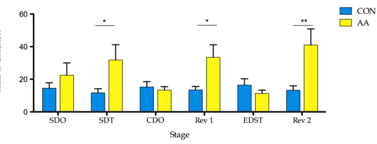
Figure 4.2 Effects of the gliotoxin L-α-AA. 33
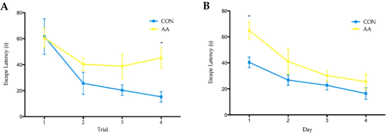
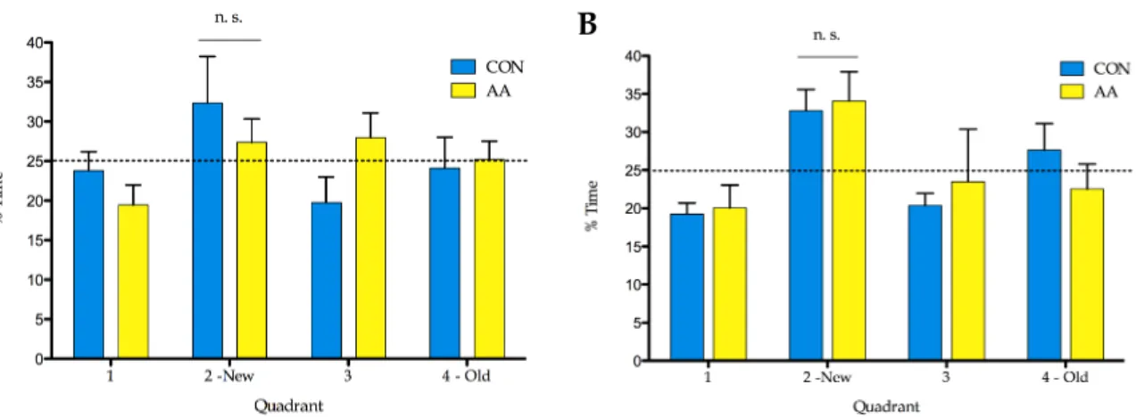
Figure 4.3 Trials to criterion along ASST stages. 34 Figure 4.4 Indexes of Learning and Reversal. 34 Figure 4.5 Escape latencies (s) in water maze tests. 35 Figure 4.6 Performance of the rats in the reversal learning task, RLT and probe task of
the water maze tests.
36
Figure 4.7. Mice genotyping. 37
Figure 4.8. Time spent (as percentage of total) in the arms and hub of the EPM. 38 Figure 4.9. Number of entrances in open arms and closed arms of the EPM. 39 Figure 4.10 Number of explorations in open arms and closed arms of the EPM. 39 Figure 4.11 Distance travelled (cm) in the open field arena. 40 Figure 4.12 Velocity (cm/s) in the open field arena. 41
Figure 4.14 Time (as percentage of total) spent in the center of the open field arena. 42 Figure 4.15 Escape latency (s) in the spatial reference memory task, RMT. 43 Figure 4.16 Performance in the reversal learning task, RLT. 44
Figure 4.17 Arm alternation (alternation score, %) in the continuous spontaneous alternation task, performed in the Y-maze 45
Figure 4.18. Total arm entries in the continuous spontaneous alternation task, performed
in the Y-maze. 45
Tables Tables Tables Tables
Table 3.1 PCR conditions used for the amplification of tTA and tet.O sequences. 17 Table 3.2 Order of discrimination stages, relevant and irrelevant dimensions, and
stimuli combinations of ASST presented to the animals. 19 Table 3.3 Stimuli combinations and sequence of presentation along 10 trials of the
simple discrimination, stages on test day one.
20 Table 3.4 Stimuli combinations and sequence of presentation along 10 trials of the test
first compound discrimination and its reversal stages.
20 Table 3.5 Stimuli combinations and sequence of presentation along 10 trials of the
test second compound discrimination and its reversal stages.
21 Table 3.6 . Timeline followed in the water maze tests used for rat models of astrocytic
ablation.
23 Table 3.7 Timeline followed in the water maze tests used for the mice models of
astrocytic dysfunction.
26
L
IST OFA
BBREVIATIONS♀ Female mice
♂ Male mice
AA Group of animals injected with L-α-aminoadipate, in the pharmacological rat model
Aβ amyloid-β
aCSF Artificial Cerebrospinal Fluid
AMPA α-amino-3-hydroxy-5-methyl-4-isoxazolepropionic acid
ASST Attentional Set-Shifting Task ATP Adenosine 5’-triphosphate
Ca2+ Calcium ion
CDO Compound discrimination of odours Cg Cingulate cortex
CON Group of animals injected with aCSF, in the pharmacological rat model
dnSNARE dominant-negative N-ethylmaleimide-sensitive factor attachment protein receptor
EDST Extradimensional shifting for textures
EGFP Enhanced Green Fluorescent Protein
EPM Elevated Plus Maze FBS Fetal Bovine Serum GABA γ-amino-butyric acid
GFAP Glial Fibrillary Acidic Protein GLUT1 Glucose transporter 1
Gt Gliotransmitters
H+ Hydrogen ion i.p. intraperitoneal
IL Infralimbic cortex
IP3 Inositol (1, 4, 5)-triphosphate
IVCS Life Sciences Research Institute K+ Potassium ion
L-α-AA L-α-Aminoadipate
mPFC medial prefrontal cortex
NCX Sodium/Calcium exchanger
NMDA N-Methyl-D-aspartic acid
Nt Neurotransmitters
OF Open Field
PBS Phosphate Buffer Saline
PCR Polymerase Chain Reaction
PFC Prefrontal Cortex
PrL Prelimbic cortex
Q1 water maze imaginary quadrant 1
Q2 water maze imaginary quadrant 2
Q3 water maze imaginary quadrant 3
Q4 water maze imaginary quadrant 4
Rev1 Reversal of the compound discrimination stage
Rev2 Reference Memory Task
RLT Reversal Learning Task
RMT Reference Memory Task
SDO Simple discrimination of odours SDT Simple discrimination of textures
SOCE Store-operated Ca2+ entry
TAE Tris-Acetate-EDTA TC Trials to criterion
tetO tet Operator
TNFα Tumor necrosis factor α
tTA Tetracycline-controlled transactivator protein VAMP 2 Vesicle-associated membrane protein 2
VAMP 3 Vesicle-associated membrane protein 3
VGCC Voltage-gated Ca2+ channels
WMT Working Memory Task
1.
I
NTRODUCTION1.1. Astrocytes in the brain
1.1.1. Arousal and evolution of the astrocytic concept
More than 150 years have passed since Rudolf Virchow shared his primordial ideas on the brain connective tissue, the “nervenkitt” or nerve-cement which he named “neuroglia” [1]. By neuroglia, that he would not believe to contain cellular elements, Rudolf Virchow referred to the supportive tissue “which lies between the proper nervous parts, hold them together and gives the whole its form in a greater or less degree” [2]. Camillo Golgi did not linger to establish the cellular nature of neuroglia [3, 4] and incredibly, this division between nerve elements and supportive tissue still persists to a large degree in the convictions of many neuroscientists [5]. Later in 1893, Michael von Lenhossek proposed the term “astrocyte”, further categorised in fibrous or protoplasmatic, located in white and grey matter, respectively [6, 7]. These studies were followed by Santiago Ramón y Cajal who further characterised astrocytes in brain tissue and anticipated that these cells should interact closely with neurons [8]. In addition to astrocytes oligodendrocytes and microglia, the other non-neural cells, were also included into the neuroglia concept by Pio del Rio-Hortega [9-11].
The concept of neuroglia (also termed glia) is now more than 150 years-old. However, our knowledge on the diversity and features of glial cells, especially astrocytes, has been dramatically changing. Despite many years of research that led to the arousal of new concepts attributing new roles to glial cells in brain function, their whole range of actions in the nervous system is still far from understood [5]. The first attributed role to glial cells, presented by the filing concept [2, 12]had a simple connotation because neuroglia was given a passive role as an element that fills “space not occupied by neurons”. Concerning the Virchow’s idea that the neuroglia holds nervous elements “together and gives the whole its form”, it was already proven that the architecture of the grey matter is defined by the astrocytes: protoplasmatic astrocytes occupy a certain territory and canton grey matter covering all neuronal elements within that domain [13, 14]. Moreover, astrocytes have been considered a homogenous cell population with a distinctive star-shaped morphology due to the numerous processes that extend to neurons and blood vessels, and that contain intermediate filaments (glial fibrils) [15].
Despite the fact that astrocytes lack axons and are not capable of generating actions potentials, several crucial functions have been attributed to these glial cells, including the
maintenance of the neuronal activity through extracellular homeostasis of potassium (K+)
and hydrogen (H+) ions [16], maturation, survival during development and nutrient supply
for neurons, release of growth factors, neurotransmitters uptake from the synaptic cleft, among others. Even though these functions are well-accepted, neuroscientists still believed that there was much more to be clarified [15].
1.1.2. Astroglia in brain metabolism
Golgi suggested that the purpose of astrocytes was to provide an environment suitable for neuronal function. The complex ultrastructure of astrocytes, makes them capable of being actively involved in neurotransmitter homeostasis [14, 17-20]. As a matter of fact, it is already recognised that astrocytes take up glutamate from the synaptic cleft and metabolise it into glutamine, via glutamine synthetase [21, 22], that is then given back to neurons for de novo synthesis of glutamate.
Besides buffering glutamate, astrocytes are also able to bridge the nutrient passage from blood to neurons - fig. 1.1. In fact, astrocytes present specialised processes, named endfeet, that project to the brain vasculature ensheathing
the blood vessel walls [23, 24]. This astrocytic endfeet location at the capillaries combined with the abundant expression of the glucose transporter GLUT1 [25], create the perfect conditions for astrocytes to access glucose supply from blood. However, the flow of energetic substrates is not that straight forward. Another two substrates, glycogen and lactate are involved in the astrocytic regulation of neuronal metabolic activity. The synthesis of glycogen may occur as a n a l t e r n a t i v e o f g l u c o s e - 6 - p h o s p h a t e metabolisation, as a part of the glycogen-shunt [26, 27]. Apparently, there is an interdependence of glycogen-shunt activity and glycolysis that plays an important role in the supply of energy for the maintenance of glutamatergic activity [26,
28]. As reviewed by Brown and Ransom [29], upon increasing brain activation, when the immediate supply of neuronal glucose is depleted, glycogen metabolism can provide a rapid energy source that rate-limited the step of glucose phosphorylation via hexokinase. Equally
Figure 1.1. Astrocytes endfeet connecting
brain capillaries to neurons to bridge the
nutrient passage from blood [23].
important is the lactate as an oxidative substrate for energy metabolism, once it is generated in astrocytes during glutamate-glutamine cycle as consequence glutamate amidation [30, 31]. Because of these findings, a model coupling neuronal activation and glucose utilisation was proposed: the astrocyte-neuron lactate shuffle hypothesis [31]. Although research on brain metabolic pathways has been controversial, it is important to recognise that upon different conditions neurons may rely on glucose, glycogen or lactate to support brain function, as these energetic pathways are not mutually exclusive [15].
1.1.3. Modulation of the synaptic transmission: the tripartite synapse
The emerging of astrocyte-neuron interaction biology has been changing our perspective on the physiology of the nervous system. In fact, the classically accepted paradigm that brain function results exclusively from the neuronal activity is being challenged by recent findings, which rather strongly suggest that brain function arises from the concerted activity and crosstalk between neurons and astrocytes. In that context, the concept of “tripartite synapse” was proposed to conceptualise the occurrence of bidirectional communication between neurons and astrocytes [32]: neurotransmitters released from the presynaptic terminals of neurons can activate not only receptors on postsynaptic elements, but also on neighbouring astrocytes which in turn signal back to n e u ro n s m o d u l a t i n g t h e s y n a p t i c transmission [33, 34] - fig 1.2. More precisely, the cellular mechanisms that underlie the tripartite synapse concept are: (1) Neurotransmitters, such as glutamate, released from the presynaptic terminals of neurons can activate receptors on neighbouring astrocytes through the partial bounding to the metabotropic glutamate receptors (mGluR); (2) this activation leads to the production of inositol (1, 4, 5)-triphosphate (IP3) and release on calcium ions (Ca2+) from the
endoplasmatic reticulum to the cytoplasm of astrocytes; (3) these increases in intracellular Ca2+ concentration- a form of
astrocytic excitability- are transmitted to
F i g u r e 1 . 2 . T h e t r i p a r t i t e s y n a p s e . Neurotransmitters (Nt) released in the synaptic cleft activate surrounding astrocytes that respond with Ca2+ elevations , leading to the modulation of the synaptic activity through the release of gliotransmitters (Gt). Adapted from Perea et al [34].
neighbour astrocytes as intercellular calcium waves, through gap junctions between astrocytes, and ATP, as diffusible messenger of Ca2+ to non-contiguous astrocytes [35, 36]; (4)
the astrocytic excitability results in the release of gliotransmitters, including glutamate and ATP to the extracellular space; (5) the gliotransmitter release feeds back onto pre- and postsynaptic terminals culminating in the modulation of the synaptic activity. [37-41]. This model of tripartite synapse emphasises the fact that astrocytes are able to sense neuronal activity, considering that intra- and intercellular waves of Ca2+ are generated by neuronal
signals [40, 42].
1.1.4. Integration of neuron-glia circuits: from gap junctions to gliotransmission
Gap junctions are key components on this remarkable feature of astrocytic excitability, and are thought to play a crucial role in the integration of neuronal-glial circuits. Indeed, most gap junctions in the brain occur in glial cells, and in particular in astrocytes, with a very high coupling strength. That astroglial coupling intensity confers a functional astrocytic syncytium, that not only forms a metabolic network that can provide energy supply to active neurons, but also implies high coordination in astroglial responses so synaptic events [41] - fig 1.3.Intercellular Ca2+ waves are a form of astrocyte signalling that not only occurs in response
to neuronal activity, but may also appear spontaneously. Even so, they are believed to
Figure 1.3. Modulation pathways of the synaptic transmission mediated by gap junctions. Either between astrocytes (1) or processes of the same cell (2), gap junctions facilitate the passage of ions, second messengers and nutrients. Synaptic activity (3) causes the production of IP3 (4, grey) and the release of Ca2+ (4, green) and the astrocytic excitability is transmitted through gap junctions to the neighbor astrocytes as calcium wave. Gliotransmission occurs as soon as the calcium wave is spread to the closest astrocyte in contact with a synapse, and culminates in the modulation of the synaptic activity (5). Adapted from Parpura et al [41].
provide a modulation pathway between domains of separate neurons [41]. That is because a single astrocyte can contact over one hundred thousand synapses, yet in a way that individual astrocytes occupy distinct and non-overlapping domains in the brain [13, 43]. Here arises the evidence that astrocytes respond to neuronal domains defined by them and not to individual neurons.
The bidirectional communication between neurons and astrocytes frequently involves modulation of synaptic transmission (by tripartite synapse) and plasticity (through dynamic GFAP expression upon different physiological conditions, such as synaptic receptor activation [44], and secondary messengers, like Ca2+ [45]) [32, 37, 46, 47]. The process by
which astrocytes communicate with surrounding cells by the release of transmitters is called gliotransmission [48]. Those heterocellular signalling events in astrocytes often imply regulated exocytosis [49, 50], a process that requires vesicles containing a chemical transmitter or a blend of them. The secretion of gliotransmitters to the extracellular space occurs by the fusion of the vesicular and plasma membranes, upon a membrane merger. Once exocyted, gliotransmitters can exert paracrine function, on adjacent cells, or autocrine function, on the cell which secreted them [51]. Moreover, gliotransmitters have been reported to act pre- and/or postsynaptically, modulating the synaptic transmission in a transient or long-lasting manner [41]. What determines the regulation of exocytosis is the increase of cytosolic [Ca2+] that, in the case of astrocytes appears to be slower than in neurons [51].
Exocytotic vesicles released from astrocytes were described to contain diverse chemical transmitters, such as glutamate, D-serine, ATP, adenosine, GABA, tumor necrosis factor α (TNFα), prostaglandins, proteins and peptides that can influence neuronal and synaptic physiology [52], blood flow, the permeability of the blood brain barrier and also provide metabolic support [5, 15, 34]. Glutamate is synthesised de novo in astrocytes as a by-product of Krebs cycle [21, 53]; D-serine is generated through the stereoisomeric conversion from L-serine by racemase, an enzyme found in astrocytes [54-56] ; ATP production results from aerobic respiration and, as stated above, can mediate intercellular signalling through purinergic receptors; but it is more frequent the hydrolysis of ATP by membrane-bound ecto-nucleotidases to adenosine-diphosphate so that adenosine can act on diverse plasma membrane receptors [57].
The synaptic transmission causes also the activation of strategically positioned ion flux pathways in astroglial perisynaptic processes. Upon activation through the release of ATP and/or glutamate, the ionotropic (P2X) and metabotropic receptors ( α-amino-3-hydroxy-5-methyl-4-isoxazolepropionic acid, AMPA, and N-Methyl-D-aspartic acid, NMDA, receptors), glutamate transporters and sodium/calcium exchanger (NCX) [58-62] act in a concerted manner resulting in the modulation of the synaptic transmission and plasticity by affecting the time kinetics of glutamate removal from the synaptic cleft [41].
The sources of Ca2+ necessary for gliotransmission in astrocytes are diverse and complex.
As a matter of fact there are multiple molecular entities acting to provide Ca2+ to drive Ca2+
-dependent regulated exocytosis. Calcium ions can be delivered from: (1) the ER lumen to the cytosol through IP3- and ryanodine-sensitive receptors that serve as channels [63]; (2) through plasmallemal channels, across the astrocytic plasma membrane - in a process named store-operated Ca2+ entry (SOCE) [64, 65]-, ligand- and voltage-gated Ca2+ channels (VGCC)
[66], and NCXs [67, 68] at the plasma membrane that mediate Ca2+ entry from the
extracellular space to the cytosol, when ER Ca2+ is depleted; (3) mitochondrial matrix, as free
Ca2+ exits through the mitochondrial NCX and transient openings of the mitochondrial
permeability transition pore to the cytosol [69].
Recent findings demonstrated that gliotransmission can be modulated downstream to Ca2+ waves generation, most likely at the secretory machinery level, as observed in the case
of glutamatergic output from astrocytes [70]. In fact, astrocytes express exocytotic secretory machinery proteins, specially the N-ethylmaleimide-sensitive factor attachment protein receptor (SNARE) complex, composed of: synaptobrevin 2- also called vesicle-associated membrane protein 2 (VAMP 2)- and its homologue cellubrevin - also called vesicle-associated membrane protein 3 (VAMP 3); syntaxins 1, 2 and 4; and synaptossome-associated protein of 23kDa (SNAP-23) [71, 72] - fig. 1.4.
Altogether, these astrocytic features allow direct or indirect interference with neural communication. Many reports (see section 1.1.3) have shown that astrocytes are able to modulate synaptic function, leading to and interference of the information processing at the network level, and consecutively in the network-output generation. Therefore it is highly predictable that astrocytes play a role in construction of behaviour outputs of a specific brain region. This aspect is relevant both in health and pathological states.
6
complexity of Ca2+ sources using an example of exocytotic glutamate release, since it has been studied in most details. Hence, the majority of cytosolic Ca2+necessary for astrocytic
glutamate release originates from the ER store as determined using a blocker of ER-specific Ca2+-ATPases (Araque et al. 1998; Bezzi et al. 1998; Innocenti et al. 2000; Hua et al. 2004; Montana et al. 2004); based on further pharmacolog-ical evidence both inositol 1,4,5-trisphosphate (InsP3)- and
ryanodine-sensitive receptors serve as channels for delivery of Ca2+from the ER lumen to the cytosol (Hua et al. 2004) (Fig. 4). It should be noted, however, that the functionality of ryanodine receptors in astrocytes in situ is still debated (Beck et al. 2004).
The Ca2+ entry across the astrocytic plasma membrane
into to the cytosol eventually represents the source of this ion for (re)filling of the ER Ca2+store. This can occur via store-operated Ca2+ entry. Plasmallemal channels mediating this
event become activated when ER Ca2+ is depleted
(Takem-ura and Putney 1989; Golovina 2005). In particular, the canonical transient receptor potential 1 protein containing channels contribute to Ca2+-dependent glutamate release from astrocytes (Malarkey et al. 2008) (Fig. 4).
In addition to store-operated Ca2+ entry, Ca2+ entry from
the ECS to the cytosol can be mediated by plasma membrane voltage- and ligand-gated Ca2+channels. Astrocytes in acute slices from ventrobasal thalamus showed intrinsic cytosolic Ca2+oscillations, which lead to glutamate release (Parri et al.
2001); these oscillations dually draw Ca2+from the ER store
and the ECS by the Ca2+ entry via voltage-gated Ca2+
channels. Whether the activation of astrocytic ionotropic transmitter receptors, which leads to cytosolic Ca2+ influx (reviewed in Lalo et al. 2011a), plays a role in exocytotic gliotransmission remains to be determined. Another pathway
for Ca2+ entry across the plasma membrane into the
astrocytic cytosol is offered by NCXs operating in the reverse mode (Kirischuk et al. 1997; Rojas et al. 2008). Resulting cytosolic Ca2+ increases can cause exocytotic
glutamate release from astrocytes (Paluzzi et al. 2007; Reyes and Parpura 2009; Reyes et al. 2012).
Mitochondria represent a source/sink of intracellular Ca2+ which can modulate the magnitude of glutamatergic exocy-tosis in astrocytes (Reyes and Parpura 2008; Reyes et al. 2011). At elevated cytosolic Ca2+ levels, these organelles
take up Ca2+ into the matrix via Ca2+ uniporter. As the cytosolic Ca2+ declines, free Ca2+ exits the matrix through the mitochondrial Na+/Ca2+ exchanger as well as via transient openings of the mitochondrial permeability transi-tion pore. Indeed, the resulting buffering of cytosolic Ca2+
levels, caused by activity of these mitochondrial proteins, reflects in modulation of the magnitude of exocytotic glutamate release from astrocytes (Reyes and Parpura 2008). The contribution of cytosolic proteins that can buffer Ca2+ and buffering capacities of the nucleus in such
modulation remains elusive at the moment.
Taken together, a concerted effort of various molecular entities located at the plasma membrane, the ER and mitochondria regulate cytosolic Ca2+ levels which in turn
drive Ca2+-dependent regulated exocytosis. Interestingly,
there could be additional levels of modulation of this process. It has been demonstrated that cytosolic glutamate concen-trations as well as the availably of vesicular glutamate transporter (VGLUT) 3, that mediates packing of this gliotransmitter into vesicles (see below), can affect the magnitude of exocytotic release (Ni and Parpura 2009). Furthermore, there has been demonstration of the immuno-phillin-mediated enhancement of the glutamatergic output from astrocytes, which had been attributed to a downstream of Ca2+modulation, likely at the level of secretory machinery
(Reyes et al. 2011).
Indeed, astrocytes express proteins of the exocytotic secretory machinery, in particular the soluble N-ethyl maleimide-sensitive fusion protein attachment protein Fig. 4 Ca2+- dependent vesicular release of gliotransmitters from
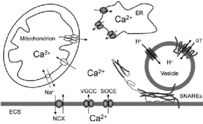
astrocytes. The sources of Ca2+for cytosolic Ca2+increase are: the endoplasmic reticulum (ER) and the extracellular space (ECS). Cytosolic Ca2+accumulation could be caused by the entry of Ca2+ from the ER store that possess inositol 1,4,5 trisphospate and ry-anodine receptors. Store specific Ca2+-ATPase fills these stores with Ca2+. Ultimately, this (re)filling requires Ca2+ entry from the ECS through store-operated Ca2+entry. Additional Ca2+entry from the ECS to the cytosol can be mediated by plasma membrane voltage -gated Ca2+channels and Na+/Ca2+exchangers, the latter operating in the reverse mode. Mitochondria represent a source/sink of cytosolic Ca2+. Mitochondrial Ca2+uptake from the cytosol to its matrix is mediated by the Ca2+uniporter. Free Ca2+exits the mitochondrial matrix through the Na+/Ca2+exchanger and also by brief openings of the mitochon-drial permeability transition pore. Increase in cytosolic Ca2+is suffi-cient and necessary to cause vesicular fusions and release of gliotransmitters (GT). This process requires the activity of the ternary SNARE complex consisting of synaptobrevin 2 and/or cellubrevin lo-cated at vesicular membrane, and the binary cis complex pre-formed at the plasma membrane and composed of syntaxin (shown in an open form) and synaptosome-associated protein of 23 kDa. Astrocytic vesicles are filled by various vesicular GT transporters, which for this action utilize the proton gradient generated by the vacuolar type H+
-ATPase. Drawing is not to scale.
12 | V. Parpuraet al.
Journal of Neurochemistry! 2012 International Society for Neurochemistry, J. Neurochem. (2012) 121, 4–27 ! 2012 The Authors
Figure 1.4. Calcium-dependent vesicular release of gliotransmitters from astrocytes: sources of Ca2+ and the formation of the SNARE complex necessary for gliotransmission in astrocytes [41].
1.2. Astrocytes in brain disorders
Astrocytic structure and protein expression were already shown to be altered in the context of brain disorders like depression, Parkinson's disease, Alzheimer’s disease, epilepsy and schizophrenia [73-86]. What remains to understand is if astrocytic dysfunction constitutes the cause, providing detrimental signals that contribute to the disorder, or the consequence, performing a supportive function in the attempt to reverse and prevent the disorder. Hence it is important to investigate the contribution of astrocytes to network function in order to understand their role in pathophysiological states. Since gliotransmission is now known as a modulatory factor of synaptic communication (see section 1.1.3) it may be considered an important mechanism underlying pathological processes.
Assuming the tripartite nature of central nervous system synapses [32] it is possible that the astrocytic dysfunction, compromising the neuronal activity support and/or gliotransmission, disturbs synaptic transmission and plasticity or neuronal excitability. These events at the cellular level are translated into distortion of brain physiology that lead to behavioural abnormalities [38]. Rat models of depression exhibit deceasing levels of GFAP expression and 13C-acetate metabolism reflecting glial metabolism [75, 77]; also in rat, glial
loss in the prefrontal cortex (PFC) is sufficient to induce depressive-like behaviour [76]. In the context of epilepsy, GFAP immunoreactivity evidenced astrocytic hypertrophy (reactive astrocytosis) and proliferation [78]: in mesial temporal lobe epilepsy, GFAP immunoreactivity is increased in a way that might be correlated with seizure frequency [79]. Moreover, astrocytic Ca2+ oscillations were observed to increase in frequency in isolated
brain slice models that exhibit epileptiform activity [80]. However, while enhanced excitatory gliotransmission, for example in terms of Ca2+ oscillations and glutamate release, can
contribute to epileptiform activity and seizures, reduced gliotransmission and D-serine release can contribute to schizophrenia. Also contrasting is the decreased GFAP expression in tissue isolated from schizophrenic patients [81-83]. In Alzheimer’s disease, increased levels of amyloid-β (Aβ) peptides and their subsequent deposition in Aβ plaques lead to the activation of the surrounding microglia and astrocytes that release diverse pro- and anti-inflammatory mediators resulting in the chronicle parenchymal inflammation in the brain [84, 85]. In Parkinson’s disease, and other neurological disorders, occurs an abnormal neuronal aggregation of α-synuclein, that is directly transmitted from neurons to astrocytes, causing inflammatory responses [86]
1.3. Astrocytic function in vivo: behaviour and cognition
The discovery that astrocytes respond to neuronal transmitters through Ca2+ elevations
[87, 88] have boosted the studies on astrocytic function attempting to understand more about the implications of these glial cells in brain activity and its output. Initially, the research tools limited the scientific progress on this subject, but successive studies have been giving input for a new insight into the role of glial in the brain function.
Functional and metabolic studies with toxins targeting selectively astrocytes have provided simple approaches to study the effects of astroglial dysfunction. Intracerebral injections of the gliotoxin L-α-aminoadipate, caused the disruption of the astrocytic network when injected intracerebrally in Long Evans Hooded and Sprague Dawley rats, since it irreparably damaged the astrocytes in the vicinity of injections [89]. Using this model of glial loss, Banasr and colleagues were able to induce depressive–like behaviour in rats [77].
Additionally, a recent study proved that astrocytes modulate the accumulation of sleep pressure and its cognitive consequences. These conclusions were achieved using the dnSNARE transgenic mice model that conditionally prevents astrocyte-dependent action on pre-synaptic A1 receptors through the conditional blockade of vesicular release. Because adenosine is involved in homeostatic drive of sleep following prolonged wakefulness, sleep pressure was significantly reduced when dnSNARE transgene was expressed in astrocytes [47]. Another study based in the same animal model followed with the evidence that astrocytic-derived ATP acting on A1 receptors via adenosine contributes to the effects of sleep deprivation on hippocampal plasticity and hippocampus-dependent memory [90].
Although these studies unveil a putative role of astrocytes in the regulation or modulation of behaviour outputs much more in this field should be cleared. By implementing different animal models of astrocytic dysfunction we expect to assess the implications of astrocytic function in the generation or modulation of cognitive function.
1.4. The prefrontal cortex
In this project we focused in the cognitive function and we focused in the prefrontal cortex because (1) it is a region with major relevance for cognitive processes (see section 1.4.1), (2) it is a large region in the brain, and therefore relatively easy to target for injections of pharmacological tools (aminoadipate rat model, see section 3.1.1)
1.4.1.
Cognitive functions of thethe prefrontal cortex: learning and memory
The prefrontal cortex (PFC), which is among the most studied regions of the brain, is intimately related to the computation of complex cognitive functions. Indeed, various cognitive and executive processes have been associated to the PFC, such as working memory, decision making, planning and behavioural flexibility, attentional set-shifting and inhibitory response control [91, 92]. The PFC has been shown to be implicated in the organisation of delayed responses and consequently in working memory function - a memory system composed of distinct, but overlapping cognitive processes used for the active maintenance and elaboration of task-relevant information, as it involves temporary storage and manipulation of information [93]. Functional neuroimaging in brain-damaged humans and healthy volunteers have confirmed the role of the PFC in working memory processes [94]. Neuropsychological and connectional evidences indicate the homology of the medial prefrontal cortex (mPFC) in rats and the dorsolateral prefrontal cortex (dlPFC) in primates. [95-98] - fig. 1.5. As the dlPFC in primates, the mPFC in rats is involved in other executive functions, besides working memory: temporal organisation of behaviour [99] and attentional control in strategy switches [98, 100, 101]. In fact, lesions in the rat mPFC - prelimbic (PrL), infralimbic (IL), and with partial damage to the cingulate cortex areas 1 and 2 (Cg1 and Cg2) - impair selectively the extradimensional set-shifting [98]. Moreover, the rat mPFC was proposed to be important in the representation of an abstract and more hierarchically-organised memory useful in the decision making, complementing the hippocampus in spatial navigation [102]. It is not surprising, since the IL and PrL regions of the PFC, constituting the ventral portion of the rat mPFC, are linked to the hippocampus by an axonal pathway originated in the subiculum and ventral CA1 [103].
Figure 1.5. Highlights of the PFC and mPFC, in human brain evidencing the homology of mPFC and dlPFC brain regions in rat and human, respectively [92]. A - lateral view of human brain; and B, left - sagittal view of rat brain; B, right coronal section at Bregma 3.0. PFC dlPFC mPFC
A
2.
A
IM OFW
ORKThe rationale of this project is based in the common accepted idea that specific brain regions compute specific tasks and are responsible for specific behavioural outputs. In this scope, we believe that affecting the astrocytic function in a brain region, mimicking what happens in pathological states [75-85], and measuring the behaviour output of that region we would be able to dissect the astrocytic component of that behaviour output.
Despite the many years of research carried out so far in the astroglial function in the brain, most of theses studies were focused on metabolic and physiologic features and little was done in what concerns to behaviour implications, namely in cognitive function, involving learning and memory, in the context of altered astrocytic function.
The aim of this work is to implement two animal models whose astrocytes are affected in two different manners to allow in vivo study of astrocytic function and its implication in behaviour outputs. Hence, (1) to mimic pathologies based on the glial loss we implemented a rat model on which astrocytes were ablated through the gliotoxin L-α-aminoadipate; and (2) to disclose the role of the exocytotic release of gliotransmitters in cognitive processes, we implemented the recently reported genetic mouse model, dnSNARE.
In this project we focused in the cognitive function linked to the prefrontal cortex because (1) it is a region with major relevance for cognitive processes (see section 1.4), (2) it is a large region in the brain, and thus relatively easy to target for injections of pharmacological tools (aminoadipate rat model). By lesioning astrocytes in the mPFC and blocking the gliotransmitter release by astrocyte we expect to observe a compromise in learning and memory functions mostly dependent on the PFC and therefore dissect the astrocytic component in the computation of these tasks.
3.
M
ATERIALS ANDM
ETHODS3.1. Animals and Treatments
Experiments were conducted in accordance with local regulations (European Union Directive 86/609/EEC) and National Institutes of Health guidelines on animal care and experimentation.
3.1.1. Aminoadipate rat model of astrocytic depletion
Male Wistar-Han rats (Charles River Laboratories, Barcelona, Spain) , ten-weeks-old, were housed in groups of two, under standard laboratory conditions (room temperature 22ºC; food and water ad libitum; 12h dark/light cycle, lights on at 8:00 AM). One group was subjected to pharmacological ablation of astrocytes in the mPFC through a single bilateral intracerebral microinjection of the selective astrocytic toxin L-α-aminoadipate (L-α-AA; see section 3.1.1.B) - treatment group (AA). As control animals (CON) for the aminoadipate pharmacological model, other set of rats received a single bilateral intracerebral microinjection of artificial cerebrospinal fluid (aCSF), the vehicle of the aminoadipate solution, in the same region, using the same procedure.
3.1.1.A. Surgical procedure for the establishment of the pharmacological rat model
For the establishment of the aminoadipate pharmacological model, rats were subjected to a surgical procedure for the implantation of bilateral cannula guides in the prelimbic region of the prefrontal cortex. Animals were deeply anaesthetised with a intraperitoneal (i.p.) mix of ketamine (75 mg/kg - Imalgene 1000, Merial) and medetomidine (0,5 mg/kg - Dorbene Vet, Pfizer) and bilateral brain cannulas (26 GA, Plastics One) were stereotaxically implanted in the prelimbic region of the prefrontal cortex. The coordinates used were: 3.0 mm posterior to bregma, ± 0.6 mm lateral to the midline, and 2.5 mm ventral to the skull surface, based on the Paxinos and Watson rat brain atlas [104]. This position represents a spot 1 mm above the target position, which will be achieved by the internal cannulas, used to inject the solution of interest. The fixation of the bilateral cannula guides in the skull was achieved by the placement of two screws in anterior and posterior positions to relative to the cannula, and by the application of an acrylic resin (Pattern Resin LS, GC) covering both screws and the basal portion of the cannula guide pedestal. At the end of the surgical procedure the anaesthesiawas reverted with atipamezol, i.p. (2 mg/kg - Antisedan, Pfizer) and animals were housed individually to avoid damage of the implant. Animals were allowed to recover from this surgical procedure for at least 7 days.
3.1.1.B. Drug preparation and administration
A 124 mM L-α-aminoadipate drug solution was prepared through the solubilisation in aCSF as previously described [89], by simply dissolve L-α-aminoadipate (A7275, Sigma-Aldrich) in aCSF and adjusting the pH to 7.4 in order to help to dissolve the aminoadipate.
Concerning the drug infusion protocol, rats were first anaesthetised with mix of propofol (70 mg/kg - Vetofol, Esteve) and medetomidine (0,2 mg/kg - Dorbene Vet, Pfizer) administered intraperitoneally. Such anaesthesia protocol based on Alves et al, 2010 provides suitable level of sedation ideal for restraint and non-painful drug infusion [105]. Five microliters of AA (620 μmol), or aCSF were administered using 25 μl Hamilton syringes, connected to a double internal cannula (that projects 1 mm from the cannula guide) through a polyethylene tubbing, at the constant rate of 0.5 μl/min controlled by an micro-pump (53100V, Stoeling). The double internal cannula was slowly withdrawn 5 minutes after infusion completion to avoid the displacement of the injected fluid by capillarity. Once this procedure was complete, anaesthesia was reverted with atipamezol (1 mg/kg - Antisedan, Pfizer) and animals returned to their home-cages.
As the selective gliotoxin L-α-aminoadipate exerts effect 4 hours following injection [89] and the astrocyte ablation effect persists up to 7 days, this drug was infused in the rats brains on the day before the behaviour tests. In this way, rats preformed the behaviour tests within the window of time of L-α-aminoadipate effect.
3.1.2. Transgenic dominant-negative SNARE mice (dnSNARE) genetic model
For the purpose of this project we obtained the dnSNARE transgenic mice strain from Prof. Philip Haydon (Tufts University, Boston) that kindly supplied the founders, and implemented the colony at the IVCS. In this strain the SNARE domain of the synaptobrevin II (dnSNARE) expression is conditionally suppressed in astrocytes, by means of a “tet-Off” expression system. The dnSNARE mice strain was generated by Pascual and colleagues[106], by expressing the cytosolic portion of the SNARE domain of synaptobrevin II (amino acids 1 to 96) selectively in GFAP-positive astrocytes, which blocks the exocytotic release of
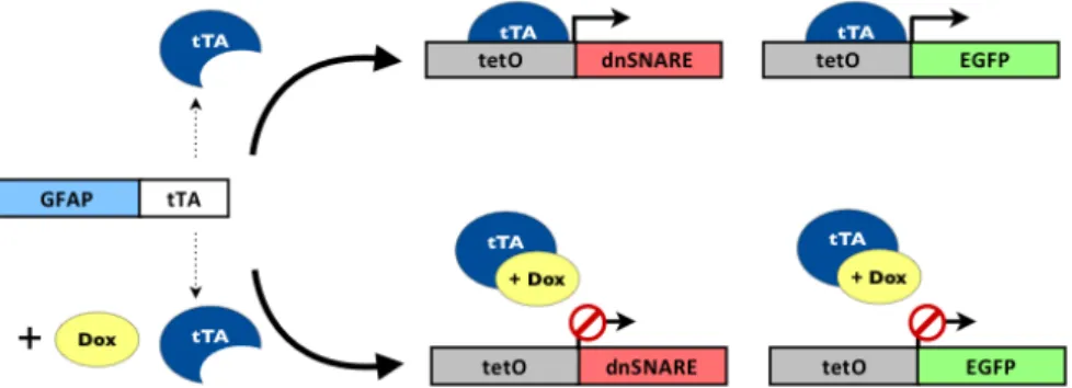
gliotransmitters, a process dependent on the formation of SNARE complexes [107]. For that purpose, they developed two separate mice lines GFAP.tTA and tetO.dnSNARE that when crossed yield dnSNARE mice. The GFAP.tTA mice line contains the astrocyte-specific glial fibrillary acidic protein (GFAP) promotor that drives the expression of the tetracycline-controlled transactivator protein (tTA), while the tetO.dnSNARE mice line contains a SNARE domain regulated by a tet operator promotor (tetO) and the reporter gene enhanced green fluorescent protein (EGFP) - fig. 3.1.
3.1.2.A. Colony implementation: strain lines and matings
To implement this transgenic mice strain in the ICVS, the two mice lines GFAP.tTA and tetO.dnSNARE, were first maintained heterozygous under C57/Blk6 genetic background. Mice originated by the backcrosses of those two separate lines were crossed (GFAP.tTA x tetO.dnSNARE) to produce dnSNARE mice for experimentation. The double negative and single positive offspring were kept as controls. In the GFAP-positive astrocytes of these animals the expression of transgenes SNARE, LacZ and EGFP is suppressed by the tetracycline doxycycline (Dox). To prevent potential developmental effects of the transgene expression, mice were bred and raised until weaning, at three weeks of age, under the action of Dox (Sigma, 25 μg/ml in drinking water). Once animals were weaned, Dox administration was maintained until 3 weeks prior to the behavioural studies. Transgene-expressing and wild type mice were visually indistinguishable [106]. The double negative, single and double positive offspring were tested in behaviour paradigms described below (see sections 3.2.2).
Figure 3.1. Schematic representation of GFAP promotor driving the expression of the target gene dnSNARE and reporter gene EGFP in astrocytes. Tetracycline doxycycline (Dox) suppresses the expression of transgenes by preventing the binding of tTA to tetO. Adapted from Florian et. al, 2011 and Halassa et. al, 2009 [40, 90]
3.1.2.B. Colony management and genotyping
In the context of colony management, the genotyping of the offspring generated by the mattings was accomplished by means of polymerase chain reaction (PCR) of DNA extracted from mice tails (about 3 mm of the tip). First, the tails were digested in 380 μl of cell lysis buffer (0,5 mM Tris, pH 8.0; 10 mM EDTA; 0,5 % SDS) with 10 μl of proteinase K (15 mg/ml), overnight and in a heating block at 55ºC. In the next morning, samples were centrifuged during 10 minutes at 13000 rpm and the supernatant was collected. The DNA precipitation was achieved b the addiction of 300 μl of isopropanol to the collected supernatant, followed by soft shaking of the sample tubes. After additional 10 minutes centrifugation at 13000 rpm, the supernatant was discarded, while the pellet was rapidly washed with 100 μl of ethanol 70%. The tubes containing the DNA sample were then left air drying at the bench for 90 minutes, after which DNA hydration was attained by the addiction of 50 to 100 μl of elution buffer (10 mM Tris, pH 8.0; 1 mM EDTA) and a short vortex.
The genotyping itself was then carried out by the multiplex PCR technique, on which the two pairs of primers, for tTA (tTA forward: 5’- ACT CAG CGC TGT GGG GCA TT - 3’; tTA reverse: 5‘ - GGC TGT ACG CGG ACC CAC TT - 3’) and tet.O (tet.O forward: 5’- TGG ATA AAG CTC ATT AAT TGT CA - 3’; tet.O reverse: 5‘ - GCG GAT CCA GAC ATG ATA AGA - 3’) sequences, were used in the same PCR mixture. The PCRs were performed in a MyCycle thermal cycler (Bio-Rad); the composition of the PCR mix and conditions used are summarised in the table 3.1. Amplified PCR products were separated on a 1% agarose gel prepared in Tris-Acetate-EDTA (TAE) running buffer, stained with ethidium bromide and compared to the DNA size marker GeneRuler™ 1kb Plus DNA Ladder, 75-20,000bp (#SM1331, Fermentas). Electrophoresis run for 40 minutes at 120V and gel images were captured with a transilluminator (Alpha Innotech Corporation, Bio-Rad).
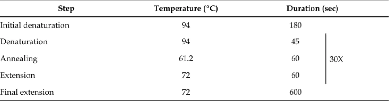
Table 3.1. PCR conditions used for the amplification of tTA and tet.O sequences. Table 3.1. PCR conditions used for the amplification of tTA and tet.O sequences. Table 3.1. PCR conditions used for the amplification of tTA and tet.O sequences. Table 3.1. PCR conditions used for the amplification of tTA and tet.O sequences. Table 3.1. PCR conditions used for the amplification of tTA and tet.O sequences.
Reaction mix (20 μl) components Reaction mix (20 μl) components Reaction mix (20 μl) components Reaction mix (20 μl) components Reaction mix (20 μl) components Taq buffer, with KCl (part of #EP0402, Fermentas)
Taq buffer, with KCl (part of #EP0402, Fermentas) 1x1x1x
MgCl2 (part of #EP0402, Fermentas)
MgCl2 (part of #EP0402, Fermentas) 1.5 mM1.5 mM1.5 mM
dNTP mix (#R0192, Fermentas)
dNTP mix (#R0192, Fermentas) 0.2 μM0.2 μM0.2 μM
Primer tTA forward
Primer tTA forward 0.5 μM0.5 μM0.5 μM
Primer tTA reverse
Primer tTA reverse 0.5 μM0.5 μM0.5 μM
Primer tet.O forward
Primer tet.O forward 0.5 μM0.5 μM0.5 μM
Primer tet.O reverse
Primer tet.O reverse 0.5 μM0.5 μM0.5 μM
Taq DNA Polymerase (#EP0402, Fermentas)
Taq DNA Polymerase (#EP0402, Fermentas) 0.5 U0.5 U0.5 U
DMSO DMSO 2%2%2% Template DNA Template DNA 100 ng100 ng100 ng Amplification program Amplification program Amplification program Amplification program Amplification program
Step Temperature (ºC) Duration (sec)Duration (sec)Duration (sec)
Initial denaturation 94 180 30X Denaturation 94 45 30X Annealing 61.2 60 30X Extension 72 60 30X Final extension 72 600 30X
3.2.
Behaviour Tests
3.2.1. Aminoadipate rat model of astrocytic depletion
The behavioural consequences of astrocyte depletion in the medial prefrontal cortex, induced by intracranial injection of L-aminoadipate (see section 3.1.1) were assessed by the attentional set-shifting task and by water maze-based tests. These tests were used to measure the cognitive performance of animals with astrocytic depletion and the respective controls, and are described in detail below.
3.2.1.A. Attentional set-shifting task
To assess the non spatial working memory, the attentional set-shifting and the reversal learning of rats subjected to the intracranial bilateral injections of AA we used the attentional
set-shifting task (ASST). The protocol of this cognitive test was adapted from the ASST paradigm originally described by Birrell and Brown [98]. The task was conducted in a custom made black Pexiglass apparatus (60 cm L x 40 cm W x 20 cm H). One third of the apparatus was separated by the rest of the area by a removable divider, creating a holding area. On the opposite side, two adjacent plastic pots filled with sawdust, separated by a vertical divider, were used as digging bowls and their rims were scented with different oil essences, producing a lasting odour - fig. 3.2.
In each trial the pot associated with the correct answer contained a small food reward (Nestle Cheerio ring cut in two parts) buried underneath the digging media, as rats could be trained to dig in small bowls filled with sawdust to retrieve food reward [108]. The reward was predicted either by an odour in the bowl or by a different texture before reaching the bowl. Because this test involves a food reward to correct answers, all animals were food restricted (1 h of access to food per day) during the prior 6 days. During this food restriction period, 6 Nestle Cheerios were given to the rats per home cage. The task was conducted in a room with dim light, controlled by a rheostat. Animals were brought to the test room 15 minutes before its start, to acclimatise them to those room conditions.
On the two days before the test animals were acclimatised to the apparatus, with food rewards in both digging bowls, for 1 hour each day. On the day 1 of the task animals have to discriminate from one stimulus, while on the day 2 of the task the animals are presented with 2 different dimensions of stimuli, odours or textures and have to learn which stimulus leads Figure 3.2. Representation of the ASST apparatus. The combinations of stimuli are located in one third of the apparatus area divided in two sections, where the stimuli combinations are presented at each trial. The opposite third of apparatus separated by a removable divider constitutes a holding area where the rat rests between trials.
removable divider
digging bowl with odour scented rim stimuli section divider
texture
left right
inter-trial holding area
to the reward. On the day 1 of the task, after 5 minutes of habituation to the apparatus, animals were first trained in two stages of simple discrimination (SD): odours (lemon vs. vanilla)- SDO- and textures (sandpaper vs. blue plastic)- SDT. The second day of the task consisted of compound discrimination of odours- CDO- (papaya vs. eucalyptus and cardboard vs. brown plastic), reversal compound discrimination- Rev1; extradimensional shift- EDST- (mango vs. lotus and velvet vs. plastic grass); and reversal extradimensional shift- Rev2. In the reversal stages, the previous positive stimulus, i.e. associated with the reward, became negative - table 3.2. The stages were only completed when the animals reached 6 correct consecutive trials- when it is considered that the animal learned to associate the positive stimulus to the reward [98]- apart from the first 2 trials, when animals are allowed to freely explore the both digging bowls. Stimuli were presented in a random sequence of 10 trials (equal to all animals), repeated if necessary - tables 3.3, 3.4 and 3.5. As in task day 1, animals were allowed to habituate to the apparatus during 5 minutes.
Once task was completed animals returned to their home cages, remaining at that same room until sacrifice.
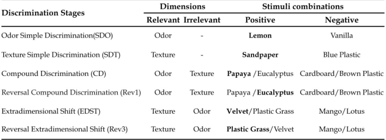
Table 3.2. Order of discrimination stages, relevant and irrelevant dimensions, and stimuli combinations of ASST presented to the animals. Reward associated stimuli are bold and minus sign indicates absence of stimulus.
Table 3.2. Order of discrimination stages, relevant and irrelevant dimensions, and stimuli combinations of ASST presented to the animals. Reward associated stimuli are bold and minus sign indicates absence of stimulus.
Table 3.2. Order of discrimination stages, relevant and irrelevant dimensions, and stimuli combinations of ASST presented to the animals. Reward associated stimuli are bold and minus sign indicates absence of stimulus.
Table 3.2. Order of discrimination stages, relevant and irrelevant dimensions, and stimuli combinations of ASST presented to the animals. Reward associated stimuli are bold and minus sign indicates absence of stimulus.
Table 3.2. Order of discrimination stages, relevant and irrelevant dimensions, and stimuli combinations of ASST presented to the animals. Reward associated stimuli are bold and minus sign indicates absence of stimulus.
Discrimination Stages DimensionsDimensions Stimuli combinationsStimuli combinations Discrimination Stages
Relevant Irrelevant Positive Negative
Odor Simple Discrimination(SDO) Odor - Lemon Vanilla
Texture Simple Discrimination (SDT) Texture - Sandpaper Blue Plastic Compound Discrimination (CD) Odor Texture Papaya /Eucalyptus Cardboard/Brown Plastic
Reversal Compound Discrimination (Rev1) Odor Texture Papaya /Eucalyptus Cardboard/Brown Plastic
Extradimensional Shift (EDST) Texture Odor Velvet/Plastic Grass Mango/Lotus Reversal Extradimensional Shift (Rev3) Texture Odor Plastic Grass/Velvet Mango/Lotus
![Figure 1.5. Highlights of the PFC and mPFC, in human brain evidencing the homology of mPFC and dlPFC brain regions in rat and human, respectively [92]](https://thumb-eu.123doks.com/thumbv2/123dok_br/17799499.840716/26.892.208.723.817.1079/figure-highlights-human-brain-evidencing-homology-regions-respectively.webp)