Universidade de Lisboa
Faculdade de Medicina Dentária
Clinical Implications of Antithrombotic
Therapy in Dentistry: A Literature Review
Joel David Martinho Seguro
Dissertação
Mestrado Integrado em Medicina Dentária
Universidade de Lisboa
Faculdade de Medicina Dentária
Clinical Implications of Antithrombotic
Therapy in Dentistry: A Literature Review
Joel David Martinho Seguro
Dissertação Orientada
Pelo Prof. Doutor João Caramês
E Coorientada
Pela Prof.ª Doutora Helena Francisco
Mestrado Integrado em Medicina Dentária
2016
2
Abstract
Introduction: The therapeutical approach of patients under chronic antithrombotic therapy needing dental procedures causes great controversy. The increased hemorrhage risk associated with these procedures when the patient is undergoing this therapy must be taken into account relatively to the increased thrombotic risk caused by its interruption. The aim of this study was to review the current literature regarding antithrombotic therapy and its implications in medical dentistry.
Materials and Methods: Research was performed from November 2015 until May 2016 on the databases Cochrane Library, Medline (via PubMed) and Google Scholar, with the key-words: Anticoagulants Dental Surgery; Hemostasis; Cell-based Model Hemostasis; Antiplatelet Therapy Oral Surgery; New Oral Anticoagulants Dental Procedures; filtered by the following formats: Meta-analysis, Systematic Reviews and Randomized Control Trials, which were extended afterwards to include Reviews, Cohorts and Guidelines. Inclusion criteria consisted of articles published in the last 10 years, available in English or Portuguese, limited to human samples. Articles were also retrieved after analyzing the reference list of articles previously obtained.
Results: The search yielded 1242 results, of which 47 were selected for inclusion, and 21 additional articles were obtained from the reference lists of other articles. Results point to the fact that, currently, several authors suggest the execution of the procedures without therapy modification or discontinuation, emphasizing preventive local hemostatic measures, with the interruption of antithrombotic therapy before surgical interventions possibly leading to severe consequences.
Conclusion: Minor oral surgery procedures are considered to be of low hemorrhagic risk. Therefore, there is no indication for the interruption of antithrombotic therapy, provided there are no additional risk factors, the patient is stably anticoagulated, and local hemostatic measures are properly applied. However, patients requiring extensive oral/maxillofacial surgery may need discontinuation of oral anticoagulants preoperatively, but always in conjunction with the treating physician.
3
Resumo
Introdução
Problemas sistémicos como embolia pulmonar, fibrilhação atrial, válvula cardíaca artificial, acidente vascular cerebral, entre outras patologias sistémicas, estão na base do tratamento profilático com terapia antitrombótica (Oake et col, 2008; António et col, 2008; Nematullah et col, 2009; Araújo et col, 2010; Van Diermen et col, 2013).
Desta terapêutica destacam-se os antiagregantes plaquetários e os anticoagulantes. Os antiagregantes plaquetários, como o AAS, atuam na hemostase primária, correspondente à formação de trombo plaquetário. Os anticoagulantes, como a varfarina, atuam na hemostase secundária, relativa à formação da rede de fibrina e coágulo (Filho et col, 2013; Napeñas et col, 2013).
Os anticoagulantes orais convencionais, ou VKAs - antagonistas da vitamina K (essencial na produção de vários fatores de coagulação) – constituem, há mais de 60 anos, a terapia de primeira linha para a profilaxia de fenómenos tromboembólicos (Mookadam et col, 2015). São fármacos com baixo custo, eficácia comprovada e com antídoto específico em caso de hemorragia, que possibilita suspender a medicação em casos de urgência (Cid-Conde & Lopez-Castro, 2013). No entanto, apresentam limitações que afetam a qualidade de vida dos pacientes e aumentam a morbilidade: janela terapêutica estreita, resposta imprevisível, necessidade sistemática de controlo da hemorragia, requerem um acompanhamento de rotina, ajustes de dose frequentes e apresentam numerosas interações medicamentosas e alimentares (Cid-Conde & Lopez-Castro, 2013; Mookadam et col, 2015).
Neste sentido, os novos anticoagulantes orais (NOACs), não dependentes da vitamina K, atuando diretamente sobre fatores de coagulação específicos, mostram promessa como uma alternativa ideal à terapia convencional (Mookadam et col, 2015). Na Medicina Dentária, as principais preocupações no tratamento de pacientes que tomam NOACs - inibidores diretos da trombina e inibidores do fator Xa - é o risco de hemorragia e a ausência de um antídoto específico (Isola et col, 2015; Costantinides et col, 2016).
O crescimento do número de indivíduos submetidos a terapêutica antitrombótica reflete-se nos consultórios dentários (Araújo et col, 2010; Isola et col, 2015; Paraschiv et col, 2015). A abordagem aos doentes a realizar terapêutica crónica antitrombótica com necessidade de intervenções estomatológicas continua a suscitar grande controvérsia
4
(Sacco et col, 2007; António et col, 2008; Van Diermen et col, 2013; Isola et col, 2015). A maioria dos profissionais recomenda suspender ou diminuir a dosagem dos anticoagulantes, mas trabalhos recentes mostram que esta conduta causa um risco aumentado para o paciente (Araújo et col, 2010; Van Diermen et col, 2013). O aumento do risco hemorrágico, associado aos procedimentos realizados em medicina dentária em pacientes em que esta terapêutica é mantida, deve ser pesado relativamente ao risco trombótico acrescido causado pela sua interrupção (Nematullah et col, 2009; Wahl, 2014; Paraschiv et col, 2015). Atualmente, vários autores sugerem a execução das intervenções dentárias sem modificação ou interrupção da terapia, dando ênfase às medidas hemostáticas locais de prevenção, executadas no pré, peri e pós-operatório, para que a prevenção de hemorragia seja conseguida (Madrid & Sanz, 2009; Nematullah et col, 2009; Araújo et col, 2010; Firriolo & Hupp, 2012).
Os objetivos deste estudo passam por fazer um levantamento dos principais riscos associados a pacientes submetidos a esta terapia e dos protocolos de abordagem para estes pacientes em Medicina Dentária, relativamente à interrupção ou não do tratamento aquando da realização de intervenções nesta área, e outras modificações ao plano de tratamento, possibilitando uma prática segura para médico e paciente.
Materiais e Métodos
A pesquisa de evidência pertinente ao tema foi realizada a partir de Novembro de 2015 até Maio de 2016, nas bases de dados Cochrane Library, Medline (via PubMed) e Google Scholar, com as palavras-chave: Anticoagulants Dental Surgery; Hemostasis;
Cell-based Model Hemostasis; Antiplatelet Therapy Oral Surgery; New Oral Anticoagulants Dental Procedures.
Os seguintes formatos foram considerados para inclusão: Meta-análises, Revisões Sistemáticas e Randomized Control Trials, que foram estendidos mais tarde para incluir Revisões Literárias, Coortes e Guidelines. Os critérios de inclusão consistiram de artigos publicados nos últimos 10 anos, disponíveis em Inglês ou Português, limitado a amostras humanas. Mais artigos foram obtidos após análise das referências bibliográficas dos artigos previamente obtidos.
5
Resultados
A pesquisa resultou em 1242 resultados, dos quais 47 foram selecionados para inclusão, e 21 artigos adicionais foram obtidos a partir das referências bibliográficas de outros artigos.
Os resultados apontam para o fato de que, apesar do risco hemorrágico, a suspensão de fármacos antiagregantes aumenta a morbilidade perioperatória, não só devido aos efeitos diretos da sua suspensão, mas também por causa de um possível efeito
rebound, como é o caso da síndrome de interrupção da aspirina, promovido pelo aumento
de tromboxano A2 e redução da fibrinólise. Consequentemente, suspender a terapia antiagregante antes de realizar procedimentos médico-cirúrgicos pode causar efeitos catastróficos (Van Diermen et col, 2009; Coelho, 2015).
Vários estudos confirmaram a não existência de um aumento clinicamente significativo do risco hemorrágico pós-operatório após intervenções dentárias invasivas em pacientes sob mono ou dupla terapia antiagregante plaquetária (Morimoto et col, 2008; Morimoto et col, 2009; Napeñas et col, 2009; Lillis et col, 2011; Park et col, 2012; Dézsi et col, 2015).
Outros estudos mostraram que as complicações hemorrágicas associadas com a anticoagulação são três vezes menos prováveis de ocorrer do que complicações tromboembólicas graves, incluindo a morte, em pacientes cuja terapia anticoagulante foi interrompida (Al-Mubarak et col, 2007; Araújo et col, 2010). Para pacientes que tomam varfarina e que apresentam um INR estável entre 2-4, os estudos indicam que o risco de hemorragia significativa é muito pequeno (Bajkin et col, 2009; Madrid & Sanz, 2009; Nematullah et col, 2009; Okamoto et col, 2014; Bajkin et col, 2014; Isola et col, 2015).
As indicações mais atuais sobre a interrupção de NOACs sugere que os pacientes que os tomam podem submeter-se a procedimentos dentários invasivos sem alteração da dose (O'Connell, 2014; Tsolka, 2014; Paraschiv et col, 2015; Costantinides et col, 2016). Quase todos os estudos propõem medidas de hemostase local para prevenção e controlo da hemorragia, incluindo: aplicação de pressão e sutura, anestesia com vasoconstritor, usando as técnicas infiltrativa e intraligamentar quando possível, esponjas de gelatina com solução de trombina, bochechos com soluções hemostáticas, (Sacco et col, 2007; Bajkin et col, 2009; Firriolo & Hupp, 2012; O'Connell, 2014; Okamoto et col, 2014; Paraschiv et col, 2015).
6
Conclusão
A maior parte das intervenções em Medicina Dentária são consideradas de baixo risco hemorrágico. Portanto, não há indicação para a interrupção de antiagregantes plaquetários, incluindo a terapia antiagregante dupla, antes de procedimentos dentários invasivos (Guyatt et col, 2012; Gerstein et col, 2012; Guyatt et col, 2012; Van Diermen et col, 2013; Wahl, 2014). Verificou-se também que os estudos mais recentes defendem a ideia de que a terapia com VKAs não deve ser interrompida (Bajkin et col, 2009; Madrid & Sanz, 2009; Nematullah et col, 2009; Firriolo & Hupp, 2012; Van Diermen et col, 2013), desde que não existam fatores de risco adicionais e que o paciente esteja estavelmente anticoagulado (Araújo et col, 2010).
Concluiu-se ainda que, em pacientes com função renal normal tomando NOACs, procedimentos dentários invasivos podem ser realizados sem interrupção da medicação (O'Connell, 2014; Tsolka, 2014; Paraschiv et col, 2015; Costantinides et col, 2016).
No entanto, pacientes que necessitam de cirurgia oral extensa ou maxilofacial, podem precisar de interromper os anticoagulantes orais no pré-operatório, sendo que esta decisão não deve ser tomada sem consultar o médico assistente (Firriolo & Hupp, 2012; O’Connell, 2014; Tsolka, 2014; Paraschiv et col, 2015; Costantinides et col, 2016).
Várias publicações provam a importância e a eficácia de medidas hemostáticas locais pré, peri e pós-operatórias, na prevenção e controlo do risco hemorrágico (Araújo et col, 2010; Paraschiv et col, 2015).
Palavras-chave:
Terapia antitrombótica, Procedimentos Dentários, Risco trombótico/hemorrágico.7
Agradecimentos
Encontrando-me neste momento a terminar uma fase muito importante da minha vida, aproveito para deixar um profundo agradecimento a todos aqueles que, de forma direta ou indireta, me ajudaram a percorrê-lo com todo o sucesso que alcancei.
Aos professores, colegas, assistentes e funcionários, agradeço o vosso contributo.
Ao Professor Doutor João Caramês e à Professora Doutora Helena Francisco, agradeço a disponibilidade e generosidade ao aceitarem o meu pedido para serem meus orientadores, dando-me a oportunidade de realizar uma monografia sobre o tema que pretendia, e tendo-me apoiado na sua elaboração.
.
Aos meu caríssimos amigos, João Rodrigues, Andreia Luís Vieira e Luana Amorim, por moldarem este percurso com as suas ações, carinho e palavras de encorajamento e sabedoria, tendo tornado estes anos numa fase da minha vida que nunca esquecerei. Um especial agradecimento ao Pedro Gomes, meu colega, minha dupla na clínica, meu parceiro em tantas aventuras, e especialmente, meu amigo, por tudo aquilo que faz por mim e por toda a ajuda que me dá.
À minha família, que tudo fez para me proporcionar a oportunidade de me tornar naquilo que sou hoje, tanto a nível pessoal como académico e profissional. Nada que eu faça alguma vez será suficiente para retribuir todos os sacrifícios que fizeram por mim e o apoio que me deram para que seguisse o meu caminho.
Por fim, um agradecimento muito especial à Joana Guerra, por tudo o que significa para mim, pelo seu amor e apoio incondicional, por nunca me deixar baixar os braços, por todas as noites em claro a ajudar-me a estudar, pela motivação que me dá, e por ser a maior constante da minha vida desde que entrei na Faculdade. Palavras não descrevem aquilo que faz por mim todos os dias.
8
Index
1. Introduction ... 9 1.1 Hemostasis ... 9 1.2 Cellular Model ... 9 1.2.1 Initiation ... 9 1.2.2 Amplification ... 10 1.2.3 Propagation ... 101.3 Endogenous Regulation of the Coagulation System ... 11
1.4 Current Concepts of Antithrombotic Therapy ... 11
2. Materials and Methods ... 15
3. Results ... 16
4. Antithrombotic Therapy... 17
4.1 Antiplatelet Drugs ... 17
4.2 Conventional Anticoagulants ... 18
4.3 Novel Oral Anticoagulants ... 19
4.3.1 Direct Thrombin Inhibitors... 21
4.3.2 Factor Xa Inhibitors... 22
5. Treatment Planning Alterations/Recommendations ... 24
5.1 Antiplatelet Drugs ... 24
5.2 Conventional Anticoagulants ... 27
5.3 Novel Oral Anticoagulants ... 30
5.3.1 Direct Thrombin Inhibitors... 31
5.3.2 Factor Xa Inhibitors... 32
5.4 Local Hemostatic Measures ... 33
6. Conclusion ... 35
9
1. Introduction
1.1 Hemostasis
Hemostasis includes a set of well-regulated processes governing blood coagulation, platelet activation and vascular repair. It is fundamentally responsible for the spontaneous arrest of bleeding after a vascular lesion, by initiating a series of vascular events and activating extracellular receptors which work in harmony in order to remedy the damage (Versteeg et al, 2013; Yau et al, 2015). Coagulation is subsequently attenuated by the plethora of inhibitors that prevent excessive clot formation and eventual thrombosis. Thus, the formation and dissolution of a blood clot consists of physiological processes that result in a dynamic balance between procoagulant and anticoagulant mechanisms (Yau et al, 2015).
Traditionally, the occurrence of blood coagulation is described in two phases – primary hemostasis and secondary hemostasis – in which initially occurs platelet aggregation to form a platelet plug, and then follows the activation of the coagulation system, which leads to the formation fibrin clot (Cañigral et al, 2010; Yau et al, 2015). However, the representations of this process evolved significantly, currently demonstrating the complex interrelationship between platelets, coagulation system and vessel walls, a relationship essential in the formation of the clot. This intrinsic network can be described as the cellular model of coagulation. In this model, hemostasis is described in three stages: initiation, amplification and propagation (Yau et al, 2015).
1.2 Cellular Model
1.2.1 Initiation
Initiation occurs after vascular injury with the activation of the endothelium and the exposure of sub-endothelial cells. The adhesion of platelets to these components of the sub-endothelium requires specific receptors, which are located in the platelet’s cell membrane. Major membrane receptors are designated glycoprotein (GP) receptors I, II, III, and IV. GPIb receptor has been identified as binding von Willebrand’s factor (VWF), which is essential for platelet adhesion. Another major GP receptor is the IIb-IIIa complex, which is considered the most important adhesive receptor for platelet aggregation. In its active conformation, GPIIb-IIIa binds to several bivalent ligands,
10
particularly fibrinogen, VWF and fibronectin. GPIIb-IIIa activating agents comprise several soluble agonists, which can activate and trap nearby platelets. These agents include, among others, ADP (released from platelet-dense granules), thromboxane A2 (produced by the platelet), and thrombin (formed at the platelet’s surface) (Monroe & Hoffman, 2006; Versteeg et al, 2013).
Platelets can also become activated from soluble agonists forming at the site of the thrombus. Those soluble agonists released from dense granules of activated platelets include small molecules such as ADP, ATP, GDP, 5-HT, pyrophosphate, magnesium and calcium (essential regulator of platelet activation), which precipitate a number of positive feedback cascades leading to rapid activation of large numbers of platelets (Golebiewska & Poole, 2015). In parallel to these events, activated endothelial cells express a procoagulant molecule, the tissue factor (TF), which binds to the FVII. TF acts as a cofactor for FVII in order to promote proteolysis and activation of the latter to become FVIIa, with TF also binding to FVIIa to form the complex TF/FVIIa. This complex will produce the proteolytic cleavage of FIX and FX in order to obtain FIXa and fXa. The first serves to generate more FXa, which in turn generates thrombin (Monroe & Hoffman, 2006; Yau et al, 2015).
1.2.2 Amplification
At this stage, circulating platelets adhering to the injury site become activated by thrombin to form a platelet aggregate. This provides a surface for the activation of other procoagulant factors. Proteolytic cleavage of FV and FVIII into FVa and FVIIIa by thrombin occurs concomitantly. In addition, thrombin will also convert FXI into FXIa, which will promote more generation of FIXa (Romney & Glick, 2009; Versteeg et al, 2013; Yau et al, 2015).
1.2.3 Propagation
In this final stage occurs the amplification of thrombin generation on the surface of activated platelets. FVa binds to FXa to form a prothrombinase complex, while FVIIIa binds to FIXa to form the tenase complex. These two complexes serve to increase the action of FXa and FIXa generating sufficient amounts of thrombin to produce a large amount of insoluble fibrin (Monroe & Hoffman, 2006; Yau et al, 2015). Still at this stage,
11
the thrombin will cleave FXIII into FXIIIa, which will connect fibrin strands through covalent cross-links to form a large network of fibrin. This network, along with the previously formed platelet aggregate, produce a stable clot that will seal the wound site and prevent excessive blood loss (Romney & Glick, 2009).
1.3 Endogenous Regulation of the Coagulation System
Since the blood coagulation system is a potent, highly effective process, tight regulation of the blood coagulation system is essential to prevent unnecessary clot formation. Any perturbations of the regulatory pathways can accordingly culminate in thrombosis (Chapin & Hajjar, 2015; Yau et al, 2015).
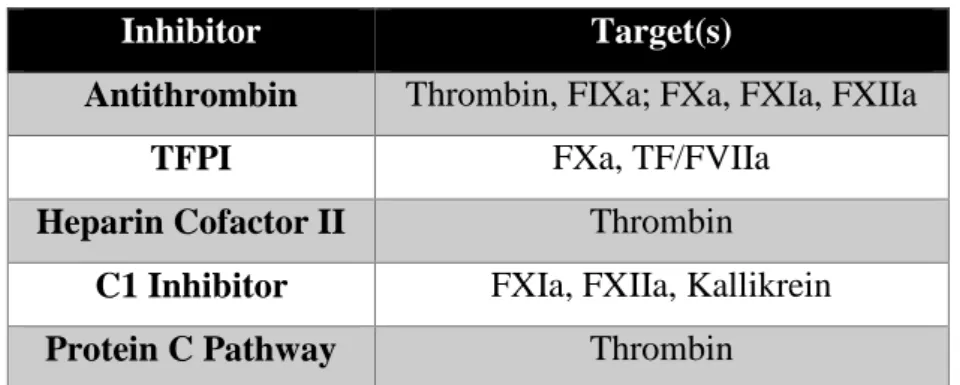
First, circulating protease inhibitors, such as antithrombin, heparin cofactor II, tissue factor pathway inhibitor (TFPI), and C1 inhibitor, eliminate activated coagulation factors by attacking their active sites. The second anticoagulant modality is provided by the enzyme-based protein C/protein S pathway, which is implicated in endothelial-based pathways of coagulation inactivation (Versteeg et al, 2013). In this pathway, fVa and fVIIIa are inactivated in the presence of thrombomodulin catalyst and endothelial protein C receptor (EPCR) (Yau et al, 2015). Collectively, these individual events come together to ensure that the blood coagulation system is highly regulated. Table 1 summarizes the respective targets for all the aforementioned inhibitors.
Inhibitor Target(s)
Antithrombin Thrombin, FIXa; FXa, FXIa, FXIIa
TFPI FXa, TF/FVIIa
Heparin Cofactor II Thrombin
C1 Inhibitor FXIa, FXIIa, Kallikrein
Protein C Pathway Thrombin
Table 1: Targets of Coagulation Inhibitors
1.4 Current Concepts of Antithrombotic Therapy
Systemic problems such as pulmonary embolism, atrial fibrillation (AF), artificial heart valve, stroke, arterial or deep vein thrombosis, acute myocardial infarction, among other systemic pathologies, constitute the basis for the need of prophylactic treatment
12
with antithrombotic therapy (Oake et al, 2008; António et al, 2008; Nematullah et al, 2009; Araújo et al, 2010; Van Diermen et al, 2013). In this therapy, the drugs that stand out are the antiplatelet agents and anticoagulants. Antiplatelet drugs, such as aspirin, work in primary hemostasis, corresponding to the formation of the platelet thrombus. Anticoagulants, such as heparin and warfarin, work in secondary hemostasis, relative to the formation of the fibrin network and blood clot (Filho et al, 2013; Napeñas et al, 2013).
Conventional oral anticoagulants, or VKAs – vitamin K antagonists (vitamin K is essential in the production of several coagulation factors) – constitute the first line therapy in the prophylaxis of thromboembolic events such as AF (Mookadam et al, 2015). For over 60 years, VKAs, especially warfarin and acenocoumarol, have been shown, in several studies, to reduce the risk of stroke in 70% in patients with atrial fibrillation when correctly anticoagulated compared with a reduction of only 22% provided by antiplatelet drugs. Therefore, oral anticoagulants are recommended in AF patients at moderate-high risk for stroke and tromboembolism (Cid-Conde & Lopez-Castro, 2013; Johnston, 2015).
VKAs are drugs with proven efficacy and a specific antidote, which can be given in case of bleeding. Moreover, there is the possibility of discontinuing medication urgently and at low cost (Cid-Conde & Lopez-Castro, 2013). However, VKAs have limitations that affect the quality of life of patients and increase morbidity: narrow therapeutic window (International Normalized Ratio, INR 2.0-3.0), unpredictable response, systematic control of bleeding, require routine monitoring, frequent dose adjustments and numerous food and drug interactions (Cid-Conde & Lopez-Castro, 2013; Mookadam et al, 2015). In this sense, the novel oral anticoagulants (NOACs), non-vitamin K dependent, acting directly on specific coagulation factors, show promising results as an ideal alternative to conventional therapy (Mookadam et al, 2015). However, it is essential to understand how they differ from VKAs, how they should be monitored, and which precautions should be taken regarding the therapeutic adjustments when medical and dental procedures are performed.
In dentistry and oral surgery, the major concerns in treatment of patients taking NOACs (direct thrombin inhibitors and factor Xa inhibitors) are the risk of hemorrhage and the absence of a specific reversal agent. The degree of renal function, the complexity of the surgical procedure and the patient’s risk of bleeding due to other concomitant
13
causes, constitute the most important factors to consider during surgical dental treatment of patients taking NOACs (Isola et al, 2015; Costantinides et al, 2016).
It is paramount that the clinician considers that the number of patients taking NOACs is rapidly increasing. In addition, since available data is not sufficient to establish an evidence-based dental management, the dentist must use caution and attention when treating patients taking these drugs (Costantinides et al, 2016).
There has been an increase in the number of patients undergoing antithrombotic therapy and in need of medical and dental care (Araújo et al, 2010; Isola et al, 2015; Paraschiv et al, 2015).This is easily justified by the fact that there is an increasingly aging population, increasing the number of anticoagulated patients, along with the current paradigm based on the dentition conservation as much as possible (António et al, 2008; Isola et al, 2015; Paraschiv et al, 2015). It is expected, therefore, a progressive increase of anticoagulated patients seeking dental interventions (António et al, 2008).
The approach of patients under chronic antithrombotic therapy needing to undergo dental procedures continues to cause great controversy (Sacco et al, 2007; António et al, 2008; Van Diermen et al, 2013; Isola et al, 2015). Most professionals recommend the suspension or reduction of the dosage of anticoagulants, but recent studies show that this behavior causes an increased risk for the patient (Araújo et al, 2010; Van Diermen et al, 2013). Furthermore, the dentist should account for the fact that dental surgery is unlike other types of surgery, as major vessels are unlikely to be encountered, and the peri and postoperative surgical sites are easily accessible to perform local hemostatic measures, such as biting on gauze, absorbable gelatin sponges, and sutures (Wahl, 2014).
The increased bleeding risk associated to procedures performed in dentistry when the patient is under oral anticoagulants or antiplatelet agents must be taken into account relatively to the increased risk of thrombosis caused by the interruption of antithrombotic therapy (Nematullah et al, 2009; Wahl, 2014; Paraschiv et al, 2015). After therapy interruption and its reintroduction a few days later, with initial high doses, it has been reported to occur a phenomenon of rebound hypercoagulation, derivative of the fact that the inhibition of proteins C and S occurs earlier than the inhibition of the prothrombotic factors, leading to a "paradoxical" thrombotic effect (António et al, 2008).
14
Several studies indicate serious embolic complications, including death, are three times more likely to occur in patients whose anticoagulant therapy is interrupted, than are bleeding complications in patients whose anticoagulant therapy is continued (and whose anticoagulation levels were within or below therapeutic levels) (Araújo et al, 2010). Currently, several authors suggest the execution of the procedures without therapy modification or discontinuation, with emphasis on preventive local hemostatic measures pre, peri and postoperatively, so that prevention of bleeding is accomplished (Madrid & Sanz, 2009; Nematullah et al, 2009; Araújo et al, 2010; Firriolo & Hupp, 2012).
The lack of standardization of medical protocols in situations of patients taking antithrombotic therapy makes communication between the dentist and the attending physician essential, in order to avoid errors from both parts, due to lack of knowledge, or caused by myths on how to act in these cases (Araújo et al, 2010).
The aim of this study was to review the current literature regarding antithrombotic therapy and its implications in medical dentistry.
15
2. Materials and Methods
2.1 Research Methods
Search for relevant literature was performed on the primary databases Cochrane Library (via cochranelibrary.com) and Medline (via PubMed), and the secondary database Google Scholar (via scholar.google.com), from November 2015 until May 26th 2016. The following key-words were searched: Anticoagulants Dental Surgery; Hemostasis; Cell-based Model Hemostasis; Antiplatelet Therapy Oral Surgery; New Oral Anticoagulants Dental Procedures.
2.2 Types of Studies Included
The following formats were considered for inclusion in this review: Meta-analysis, Systematic Reviews and Randomized Control Trials. However, due to insufficient evidence of high scientific value, the search was extended to include Reviews, Cohorts and Guidelines, in order to obtain the best evidence possible.
2.3 Inclusion Criteria
Inclusion criteria consisted of articles published in the last 10 years, limited to those written in English or Portuguese, with the sample limited to humans.
2.4 Article Selection
Articles were selected independently by the author, with initial screening being accomplished by reviewing the titles and abstracts. The full versions of the articles that were considered relevant or whose relevance needed to be assessed were obtained. In addition, the reference lists of retrieved articles on the subject were reviewed, and more articles were retrieved, in accordance to the inclusion criteria.
2.5 Data Collection Process
After the initial selection and collection of the articles, they were thoroughly analyzed by the author in order to obtain the relevant data needed for the elaboration of this review.
16
3. Results
According to the aforementioned criteria, the search yielded 1242 results, of which 47 were selected for inclusion in this paper after analyzing the titles, abstracts and full text. An analysis of the reference lists of the previously selected articles was also performed in order to obtain further data, beyond the data collected from the databases, which resulted in the inclusion of 21 additional articles pertinent to the subject, in accordance to the inclusion criteria. The 68 articles obtained are divided into: 4 Systematic Reviews, 2 Meta-Analysis, 11 Randomized Control Trials (RCTs), 27 Reviews, 15 Cohorts and 9 Guidelines. The following scheme shows the data collection process and results yielded from the search.
Medline
via pubmed.govCochrane Library
via cochranelibrary.comGoogle Scholar
via scholar.google.com1242 Articles
Inclusion Criteria47 Articles
Selected
Assessment of
Relevance:
Tittle Abstract Full Text21 Articles
From Reference Lists of Selected Articles 27 Reviews 4 Systematic Reviews 15 Cohorts 2 Meta-Analysis 11 RCTs 9 Guidelines
17
4.
Antithrombotic Therapy
4.1 Antiplatelet Drugs
The use of antiplatelet agents is indicated for the secondary prevention of cardiac and cerebrovascular diseases, more significantly for the prevention of arterial and venous thrombosis in patients with certain systemic conditions, such as ischemic heart disease, prosthetic heart valves, and those at risk for ischemic cerebrovascular accidents (Napeñas et al, 2013; Bajkin et al, 2014). They exert their action in the primary phase of coagulation, specifically in the platelet plug formation, by preventing platelets from aggregating to each other and adhering to blood vessel walls. This is enabled by several mechanisms (Pototski & Amenábar, 2007; Cardona-Tortajada et al, 2009; Guyatt et al, 2012).
Cyclooxygenase inhibitors (COX-1 and COX-2) inhibit thromboxane A2-dependent platelet activation by reversible competitive blocking of COX activity. Cyclooxygenase inhibitors include: acetylsalicylic acid (ASA) – COX-1 irreversible inhibitor; nonsteroidal anti-inflammatory drugs (NSAID) and triflusal – both COX-1 reversible inhibitors (Pototski & Amenábar, 2007; Napeñas et al, 2013; Coelho, 2015). ASA creates permanent changes by blocking platelet aggregation, which last for the life span of the platelet (7-10 days) (Madrid & Sanz, 2009; Filho et al, 2013; Napeñas et al, 2013; Bajkin, Urosevicb et al, 2014). Platelet aggregation and bleeding time typically do not return to baseline until new platelets, which were not exposed to the medication, are produced (Madrid & Sanz, 2009; Guyatt et al, 2012).
Thienopyridines, such as clopidogrel, are irreversible blockers of P2Y12, an ADP
receptor present in the platelets’ surface, which is necessary for the activation of the receptor GPIIb/IIIa complex during platelet aggregation (Napeñas et al, 2013; Coelho, 2015).Prasugrel is a new oral antiplatelet agent (NOAP), which is also a thienopyridine with the same mechanism of action of clopidogrel, but with a faster onset of action, more potent antiplatelet effect, reduced variability and fewer drug interactions. Another NOAP, ticagrelor, also blocks the P2Y12 platelet receptor, but binds reversibly, which enables a
rapid offset as well as onset of action (Dézsi et al, 2015; Johnston, 2015). Thienopyridines are also effective during the lifetime of the platelet (Bajkin, Urosevicb et al, 2014).
18
Dipyridamole inhibits phosphodiesterase activity, blocking cAMP decomposition and reducing intracellular calcium, therefore inhibiting platelet activation and aggregation. It is frequently used concomitantly with ASA (Pototski & Amenábar, 2007; Napeñas et al, 2013; Coelho, 2015; Johnston, 2015).
GPIIb/IIIa receptor antagonists (tirofiban, abciximab), which are used intravenously, inhibit the final common pathway of platelet aggregation (Napeñas et al, 2013; Coelho, 2015).
Recently there has been a significant effort to develop tests to assess the effects of antiplatelet drugs on platelet function. There are several tests available to measure the platelet effects of aspirin, clopidogrel and other P2Y12 inhibitors. The clinical significance
of these tests is, however, uncertain, and there is a strong correlation between their results and the clinical outcome. Consequently, the use of laboratory tests is not recommended routinely in the management of these patients (Fonseca et al, 2014).
4.2 Conventional Anticoagulants
Currently, the most commonly used VKAs are heparin, warfarin and acenocoumarol (Sacco et al, 2007; Madrid & Sanz, 2009). Heparin has a very short half-life and thus it is usually administered intravenously for short-term use. It interferes with the thrombin–antithrombin pathways, by activating antithrombin and forming a complex which will accelerate the inhibition rate of the coagulation enzymes, specially thrombin and FXa, leading to a decrease in fibrin formation (Madrid & Sanz, 2009; Coelho, 2015; Isola et al, 2015). Although its pharmacokinetics have been improved with the development and introduction of low-molecular-weight heparin (LMWH), which may be used on an outpatient basis and have a more predictable anticoagulant response, it still requires narrow medical surveillance (Madrid & Sanz, 2009; Coelho, 2015).
Warfarin is a coumarinic oral anticoagulant, which prevents the reduction of vitamin K into its active forms. It is a competitive vitamin K epoxide redutase inhibitor, thus inhibiting the formation of several vitamin K dependent coagulation factors (II, VII, IX, X), and proteins C and S. The maximum anticoagulant effect of warfarin takes 48-72 hours to develop. It has a half-life of about 36 hours and a duration of action for a single
19
dose between 2-5 days (Al-Mubarak et al, 2007; Madrid & Sanz, 2009; O’Connell, 2014). Another coumarinic anticoagulant is acenocoumarol, but warfarin is the gold-standard (Madrid & Sanz, 2009; Paraschiv et al, 2015). They demand anticoagulation monitoring by measuring the INR, which was developed to incorporate the international sensitivity index (ISI) values and attempt to make prothrombin time (PT) results uniformly useable (Al-Mubarak et al, 2007; Sacco et al, 2007; Fakhri et al, 2013). INR therapeutic ranges depend on the disease: 2 to 3 for venous thromboembolism, stroke and atrial fibrillation; 2.5 to 3.5 for patients with prosthetic valves (Sacco et al, 2007; Oake et al, 2008; Madrid & Sanz, 2009; Paraschiv et al, 2015). The incidence of bleeding is dose-dependent and influenced by age and the presence of comorbidities, including some medical conditions, polimedication, hypertension and renal or hepatic failure (Paraschiv et al, 2015).
The risk of bleeding is an essential factor for the clinician’s decision of discontinuing the therapy or not (Jiménez et al, 2008). Thus INR monitoring is paramount. In spite of this, in certain non-invasive procedures, INR evaluation is not necessary. These procedures include: supragingival scaling, simple restorative treatment, local anesthetic injections (buccal infiltration, intraligamentary or mental block), impressions and other prosthetic procedures (Perry et al, 2007; Tsolka, 2014). Potentially invasive procedures, in which bleeding is predictable, will require previous evaluation of the aforementioned parameters, especially INR. These procedures are: local anesthesia by inferior alveolar or other regional nerve blocks or floor of mouth infiltrations, subgingival scaling and root surface instrumentation, subgingival crown and bridge preparations, endodontics - standard root canal treatment, simple extractions (up to 3 teeth), incision and drainage of swellings, biopsies – which are considered moderate risk procedures – and extensive maxillofacial surgery, periodontal surgery, alveolar surgery (bone removal) and multiple extractions – which are considered high risk procedures (Perry et al, 2007; Douketis et al, 2008; Tsolka, 2014).
4.3 Novel Oral Anticoagulants
As they are relatively new and not as commonly used as warfarin is today, this chapter will focus on the mechanism of action and monitoring of the novel oral anticoagulants.
20
As it was mentioned before, VKAs present various disadvantages and limitations: narrow therapeutic window, unpredictable response, systematic control of bleeding, frequent dose adjustments and numerous food and drug interactions, which results in the necessity of routine monitoring. They also take several days to reach a satisfactory therapeutic concentration and present a complex overlap with parenteral anticoagulants (Cid-Conde & Lopez-Castro, 2013; Isola et al, 2015; Mookadam et al, 2015). These disadvantages created an impetus for the development of novel oral anticoagulants with a wider therapeutic index, less interactions, and a predictable level of anticoagulation at a specific dose. In the last few years, two new classes of oral anticoagulants – direct thrombin inhibitors (e.g. dabigatran etexilate), and factor Xa inhibitors (e.g. rivaroxaban, apixaban and edoxaban) – have been approved for use (Firriolo & Hupp, 2012; O’Connell, 2014; Tsolka, 2014; Costantinides et al, 2016).
These NOACs are non-vitamin K dependent and, unlike warfarin, dabigatran and rivaroxaban are relatively small molecules that function as anticoagulants by targeting specific steps of the coagulation cascade (Cid-Conde & Lopez-Castro, 2013; Giugliano et al, 2013; O’Connell, 2014; Costantinides et al, 2016). Furthermore, they are reported to have fewer drug interactions, no significant food interactions, and have predictable pharmacokinetics, which means that they provide predictable anticoagulation at a specific dose, without the need for regular laboratory monitoring and adjustments of dosage (Granger et al, 2011; Cid-Conde & Lopez-Castro, 2013; O’Connell, 2014; Tsolka, 2014; Paraschiv et al, 2015; Costantinides et al, 2016).
Both dabigatran and rivaroxaban are licensed for short-term primary prevention of venous thromboembolic events in adult patients who underwent hip or knee replacement surgeries, while dabigatran is also licensed for prevention of stroke and systemic embolism in adult patients with non-valvular atrial fibrillation (Connolly et al, 2009; Patel et al, 2011; Cid-Conde & Lopez-Castro, 2013; O’Connell, 2014).
Due to having more predictable pharmacokinetics and pharmacodynamics than warfarin, their use in the prevention of thromboembolic complications has shown to be as effective as, or in some cases even more effective than warfarin, with lower rate of major hemorrhage (Connolly et al, 2009; Patel et al, 2011; Paraschiv et al, 2015).
21
4.3.1 Direct Thrombin Inhibitors
Dabigatran is a specific, reversible, direct thrombin inhibitor that inhibits both free and fibrin-bound thrombin. It prevents the conversion of fibrinogen into fibrin by binding itself to the active site on the thrombin molecule (factor IIa) (Firriolo & Hupp, 2012; Cid-Conde & Lopez-Castro, 2013; O’Connell, 2014; Isola et al, 2015; Johnston, 2015). It has a rapid onset of action with a peak plasma concentration at 0.5-4 hours, with a half-life elimination is 12-14 hours in healthy patients, 14-17 hours in the elderly, and up to 27 hours in patients with severe renal impairment (i.e., creatinine clearance <15-30ml/min) (Firriolo & Hupp, 2012; Cid-Conde & Lopez-Castro, 2013; O’Connell, 2014).
Owing to the reversal of dabigatran’s effects, due to its short half-life, discontinuation of the drug may be sufficient to resolve minor hemorrhage, with the exception of patients with renal impairment (Firriolo & Hupp, 2012; O’Connell, 2014; Paraschiv et al, 2015). Supportive strategies to control more severe bleeding include oral charcoal application (for recent ingestion of dabigatran) (Costantinides et al, 2016), mechanical compression, surgical hemostasis, fluid replacement and transfusion of blood products (packed red cells or fresh frozen plasma). If these measures fail to control bleeding, then recombinant activated factor VII (rFVIIa) and/or hemodialysis can be considered (Firriolo & Hupp, 2012; Isola et al, 2015; Paraschiv et al, 2015).
In an initial clinical study, part of an ongoing, multicenter, prospective cohort, published in June 2015, Pollack et al showed that idarucizumab – a monoclonal antibody fragment which rapidly and completely reversed the anticoagulant activity of dabigatran in 88 to 98 % of patients – is an effective reversal agent (Pollack et al, 2015). The U.S. Food and Drug Administration (FDA), in October 2015, approved idarucizumab for the treatment of patients taking dabigatran when reversal of its anticoagulant effects is needed for emergency procedures, or in life-threatening or uncontrolled bleeding (U.S. Food and Drug Administration, 2016). The European Medicines Agency (EMA), in December 2015, decided that idarucizumab’s benefits are greater than its risks and recommended that it be approved for use in the EU (European Medicines Agency, 2016). As of today, its use is authorized in Portugal (Infarmed, 2016).
22
As it was mentioned above, unlike warfarin, routine monitoring of the anticoagulant effect of dabigatran is not required (Firriolo & Hupp, 2012; O’Connell, 2014; Paraschiv et al, 2015). However, certain situations, such as emergency surgery, may require assessment of anticoagulation. This cannot be done by resorting to the INR, as this test is not sensitive enough (Firriolo & Hupp, 2012; Paraschiv et al, 2015).
Dabigatran prolongs the activated partial thromboplastin time (aPTT), ecarin clotting time (ECT), and thrombin time (TT) at recommended therapeutic doses, but it has little effect on prothrombin time (PT)/INR at clinically relevant plasma concentrations (Castellone & Van Cott, 2010; Firriolo & Hupp, 2012; Paraschiv et al, 2015; Isola et al, 2015). Therefore, the most sensitive tests used for quantifying the anticoagulant effects of dabigatran are the thrombin clotting time (TT) and ecarin clotting time (ECT). Castellone and Van Cott state that the TT test is too sensitive for monitoring the anticoagulant effects of DTIs (Direct Thrombin Inhibitors), such as dabigatran (Castellone & Van Cott, 2010). Despite being very good from a qualitative point of view, to assess adherence to treatment, it cannot quantify levels (Van Ryn et al, 2010). The activated partial thromboplastin time (aPTT) may also be used, but it is less sensitive than the TT and ECT tests, particularly at higher dabigatran doses (O’Connell, 2014; Paraschiv et al, 2015).
4.3.2 Factor Xa Inhibitors
Rivaroxaban is an orally-administered, selective, reversible, direct inhibitor of activated factor X (factor Xa),which interrupts both the extrinsic and intrinsic coagulation pathways, thereby inhibiting thrombin formation (Cid-Conde & Lopez-Castro, 2013; O’Connell, 2014, Johnston, 2015; Costantinides et al, 2016). It can inhibit free factor Xa, clot-bound factor Xa, and factor Xa bound to the prothrombinase complex (Firriolo & Hupp, 2012, Isola et al, 2015; Costantinides et al, 2016). It has a rapid onset of action with a peak plasma concentration at 2.5-4 hours, and its half-life is 5-9 hours in healthy adults, and 11-13 hours in the elderly (Firriolo & Hupp, 2012; Cid-Conde & Lopez-Castro, 2013; O’Connell, 2014; Tsolka, 2014).
Concerning a reversal agent, there is no specific antidote for rivaroxaban (Firriolo & Hupp, 2012; O’Connell, 2014; Paraschiv et al, 2015; Costantinides et al, 2016). In cases where severe bleeding occurs, certain strategies for the reversal of the effects of
23
rivaroxaban may be considered: fluid replacement, transfusion or blood products – recombinant activated factor VII or prothrombin complex concentrate (Isola et al, 2015; Paraschiv et al, 2015).
More recently, a modified form of factor Xa is currently undergoing clinical trials to study the effectiveness of reversal of the effects of oral FXaIs, in order to be used as an antidote for these drugs (Costantinides et al, 2016).
Similarly to dabigatran, routine monitoring of rivaroxaban is not required. However, in emergency situations where the measurement of the level of anticoagulation may be necessary, anti-factor Xa assay is reportedly the most accurate measurement of the anti-coagulant effect of rivaroxaban. Moreover, FXaIs (Factor Xa Inhibitors), including rivaroxaban, are reported to slightly prolong aPTT and PT (Castellone & Van Cott, 2010, Tsolka, 2014; Costantinides et al, 2016). Therefore, some authors aPTT and PT (with rivaroxaban specific calibration) may also be used (Samama et al, 2010; O’Connell, 2014; Paraschiv et al, 2015).
Apixaban is the most recently introduced targeted oral anticoagulant. It is a reversible, highly selective and potent inhibitor of factor Xa, with the same therapeutic indications of dabigatran and rivaroxaban (Cid-Conde & Lopez-Castro, 2013; Johnston, 2015; Costantinides et al, 2016). It inhibits and delays the generation of thrombin without significantly affecting platelet aggregation (Cid-Conde & Lopez-Castro, 2013). Its half-life is about 12 hours and it also has a rapid onset of action, reaching peak plasma concentration in 1-3 hours (Tsolka, 2014; Costantinides et al, 2016). Apixaban has low renal elimination so it is preferred in elderly patients and those with decrease renal function (Paraschiv et al, 2015; Johnston, 2015).
As rivaroxaban, no specific reversal agent exists for apixaban. In situations of normal bleeding, the delay or discontinuation of the next dose can be sufficient to resolve bleeding. In emergency situations, when severe bleeding occurs, recombinant factor VIIa, recombinant factor Xa or activated thrombin complexes can be used (Costantinides et al, 2016).
24
5. Treatment Planning Alterations/Recommendations
5.1 Antiplatelet Drugs
There is some controversy over whether antiplatelet therapy should be maintained or suspended prior to elective dental procedures (Grines et al, 2007; Napeñas et al, 2013). Girotra et al, in 2013, evaluated the association between postoperatory risk of prolonged bleeding and various types of minor surgical interventions. They found a subtle, yet statistically significant, association between these variables, verifying that prolonged bleeding occurred independently of the type of minor procedure. (Girotra et al, 2014).
In spite of the hemorrhagic risk, the suspension of antiplatelet drugs increases perioperatory morbidity, not only due to the direct effects of their suspension, but also because of a possible rebound effect, as is the case of aspirin discontinuation syndrome, promoted by the increase of thromboxane A2 and the reduction of fibrinolysis. Consequently, suspending antiplatelet therapy before performing medico-surgical procedures might cause catastrophic effects (Van Diermen et al, 2009; Coelho, 2015). Moreover, in patients with coronary artery disease, discontinuation of chronic antiplatelet therapy poses a significant risk for acute coronary events, which can result in death (Napeñas et al, 2013).
The American Heart Association (AHA) and the American College of Cardiology (ACC) recommend not to discontinue the therapy in patients undergoing antiplatelet monotherapy with ASA, i.e., in patients treated with a single antiplatelet drug. In cases where patients are in a therapeutic regimen with dual antiplatelet therapy, i.e., treated with two antiplatelet agents, needing to undergo elective procedures in which there is high risk of bleeding, they recommend postponing the surgery, if possible, until the appropriate course of antiplatelet therapy is completed (Grines et al, 2007).
According to the American College of Chest Physicians (ACCP), in their 2012 guidelines, in procedures of minor hemorrhagic risk, such as most dental procedures, it is recommended to keep the antiplatelet therapy with ASA in cases of secondary prevention (Guyatt et al, 2012).
25
In Annals of Surgery, Gerstein et al, in an article published in 2012, stated that in patients undergoing antiplatelet treatment, during dental procedures, the risk of bleeding increases only by 1.5 times, which does not compromise the viability of surgery, so the empirical discontinuation of ASA has no indication (Gerstein et al, 2012).
In their guidelines from 2014, the Portuguese Society of Anesthesiology recommends that patients on monotherapy with ASA or under dual antiplatelet therapy consisting of ASA and a P2Y12 inhibitor, undergoing procedures with low risk of
bleeding, as is the case of most dental procedures, should not suspend the therapy; the importance of first assessing the thrombotic and hemorrhagic risk along with the urgency of the procedure is also highlighted. In patients under dual antiplatelet therapy, in cases of high risk of bleeding and an intermediate to high thrombotic risk, the recommendation is to defer elective procedures and to maintain ASA and suspend the P2Y12 inhibitor in
emergency procures. They also suggest considering bridging therapy with GPIIb/IIIa inhibitors when the thrombotic risk is high (Fonseca et al, 2014).The reintroduction of the P2Y12 inhibitor should occur within 24 hours after surgery. When the antiplatelet
agent is clopidogrel, which reaches a maximum effect at 5-10 days, if there is an elevated risk of thrombosis, a loading dose (300 to 600 mg), which is a higher dose sometimes recommended for drugs which take a longer time to reach their maximum effect, may be taken in order to obtain a faster effect (Fonseca et al, 2014; Coelho, 2015).
The Italian cardiological, surgical and anaesthesiological societies, in an article from 2014, are in agreement with most recommendations from the Portuguese Society of Anesthesiology. However, they recommend keeping ASA and P2Y12 combined therapy
in patients with intermediate to high thrombotic risk undergoing non-deferrable procedures which present an intermediate risk of hemorrhage (Rossini et al, 2014).
Results from a clinical study by Gröbe et al suggest that, despite its more invasive nature, oral osteotomy can be performed safely under continued antiplatelet monotherapy with clopidogrel or dual therapy with clopidogrel and ASA (Gröbe et al, 2015). Hanken et al reached the same conclusion regarding continued ASA therapy (Hanken et al, 2015).
Morimoto et al, in two different studies, concluded that postoperative hemorrhage after periodontal surgery and teeth extractions for patients on single or dual antiplatelet
26
therapy was significantly lower than for those on combined antiplatelet/warfarin therapy and warfarin therapy alone (Morimoto et al, 2008; Morimoto et al, 2009). As several studies suggest keeping warfarin therapy for patients undergoing most dental procedures (which will be discussed in the next section), when the INR is below 4, it is possible to infer that antiplatelet therapy should not be suspended (Napeñas et al, 2013). In reality, the hemorrhagic risk may be a rare occurrence after dental procedures, with a low to negligible frequency for patients undergoing mono or dual antiplatelet therapy, as it has been reported in only a few cases after periodontal surgery or extensive oral surgery, when an excessive dosage of ASA was taken (Napeñas et al, 2009).
It is also important to consider that a growing number of patients are taking combined oral anticoagulant and antiplatelet therapy, which is strongly recommended to be used only in patients with prosthetic heart valves. A prospective study by Bajkin et al, in 2012, concluded that teeth extractions can be performed safely while patients continue to receive combined anticoagulant-aspirin therapy, as long as their INR values are within therapeutic range and local hemostasis measures are provided (Bajkin et al, 2012).
Various studies found no clinically significant increased risk of postoperative bleeding complications from invasive dental procedures in patients on either single or dual antiplatelet therapy, which supports the recommendation that there is no indication to alter or suspend this therapy (Morimoto et al, 2008; Morimoto et al, 2009; Napeñas et al, 2009; Lillis et al, 2011; Park et al, 2012; Dézsi et al, 2015).
To sum up, the risks of altering or discontinuing the use of antiplatelet drugs far outweigh the risk of postoperative oral bleeding (Napeñas et al, 2009; Wahl, 2014). In the rare event of postoperative bleeding, local hemostatic measures are effective in addressing this complication (Morimoto et al, 2009; Napeñas et al, 2013).
Regarding new oral antiplatelet agents, such as prasugrel and ticagrelor, further evidence is required to establish whether a period of discontinuation is necessary for minor oral surgery procedures (Johnston, 2015). However, Dézsi et al, in a study published in 2015, refer that dental extractions can be performed safely with due precaution in patients under dual antiplatelet therapy combining either clopidogrel and ASA or prasugrel and ASA (Dézsi et al, 2015).
27
5.2 Conventional Anticoagulants
The management of dental procedures in patients undergoing oral anticoagulation must be individualized depending on the type of procedure, bleeding risk and thrombotic risk (Paraschiv et al, 2015). Current available information suggests that the risk of hemorrhage in patients undergoing invasive dental procedures (e.g. up to three dental extractions, up to three dental implants and periodontal surgery) is low (Van Diermen et al, 2013; Tsolka, 2014).
Several strategies have been used for the management of anticoagulated patients during dental procedures. One approach is to withhold VKAs for 3–5 days prior to the intervention, without substitution with additional anticoagulation (Patatanian & Fugate, 2006; Pototski & Amenábar, 2007; Sacco et al, 2007; Madrid & Sanz, 2009). However, this approach exposes the patient to increased thromboembolic risk for over a week while the INR is subtherapeutic (Patatanian & Fugate, 2006), as it would take about 4 days for the INR to reach the value of 1,5, and about 3 days to reach therapeutic values after being resumed (Pototski & Amenábar, 2007; Nematullah et al, 2009). In addition, the risk of thromboembolism may be increased by other factors, such as rebound hypercoagulable state due to discontinuation of oral anticoagulation and the hemostatic changes of the surgical intervention itself (Patatanian & Fugate, 2006; António et al, 2008; Madrid & Sanz, 2009). Therefore, the drug becomes unpredictable and a higher probability of occurrence of a thromboembolic event is reported (Nematullah et al, 2009; Madrid & Sanz, 2009; Isola et al, 2015).The scientific evidence does not support this protocol as it may increase the thromboembolic risk, without significantly reducing bleeding after the procedures (Madrid & Sanz, 2009; Van Diermen et al, 2013; O’Connell, 2014).
Another approach is to withhold VKAs for 3–5 days before the procedure, while bridging with a parenteral anticoagulant such as LMWH, which has the advantage of minimizing thromboembolic risk (Patatanian & Fugate, 2006; Sacco et al, 2007; Bajkin et al, 2009; Nematullah et al, 2009). Nevertheless, it has disadvantages such as increased risk of bleeding from dual anticoagulation therapy perioperatively, expense of an additional anticoagulant, inconvenience of injections, and higher complexity of the therapeutic regimen (Patatanian & Fugate, 2006). Various studies concluded that there is no need for bridging therapy with LMWH in patients undergoing minor dentoalveolar
28
procedures, although this approach can be used in patients going through major oral surgical interventions (Pettinger & Owens, 2007; Bajkin et al, 2009). Consequently, this method is advocated only for patients at intermediate to high thromboembolic risk who are undergoing invasive procedures or surgery necessitating therapy interruption (Patatanian & Fugate, 2006; Pettinger & Owens, 2007). This protocol requires very subtle monitoring of the therapy. Moreover, the risk of thromboembolic events may be even higher than when warfarin is discontinued without bridging therapy, due to the fact that heparin may induce thrombocytopenia, which may further impair the patient’s hemostasis (Madrid & Sanz, 2009).
A third approach is to continue oral anticoagulation perioperatively, in order to avoid compromising the thromboembolic protection. Several studies and guidelines support this practice (Patatanian & Fugate, 2006; Al-Mubarak et al, 2007; Perry et al, 2007; Douketis et al, 2009; Bajkin et al, 2009; Madrid & Sanz, 2009; Nematullah et al, 2009; Firriolo & Hupp, 2012). The literature does not describe any cases of severe postoperatory bleeding in patients who continued the therapy, whereas several cases of fatal thromboembolic complications were reported after cessation of oral anticoagulants before procedures such as dental extractions (Sacco et al, 2007; Bajkin et al, 2009). Recent studies have suggested that VKAs should not be discontinued in the majority of patients requiring outpatient dental procedures (including dental extractions and implant placement – as long as it does not involve autogenous bone grafts, extensive flaps or osteotomy preparations extending outside the bony envelope), provided local hemostasis is adequately maintained (Al-Mubarak et al, 2007; Perry et al, 2007; Douketis et al, 2009; Madrid & Sanz, 2009; Van Diermen et al, 2013; Bajkin et al, 2014).
Due to the fact that dental implants have become the standard of care regarding the substitution of missing teeth, it is inevitable to treat an anticoagulated patient needing implant placement, which could be considered as comparable with or even less traumatic than the extraction of three teeth. Consequently, evidence does not support that dental implant placement is contraindicated in these patients (Madrid & Sanz, 2009).
The difficulty in predicting the drop in the INR value when warfarin therapy is suspended and the associated risk of thromboembolism outweighs the risk of postoperative bleeding in patients who maintain the therapy (Al-Mubarak et al, 2007;
29
Madrid & Sanz, 2009; Okamoto et al, 2014). Moreover, other studies have shown that bleeding complications associated with anticoagulation are three times less likely to occur than serious embolic complications (including death) in patients whose anticoagulant therapy was interrupted (Al-Mubarak et al, 2007; Araújo et al, 2010).Notwithstanding, there is a concern for higher hemorrhagic risk when continuing the therapy. However, this can be managed by performing local hemostatic measures, such as applying local pressure, hemostatic mouthwashes and suturing (Patatanian & Fugate, 2006; Sacco et al, 2007; Madrid & Sanz, 2009; Fakhri et al, 2013), which will be addressed ahead.
For patients under warfarin therapy who present a stable INR between 2-4, studies indicate that the risk of significant bleeding is very small, hence the medico-surgical intervention can be performed without suspending the therapy (Perry et al, 2007; Al-Mubarak et al, 2007; Bajkin et al, 2009; Madrid & Sanz, 2009; Nematullah et al, 2009; Okamoto et al, 2014; Bajkin et al, 2014; Isola et al, 2015). Furthermore, according to Oake et al, in a review from 2008, the risks of hemorrhage and thromboembolism are both minimized at INR ratios of 2–3, while ratios that are moderately higher than this therapeutic range appear to be safer than subtherapeutic ratios (Oake et al, 2008). Nonetheless, when INR values approach 4, an appreciation of the difficulty of surgery and the experience and surgical skills of the dentist is recommended when assessing the risk of bleeding. For patients who are stably anticoagulated, the determination of INR is recommended 72 hours prior to dental surgery (Perry et al, 2007; Johnston, 2015). When this is not the case, INR should be checked 24 hours before the procedure (Douketis et al, 2012).
Concerning drug interactions, patients undergoing dental surgery may be prescribed antibiotics to prevent endocarditis. According to Perry et al, in their 2007 guidelines, a single dose of an antibiotic is unlikely to have any significant effect upon the INR. Individuals who are prescribed more than a single dose of antibiotics should have the INR measured 2-3 days after starting treatment (Perry et al, 2007). Macrolide antibiotics, including erythromycin, clarithromycin, and possibly azithromycin, are reported to cause significant episodes of bleeding in patients taking warfarin (Firriolo & Hupp, 2012). In terms of analgesic therapy, non-steroidal anti-inflammatory drugs (NSAIDs) and COX-2 selective inhibitors are avoided in patients receiving oral anticoagulants because of their antiplatelet action and the risk of over-anticoagulation and
30
hemorrhage (Perry et al, 2007; Van Diermen et al, 2013; Isola et al, 2015). Metronidazole and other azole antifungals, including ketoconazole, fluconazole and itraconazole are reported to significantly interact with warfarin via CYP inhibition, thus augmenting its effects (Firriolo & Hupp, 2012; Van Diermen et al, 2013).
Furthermore, in patients taking oral anticoagulants, block anesthetic techniques are not recommended. However, inferior alveolar nerve block is accepted in patients taking warfarin with INR<3. Owing to local anesthesia, intraligamentary and intraseptal techniques produce less bleeding complications (Paraschiv et al, 2015).
Finally, patients on oral anticoagulants with co-existing systemic conditions, including liver disease, renal disease and thrombocytopenia, or who are taking antiplatelet drugs, present an increased risk of bleeding. Therefore, these patients should not go through surgical dental procedures in the primary care setting. In these cases, or in cases where patients do not present stable INR values, the treating physician should always be consulted on whether to suspend the therapy or not (Perry et al, 2007).
5.3 Novel Oral Anticoagulants
Similarly to VKAs, NOACs also increase the risk of bleeding complications during and after surgical procedures (Heidbuchel et al., 2013). However, Firriolo and Hupp, in 2012,concluded that there should not be a significant risk for serious bleeding after dental treatment, including most uncomplicated dental extractions, in patients with normal renal function and without other risk factors for bleeding (Firriolo & Hupp, 2012). This risk assessment is based on the type, complexity and invasiveness of the surgical procedure, the presence and degree of any renal impairment, and the presence of other risk factors for impaired hemostasis (Firriolo & Hupp, 2012; Heidbuchel et al., 2013). Due to lack of data in the literature, there are currently no definitive dental management recommendations for patients on NOACs (Paraschiv et al, 2015; Isola et al, 2015).
Heidbuchel et al, in their guidelines from the EHRA (European Heart Rhythm Association), suggest that even in low or medium risk procedures it may be more advisable to schedule the intervention 18–24 hours after the last intake, and then restart 6 hours later, skipping one dose for oral anticoagulants which are usually administered
31
twice a day (Heidbuchel et al, 2013). For procedures with a minor bleeding risk, they recommend the discontinuation of NOACs (DTIs and FXaIs alike) 24 hours before elective procedures in patients with a normal kidney function, and to take the last NOAC 48 hours before procedures that carry a risk for major bleeding (Heidbuchel et al, 2013).
5.3.1 Direct Thrombin Inhibitors
Various studies demonstrated that the risk of hemorrhage for patients taking DTIs is similar to those taking warfarin when the INR value ranges between 2 and 3 (Connolly et al, 2009; Healey et al., 2012). For patients requiring simple dental extraction or minor oral surgery procedures (surgical dental extraction, localized periodontal surgery, apicectomies, incisional biopsies or excision of localized mucosal lesion), it can be assumed that the risk is similar to that of patients taking VKAs with the aforementioned INR values (Isola et al, 2015; Costantinides et al, 2016).
The most current information concerning the interruption of dabigatran suggests that patients taking it can undergo invasive dental procedures without alteration of dose (O’Connell, 2014; Tsolka, 2014; Paraschiv et al, 2015). In order to reduce the risk of bleeding, the surgical procedures should be performed as late as possible after the most recent intake (Costantinides et al, 2016). Nonetheless, certain situations, as is the case of oral/maxillofacial surgical procedures, may require the temporary discontinuation of dabigatran due to concerns for possible complications resulting from excessive bleeding and/or impaired hemostasis. In those cases, dabigatran should be discontinued 12-24 hours before surgery, or longer, depending on the risk of bleeding (Firriolo & Hupp, 2012; Tsolka, 2014; Paraschiv et al, 2015).
Heidbuchel et al, in the previously mentioned guidelines, consider that in patients with chronic kidney disease (CKD), with a creatinine clearance (CrCl) between 50-80 ml/min, the discontinuation of dabigatran should occur 36 hours prior to procedures with minor risk of bleeding and 72 hours prior to high risk interventions (Heidbuchel et al, 2013). Additionally, consideration should be given to performing a TT or aPTT 6 to 12 hours prior to surgery, which, if normal, indicates that the coagulation is normal and that the anticoagulant effect of dabigatran has resolved (Firriolo & Hupp, 2012).
32
Owing to the risk of thromboembolism, dabigatran should never be discontinued without prior consultation with the treating physician (O’Connell, 2014; Tsolka, 2014; Paraschiv et al, 2015). Dabigatran should only be recommenced postoperatively once a stable clot has formed, usually within 24–48 hours following surgery (Costantinides et al, 2016), as its onset of effect is predictable and rapid, thereby minimizing the risk of bleeding (Firriolo & Hupp, 2012; O’Connell, 2014).
Concerning drug interactions, drugs such as ketoconazole, verapamil, and amiodarone may increase the anticoagulant effect of dabigatran, whilst rifampicin, dexamethasone and carbamazepine may decrease its effect. Polimedication with antiplatelet agents, salicylates and other anticoagulants may increase the risk of bleeding. Non-cox-selective nonsteroidal anti-inflammatory drugs (NSAID) inhibit platelet aggregation and are associated with gastro-intestinal bleeding; therefore, it may be prudent to avoid their use in patients taking dabigatran. Paracetamol and opioid analgesics are suitable alternatives in cases such as dental extractions post-ops (Firriolo & Hupp, 2012; O’Connell, 2014; Paraschiv et al, 2015; Isola et al, 2015; Costantinides et al, 2016).
5.3.2 Factor Xa Inhibitors
In general, the considerations presented above in relation to dabigatran are also applicable to rivaroxaban (O’Connell, 2014; Costantinides et al, 2016). Therefore, there is no indication to discontinue rivaroxaban for uncomplicated extractions and other similar invasive dental procedures in patients with normal renal function (O’Connell, 2014; Tsolka, 2014; Costantinides et al, 2016).
Similarly to dabigatran, rivaroxaban should be discontinued at least 24 hours before oral and maxillofacial surgery in patients with impaired hemostasis or with high risk of bleeding. Patients with severe renal impairment may need to discontinue its intake for more than 24 hours, as they show a significant increase in maximum plasma concentration and longer half- life of rivaroxaban (Firriolo & Hupp, 2012; Costantinides et al, 2016). In patients with chronic kidney disease (CKD), Heidbuchel et al suggest that it is advisable to suspend FXaIs 24 hours before low-risk and 48 hours before high-risk procedures (Heidbuchel et al, 2013). After being discontinued, rivaroxaban should only be restarted within 24–48 hours after the procedure, due to its rapid onset (Firriolo &