The artery of Mouchet: blood supply of the
septomarginal trabecula in 50 human hearts
Carlos Chagas1,2 Lucas Pires1 Tulio Leite3 Marcio Babinski1
1. Morphology Department, Fluminense Federal University, Niterói /RJ, Brasil. 2. Carlos Chagas Postgraduate Medical Institute, Rio de Janeiro/RJ, Brasil. 3. Interventional Radiology Unit, São Paulo University, São Paulo/SP, Brasil.
http://dx.doi.org/10.1590/1806-9282.64.10.916
SUMMARY
The septomarginal trabecula is a muscular structure which transmits the right branch of the atrioventricular bundle. It is usually sup-plied by a branch from the second anterior septal artery. Anastomoses between the right and left coronary arteries may happen on the septomarginal trabecula. They are of great significance in order to prevent ischemia during a myocardial infarction. Surgeries such as Konno’s and Ross’ procedures implies in knowledge of these vessels anatomy. The coronary arteries of 50 human hearts were injected with latex and subsequentely dissected with the purpose of identifying the arterial branch that supplied the septomarginal trabecula. The trabecular branch arose from the second anterior septal artery in 38% of cases, and the branch arose from the first anterior sep-tal artery in 26%. One of the hearts had its septomarginal trabecula supplied by the conus arteriosus arteryliterature. Anastomoses between the right and left coronary arteries were found inside the septomarginal trabecula. The right branch of the atrioventricular bundle is subject to a great number of clinical conditions and is often manipulated during surgery, thus, the study of the septal branches of the coronary arteries and the trabecular branch is essential.
KEYWORDS: Heart/anatomy & histology. Coronary circulation. Heart ventricles/anatomia & histologia.
DATE OF SUBMISSION: 22-Jan-2018
DATE OF ACCEPTANCE: 27-Jan-2018
CORRESPONDING AUTHOR: Carlos Alberto Araujo Chagas
Morphology Department, Fluminense Federal University (UFF) - Rua Professor Ernani Mello, 101, São Domingos - Niterói - Rio de Janeiro - Brasil CEP: 24210-150 Telefone: +55 (21) 2629-2335 E-mail: chagascaa@gmail.com
INTRODUCTION
The septomarginal trabecula (ST) was originally described by Leornardo Da Vinci in 1573 as a fleshy pons inside the right ventricle which originated from the interventricular septum wall and reached the right ventricular anterior wall, either ending on this wall or at the base of the anterior papillary muscle. Da Vinci named it as “arcuate trabecula”1,2.
The ST is described in modern literature as an ex-tremely important myocardial projection that rises from the septal wall of the right ventricle below to the pulmonary orifice and reaches the anterior papillary muscle3-5. Its function was believed to prevent over
distension of the right ventricle, although nowadays it is stated that the ST is responsible to transmit the right branch of the atrioventricular bundle to the right
ventricle, and consequently, the electric impulse2,3,5,6.
The interventricular septum is supplied by septal branches of the anterior interventricular artery and septal branches of the posterior interventricular ar-tery. The ST is usually supplied by the second anterior septal branch of the anterior interventricular artery, although this disposition is prone to vary5,7-12. The
an-terior interventricular artery is a branch of the left coronary artery, while the posterior interventricular artery is a branch of the right coronary artery3,5,6.
circulatory phenomenon of myocardial hypertro-phy12,13. Furthermore, pathologies of ischemic nature
can compromise the ST and consequently the right branch of the artrioventricular bundle4.
This study aims to perform macroscopic analysis of the septal branches of the right and left coronary arteries on human hearts, emphasizing the blood supply of the ST.
MATERIALS AND METHODS
The coronary arteries of 50 fresh human hearts were injected with latex (green for the right coronary artery and red for the left coronary artery) and fixed in a solution of 3% formaldehyde, 3% absolute alcohol, and 2% ethylene glycol. The causa mortis of the donors was unrelated to the cardiovascular system and the hearts were free of damage. The present study was conducted at the Anatomy Laboratory of the Rio de Ja-neiro University and the Morphology Department of the Fluminense Federal University.
The right coronary artery and its main branches (conus arteriosus artery, sinoatrial nodal artery, mar-ginal arteries, posterior interventricular artery, and the atrioventricular nodal branch) as well as the left coronary artery and its main branches (circumflex ar-tery, anterior interventricular arar-tery, diagonal branch-es) were carefully dissected.
With the help of a surgical microscope, the anterior septal branches of the anterior interventricular artery were dissected. Collateral branches of the right coronary artery that anastomosed with the anterior septal branch-es were also dissected. The number of anterior septal branches and which branch gave the ST supply was ob-served and measured with the aid of a digital caliper.
The left coronary artery was divided in three seg-ments according to the ventricular area: superior, middle and inferior.
Descriptive statistics (mean and standard devi-ation) were analyzed with GraphPad Prism 6 soft-ware. All pictures were taken with a Sony Alpha ILCE-3000K (20.1 Megapixels).
RESULTS
The number of anterior septal branches ranged from six to fifteen. All hearts had at least one arterial branch to the ST.
In 26% of cases (13 hearts), the blood supply came from the first anterior septal artery. In 38% (19 hearts),
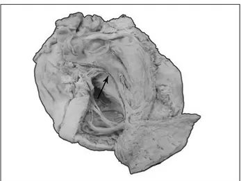
the artery arose from the second anterior septal artery. Seven hearts (14%) had the ST supplied by a branch from the third anterior septal artery. Abnormally, the artery that supplied the ST arose in one case from the conus arteriosus artery (Figure 1). Other results are summarized in Table 1.
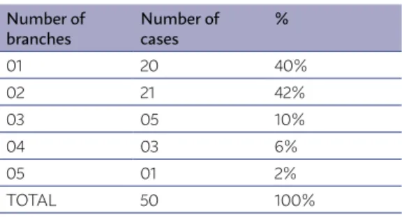
Two branches of the trabecular artery were found in 21 cases (42%). In 20 cases (40%) there was one branch. In 5 cases (10%) there were three branches. In 3 cases (6%), four branches. Only one case (5%) pos-sessed five branches inside the ST, which was not previously described in the literature. The results are summarized in Table 2.
The vessels reached the ST through three different patterns: the first pattern was the usual sub-endocar-dic disposition; the second pattern was through an intramuscular disposition, beneath the center of the ST; and the third pattern was also with an intramus-cular disposition, although it was beneath the inferior
TABLE 1. ORIGIN OF THE TRABECULAR ARTERY.
Origin Number
of cases %
1st anterior septal 13 26% 2nd anterior septal 19 38% 3rd anterior septal 07 14%
4th anterior septal 01 2%
5th anterior septal 03 6%
6th anterior septal 01 2%
7th anterior septal 02 4%
2nd and 6th anterior septal 01 2% Left coronary artery 01 2% 1st anterior septal with Right coronary
branches
01 2%
Conus arteriosus artery 01 2%
TOTAL 50 100%
surface of the ST. 58% of the sample had the pattern 1, 34% had the pattern 2, while 6% had the third pat-tern. One of the cases (2%) had two ST branches, one of them presented the pattern 1 and the other branch possessed the pattern 2.
The ventricular area ranged from 65 to 100 mm, with a mean of 85 mm. Thus, the three segments (su-perior, middle and inferior) had a mean of 28.3 mm. In 38 hearts (76.%), the ST supply came from the middle segment. In 9 hearts (18%) it came from the superior segment. In only three cases (6%), the branches for the ST blood supply came from the inferior segment.
The length of the ST branch in different hearts var-ied from 10 to 68 mm.
DISCUSSION
The ST is classified as a trabecula of second order by Sappey14, Cunningham15, Llorca16, and Testut and
Latarjet5. It can be seen at the ninth week of fetal age, together with the anterior papillary muscle4.
Dewitt17 observed the disposition and arrange-ment of the conduction system in mammals’ hearts and concluded that there is a constant presence and distribution of the right branch of the atrioventricular bundle and its relation to the ST in all studied hearts.
The blood supply of the anterior pillar is target to only a few anatomical variations, as stated by Winck-ler18 and Llorca16 although both authors emphasizes
the fact that the anterior interventricular artery gives origin to a dozen of branches that penetrates the ven-tral portion of the interventricular septum. Cunning-ham15, Schlesinger et al.19, and Testut and Latarjet5
also support this description.
These vessels have a well-defined vascular terri-tory. The anterior septal arteries that arise from the superior third of the anterior interventricular artery possess a descending and posterior trajectory, while the vessels that originates from the middle third have a horizontal trajectory, and the arteries that arise from the inferior third have an ascending and
poste-rior, slightly recurrent trajectories5,15,16,20. Those
ves-sels, according to Schlesinger et al.19, Farrer-Brown
and Rowles21, and Hadziselimović22, runs to the
in-terventricular septum and forms anastomosis with posterior septal arteries.
Usually, the second anterior septal artery reaches the base of the anterior papillary muscle on the right ventricle and gives origin to a small branch that pene-trates the ST, although this branch can originate from the first, third or fourth anterior septal arteries7-11,16,18,23.
Names such as “artere du pilier anterieru du ventricle droit de Mouchet”5,6,10, “ramus limbi dextri de Gross”5,23,
“anterior pilar artery”6 “ramus trabeculae
supra-mar-ginalis”18, “artere de la branche droite du faisceau de
His”18 have been used to refer to the trabecular branch
of the second anterior septal artery.
Our results showed that the “trabecular artery” can arise in rare cases from the fifth, sixth, and sev-enth anterior septal arteries, as well as directly from the right coronary artery, from the conus arteriosus artery, or even from two anterior septal arteries (sec-ond and sixth), facts never reported in the literature.
Testut and Latarjet5 stated that in rare cases, the
first and third anterior septal arteries would be sponsible for the ST supply, in contraposition, our re-sults showed that in 26% it arose from the first and in 14% it came from the third.
The anterior septal branches would be also respon-sible for the most part of the IVS blood supply, togeth-er with the ST, the right branch of the atrioventricular bundle, and the Purkinje fibers, due to the low pres-sure values of the right ventricle24.
Hadziselimović22 studied 71 human hearts (from
neonatal to 81 years old and of both sexes) and his re-sults showed that intramyocardic anastomosis were found in all portions of the heart, particularly on the interventricular septum and adjacent areas.
Moscovici25 in a study of 80 human hearts also
stat-ed the presence of intercoronary anastomosis on the interventricular septum, and their importance regard-ing the subendocardic plexus. The author also gives im-portance to the vessels that reach the papillary muscles through their implantation on the ventricular wall, as these vessels use the fleshy trabeculas and myocardic pons to reach them, according to his results, those ar-teries would also provide intercoronary anastomoses.
Despite the presence of the anterior septal arteries, Correia23 reported the presence of smaller branches
from the anterior interventricular artery that pene-trated the ST at different locations, thus, the author
TABLE 2. NUMBER OF BRANCHES INSIDE THE ST.
Number of branches
Number of cases
%
01 20 40%
02 21 42%
03 05 10%
04 03 6%
05 01 2%
proposed that the ST blood supply should be classified as segmental.
In a study of 651 human hearts, Schlesinger et al.19 found that in 50% of his sample had an artery
rising from the right coronary sinus, together with the right coronary artery: the “conus artery”, as he named. The territory supplied by this vessel, accord-ing to the author, was the superior portion of the in-terventricular septum, close to the supraventricular crest, although this territory can also extend itself to larger portions of the septum, thus, its role is fun-damental regarding collateral circulation in cases where the right and left coronary arteries are ob-structed. Furthermore, the author reported six large arteries that penetrated the interventricular septum (branches of the anterior interventricular artery) with an intimate trajectory with the right margin of the IVS’s endocardium.
Schlesinger et al.19 also stated that some of the
anterior interventricular artery branches - especial-ly the ones next to the cardiac apex - diverted their usual trajectory in order to supply or anastomose with nutricious branches of large trabeculas or large myocardic bands (such as the supramarginal crest). Regarding the anastomotic branches, the author states that their origins can be from the anterior in-terventricular artery, the circumflex artery or the right coronary artery. The branches of the anterior interventricular artery usually have 70 to 800 mm of length, according to Schlesinger et al.19,
A study by Zapedowski26 showed that the territory
supplied by posterior septal branches would receive collateral vessels from the right and left coronary ar-teries, thus, again, proving that the anastomosis on the interventricular septum and the papillary muscles can play a large role in pathophysiological and mor-phological changes.
Hadziselimovic et al.27 investigated 200
hu-man hearts through coronariography and dissec-tions. Their results displayed the role of the many anastomoses between the collateral and terminal branches of both coronary arteries in respect to coronary artery diseases.
A study conducted by Melo et al.28 showed that
the human heart possess 7 anterior septal branch-es on average, although some hearts displayed lbranch-ess than 5 branches, while others possessed 13 branch-es, in contrast with a study performed by Hossein-pour et al.29 which had the presence of 4 anterior
septal branches, on average.
In 2% of cases, the ST blood supply originated from an anastomosis between the first anterior sep-tal artery and branches of the right coronary artery. There are no descriptions regarding this variation in the literature, although Campbell7 stated the ex-istence of anastomoses between the right and left coronary arteries in 20% of his specimens. Accord-ing to the author, those anastomoses could reduce the consequences of ischemia on the right branch of the atrioventricular bundle in cases of anterior septal arteries obstruction. Likewise, a great num-ber of authors reiterate the clinical significances of those anastomoses19,24,25,27 .
Pino and Prates1,30 stated that the origin of the
tra-becular artery was mainly from the superior segment of the sternocostal face, in contraposition, our results showed that this vessel often came from the middle segment. Furthermore, the authors did not found ar-teries arising from the inferior segment.
The present work showed the presence of one to five arteries inside the ST. In 21 hearts (42% of cases), it was found 2 arteries inside the trabecula, in accor-dance to the results of Mouchet10,11, Correia23, and
Pino and Prates1,30. Other authors such as Lascano31
and Truex and Conpenhaver32 described the presence
of one main branch accompanied by smaller vessels. Furthermore, Pino and Prates1,30 found four arteries
inside the ST, whereas the results of the present study showed a ST with five arteries on its inside, a fact nev-er reported in the litnev-erature.
As previously stated, the vessels that supplied the ST reached the band in three different patterns, although a review of the literature only showed the description of the subendocardic pattern1,8-11,16,18,30,33.
In the present study, it was found two new patterns: an intramuscular pattern in which the vessel ran through inside the ST (34% of cases) and another in-tramuscular pattern in which the artery ran through the inferior margin of the trabecula (6% of cases).
The mean distance between the origin of the cor-onary vessels and the trabecular branch of Pino and Prates1,30 studies had similar results to ours, although
the authors only found branches from the first five an-terior septal arteries.
A study performed by Possatti et al.12 in 40 hearts
the present study, as the second anterior septal ar-tery was the main responsible for the ST blood sup-ply (38% of cases) and the first anterior septal artery was responsible in 26% of cases.
According to Hosseinpour et al.29 there are slight
differences among the pattern and trajectory of the anterior septal branches in normal hearts in com-parison to congenitally malformed hearts, especial-ly with hearts that had ventricular septal defects.
Clinical features of the ST and its blood supply involves the fact that it is deeply related to the atrio-ventricular bundle. Due to this anatomy, removal of this structure in order to treat low defects on the in-terventricular septum may cause dynamic changes and disruption of the conduction system4. Further-more, variations on the length and girth of the ST may cause surgical difficulty4.
Knowledge of the anterior septal branches anat-omy is significant to operations such as Ross proce-dure, Konno procedure (correction of Fallot’s tetral-ogy), resection of obstructive muscular subaortic stenosis, and enlargement of restrictive ventricular septal defects, as they require incisions on the up-per portion of the interventricular septum28,29.
In-jury of these vessels can cause myocardial damage and arrhythmia, due to its relation with the ST and right branch of the atrioventricular bundle, in rare cases, iatrogenic injuries can cause sudden death29.
Aortic stenosis associated with myocardial bridges has shown disappearance of the ASB during systole and their reappearance during diastole12.
The anterior septal branches are also clinically rel-evant to angioplasties, as it was shown that those branches can be used to myocardial revasculariza-tion if their diameter was at least 2 mm wide12.
CONCLUSIONS
In summary, the ST is usually supplied by branch from the anterior interventricular artery. Our work showed that the first and second anteri-or septal branches have an impanteri-ortant role regard-ing this vascular supply, since they originated the trabecular artery in most cases. The conus arteri-osus artery can exceptionally provide the ST blood supply, as well as anastomotic branches between the right and left coronary arteries, facts never re-ported in the literature.
We believe this work has added to the literature new findings regarding the interventricular sep-tum, ST and right branch of the atrioventricular bundle blood supply.
The increasing rates of coronary artery diseases, the constant advances in imaging exams and surgi-cal procedures implies in a more detailed study of the distribution, branching pattern and anastomo-ses of coronary vessels.
ACKNOWLEDGEMENTS
The authors wish to pay respects and posthu-mously honor Professor Mauricio Moscivi which was responsible for guiding and supervising Dr. Car-los Chagas Master’s thesis, presented in the current study. Furthermore, the authors wish to honor his memory as Anatomy Professor of the Federal Univer-sity of Rio de Janeiro.
CONFLICTS OF INTEREST
The authors declare that they have no conflict of interests.
RESUMO
A trabécula septomarginal é uma estrutura muscular que transmite o ramo direito do feixe atrioventricular. É usualmente suprida por um ramo da segunda artéria septal anterior. Anastomoses entre as artérias coronárias direita e esquerda podem ocorrer na trabécu-la. São de grande significância especialmente na prevenção de isquemia durante um infarto do miocárdio. Procedimentos cirúrgicos como o de Konno`s e Ross implicam conhecimento anatômico desses vasos. As artérias coronárias de 50 corações humanos foram injetadas com látex e dissecadas com o propósito de identificar o ramo arterial que supria a trabécula septomarginal. Em somente 38% dos casos o ramo foi proveniente da segunda artéria septal anterior, enquanto que em 26% dos casos a artéria se originou da primeira septal anterior. Um dos corações teve a trabécula septomarginal suprida por um ramo originário da artéria do cone arterioso. Além disso, foram encontradas anastomoses entre as artérias coronárias no interior da trabécula septomarginal. Em suma, o ramo direito do feixe atrioventricular está sujeito a inúmeras condições clínicas e é alvo de manuseio em cirurgias, logo, o estudo dos ramos septais das artérias coronárias, em especial o ramo trabecular é essencial.
REFERENCES
1. Pino JA, Prates JCH. Contribuição ao estudo da irrigação da trabécula sept-marginalis no coração humano. Rev Iatros. 1985;4(1):6-9.
2. Paraskevas G, Koutsouflianiotis K, Iliou K. The first descriptions of various anatomical structures and embryological remnants of the heart: a system-atic overview. Int J Cardiol. 2017;227:674-90.
3. Goss CM. Gray’s anatomy. 29a ed. Rio de Janeiro: Guanabara Koogan; 1977.
4. Kosiński A, Kozłowski D, Nowiński J, Lewicka E, Dąbrowska-Kugacka A, Raczak G, et al. Morphogenetic aspects of the septomarginal trabecula in the human heart. Arch Med Sci. 2010;6(5):733-43.
5. Testut L, Latarjet A. Tratado de anatomía humana. Barcelona: Salvat; 1958. 6. Rouvière H. Anatomía humana: descriptiva y topográfica. 8a ed. Madrid:
Bailly-Bailliere; 1961.
7. Campbell JS. Stereoscopic radiography of the coronary system. Quart J Med. 1929;22:247-67.
8. Gross L. The blood supply to the heart in its anatomical and clinical as-pects. New York: Paul B Hoeber; 1921.
9. Gross L, Kugel MA. The arterial blood vascular distribution to the left and right ventricles of the human heart. Amer Heart J. 1933;9:165.
10. Mouchet A. Les artéries coronaires du coeur chez l’homme. 10a ed. Paris: Maloine; 1933.
11. Mouchet A, Noureddine A. L’artere du piller antérieure du ventricule droit ou artère de la branche du faisceau de His. Compt Rend Assoc Anat. 1926:415-21. 12. Possatti LL, Ramos HF, Rodrigues H, Musso F. Anatomical study of
the anterior interventricular septal branches and their relationship with the blood supply of the septomarginal trabecula. Braz J Morphol Sci. 2005;22(3):169-74.
13. Pichard AD, Meller T, Teichholz LE, Lipnik S, Gorlin R, Herman MV. Septal perforator compression (narrowing) in idiopathic hypertrophic subaortic stenosis. Am J Cardiol. 1977;40(3):310-4.
14. Sappey MPC. Traité d’anatomie descriptive. Paris: Delahaye; 1988. 15. Cunningham DJ. Anatomia humana. Barcelona: Manuel Marín; 1949. 16. Llorca FO. Anatomia humana. Barcelona: Editorial Cientifico Medica; 1952. 17. Dewitt LM. Observations on the sino-ventricular connecting system of
the mammalian heart. Anat Rec. 1909;3(9):475-97.
18. Winckler G. Étude sur les artéres coronaires du coeur chez l’homme. Arch Anat Hist Embriol. 1948;31:199-235.
19. Schlesinger MJ, Zoll PM, Wessler S. The conus artery: a third coronary ar-tery. Am Heart J. 1949;38(6):823-36.
20. Tose D. As veias atrioventriculares e ventriculoatriais do coração humano. Ohio: University of Toledo; 1984.
21. Farrer-Brown G, Rowles PM. Vascular supply of interventricular septum of human heart. Br Heart J. 1969;31(6):727-34.
22. Hadziselimović H. Age characteristics of blood vessels of the human heart. Acta Anat (Basel). 1981;109(3):231-7.
23. Correia MA. A irrigação arterial dos músculos papilares do coração huma-no. Fol Anat Univ Conimbrigensis. 1946;21(11):1-7.
24. James TN, Burch GE. Blood supply of the human interventricular septum. Circulation. 1958;17(3):391-6.
25. Moscovici M. Irrigation of the papillary muscles of the ventricle of the hu-man heart. Rev Bras Cien Morfol. 1990;7(1):55-60.
26. Zapedowski Z. Arterial branches in the region of the posterior in-terventricular sulcus in the human heart. Folia Morphol (Warsz). 1977;36(3):195-201.
27. Hadziselimović H, Dilberović F, Ovcina F. Blood vessels of the hu-man heart: coronarography and dissection. Acta Anat (Basel). 1980;106(4):443-9.
28. Melo JQ, Abecassis M, Neves J, Calquinha J, Ramos S, Martins AP, et al. Can the location of the large septal artery be predicted? Eur J Cardiothorac Surg. 1995;9(11):628-30.
29. Hosseinpour AR, Anderson RH, Ho SY. The anatomy of the septal per-forating arteries in normal and congenitally malformed hearts. J Thorac Cardiovasc Surg. 2001;121(6):1046-52.
30. Pino JA, Prates JC. Estudio mesoscopico de los vasos arteriales en la tra-bécula septomarginal del corazon humano. Rev Frontera. 1986:5-6. 31. Lascano EF. Irrigacion normal del nódulo de Tawara, haz de His y sus
ra-mas. Rev Arg Cardiol. 1943;10:23-54.
32. Truex RC, Conpenhaver WM. Histology of the moderator band in man and other mammals with special reference to the conduction system. Am J Anat. 1947;80(2):173-201.