1 – Nuclear Physician at ECOAR Diagnostic Medicine, Belo Horizonte, MG, Brazil. 2 – Orthopedist at the Vera Cruz and Life Center Hospitals, Belo Horizonte, MG, Brazil.
3 – Cardiologist at the Nuclear Medicine Service, ECOAR Diagnostic Medicine, Belo Horizonte, MG, Brazil. 4 – Cardiologist and Director of ECOAR Diagnostic Medicine, Belo Horizonte, MG, Brazil.
Work performed at ECOAR Diagnostic Medicine, Belo Horizonte, MG.
Correspondence: Rua Prof. Moraes 476/901, Bairro Funcionários, 30150-370 Belo Horizonte, MG. E-mail: [email protected] Work received for publication: October 6, 2010; accepted for publication: August 4, 2011.
INTRODUCTION
Sesamoid is a term derived from the Greek work sesamen, because of the similarity between these bo-nes and the seeds of a plant, Sesamum indicum, which was used as a purgative in ancient Greece. Around two thousand years ago, it was imagined that the se-samoids were a repository for the soul after death(1).
The sesamoids are two small accessory bones that are inserted into the tendons of the short flexor of the hallux, adjacent to the medial and lateral facets of the head of the first metatarsal. The tibial sesamoid has an average size of between 12 and 15 mm, and the fibu-lar sesamoid has an average size of between 10 and 12 mm. The upper faces of the sesamoids are covered with cartilage and articulate with the head of the first
THE BONE SCINTIGRAPHY AS A COMPLEMENTARY EXAM IN THE
DIAGNOSIS OF THE AVASCULAR NECROSIS OF THE SESAMOID
Carlyle Marques Barral1, Arnóbio Moreira Félix2, Leonardo Neuenschwander Magalhães3, Luciana Araújo Carvalho1, Fernando Santana Machado4
metatarsal and the capsule of the first metatarsopha-langeal joint, into which its tendons are inserted. The two sesamoids are firmly joined together by the thick intersesamoid ligament and to the base of the proxi-mal phalange by means of the sesamoid-phalangeal ligament. In addition, they are also fixed by means of the deep transverse intermetatarsal ligament and are therefore anatomically interlinked with the second metatarsal(2,3). A bone crest divides the plantar surface
of the head of the metatarsal into two longitudinal joint grooves, which are coated with joint cartilage and guide the movement of the sesamoid bones. The tendon of the long flexor of the hallux goes between the sesamoid bones, in close contact with the plantar face of the intersesamoid ligament(3).
The authors declare that there was no conflict of interest in conducting this work
This article is available online in Portuguese and English at the websites: www.rbo.org.br and www.scielo.br/rbort
ABSTRACT
Objective: This study aimed to present seven cases of avascular necrosis of the sesamoid and report the role of bone scintigraphy in the diagnosis of these patients. Methods: Seven patients with clinical suspicion of avascular necrosis of the sesamoid underwent three-phase bone scintigraphy with 30 mCi of 99mTc-MDP. Results: Most of the patients were young female adults with complaints of limiting pain in the forefoot, who were making use of inappropriate footwear and/ or had a history of injury with or without fracture. There was no predominance of either of the feet or between the femoral or tibial sesamoid. Two patients (28.57%) had a bipartite tibial sesamoid and one (14.29%) had splitting of the tibial and fibular sesamoids. In 100% of the patients, three-phase bone
scintigraphy, combined with other propaedeutic methods, proved to be crucial for the diagnosis. The initial procedure in all cases was conservative. In four cases (57.14%), there was no remission of symptoms, and surgical excision of the necrotized sesamoid tissue was performed. In all the patients, the therapy used was effective, with complete remission of symptoms, without complications or deformities of the forefoot. Conclusions: Three-phase bone scintigraphy becomes a cornerstone of the propaedeutics when avascular necrosis of the sesamoid is suspected, through contributing towards early and accurate diagnosis and enabling allowing appropriate specialized treatment.
The sesamoids are generally chance and asympto-matic findings in imaging examinations, but should not be ignored as sites that possibly cause pain. They absorb pressure, reduce attrition and protect and stabi-lize the metatarsophalangeal joint and the tendons of the long flexors of the hallux. They act as a fulcrum for increasing the mechanical resistance of the tendons at the time of the impulse of gait and provide a dynamic function to the hallux, through raising the head of the first metatarsal and distributing the weight-bearing in the lateral projection of the forefoot(3-5). Despite
the crucial role played by the sesamoid bones in the mechanics of the forefoot, complaints resulting from pathological conditions in these structures are often neglected or poorly diagnosed and managed.
The main blood irrigation for these small bones co-mes through the posterior tibial artery, which branches into the medial plantar artery and divides on entering the medial and lateral sesamoid bones at their proxi-mal poles. Although vessels from the peripheral soft tissues are abundant, they do not appear to penetra-te the corpenetra-tex of the sesamoid bones. Thus, the blood supply to the sesamoid bones may come from up to three vessels. Arteries penetrate the lateral and medial sesamoid bones proximally through a single vessel that proceeds distally with a network of ramifications. In the plantar projection, vessels penetrate the non-joint surfaces of the sesamoid bones, thus forming a second source of vascularization. Lastly, small vessels also penetrate the sesamoid bones by means of the medial and lateral capsular adnexa(4,6).
Disorders of the sesamoid bones are a cause of me-tatarsal pain and, because of the complex anatomy and numerous pain-sensitive structures in the region, their differential diagnosis may be challenging, taking into consideration the possible causes of congenital, trauma-tic, arthritrauma-tic, infectious and ischemic nature. Dislocation of the sesamoids may be associated with metatarsalgia, callus formation and stress fractures(2). In 1924,
Re-nander(7) was one of the first authors to draw attention
to avascular necrosis of the sesamoid. This is a very uncommon clinical entity, and its low incidence and incomplete definition may lead to erroneous diagno-ses and delayed treatment. It needs to be differentiated from other pathological conditions such as fractures, pseudarthrosis or osteomyelitis. The present study had the aim of presenting seven cases of avascular necrosis of the sesamoids and report on the role of bone scinti-graphy in diagnosing these patients.
METHODS
Seven patients with a clinical suspicion of avas-cular necrosis of the sesamoids underwent a dyna-mic study of blood flow in a high-resolution gamma chamber with a rectangular double detector, in ante-rior and posteante-rior projections of the region of interest. Sequential images were produced immediately after injection of 30 mCi of 99mTc-MDP for one minute,
followed by static images at equilibrium. After three hours of intravenous administration of the radiophar-maceutical, images of the whole body were obtained in anterior and posterior projections, along with spe-cial late-stage static images of the regions of interest.
RESULTS
The patients’ ages ranged from 20 to 46 years, with a mean of 31 and median of 32 years. Among the patients, six (85.71%) were female and one (14.29%) was male.
Three patients (42.86%) presented a pathological condition in their right foot and four (57.14%) in their left foot. All of them presented the symptom of pain in the affected forefoot, and one (14.29%) was also found to present localized edema and rubor.
In four patient (57.14%), the sesamoid affected was the tibial, and in three (42.86%), the fibular. Four patients (57.14%) presented undivided sesamoids, two (28.57%) presented bipartite tibial sesamoids and one (14.29%) patient presented bipartition of the tibial and fibular sesamoids.
All the patients were found to have been using inadequate footwear and/or they reported suffering traumatic events with or without associated fractu-ring. One of the patients was practicing ballet and another, soccer.
Two of the female patients (28.57%) were using contraceptives.
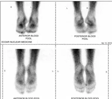
In 100% of the patients, triphasic bone scintigra-phy in association with other propaedeutic methods was shown to be fundamental for the diagnosis (Fi-gures 1, 2 and 3).
In four cases (57.14%), there was no remission of the symptoms and surgical excision of the necrotized sesamoid tissue was performed. One of the patients evolved with pain, edema and localized paresthesia, which resulted in slight claudication. This was treated conservatively with non-steroidal anti-inflammatory drugs and physiotherapy sessions.
In all the patients, the therapy used was shown to be effective, with complete remission of the symp-toms. Clinical inspection, radiological findings and, notably, scintigraphic findings demonstrated that the condition had been resolved, with complete pain relief and without complications or deformities of the forefoot.
DISCUSSION
Both metatarsal sesamoid bones are always pre-sent, and their complete ossification takes place be-tween the ages of nine and fourteen years(8,9). This
generally occurs earlier for the lateral sesamoid(1,10)
and among females(10). Ossification starting from
more than one bone center leads to partition of the sesamoid bone in around 30% of individuals(1,8,9).
The sesamoids are surrounded by a fibrous liga-ment structure that forms the sesamoid-phalangeal apparatus and moves under the head of the meta-tarsal head, thus playing an important role as shock absorbers and thereby facilitating gentle footfall from the heel to the extremity of the toes. They also increase the muscle strength at the impulsion stage of gait and protect the metatarsophalangeal joint and the tendon of the long and short flexors of the hallux(4).
Regarding the physiopathology of the osteo-necrosis, changes to the vascular supply to the accessory center of the sesamoid or fragility of the ossification centers have been reported. Re-peated trauma affecting the tendons and serous membranes of the sesamoid-phalangeal apparatus and fracturing of the sesamoid bone may result in ischemia with osteonecrosis. The most frequent precipitating factors are microtrauma, sports acti-vities such as athletics and dancing, and alignment disorders of the hind foot, such as pes cavus or pes valgus(2,8).
Osteonecrosis of the sesamoid bones has unknown prevalence and is probably underdiagnosed. Most of the patients are adolescents or young adults, and
Figure 1 – Images of blood flow in the feet in anterior and posterior
pro-jections, showing slightly increased flow in the projection of the medial
bone of the right metatarsal. Blood flow
anterior
Blood flow posterior
Figure 2 – Images at equilibrium (vascular permeability) with the feet in
anterior, posterior and plantar projections of the medial sesamoid of the
right metatarsal.
Figure 3 – Late-stage images of the whole body and static images of the
feet in anterior, posterior and plantar projections, showing heterogenous
distribution of the radiopharmaceutical, with focal high uptake of the drug in the medial sesamoid of the right metatarsal.
R L L R
ANTErIOr BLOOd POOL ECOAr NUCLEAr MEdICINE
POSTErIOr BLOOd POOL
ANTErIOr BLOOd POOL POSTErIOr BLOOd POOL
R L L R
Apr 12, 2010
BONE SCINTIGrAPHy NUCLEAR MEDICINEE C O A r
women are more affected than men(2,8). Although
both of the sesamoids may be affected, the tibial sesamoid is subjected to greater loads and is therefore more susceptible to this condition(1,3,5,10).
Another cause of this condition could be the natural pronation of the first metatarsal, which places the tibial sesamoid in a more prominent position(2).
It also has to be taken into consideration that sesamoid partition is more common in the tibial than in the fibular sesamoid(1-3) and more common
in women than in men(11), and that bipartite
sesamoid bones fracture under lower force than do undivided sesamoids(1,3,9). The incidence of
unilaterally bipartite medial sesamoids is around 10.7%, while bilaterally bipartite medial sesamoids occur in around 25% to 85%(2).
Regarding blood perfusion, Pretterklieber and Wanivenhaus(12) demonstrated that the tibial
sesa-moid is fed by a single vessel in 64% of women and 43% of men. The fibular sesamoid is irrigated by a single vessel in 57% of women and 50% of men. This may also explain the greater incidence of avas-cular necrosis in tibial sesamoids and in women. In studying the vascular supply of the sesamoids, these authors also demonstrated that the sesamoids of the left foot tend to be smaller and denser than those in the right foot, and those of males tend to be bigger than those of females. They also showed that the sesamoids of the left foot have a greater blood supply than those of the right foot and that the sesamoids of males have a greater blood supply than those of females. These authors believed that this explained the difference in size between the sesamoids obser-ved in these groups.
The differential diagnoses include nonspecific sesamoiditis; osteomyelitis; trauma with fractures; pseudarthrosis; bursitis; sympathetic-reflex dystro-phy syndrome; gout and other diseases with depo-sition of crystals, such as hyperuricemia; joint in-flammation diseases such as rheumatoid arthritis, psoriatic arthritis and reactive arthritis; and abnormal alignment, dislocation and osteoarthritis of the sesa-moid bone(1,2,5,8,10).
The main symptom is mechanical pain that starts gradually and is reflected in the plantar surface of the head of the first metatarsal, on palpation, on putting weight on the hallux and in the final phase of the gait cycle. It is worsened by forced dorsiflexion of the
hallux until becoming incapacitating. Antalgic supi-nation of the forefoot while walking is noted(2-4,8,10).
Bone scintigraphy is fundamental for early diag-nosis, since scintigraphic abnormalities often prece-de the radiographic findings. Areas with very high uptake of radiopharmaceutical, or even with very low uptake, can be observed at the beginning of the process of necrosis(1,3,8,10,13).
The initial treatment is based mainly on eli-mination of weight-bearing and support for the metatarsal arch by means of personalized footwear and molded insoles, with insertion of a pressu-re point posteriorly to the head of the metatarsus and an opening under the affected sesamoid(2,8).
Non-steroidal anti-inflammatory drugs and tem-porary immobilization may be necessary(1). There
is controversy regarding the use of intra-articular injections of glucocorticoids(2). If the pain lasts
for more than six months and is refractory to ap-propriate conservative treatment, partial or total sesamoidectomy with excision of the necrosed part becomes an alternative. Preference is given to a dorsal approach(8) in order to avoid painful scars or
formation of cheloids in weight-bearing areas. Sur-geons should take care to protect the neurovascular bundle in repositioning the intrinsic tendons and ligaments. Anatomical knowledge of the course and distribution of the vessels becomes necessary for understanding the pathogenesis of avascular necrosis, so that orthopedists are knowledgeable about correct use of the surgical technique. It is important to keep the contralateral sesamoid bone and the surrounding fibrous structure, which stabi-lizes the metatarsophalangeal joint(8). Occasionally,
patients may develop hallux varus after complete excision of the fibular sesamoid, or hallux valgus after excision of the tibial sesamoid(2,4), which may
be followed by pain in the contralateral sesamoid and may even require a second sesamoidectomy(4,8).
Plantar pain in the first metatarsal(4) or a “claw”
deformity of the interphalangeal joint due to dimi-nished strength of the short flexor of the hallux(2,8)
CONCLUSIONS
The recent literature provides support for initial non-surgical management of cases of avascular ne-crosis of the sesamoid, using therapy consisting of anti-inflammatory medications, adequate footwear and elimination of weight-bearing. After the condition has continued for more than six months, surgical interven-tion should be considered.
Triphasic bone scintigraphy plays an important role in
the propaedeutics of avascular necrosis of the sesamoid. Scintigraphic studies become crucial through contributing towards an early and accurate diagnosis of the complex disorders of the sesamoid bone, which is invariably a challenge for specialists. Scintigraphy is therefore an important tool for guiding physicians regarding the appropriate treatment, thereby avoiding potentially harmful dysfunctions that drag on for a long time and especially comprise the patient’s social and working lives.
REFERENCES
1. Julsrud ME. Osteonecrosis of the tibial and fibular sesamoids in an aerobics instructor. J Foot Ankle Surg. 1997;36(1):31-5.
2. Waizy H, Jäger M, Abbara-Czardybon M, Schmidt TG, Frank D. Surgical treat-ment of AVN of the fibular (lateral) sesamoid. Foot Ankle Int. 2008;29(2):231-6.
3. Taylor JA, Sartoris DJ, Huang GS, Resnick DL. Painful conditions affecting the first metatarsal sesamoid bones. Radiographics. 1993;13(4):817-30.
4. Williams G, Kenyon P, Fischer B, Platt S. An atypical presentation of hallucial se-samoid avascular necrosis: a case report. J Foot Ankle Surg. 2009;48(2):203-7.
5. Kanatli U, Ozturk AM, Ercan NG, Ozalay M, Daglar B, Yetkin H. Absence of the medial sesamoid bone associated with metatarsophalangeal pain. Clin Anat. 2006;19(7):634-9.
6. Chamberland PD, Smith JW, Fleming LL. The blood supply to the great toe sesamoids. Foot Ankle. 1993t;14(8):435-42.
7. Renander A. Two cases of typical osteochondropathy of the medial sesamoid bone of the first metatarsal. Acta Radiol. 1924;3:521-7.
8. Toussirot E, Jeunet L, Michel F, Kantelip B, Wendling D. Avascular necrosis of the hallucal sesamoids update with reference to two case-reports. Joint Bone Spine. 2003;70(4):307-9.
9. Kewenter Y. Die Sesambeine des 1: Metatarsophalangealgelenks des Mens-chen. Acta Orthop. Scand. 1936;2:1-113.
10. Fleischli J, Cheleuitte E. Avascular necrosis of the hallucial sesamoids. J Foot Ankle Surg. 1995;34(4):358-65.
11. McBryde AM Jr, Anderson RB. Sesamoid foot problems in the athlete. Clin Sports Med. 1988;7(1):51-60.
12. Pretterklieber ML, Wanivenhaus A. The arterial supply of the sesamoid bones of the hallux: the course and source of the nutrient arteries as an anatomical basis for surgical approaches to the great toe. Foot Ankle. 1992;13(1):27-31.