468
The clinical application of the anatomical study of the ventri-cular branches of coronary arteries is the execution and interpre-tation of examination methods and the planning and performing of cardiovascular disease treatment 1-6.
In 1940, Schlesinger 1 developed the anatomical concept of balance or predominance in coronary artery circulation in the dia-phragmatic face of the heart, using the crux cordis region as a reference. The crux cordis is defined as the point where the coro-nary sulcus meets the interatrial and interventricular sulci. Accor-ding to the author 1, the right coronary artery was predominant when it provided the posterior interventricular branch and supplied blood for most of the left ventricular posterior wall. However, this criterion did not quantify the limit of this region in the posterior wall. In the balanced type, the right coronary artery irrigated only the right ventricle and the posterior part of the interventricular septum, and did not provide significant branches for the left ven-tricle, which was irrigated by the left coronary artery. The criterion remained subjective, because it did not define the concept of significant branches, because, for example, in left dominance, the left coronary artery provided, along with the right coronary artery, parallel posterior interventricular branches, or only the left coronary artery provided a posterior interventricular branch, some-times sending branches to the right ventricle.
Blunk and DiDio 7 considered dominance of the right coronary artery to be all cases in which the right coronary artery provided branches for the posterior face of the right ventricle. More recent studies by Falci Junior et al 8 reported that in right dominance, the right coronary artery reached and extended beyond the crux cordis, providing 1 or more branches to the left ventricle; in balanced circulation, the right coronary artery reached that point, but did not extend beyond it; and, in left dominance, the left coronary artery reached the crux cordis, originating, or not, branches to the right ventricle. For this reason, they did not consider the possibility that the branches extending beyond the crux cordis were of little significance.
In addition, these classifications did not consider the fact that sometimes the anterior interventricular branch surrounded the apex of the heart and extended upward in the posterior interventricular sulcus, which, according to Cavalcanti et al 9 occurred in 28.18% of the cases, and according to Lima Júnior et al 10 in 50%. Therefore, we found it pertinent to include the analysis of the anterior interventricular branch among our classification criteria.
Pino et al 6 reported a division of the ventricles between the coronary sulcus and the apex of the heart into superior, middle, and inferior thirds. However, we believe that the addition of 1
Original Article
The Posterior Ventricular Branches of the
Coronary Arteries in the Human Heart
José Roberto Ortale, Luisa Carolina Borges Keiralla, Luciana Sacilotto
Campinas, SP - Brazil
Pontifícia Universidade Católica de Campinas
Mailing address: José Roberto Ortale - Rua Francisco de Assis Pupo, 245/32 - 13035-000 – Campinas, SP, Brazil – E-mail: [email protected]
Received: 4/30/03 Accepted: 8/5/03
English version by Stela Maris C. e Gandour
Objective
To describe the trajectory of the posterior ventricular branches of the coronary arteries in the epicardial adipose tissue, and to propose a new criterion for analyzing the distribution of these branches, according to the traditional classification, to determine the predominance or balance of the coronary arteries in the arterial supply to the heart.
Methods
Forty hearts obtained from autopsies of adults were dissected and fixed in a formol solution. The posterior face of each ventricle was divided into 8 approximately equal areas for themorphological classification of the coronary circulation. The following 3 tradi-tional types were considered: A) right dominance, B) balanced, and C) left dominance. The number, diameter, and terminal areas of the posterior ventricular branches in the epicardium were analyzed.
Results
The following branches and respective frequencies were found: left marginal branch - 100%; posterior left ventricular branches: lateral 75%, intermediate 82.5%, and medial -87.5%; interventricular posterior branch - 95%; posterior right ventricular branch: medial - 40%, intermediate - 32.5%, and lateral 40%; posterior diagonal branch of the right ventricle -17.5%; right marginal branch - 95%. In regard to dominance, the following values were found: dominance of the right coronary artery - 62.5%; balanced type - 25%; and dominance of the left coronary artery - 12.5%.
Conclusion
The method adopted allowed a more precise classification of the types of coronary artery distribution found. The right coronary artery dominance type was the most prevalent, followed by the balanced type and the left coronary artery dominance.
Key words
469
subdivision to each third would make the determination of thelimits of distribution of the posterior interventricular branches more precise.
Therefore, our objective was to describe the trajectory of coro-nary artery branches in the diaphragmatic face of the heart, and to propose a criterion for classifying the predominance or balance of the coronary circulation based on the studies cited, adding the division of the ventricles into areas.
Methods
The coronary arteries and their posterior ventricular branches in the epicardium of the diaphragmatic face of 40 hearts obtained from autopsies of adults were dissected, fixed in a 5% formol solution and conserved in a 10% formol solution. These hearts came from the Legal Medical Institute of Campinas and from the Laboratory of Anatomy of the Center of Life Sciences of the Pontifícia Universidade Católica of Campinas, in the state of São Paulo.
For classifying the distribution of the posterior ventricular bran-ches of the coronary arteries according to their superficial trajec-tory, the posterior face of each ventricle was divided into 8 areas by 2 longitudinal lines, which separated the medial, intermediate, and lateral thirds, and 2 transverse lines, which subdivided each third into approximately equal superior, middle, and inferior areas (fig.1).
According to their distribution to the medial, intermediate, or lateral third, the posterior branches of each ventricle received their respective names 11. When more than 1 branch existed in the same area, roman numerals were added to the name 12. For example: left ventricular posterior intermediate I branch, left ven-tricular posterior intermediate II branch, and so forth. On the other hand, the branch originating from the right coronary artery and obliquely crossing the left ventricular posterior face towards the interventricular sulcus was named the right ventricular poste-rior diagonal branch 13.
The following diameters were measured: at the point of origin of the right and left coronary arteries; of the anterior interventricular and circumflex branches of the left coronary artery; of each super-ficial branch present in the posterior face of the ventricles. The areas in which the branches deepened in the myocardium were recorded. The presence or absence of anastomosis due to inoscu-lation between the anterior and posterior interventricular branches was verified.
For establishing the 3 basic types of coronary circulation, we followed the criteria of Schlesinger 1 for studying dominance or balance in the distribution of coronary arteries: A) right dominance; B) balanced type; and C) left dominance.
In the right dominance type of circulation, the right coronary artery originates the posterior interventricular branch, extends beyond the crux cordis, and branches towards at least the middle medial part (areas 1 and 4) of the left ventricular posterior face (fig. 1A).
In the cases of balanced circulation and left dominance, the development of new criteria is required for making the classification of the types of coronary artery distribution more accurate. In the balanced type, 2 subtypes were determined: B1) the right coronary artery originates the posterior interventricular branch that extends
to the inferior third of the posterior interventricular sulcus, but in some cases it originates small branches to the medial superior area (area 1) of the left ventricular posterior face (fig. 1 B1 and B2) the right coronary artery ends by bifurcating into the posteri-or interventricular branch to half of the sulcus and the posteriposteri-or branch of the medial left ventricle to areas 1 and 4 of the left ventricular posterior face, while the anterior interventricular branch of the left coronary artery ends in the inferior third of the posterior interventricular sulcus (fig. 1 B2).
The type of dominance of the left coronary artery also comprises 2 subtypes: C1) the left coronary artery originates the posterior interventricular branch, independently of originating, or not origina-ting, branches to the right ventricular posterior face (fig. 1 C1 and C2) the anterior interventricular branch of the left coronary artery extends beyond the apex of the heart and ends in the inferior third of the posterior interventricular sulcus, while the superior two thirds of the sulcus are irrigated by the terminal branches of the right coronary artery (diagonal branch of the right ventricle and posterior interventricular branch), and may or may not branch to the superior medial area (area 1) of the left ventricle (fig. 1 C2).
Finally, in each specific type of coronary circulation, the number of branches of the right and left coronary arteries extending beyond the crux cordis and running through the diaphragmatic face of the opposite ventricle was assessed.
The measurements were performed with a Mitutoyo digital pachymeter, and the values were reported as minimum, maximum, mean, and standard deviation. A diagrammatic representation was performed for each specimen.
Results
The right coronary artery, present in all cases, originated from the right aortic sinus, with diameters ranging from 1.8 to 5.3 mm (mean of 3.6±0.8 mm). The left coronary artery, present in all cases, originated from the left aortic sinus, with diameters ranging from 3.1 to 7.1 mm (mean of 4.6±0.9 mm) and lengths ranging from 4.5 to 18.4 mm (mean of 11.5±3.9 mm). In all cases, its anterior interventricular branch was present, with
dia-Fig. 1 - Division of the posterior face of the left (LV) and right (RV) ventricles into 8 areas and types of coronary circulation: A) dominance of the right coronary artery; B1/B2) balanced circulation; C1/C2) dominance of the left coronary artery. rc- right coronary artery; lc- left coronary artery; ai- anterior interventricular branch; cx- circumflex branch; pi- posterior interventricular branch; rm- right marginal branch; lm- left marginal branch; rl, ri, rm- lateral, intermediate, and medial posterior branches of the RV, respectively; ll, li, lm- lateral, intermediate, and medial posterior branches of the LV, respectively; dg- posterior diagonal branch of the RV.
llI llII lmII
lmI
l l
l l
lmI
lmI
lmIII lmII lmI
lmI
lmII lm lm
lm lm
lm
r c r c
470
meters ranging from 2.4 to 5.9 mm (mean of 3.6±0.8 mm), as was its circumflex branch, with diameters ranging from 1.9 to 6.2 mm (mean of 3.5±0.9 mm). Table I shows the frequency, origin, diameter, and number of the posterior ventricular branches. Based on the distribution of the coronary artery branches loca-ted in the epicardium of the posterior face of the ventricles, 3 types of coronary circulation were considered: A -right dominant; B - balanced; and C - left dominant (fig. 1).
Table II shows the origin and diameter of the posterior ventri-cular branches in the 3 types of coronary circulation: dominance of the right coronary artery, balanced, and dominance of the left coronary artery. Figures 2 A to D summarize the location of the points where the posterior ventricular branches penetrate the myocardium in the 3 types of coronary circulation.
Right coronary artery dominance (fig. 1A) was found in 25/40 cases (62.5%). In these cases, the right coronary artery extended beyond the crux cordis. In 21/25 cases, the right coronary artery provided the posterior branch of the medial left ventricle; in 19/ 25 cases, it provided the posterior branch of the intermediate left ventricle; and, in 4/25 cases, it provided the posterior branch of the lateral left ventricle.
Balanced coronary circulation (fig. 1 B1 and B2) was observed in 10/40 cases (25%), being of the B1 subtype in 9/10 cases and of the B2 subtype in 1/10 cases. The right coronary artery extended
beyond the crux cordis, and, in 8/10 cases, provided the posterior branch of the medial left ventricle, which branched in the medial superior area (area 1) of the left ventricle. In 2/10 cases, this branch originated from the left coronary artery and ran until area 4 of the left ventricle.
Dominance of the left coronary artery (fig. 1 C1 and C2) was observed in 5/40 cases (12.5%), being of the C1 subtype in 3/5 of the cases, and of the C2 subtype in 2/5 of the cases, of which, in 1 case, branches of this artery were destined to area 1 of the posterior face of the right ventricle. In 3/5 cases, the left coronary artery originated the posterior interventricular branch, which, in 2/3 cases, ran through the superior third of the sulcus, and, in 1/ 3 cases, extended until the middle third. In 2/5 cases, the ante-rior interventricular branch of the left coronary artery surrounded the apex of the heart and irrigated the inferior third of the poste-rior interventricular sulcus. Of these 2/5 cases, in 1 case, the posterior interventricular branch originated from the right coronary artery and ran through the superior third of the sulcus, while the diagonal branch of the right coronary artery ran towards its middle third (fig. 1 C2). In the other case, the posterior interventricular branch did not exist, and the diagonal branch of the right coronary artery ran through the middle third of the sulcus. In regard to the posterior branch of the medial right ventricle in the cases of left dominance, it was present in the medial superior area (area 1) of
Table I - Origin, diameter, and number of the coronary artery branches in the posterior face of the ventricles (n = 40 cases).
Origin* Diameter Number of branches 0 1 2 3 4
Branch n % rc cx mm n n n n n
Left marginal 40 100.0 - 40# 0.7-4.4 (2.2±0.9) - 38 2 -
-Lateral posterior LV 30 75.0 4 26 0.8-3.5 (1.9±0.6) 10 24 5 1 -Intermediate posterior LV 33 82.5 19 14 0.7-2.9 (1.8±0.6) 7 21 8 4 -Medial posterior LV 35 87.5 30 5 0.8-2.7 (1.4±0.4) 5 12 16 4 3 Posterior interventricular 38 95.0 35 3 1.2-3.3 (2.3±0.5) 2 35 3 - -Medial posterior RV 16 40.0 15 1 0.6-2.3 (1.4±0.5) 24 15 1 - -Intermediate posterior RV 13 32.5 13 - 0.7-1.6 (1.1±0.3) 27 13 - - -Lateral posterior RV 16 40.0 16 - 0.6-2.8 (1.1±0.5) 24 14 2 - -Diagonal RV 7 17.5 7 - 1.1-2.6 (2.0±0.4) 33 7 - - -Right marginal 38 95.0 38 - 0.8-2.9 (1.5±0.5) 2 38 - - -RV- right ventricle; LV- left ventricle; rc- right coronary artery; cx- circumflex branch of the left coronary artery. * When, in the same case, a certain branch had more than 1 different origin, the origin considered was that of the greatest diameter. #In 2 cases, the left marginal branch originated from the posterior branch of the lateral
left ventricle.
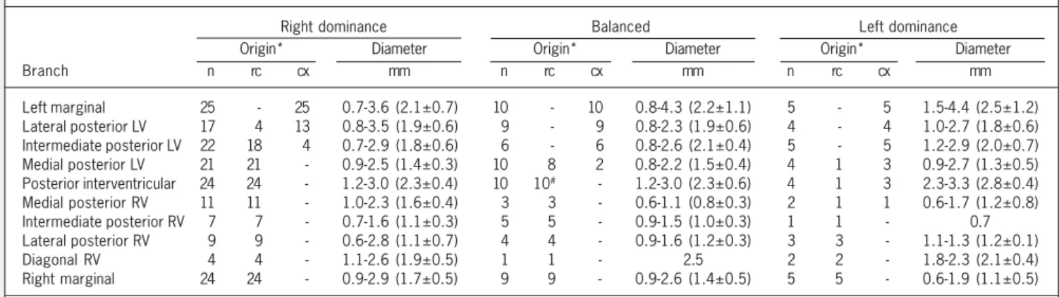
Table II – Origin and diameter of the posterior ventricular branches in the types of coronary artery circulation: A - dominance of the right coronary artery (25 cases), B - balanced (10 cases), and C - dominance of the left coronary artery (5 cases).
Right dominance Balanced Left dominance Origin* Diameter Origin* Diameter Origin* Diameter Branch n rc cx mm n rc cx mm n rc cx mm Left marginal 25 - 25 0.7-3.6 (2.1±0.7) 10 - 10 0.8-4.3 (2.2±1.1) 5 - 5 1.5-4.4 (2.5±1.2) Lateral posterior LV 17 4 13 0.8-3.5 (1.9±0.6) 9 - 9 0.8-2.3 (1.9±0.6) 4 - 4 1.0-2.7 (1.8±0.6) Intermediate posterior LV 22 18 4 0.7-2.9 (1.8±0.6) 6 - 6 0.8-2.6 (2.1±0.4) 5 - 5 1.2-2.9 (2.0±0.7) Medial posterior LV 21 21 - 0.9-2.5 (1.4±0.3) 10 8 2 0.8-2.2 (1.5±0.4) 4 1 3 0.9-2.7 (1.3±0.5) Posterior interventricular 24 24 - 1.2-3.0 (2.3±0.4) 10 10# - 1.2-3.0 (2.3±0.6) 4 1 3 2.3-3.3 (2.8±0.4)
Medial posterior RV 11 11 - 1.0-2.3 (1.6±0.4) 3 3 - 0.6-1.1 (0.8±0.3) 2 1 1 0.6-1.7 (1.2±0.8) Intermediate posterior RV 7 7 - 0.7-1.6 (1.1±0.3) 5 5 - 0.9-1.5 (1.0±0.3) 1 1 - 0.7 Lateral posterior RV 9 9 - 0.6-2.8 (1.1±0.7) 4 4 - 0.9-1.6 (1.2±0.3) 3 3 - 1.1-1.3 (1.2±0.1) Diagonal RV 4 4 - 1.1-2.6 (1.9±0.5) 1 1 - 2.5 2 2 - 1.8-2.3 (2.1±0.4) Right marginal 24 24 - 0.9-2.9 (1.7±0.5) 9 9 - 0.9-2.6 (1.4±0.5) 5 5 - 0.6-1.9 (1.1±0.5) RV- right ventricle; LV- left ventricle; rc- right coronary artery; cx- circumflex branch of the left coronary artery. * When, in the same case, a certain branch had more than 1 different origin, the origin considered was that of the greatest diameter. # In 1 case, the posterior interventricular branch was the continuation of the diagonal
471
the right ventricle in 2/5 cases, originating, in 1 case, from theright coronary artery, and, in another, from the circumflex branch of the left coronary artery.
In 4/25 cases of right coronary artery dominance, 1 anastomosis due to inosculation existed between the anterior and posterior interventricular branches.
Discussion
In our results, the mean diameter of the left coronary artery was greater than the mean diameter of the right coronary artery, in accordance with James’ results 3. The circumflex branch of the left coronary artery originated from the posterior interventricular branch in 3/40 or 7.5% of the cases, a frequency between those obtained by other authors 3,5,7,10,14.The anterior interventricular branch ended in a termino-terminal anastomosis with the poste-rior interventricular branch of the right coronary artery in a fre-quency intermediate between those reported by Hadziselimovic and Secerov 15 and Cavalcanti et al 9.
In no case was the anatomical variation of a left single coronary artery observed irrigating the entire heart with the absence of the right coronary artery, as reported by Koizumi et al 16.
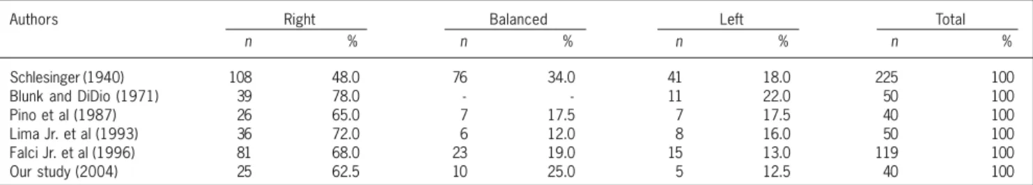
In regard to the classification of the types of coronary circulation into right or left dominance, and balanced, table III shows the comparison between the frequency of types of coronary circulation observed by us and other authors 1,6-8,10. Our percentages were similar to those reported by Pino et al 6, probably because those authors established a division of the ventricles.
Our frequency of right dominance was lower than those reported by Blunk and DiDio 7, Lima Junior et al 10, and Falci Junior et al 8, because these authors included in this type all cases in which the right coronary artery branched towards the posterior face of the left ventricle. We, on the other hand, excluded the cases in which this supply was restricted to the superior medial area (area 1), because these branches had a small caliber, exerting little influence on coronary circulation distribution. In regard to Schlesinger 1,we believe that our greater percentage was due to the fact that we had fixed for right coronary artery dominance
472
1. Schlesinger, MJ. Relation of the anatomic pattern to pathologic conditions of the coronary arteries. Arch Path1940; 30: 403-15.
2. Smith, GT. The anatomy of coronary circulation. Am J Cardiol 1962; 9: 327-42. 3. James, TN. Anatomy of the coronary arteries in health and disease. Circulation
1965; 32: 1020-33.
4. Hood, JH. Anatomy of the coronary arteries. Seminars Roentgen 1973; 8: 3-17. 5. Nguyen, H, Nguyen, TD, Doutriax, M, Hong, T. Artères ventriculaires inférieures.
Bull Ass Anat 1977; 61: 369-87.
6. Pino, JH, Riffo, EO, Vargas, FM, Vargas, JE. Disposicion de las ramas arteriales ventriculares em corazones de indivíduos chilenos. An Anat Nor 1987; 5: 67-72. 7. Blunk, JN, DiDio, LJA. Types of coronary circulation in human hearts.Ohio State
Med J 1971; 67: 596-607.
8. Falci Júnior, R, Guimarães, MH, Santos, APS, Cabral, RH, Jatene, FB, Prates, NEVB. Estudo comparativo do padrão de circulação coronariana entre peças ana-tômicas e pacientes cirúrgicos. Rev Hosp Clín Fac Med S Paulo 1996; 51: 224-7. 9. Cavalcanti, JS, Oliveira, ML, Melo JR, AVP, Balaban, G., Oliveira, CLA, Oliveira, EL. Contribuição ao estudo das variações anatômicas das artérias coronárias. Arq Bras Cardiol 1995; 65: 489-92.
References
10. Lima Júnior, R, Cabral, RH, Prates, NEVB. Tipos de circulação e predominância das artérias coronárias em corações de brasileiros. Rev Bras Cir Cardiovasc 1993; 8: 9-19.
11. Baptista, CAC, DiDio, LJA, DAVIS, JT, Teofilovsci - Parapid, G. The cardiac apex and its superficial blood supply. Surg Radiol Anat1988; 10: 151-60. 12. Falci Junior, R, Prates, NEVB Anatomia das artérias coronárias. Rev. Med. São
Paulo 1994; 72: 21-4.
13. Nerantzis, CE, Gribizi, JE, Margaris, NG, Antonelis, JP, Salahas, TI, Koroxenidis, GT. Posterior right diagonal artery. Anat Rec 1994; 238: 528-32.
14. Leguerrier, A, Bourgin T, Marcade, E, Duval, JM, Rioux, C, Logeais, Y. Les branches ventriculaires de l’artère circonflexe du coeur. Bull Assoc Anat 1980; 64: 415-23.
15. Hadziselimovic, H, Secerov, D. Superficial anastomoses of blood vessels in the human heart. Acta Anat. 1979; 104: 268-78.
16. Koizumi, M, Kawai, K, Honma, S, Kodama, K. Anatomical study of a left single coronary artery with special reference to the various distribution patterns of bilateral coronary arteries. Ann Anat. 2000; 182: 549-57.
Table III - Comparative frequency of the types of coronary circulation according to different authors
Authors Right Balanced Left Total
n % n % n % n %
Schlesinger (1940) 108 48.0 76 34.0 41 18.0 225 100 Blunk and DiDio (1971) 39 78.0 - - 11 22.0 50 100 Pino et al (1987) 26 65.0 7 17.5 7 17.5 40 100 Lima Jr. et al (1993) 36 72.0 6 12.0 8 16.0 50 100 Falci Jr. et al (1996) 81 68.0 23 19.0 15 13.0 119 100 Our study (2004) 25 62.5 10 25.0 5 12.5 40 100
the minimum supply of the middle medial part (area 4) of the left ventricular posterior face. That author, on the other hand, based the supply on the largest region of that face, without specifying it. In the balanced type, according to Schlesinger 1, the right coronary artery did not originate significant branches to the posterior half of the left ventricle, while we considered little significant branches those restricted to the superior medial area or area 1 (B1 subtype), like Falci Junior et al 8 did, which justified our smaller percentage as compared with that of Schlesinger 1 and close to that of Falci Junior et al 8. Our frequency may have been a little greater than that of these authors 8, because we added to the balanced type the hearts in which the anterior interventricular branch ascended in the posterior sulcus and irrigated the infero-medial areas of the ventricles or area 7 (B2 subtype), although the right coronary artery originated branches to the left ventricular areas 1 and 4.
In regard to left dominance, Schlesinger 1 reported an incidence of 18%, while we found an incidence of 12.5%. In none of the hearts that we studied did the right coronary artery and the cir-cumflex branch of the left coronary artery end in parallel posterior interventricular branches, as reported by that author 1. In addition to the cases of left dominance, due to the presence of the poste-rior interventricular branch originating from the left coronary artery sending or not branches to the right ventricle (C1 subtype), we included into this classification the specimen in which the poste-rior interventricular branch of the right coronary artery was insuffi-cient to irrigate the entire extension of the sulcus, its inferior third being occupied by the end of the anterior interventricular branch of the left coronary artery (C2 subtype).