R E S E A R C H A R T I C L E
Open Access
Retinal pigment epithelium degeneration
caused by aggregation of PRPF31 and the
role of HSP70 family of proteins
Lourdes Valdés-Sánchez
1†, Sofia M. Calado
1,2†, Berta de la Cerda
1, Ana Aramburu
1,3, Ana Belén García-Delgado
1,
Simone Massalini
1,4, Adoración Montero-Sánchez
1, Vaibhav Bhatia
1, Eduardo Rodríguez-Bocanegra
1,5,
Andrea Diez-Lloret
1, Daniel Rodríguez-Martínez
1, Christina Chakarova
6, Shom S. Bhattacharya
6*and
Francisco J. Díaz-Corrales
1*Abstract
Background: Mutations in pre-mRNA splicing factorPRPF31 can lead to retinitis pigmentosa (RP). Although the exact disease mechanism remains unknown, it has been hypothesized that haploinsufficiency might be involved in the pathophysiology of the disease.
Methods: In this study, we have analyzed a mouse model containing the p.A216P mutation inPrpf31 gene. Results: We found that mutant Prpf31 protein produces cytoplasmic aggregates in the retinal pigment epithelium and decreasing the protein levels of this splicing factor in the nucleus. Additionally, normal protein was recruited in insoluble aggregates when the mutant protein was overexpressed in vitro. In response to protein aggregation, Hspa4l is overexpressed. This member of the HSP70 family of chaperones might contribute to the correct folding and solubilization of the mutant protein, allowing its translocation to the nucleus.
Conclusions: Our data suggests that a mechanism haploinsufficiency and dominant-negative is involved in retinal degeneration due to mutations inPRPF31. HSP70 over-expression might be a new therapeutic target for the treatment of retinal degeneration due toPRPF31 mutations.
Keywords:HSP70, PRPF31, Retinal degeneration, Retinal pigment epithelium, Retinitis pigmentosa Background
Retinitis pigmentosa (RP) is one of a diverse group of ret-inal dystrophies and one of the commonest causes of inher-ited blindness in adults, affecting around 1:4000 individuals worldwide (Verbakel et al., 2018). RP initially presents a progressive impairment and cell death of rod photorecep-tors, followed by loss of cones and retinal pigment epithelium (RPE). Clinically, RP is characterized by
night blindness, which usually starts during adoles-cence and progresses with constriction of the visual field and a marked reduction in the amplitude of electroretinogram (ERG) waves. So far, mutations in more than 80 genes have been implicated in
non-syndromic RP (Verbakel et al., 2018). Many of these
genes encode for retinal-specific proteins; however, some are ubiquitously expressed, such as splicing fac-tors PRPF3, PRPF4, PRPF6, PRPF8 and PRPF31 (Liu & Zack, 2013; Ruzickova & Stanek, 2017).
Pre-mRNA splicing is a general cellular function cru-cial for the expression of eukaryotic transcripts. It is cat-alyzed by the spliceosome, a large ribonucleoprotein complex composed of five small nuclear ribonucleopro-tein complexes (Ruzickova & Stanek,2017). In humans,
PRPF31 encodes the homolog of S. cerevisiae
pre-mRNA processing factor 31, also known as PRPF31
© The Author(s). 2019 Open Access This article is distributed under the terms of the Creative Commons Attribution 4.0 International License (http://creativecommons.org/licenses/by/4.0/), which permits unrestricted use, distribution, and reproduction in any medium, provided you give appropriate credit to the original author(s) and the source, provide a link to the Creative Commons license, and indicate if changes were made. The Creative Commons Public Domain Dedication waiver (http://creativecommons.org/publicdomain/zero/1.0/) applies to the data made available in this article, unless otherwise stated.
* Correspondence:shomi.bhattacharya@ucl.ac.uk;francisco.diaz@cabimer.es
†Lourdes Valdés-Sánchez and Sofia M. Calado contributed equally to this
work.
6Institute of Ophthalmology, University College London, 11-43 Bath Street,
London EC1V 9EL, UK
1
Regeneration and Cell Therapy Department, Andalusian Molecular Biology and Regenerative Medicine Centre-CABIMER (Junta de Andalucía), CSIC, Universidad de Sevilla, Universidad Pablo de Olavide, Avda. Americo Vespucio 24, 41092 Seville, Spain
protein (Vithana et al., 2001). PRPF31 is required for the U4/U6-U5 tri-snRNP formation and spliceosome activity (Makarova et al., 2002; Schaffert et al., 2004). Mutations in PRPF31 have been described as the sec-ond most common cause of autosomal dominant RP
(adRP) known as RP11 (Vithana et al., 2001;
Al-Maghtheh et al., 1998; Rose et al., 2016) and,
al-though PRPF31 is necessary for pre-mRNA splicing in every cell, adRP is the only clinical entity associated with these mutations.
Curiously, within the PRPF31-affected families, it is common to find asymptomatic carriers due to overex-pression of the WT allele inherited from the normal par-ent. Therefore, the differential expression of the WT allele explains the incomplete penetrance associated with this RP locus (Rose et al., 2016; Vithana et al., 2003). It has recently been described that the expression level of
PRPF31is regulated by the number of copies of a
minis-atellite repeat element-MSR1 located 200 bp upstream of the promoter. High-expressing WT alleles are found in asymptomatic carriers and low-expressing alleles are as-sociated with the disease, where the amount of WT PRPF31 protein produced is beneath its threshold for normal function (Rose et al.,2016).
Although haploinsufficiency contributes to the physio-pathology of the disease, it is still not clear how retinal degeneration occurs in patients carrying PRPF31 muta-tions. To explore disease mechanisms, two animal models were previously generated (Bujakowska et al.,
2009). One was a heterozygous knockout (KO) mouse
(Prpf31+/−) and the second a knock-in (KI) mouse
carry-ing the point mutation p.A216P (Prpf31A216P/+). This
mutation was previously identified in RP11 patients with a severe retinal phenotype (Vithana et al., 2001). How-ever, both heterozygous mouse models did not show any sign of photoreceptor degeneration and, as expected, the homozygous mutant mice were found to be embryonic lethal (Bujakowska et al.,2009). Based on these results, it was speculated that Prpf31 is essential for survival and the presence of one WT Prpf31 allele is enough to main-tain retinal function with no dominant-negative effect of the p.A216P mutation in mice.
More recently, it has been published that three
splicing-factor mouse models (Prpf3T494M/+,
Prpf8H2309P/+and Prpf31+/−)develop late-onset morpho-logical changes and dysfunction in the RPE rather than photoreceptor degeneration (Farkas et al.,2014; Graziotto et al.,2011). Therefore, in this work, we decided to study the effect of the p.A216P mutation on RPE. We found mislocalization and aggregation of the mutant Prpf31 pro-tein with concomitant depletion of normal propro-tein. These results indicate mixed haploinsufficiency and dominant-negative mechanisms involved in retinal degeneration due to mutations in PRPF31. Also, this work postulates HSP70
modulation as a new therapeutic target for the treatment of RP due to PRPF31 mutations.
Methods
Animal handling and eye samples
Eight to sixteen-month old C57BL/6 J Prpf31+/+ (WT)
and C57BL/6 J Prpf31A216P/+ (KI) mice were housed in
the Biological Resources Unit of CABIMER and kept in a temperature-controlled environment (21 ± 1 °C), with a relative humidity of 55 ± 5%, a light/dark cycle 08:00–20: 00 and given standard mouse chow and water ad libi-tum. Mouse genotyping was performed as previously
de-scribed (Bujakowska et al., 2009). Due to homozygous
Prpf31A216P/A216P mice are not viable, we use
Prpf31A216P/+and Prpf31+/+mice to obtain a similar pro-portion of KI and WT in each litter. The WT mice used as controls in each experiment belonged to the same lit-ter of the Prpf31A216P/+mice. The rd8 mutants were dis-carded in these mice using the specific primers: forward 5′-GCC CCT GTT TGC ATG GAG GAA ACT TGG AAG ACA GTC ACA GTT CTT CTG-3′ and reverse 5′-GCC CCA TTT GCA CAC TGA TGA C-3’ (Matta-pallil et al., 2012). A group of WT CD-1 mice were also used for the immunohistochemistry experiments.
All experiments described in this work were per-formed in compliance with the Spanish and European Laboratory Animal Science Association-FELASA Guide for the Care and Use of Laboratory Animals, the Euro-pean Union Council Directive 2010/63/EU for the use of animals and the Association for Research in Vision and Ophthalmology-ARVO for the use of animals in oph-thalmic and vision research. Animal manipulation and experimental methods have been approved by the Ethics Committee for Animal Experimentation of CABIMER, Seville, Spain. All efforts were made to minimize the number of animals used and their suffering. Pig and cow eye samples were obtained from a local slaughterhouse. Human eye sample for Western blotting was obtained from a deceased healthy donor, in a procedure approved by the Ethics Committee of University Hospital Virgen Macarena, Seville, Spain.
Immunohistochemistry and immunofluorescence experiments
Immunohistochemistry was performed to evaluate distri-bution of Prpf31 protein in retinal sections of WT CD-1 mice. The animals were euthanized by cervical disloca-tion and the eyes excised quickly and fixed in ice-cold 4% paraformaldehyde (PFA) in PBS, overnight, at 4 °C. The fixed eyes were then cryoprotected in 30% sucrose in PBS, and embedded in optimal cutting temperature compound for cryotome sections. Serial sections of
18μm thick were mounted in five parallel series and
sections were kept in 3% H2O2in PBS for 30 min. Sam-ples were then washed in 0.2% Triton X-100/PBS (PBS-T) and blocked in 1% BSA/PBS-T at room temperature for 1 h. Incubation with the primary antibody goat anti-PRPF31 (1:100; OriGene Technologies Inc., Maryland, USA, TA302582) and mouse anti-Rhodopsin (1:1000; Abcam, Cambridge, UK, ab190307) were performed overnight at 4 °C. After incubation, samples were washed 3 times in PBS-T, and incubated with appropriate bio-tinylated anti-goat IgG (1:500; Vector Laboratories, Cali-fornia, USA, BA9500) and anti-mouse IgG (1:250; Chemicon International, California, USA, AP124B) anti-bodies for 1 h at room temperature. The retinal sections were incubated for 1 h in avidin-biotin-peroxidase com-plex (1:500; Vector Laboratories). The immuno-reactive signals were visualized by 0.02% 3,3′-diaminobenzidine,
0.4% nickel ammonium sulphate and 0.005% H2O2 in
50 nM Tris-HCl buffer. Standard haematoxylin staining was performed in order to observe cell nuclei in the ret-inal samples. Fret-inally, samples were dehydrated and mounted with Eukitt mounting medium (Sigma-Aldrich, Missouri, USA).
Immunofluorescence experiments were performed on
eyecup sections obtained from WT and Prpf31A216P/+
mice. Serial sections of 18μm thick were mounted in
five parallel series and processed for immunofluores-cence. After 4% PFA fixation and cryopreservation, ret-inal sections were incubated overnight at 4 °C with the primary antibodies: goat anti-PRPF31 (1:100; OriGene Technologies Inc., TA302582), mouse anti-RPE65 (1: 100; Abcam, ab78036), rabbit anti-Laminin (1:250; Sigma-Aldrich, L9393), mouse anti-HSPA4L (1:100; Santa Cruz Biotechnology, California, USA, SC-137007) and rabbit anti-HSP27 (1:1000 Enzo Life Sciences, New York, USA, ADI-SPA-803). After incubation, samples were washed 3 times in 0.2% PBS-T, and incubated with appropriate AlexaFluor® secondary antibodies (Molecular Probes, Oregon, USA) at room temperature for 1 h. After 3 washes, sections were mounted with Vectashield mounting medium containing DAPI (Vector Laborator-ies). Sections of all analysed cases were processed in
par-allel following an identical protocol without the
incubation step with the primary antibody, to be used as controls for immunoreaction specificity. To detect chol-esterol accumulation, retinal sections were incubated with Filipin III (Sigma-Aldrich) for 2 h at room temperature. Whole mount of the RPE was performed as usually, and F-actin was stained with TRITC-phalloidin (Sigma-Aldrich) according to the manufac-turer’s instructions.
Immunofluorescence experiments were also performed in cells grown on glass coverslips. Cells were fixed in 4% PFA and then permeabilized and blocked with 2%
don-key serum/PBS-T for 1 h at room temperature.
Incubation with primary antibodies: goat anti-PRPF31 (1:100; OriGene Technologies Inc., TA302582) and mouse anti-HSP70 (1:100; Santa Cruz Biotechnology, SC-24) was performed for 1 h at room temperature. Cells were washed three times with PBS-T and incu-bated with AlexaFluor® secondary antibodies (Molecular Probes). Coverslips were mounted on glass slides with Vectashield mounting medium containing DAPI (Vector Laboratories). Confocal images of retinal sections and cell coverslips were captured by a spectral confocal microscope TCS SP5 (Leica, Wetzlar, Germany) with an HCX PL APO Lambda blue 63 1.4 OIL objective, at 22 °C. MetaMorph Microscopy Automation and Image Analysis Software were used to analyse the images, and quantification of colocalization signal was obtained using Mander’s overlap coefficient. Adobe Photoshop CS5.1 software was used for digital amplification of the images.
Lipofuscin staining
Retinal sections were incubated with carbol-fuchsine so-lution (4 g of fuchsine; 8 g of phenol, 20 mL of absolute ethanol and 100 mL of distilled water), for 1 h at room temperature. After 3 washes with distilled water, slides were cleared with alcohol acid solution (1% hydrochloric acid in 70% ethanol). The slides were then washed with tap water for 5 min and counterstained with 2% picric acid. Finally, slides were dehydrated with rising alcohol solutions and cleared with xylene.
Transmission electron microscopy (TEM) images
Mice were anesthetized by subcutaneous injection of ketamine hydrochloride/xylazine solution (80/12 mg/kg body weight) and perfused using a fixation solution con-taining 2.5% of PFA and 2.5% of glutaraldehyde in PBS. Eyes were enucleated and fixed overnight at 4 °C in the same fixation solution. TEM was performed by Nanoi-maging Service in BIONAND (Malaga, Spain), using a FEI Tecnai G2 29 TWIN Transmission Electron Microscope.
Dissection of mouse neuroretina and RPE for protein and mRNA extractions
The animals were euthanized by cervical dislocation and the eyes quickly excised. The cornea was cut towards corneal limbus, using a small spring scissor. Then, the back of the eye was gently pressed to remove the lens. Four cuts were made perpendicular to the corneal lim-bus and towards the optic nerve head. The eye was opened in four petals, and finally the neuroretina was carefully separated from the underlying RPE-choroid using curved forceps. Samples were collected in separate microcentrifuge tubes for subsequent protein or mRNA extraction.
Western blot
Proteins were extracted in ice-cold RIPA buffer containing protease inhibitor cocktail. Soluble/insoluble fractioning was performed as previously described (Diaz-Corrales et al.,2005). Briefly, cell lysates were incubated on ice for 60 min and the homogenates were centrifuged (19,200×g, 20 min at 4 °C). The supernatants (detergent-soluble frac-tion) were collected and the pellets (detergent-insoluble fraction) were re-suspended in resuspension buffer (60 mM Tris-HCl, 2% SDS, 2.5% 2-mercaptoethanol) and son-icated for 20 min, at 4 °C. Nuclear and cytosolic fractions were collected using the Ne-Per Nuclear and Cytoplasmic extraction reagents (Thermo Fisher Scientific). Protein content was measured by DC™ protein assay (Bio-Rad, California, USA) and samples stored at− 80 °C. Thirty mi-crograms of each extract were separated in a denaturing 10% SDS–PAGE gel and the proteins transferred to a PVDF membrane (Amersham Biosciences, Little Chalfont, UK), and blocked using Superblock Blocking buffer (Thermo Fisher Scientific, Massachusetts, USA) contain-ing 0.1% of Tween-20 (Sigma-Aldrich) for 1 h at room temperature. The primary antibodies: anti-PRF31 (1:3000, Santa Cruz Biotechnology, SC-68347), mouse Rhodopsin, (1:1000, Abcam, ab190307), mouse anti-RPE65, (1:5000, Abcam, ab78036), mouse anti-HSPA4L (1:500, Santa Cruz Biotechnology, SC-137007), mouse anti-HSP70 (1:2000, Santa Cruz Biotechnology, SC-24), mouse anti-GAPDH (1:1000, Abcam, ab9484) and mouse anti-γ-Tubulin (1:2000, Sigma-Aldrich, T-5192) were in-cubated overnight at 4 °C. The primary antibody mouse anti-FLAG® M2 (1:1500, Sigma, F3165) was incubated for 1 h at room temperature. The membrane was probed with the appropriate HRP-conjugated secondary anti-bodies for 1 h at room temperature, and the immune-reactive bands were detected by chemiluminescence using ECL plus (Amersham Biosciences). Immunoreactive bands were quantified by densitometric analysis using
ImageJ software, and normalized with GAPDH or
γ-tubulin immunoreactive bands.
Microarrays for gene expression analysis and alternative splicing
Eight-month old WT and Prpf31A216P/+mice were
sacri-ficed by cervical dislocation and total RNA from RPE was extracted using High Pure RNA tissue kit (Roche, Mannheim, Germany), according to manufacturer’s in-structions. Quality of isolated RNA was evaluated by RNA 6000 Nano assay on a 2100 Bioanalyzer (Agilent Technologies, California, USA). The RNA extracted from RPE/Choroid samples (100 ng) was used to pro-duce end-labeled biotinylated ssDNA. The labeled ssDNA was hybridized using oligonucleotide microarray GeneChip® MTA 1.0 (Affymetrix, California, USA), ac-cording to manufacturer’s instructions. The arrays were
scanned using the GeneChip® Scanner 3000 7G (Affyme-trix) and analyzed with the GeneChip® Command Con-sole Software (Affymetrix). The raw array data were pre-processed and normalized using the Signal Space Transformation-SST Robust Microarray Analysis-RMA (Irizarry et al.,2003). Genes differentially expressed (fold change linear < − 2 or > 2 and ANOVA p-value < 0.05) were selected for further analysis. Gene ontology was
evaluated through the Database for Annotation,
Visualization and Integrated Discovery (DAVID) v6.8 (Sherman & Lempicki, 2009). For alternative splicing analysis, the data was normalized by Robust Multiarray
Average-RMA and applying Detection Above the
Background-DABG method. The splicing index was de-termined to evaluate the difference of expression of a
given exon between Prpf31A216P/+and WT mice,
exclud-ing the influence of gene level expression. Exons differ-entially expressed (splicing index = fold change linear < − 2 or > 2 and ANOVA p-value < 0.05) were selected for further analysis.
RT-PCR and quantitative RT-PCR (qPCR)
Total RNA from neuroretina and RPE samples was ex-tracted using High Pure RNA tissue kit (Roche)
accord-ing to the manufacturer’s instructions. After
spectrophotometric quantification of RNA using
Nano-Drop1000 (Thermo Fisher Scientific), reverse
transcrip-tion was carried out using cDNAQuantiTect® reverse transcription kit (Qiagen, Hilden, Germany), according to manufacturer’s instructions. cDNA amplification was
performed by using 1μg of RNA as template.
Approxi-mately 100 ng of cDNA was used for qPCR. Specific primers for Prpf31 (Mm01329809_m1, Thermo Fisher Scientific), Recoverin (Mm00501325_m1, Thermo Fisher Scientific), Rpe65 (Mm00504133_m1, Thermo Fisher Scientific) and Hspa4l (Mm00495441_m1, Thermo Fisher Scientific) were used. The qPCR was performed using TaqMan® Gene Expression Real Time qPCR assays (Life-Technologies, California, USA) according to the manufacturer’s instructions, using a Thermal Cycler C100 (Bio-Rad). The average cycle threshold (CT) of fluorescence units was used to analyze the mRNA levels.
Prpf31, Recoverin, Rpe65 and Hspa4lmRNA levels were
normalized by Gapdh RNA levels. Quantification was calculated as: mRNA levels (percent of control) = 2Δ(CT) withΔ (CT) = CT(Prpf31/Recoverin/Rpe65/Hspa4l)- CT(Gapdh).
Funduscopy
Mouse retinas were evaluated in vivo using an advanced retinal-imaging microscope (MICRON III, Phoenix Re-search Laboratories, Inc., California, USA). The animals were anaesthetized by subcutaneous injection of keta-mine hydrochloride/xylazine solution (80/12 mg/kg body weight) and pupils were dilated with one drop of 10%
phenylephrine and 1% tropicamide. Additionally, eyes were locally anaesthetized with 0.1% tetracaine and 0.4% oxybuprocaine and a generous amount of 1% methylcel-lulose was placed on the mouse corneas to keep the eye moist. Correct alignment of the eye and dilatation of the pupils were checked before placing the camera lens in contact with the cornea to visualize the retina. Finally, images of the central and the peripheral regions of the retina were repeatedly captured with a three-separate charge-coupled device camera. A short wavelength exci-tation filter (486.5 nm transmission band Tavg N 90% 451.5) and a long wavelength emission filter (transmis-sion band Tavg N 93% 504.7–900 nm) were used to de-tect autofluorescence signal.
ERG recordings
ERG is used to measure the electrical response of retinal cells (photoreceptors, RPE cells, etc) to light stimuli. Whole field ERG was recorded in a Ganzfeld Color Dome (Diagnosys LCC). To assess scotopic vision, mice were dark adapted overnight. Anesthesia and pupil dila-tion of mice were performed as described above. A ring electrode made of gold wire (active electrode) was placed on the surface of the cornea which was previously treated with a wetting agent (1% methyl cellulose). Nee-dle electrodes made of stainless steel were used as refer-ence (forehead) and ground electrodes (tail). The narrow band filter was adjusted to frequencies of 0.312 to 300 Hz. A single flash white (6500 K) was used as stimulus divided in 6 stages of progressive intensity at 0.01, 0.05, 0.2, 1, 3 and 10 cd (cd).s/m2. Fifteen responses were re-corded at each stage with an interval of 15 s between each stimulus. To evaluate photopic vision, mice were adapted to light for 10 min with a background illumin-ation of 30 cd/m2. The intensity of the stimulus was 3, 5,
10, 15 and 20 cd.s/m2. The amplitude and frequency of
a- and b-waves were evaluated. To measure the c-wave the narrow and broad band filters were adjusted to 0.1 Hz and 30 Hz, respectively. The c-wave value was mea-sured at the maximum peak of the c-wave. A single
green flash of 64 cd/m2 during 200 ms was used was
used as stimulius and the recording was extended until 4 s.
Plasmids
pEGFP-N1 (Clontech, Michigan, USA) containing the CMV promoter was used as backbone. Human PRPF31 was amplified with specific primers containing NheI (5′)
and BamHI (3′) restriction sites. PRPF31A216P
was ob-tained by using a GeneArt® Site-Directed Mutagenesis System kit (Invitrogen, California, USA). The amplified fragments were cloned in pEGFP-N1. The resulting con-structs (PRPF31-GFP and A216P-GFP) were confirmed by restriction enzyme digestion and sequencing. The
pcDNA3.1-PRPF31-C(K) DYK plasmid (PRPF31-Flag) was acquired from GenScript (New Jersey, USA).
Cell culture
Human RPE cell line ARPE-19 (ATCC® CRL-2302™) was kept in culture at 37 °C in a humid chamber with 5%
CO2and grown in Dulbecco’s modified Eagle’s medium
F12 (DMEM/F12; Sigma-Aldrich) supplemented with 1% penicillin/streptomycin (Sigma-Aldrich), 1% glutamine (Sigma-Aldrich), and 10% fetal bovine serum (Sigma-Al-drich). Culture medium was changed every 2 days. Transfection was performed using Lipofectamine 2000 (Invitrogen) with a 3:1 (μL of Lipofectamine 2000/μg of DNA) ratio, according to manufacturer’s instructions. Briefly, 7.5 × 10⁵cells were seeded in a 6-cm culture dish (Orange Scientific, Belgium), and 24 h after seeding, cells
were transfected with 1μg of DNA. Twenty-four-hour
post-transfection, cells were either fixed or collected for protein isolation, depending on the experiment. Cells were also transfected with the PRPF31-GFP or A216P-GFP plasmids alone and co-transfected with the PRPF31-Flag plasmid.
Fluorescence recovery after bleaching (FRAP) assay
ARPE-19 cells transfected either with PRPF31-GFP or A216P-GFP constructs were used for FRAP experiments. FRAP assay was performed using a laser scanning con-focal microscope TCS SP5 (Leica) equipped with an en-vironmental control system for temperature (37 °C),
humidity and CO2 concentration (5%). Briefly,
trans-fected cells from each condition were selected and im-aged before bleaching. Photo-bleaching was applied in a circular region of interest-ROI of the same diameter po-sitioned in the cell nucleus of the selected cells using an Argon laser (488 nm). Pbleached images were re-corded for 3 s (1 s/frame) and the selected area was bleached for 1 s with a pulse of 488-nm laser, at max-imum intensity. After bleaching, a series of images were captured every second for 200 s. Normalization was per-formed using the values before bleaching and the first time point after bleaching.
Statistical analysis
The SSPS software was used for statistical analysis. All ex-perimental measurements were expressed as the means ± SEM or quartiles in boxplot diagrams. Normal distribution of samples was evaluated by Kolmogorov-Smirnov. The samples of both eyes were pooled in one sample for protein and mRNA extractions. Statistically significant differences between groups were estimated by t-test, one-way ANOVA or the nonparametric Mann-Whitney U-test. A p value < 0.05 was considered statistically significant.
Results
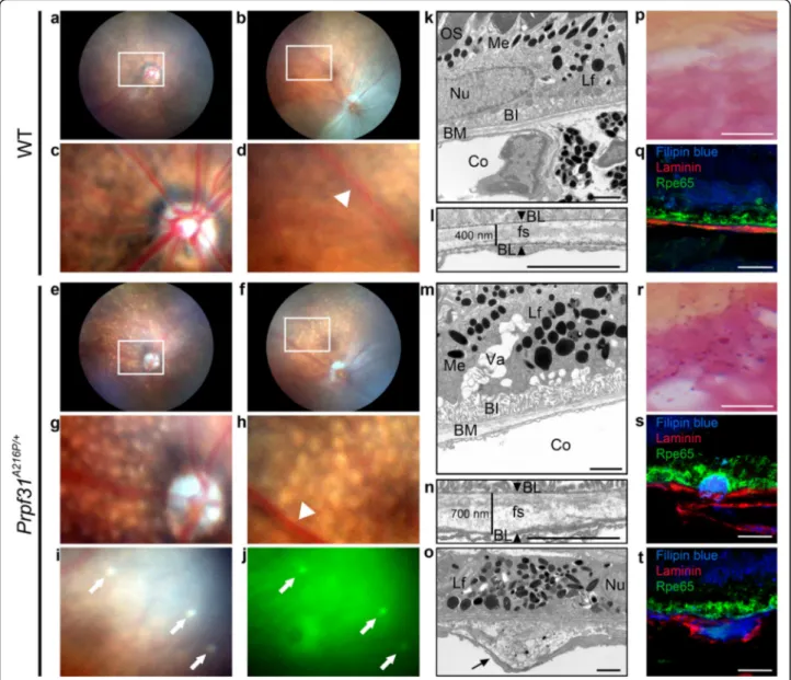
Prpf31A216P/+KI mice display RPE degeneration with drusen-like deposits
To better understand the role of Prpf31 in retinal
degen-eration, we have used heterozygous Prpf31A216P/+ KI
mice (Bujakowska et al., 2009), a mouse model which
carries the point-mutation p.A216P in the Prpf31 gene, known to be responsible for adRP in humans (Vithana
et al., 2001). However, in mice p.A216P does not
pro-duce a photoreceptor cell death phenotype as it does in
humans. On the other hand, it is known that Prpf31+/−
KO mice display an RPE-degenerative phenotype. For this reason, we decided to characterize, in detail, the RPE-degenerative phenotype of aged KI mice. We started by studying funduscopy images in 8 to 16-month-old mutant mice and their WT-littermates to evaluate the appearance of the retina. Ophthalmoscopic images of the central and peripheral regions of the retina
showed normal appearance in the WT mice (Fig. 1a-d).
The homogeneity in the surface of the central (Fig. 1a) and peripheral (Fig. 1b) retina is clearly shown, as well as the normal size of the optic nerve head (Fig. 1c) and the normal thickness of the blood vessels (Fig.1d; white arrowhead). In contrast, small round, white-yellowish, non-confluent, scattered lesions were observed through-out the retina of Prpf31A216P/+ mice like drusenoid
de-posits (Fig. 1e-h). Most of these lesions were
autofluorescent (Fig.1i-j; white arrows). The number of drusen-like deposits begins to be observed since the 8th month, and their number progressively increases in a non-homogeneous way during the retinal degenerative process. In the mutant mice evaluated we did not ob-serve any of the typical fundus features of RP, such as black pigment accumulation in the form of bone spic-ules, vascular sharpening or optic nerve head atrophy.
TEM of 8-month-old WT (Fig. 1k, l) and KI retinas
(Fig. 1m-o) was also performed to evaluate the
morph-ology of RPE cells and the Bruch’s membrane in detail. In the WT mice, the normal expected morphology for RPE was observed, with the presence of photoreceptor outer segments (OS) in contact with RPE apical micro-villi (Mv) (Fig.1k; OS and Additional file1: Figure S1a), melanin and lipofuscin granules in the cytoplasm (Fig.1k; Me, Lf) as well as basal infoldings of the RPE membrane
(Fig. 1k; BI) in contact with the Bruch’s membrane
(Fig. 1k; BM). Bruch’s membrane presented a
well-defined fundamental structure (Fig. 1l; fs) between each basal lamina, one corresponding to the RPE and the other to the endothelium of a choroidal vessel (Fig. 1l; BL, ar-rowheads). The thickness of the Bruch’s membrane mea-sured between both basal laminae was 400 nm (Fig.1l). In
contrast, Prpf31A216P/+ TEM images showed,
accumula-tion of lipofuscin granules (Fig.1m, o; Lf), vacuolization of the RPE (Fig.1m; Va), atrophy of basal infoldings (Fig.1m;
BI) and thickening of Bruch’s membrane (Fig. 1m; BM),
to an approximate size of 700 nm (Fig. 1n). Additionally, the homogeneity of the fundamental membrane structure was lost (Fig. 1n; fs) and we also found accumulation of amorphous electrodense material within the Bruch’s
membrane (Fig.1o; black arrow). The Mv and the end of
OS in mutant mice were also observed. Mv were shorter and disorganized when compared with the Mv of WT mice (Additional file1: Figure S1). Despite all these alter-ations, the Prpf31A216P/+mice did not show any damage of the photoreceptor.
In addition, specific staining methods were used to visualize lipofuscin granules (Fig.1p, r). Large accumula-tion of lipofuscin granules was observed in the RPE of
the mutant mice (Fig. 1r; dark magenta) compared to
WT (Fig. 1p). To evaluate the composition and
localization of the amorphous material observed in the Bruch’s membrane, Filipin blue staining was used to de-tect free cholesterol (Fig.1q, s, t; blue). Immunofluores-cence for laminin (Fig.1 q, s, t; red) and Rpe65 (Fig.1q, s, t; green) are shown as markers of basal lamina and RPE, respectively. In the KI mice, accumulation of free cholesterol was observed between the RPE and the
Bruch’s membrane (Fig. 1s) or between both basal
lam-inae (Fig.1t). Localization of these deposits is similar to the basal linear deposits and basal lamellar deposits de-scribed by Curcio and colleague in age-related macular degeneration (AMD) (Curcio & Millican, 1999). RPE
at-rophy, lipofuscin accumulation and thickening of
Bruch’s membrane are also features described in AMD (Curcio & Millican,1999; Ding et al.,2009).
Finally, we have monitored ERG responses in the KI mice and found that a- and b-waves, corresponding to photoreceptor electrical activity, were not affected (Add-itional file1: Figure S2a-c”). This is similar to what was previously reported for all splicing factor-mutant mouse models (Bujakowska et al., 2009; Farkas et al., 2014). Surprisingly, a defective c-wave was observed, reflecting, at a functional level, the specific degenerative changes found in the RPE layer (Additional file1: Figure S2d-d”).
Therefore, the Prpf31A216P/+ mice display RPE
degener-ation with drusen-like deposits.
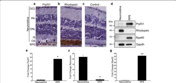
PRPF31 is highly expressed in the RPE
We have analysed expression of Prpf31 in retinal sam-ples of WT CD-1 mice to examine its distribution in the different retinal layers. Immunohistochemistry (Fig. 2) shows Prpf31 to be highly expressed in the RPE cell
layer (Fig. 2a; arrowheads) compared to neuroretina,
where almost no Prpf31 signal was detected. Rhodopsin, the photo-pigment expressed in rod-photoreceptors, was used as a positive control for the immunohistochemical staining (Fig.2b) and, as expected, a clear signal was dis-played in the photoreceptor OS. A negative control,
without primary antibody, was performed to discard non-specific binding of secondary antibody (Fig.2c).
To compare differential expression of Prpf31 protein in different layers of the mouse retina, neuroretina and RPE were manually dissected and protein and mRNA samples were obtained from each fraction. Immunoblotting results indicate high expression of Prpf31 protein compared to
the neuroretina (Fig. 2d). Antibodies against Rhodopsin and Rpe65, an enzyme of the visual cycle cascade expressed in the RPE, were used as fraction-specific markers (Fig. 2d). This result was further confirmed by qPCR (Fig. 2e), in which it is possible to observe that
Prpf31 expression level is much higher in the RPE, when
compared to its expression in the neuroretina. Recoverin Fig. 1Prpf31A216P/+mice exhibit degenerative phenotype of the RPE with drusen-like deposits. Funduscopy of WT (a-d) andPrpf31A216P/+mice (e-j) are shown. Numerous white-yellowish round lesions were observed in the retina ofPrpf31A216P/+mice (e-h). These lesions were distributed in the central (g) and peripheral retina (h) and most of them showed autofluorescence (i, j; white arrows). The optic nerve head (g) and retinal vessels (h; white arrowhead) did not show differences when compared to WT mice (c, d; white arrowhead). TEM images of RPE of 8-month-old WT (k, l) andPrpf31A216P/+mice (m-o) and the amplified images of Bruch’s membrane (BM) are displayed (l, n, o). Photoreceptor outer segments (OS) were observed in contact with the RPE microvilli in WT mice (k). Accumulation of lipofuscin granules (Lf), large vacuoles (Va) and atrophy of basal infoldings (BI) were observed in the RPE ofPrpf31A216P/+mice (m). The distance between both basal laminae (BL) was measured (l, n; arrowheads), and thickening of the BM was detected inPrpf31A216P/+mice (n). In addition, the homogeneity of the fundamental substance (fs) was lost and amorphous electrodense material was accumulated within the BM of these mice (o; black arrow). The morphology of melanin granules (Me), nuclei (Nu) and choroid (Co) was normal. Lipofuscin staining in dark magenta (p, r) showed large accumulation of lipofuscin granules in the RPE ofPrpf31A216P/+mice (r). Filipin blue dye was used to stain free cholesterol (q, s, t; blue).Prpf31A216P/+mice showed free cholesterol accumulation (s, t; blue) between the RPE and BM (s) or within the BM (t). Anti-Laminin antibodies were used to stain the BL (q, s, t; red) and the RPE was visualized by anti-Rpe65 antibody (q,s, t; green). Scale bars represent 2μm (k-o) or 12.5 μm (p-t)
and Rpe65 mRNAs were used as markers for neuroretina and RPE fractions, respectively (Fig.2f-g).
To confirm whether this differential distribution of PRPF31 along the retinal cell types is common to other vertebrates, fractions of RPE and neuroretina were ob-tained from C57BL/6 J mice, pig, cow and human eye
samples. The immunoblot for PRPF31 (Additional file1:
Figure S3) showed that protein level is comparatively higher in the RPE than in the rest of the retinal layers in several vertebrates, including humans.
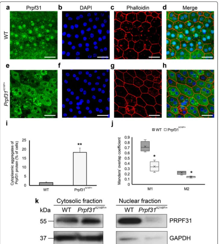
Mutant PRPF31 protein is aggregated in the cytoplasm of RPE cells
Next, we were interested in the distribution of the Prpf31 protein histologically in the RPE of the aged-KI mice. Immunofluorescence analysis of Prpf31 protein did not show clear differences between WT and KI mice in retinal sagittal sections. However, RPE whole-mount evaluation showed large cytoplasmic aggregates of Prpf31 protein in Prpf31A216P/+, which were almost not present in the WT samples (Fig.3e-h). Additionally, a dif-ference in the Prpf31 distribution within the RPE cell was observed, with a weaker staining in the KI nuclei
com-pared to the WT-littermates (Fig.3e-h). In the WT RPE
cells, most of Prpf31 is localized inside the nucleus with
some homogeneous cytoplasmic staining (Fig.3a-d) but in the Prpf31A216P/+RPE cells, most of the Prpf31 staining is shown in the cytoplasm forming rounded clumps of Prpf31 protein resembling aggregates (Fig. 3e-h). Cells that were observed harbouring protein aggregates formed clusters in the RPE layer of the KI mice. The number of cells with cytoplasmic aggregates was counted in both KI and WT mice, providing a statistically significant differ-ence, with Prpf31A216P/+ mice having 18.4 ± 2.3% of RPE cells with cytosolic aggregates compared to 1.7 ± 0.2% cells in the WT mice (Fig.3i).
To quantify the amount of Prpf31 signal in the RPE
nuclei, the Manders’ overlap coefficient for
DAPI+Prpf31/DAPI colocalization (M1) and for
Prpf31 + DAPI/Prpf31 colocalization (M2) were calcu-lated (Fig. 3j). Both coefficients were significantly lower in the mutant mice (Fig.3j), corresponding to the dimin-ished amount of Prpf31 protein in the nucleus of RPE cells as observed by histology. Besides, Western blot of cytosolic and nuclear fractions clearly showed a decrease of Prpf31 protein in the nuclear fractions of mutant mice
(Fig. 3k). Therefore, these results show that not only
Prpf31 protein in the Prpf31A216P/+mice is aggregated in the cytoplasm of the RPE cells, but also its concentration in the nuclei is decreased when compared to the WT. Fig. 2Prpf31 protein and its mRNA are highly expressed in the RPE of mouse retinas. Immunohistochemical staining showed strong Prpf31 signal in the RPE of CD-1 mouse retinas (a; arrowheads). Anti-Rhodopsin antibodies were used as positive control for immunohistochemical staining (b). A negative control without primary antibodies is also present (c). Western blot (d) and qPCR (e) analysis of Prpf31 protein and mRNA expression in the neuroretina and RPE samples showed that it is mainly expressed in the RPE (d, e). Anti-Rhodopsin and anti-Rpe65 antibodies were used as controls for the neuroretina/RPE tissue fractions, and anti-Gapdh antibody was used as a loading control (d). For qPCR,Recoverin (f) and Rpe65 (g) mRNA expression levels were used as controls for the two different tissue fractions. The bars in the graphs e-g represent means of fold change ± SEM (n = 4 replicates of 3 samples in each group). Statistically significant differences were determined by t-test (*p < 0.05, **p < 0.01). RPE = retinal pigment epithelium, OS = outer segment, IS = inner segment, ONL = outer nuclear layer, INL = inner nuclear layer, GCL = ganglion cell layer. Scale bars represent 50μm
Fig. 3 Large cytoplasmic aggregates of Prpf31 protein were observed in the RPE ofPrpf31A216P/+mice. Whole-mount of the RPE layer from WT (a-d) andPrpf31A216P/+mice (e-h) were immunostained with anti-Prpf31 antibodies (a, e). Cell nuclei were stained with DAPI (b, f), and TRITC-phalloidin was used to visualize the F-actin microfilaments (c, g). Prpf31 signal was mainly localized in the nuclei of WT RPE cells (a), while large protein aggregates stained for PRPF31 were observed in the cytoplasm of RPE cells of mutantPrpf31A216P/+mice (e). The bars in graph i represent the percentage of RPE cells with cytoplasmic aggregates of Prpf31 protein ± SEM in WT andPrpf31A216P/+whole mount RPE samples (n = 1200 cells were counted from 4 mice in each group). The boxplot j represents the Manders’ overlap coefficient of DAPI+Prpf31/DAPI colocalization (M1) and Prpf31 + DAPI/Prpf31 colocalization (M2) in WT andPrpf31A216P/+whole-mount RPE samples (n = 4 in each group). Expression of Prpf31 protein was evaluated in the cytosolic and nuclear fractions by Western blot (k). Statistically significant differences were determined byt-test or Mann-WhitneyU-test (*p < 0.01, **p < 0.001). Scale bars represent 25 μm
The antibody used to visualize Prpf31 recognized both mutant and normal Prpf31 protein, thus we were unable to determine whether the aggregates are composed solely of the mutated protein or if the WT protein is also present in the aggregates.
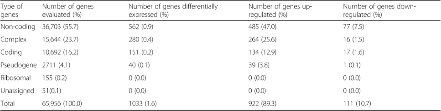
Differential gene expression and alternative splicing were affected in Prpf31A216P/+mice
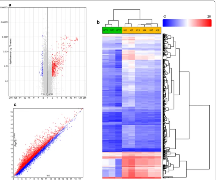
Considering that the mutant Prpf31 protein is aggregated in the cytoplasm of RPE cells in Prpf31A216P/+ mice, we decided to perform transcriptomic analysis using a Gene-Chip™ Mouse Transcriptome Array (MTA) 1.0 to evaluate differential gene expression in RPE samples of six
Prpf31A216P/+ and three WT-littermates. The number of
genes evaluated was 65,956 and, from these, a total of 1033 (1.6%) genes were differentially expressed in the Prpf31A216P/+mice. Most of these genes were upregulated
(922, 89.3%) and the rest were downregulated (Table 1;
Additional file2, Gene expression). The gene-level differ-ential expression analysis is graphically displayed in the volcano plot (Fig. 4a). Each point on the plot represents the statistical result of a single gene. Horizontal axis repre-sents fold change in log2 scale and vertical axis reprerepre-sents p-value in log10 scale. Threshold of fold change was either <− 2 (Fig.4a; blue) or > 2 (Fig. 4a; red) and ANOVA p-value < 0.05. Gray dots correspond to the genes without statistically significant change. Hierarchical clustering of 1033 genes differentially expressed in Prpf31A216P/+vs WT mice is shown in Fig.4b with an expression profile clearly different for WT and Prpf31A216P/+clusters (Fig.4b).
Gene ontology was evaluated through the informatic tool DAVID v6.8 (Sherman & Lempicki,2009) using the list of 1033 genes which were differentially expressed in
Prpf31A216P/+ mice, showing that the largest group of
genes (123 genes) affected by the mutation belongs to Pro-tein Binding (Additional file1: Table S1). This molecular function is defined as: interacting selectively and non-covalently with any protein or protein complex (GO: 0005515), including the subcategory chaperone binding (GO:0051087); a class of proteins that bind to nascent or
unfolded polypeptides to ensure correct folding or trans-port. Because the most relevant change observed in the RPE of the mutant mice was the cytoplasmic aggregation of Prpf31 protein, we decided to look for candidate genes related to molecular chaperones involved in protein fold-ing. We found that heat shock protein family A (Hsp70)
member 4 like gene (Hspa4l), which encodes the
chaperone heat shock 70 kDa protein 4 L (Hspa4l), was upregulated in Prpf31A216P/+ mice (fold change 2.26; p-value 0.009). Other chaperones involved in the unfolded protein response were not highlighted with a different gene expression level. Lists of candidate genes found to be differentially expressed which might also be involved in
RPE degeneration are shown in Additional file 1: Table
S2.
Alternative splicing analysis was also performed through the GeneChip™ MTA 1.0 in the same RPE
sam-ples of six Prpf31A216P/+ and three WT-littermates. A
total of 65,770 genes were evaluated and 92.6% (60871)
of these genes were expressed in both mice
(Prpf31A216P/+and WT mice). From these 60,871 genes,
6700 (11%) genes have, at least, one differentially expressed probe selection region or junction to indicate alternative splicing (Table 2; Additional file3, Splicing). The scatter plot (Fig. 4c) displays the number of genes that are alternatively spliced in Prpf31A216P/+ mice with lower < − 2 (Fig. 4c; red) or higher > 2 (Fig. 4 c; blue) splicing index, when compared to WT mice.
Functional categories of alternative spliced genes in
the RPE of Prpf31A216P/+ mice are listed in the
Add-itional file1: Table S3. We observed that several splicing factors, including Prpf31, present a different splicing index (Prpf31 splicing index − 2.33, p-value 0.04; Prpf18 splicing index− 2.62, p-value 0.01; Prpf39 splicing index 2.25, p-value 0.03). Apart from the aforementioned spli-cing factors, others genes of different pathways involved in retinal degeneration were also affected by the muta-tion, such as inflammamuta-tion, oxidative stress, retinol me-tabolism (Abca4), ciliogenesis (Bbs1, Bbs4, Bbs5, Bbs7, Bbs9) and cellular apoptosis (Additional file1: Table S4;
Table 1 Summary of gene level differential expression analysis in RPE samples in two different conditions (Prpf31A216P/+vs WT mice). Default filter criteria, fold change <− 2 or > 2 and ANOVA p-value < 0.05
Type of genes
Number of genes evaluated (%)
Number of genes differentially expressed (%)
Number of genes up-regulated (%)
Number of genes down-regulated (%) Non-coding 36,703 (55.7) 562 (0.9) 485 (47.0) 77 (7.5) Complex 15,644 (23.7) 280 (0.4) 264 (25.6) 16 (1.5) Coding 10,692 (16.2) 151 (0.2) 134 (12.9) 17 (1.6) Pseudogene 2711 (4.1) 40 (0.1) 39 (3.8) 1 (0.1) Ribosomal 155 (0.2) 0 (0.0) 0 (0.0) 0 (0.0) Unassigned 51(0.1) 0 (0.0) 0 (0.0) 0 (0.0) Total 65,956 (100.0) 1033 (1.6) 922 (89.3) 111 (10.7)
Fig. 4 Differences in the level of gene expression and alternative splicing in the RPE ofPrpf31A216P/+mice compared to WT controls. Volcano plot (a) showing the genes that are upregulated (red) or downregulated (blue) inPrpf31A216P/+KImice with a significance ANOVA p value < 0.05 and fold change <−2 or > 2 when compared with WT mice. Hierarchically clustered (b) genes (rows) and WT or KI mice (columns) with dendrograms and flat clusters; red in the heatmap denotes upregulation while blue denotes downregulation. Scatter plot (c) presents the genes that are alternatively spliced inPrpf31A216P/+mice with lower (red) or higher (blue) splicing index when compared with WT mice (WTn = 3 and Prpf31A216P/+n = 6)
Table 2 Summary of alternative splicing analysis in RPE samples in two different conditions (Prpf31A216P/+vs WT mice). Default filter criteria, splicing index <−2 or > 2 and ANOVA p-value < 0.05
Type of genes
Number of genes evaluated (%)
Number of genes expressed in both conditions (%)
Genes with at least one differentially expressed PSR or junction to indicate alternative splining (%)
Non-coding 36,581 (55.6) 33,587 (51.1) 929 (1.5) Complex 15,644 (23.8) 15,056 (22.9) 4344 (7.1) Coding 10,628 (16.2) 9450 (14.4) 1381 (2.3) Pseudogene 2711 (4.1) 2601 (3.9) 29 (0.1) Ribosomal 155 (0.2) 155 (0.3) 16 (0.0) Unassigned 51(0.1) 22 (0.0) 1 (0.0) Total 65,770 (100.0) 60,871 (92.6) 6700 (11.0)
Additional file 3, Splicing). The number of candidate genes with modified splicing index that can be involved
in RPE degeneration are detailed in Additional file 1:
Table S4. These results suggest that normal splicing of different genes, including splicing factors, is affected in the RPE of Prpf31A216P/+mice.
Increased Hspa4l expression and colocalization with mutant Prpf31 protein in the RPE of Prpf31A216P/+mice
Analysis of transcriptomic data show that a member of the heat shock protein 70 (HSP70) family, Hspa4l, was overexpressed in the RPE of KI mice. The HSP70 family is a ubiquitous and conserved family of molecular chaperons assisting in protein folding to prevent aggregation and to protect cells from stress (Mashaghi et al.,2016; Mayer &
Bukau, 2005). We have analyzed mRNA expression of
Hspa4lby qPCR in both RPE and neuroretina of the KI
mice and we found that Hspa4l is overexpressed in the RPE of the mutant mice, when compared to its expression in the WT mice (Fig.5a). No differences of Hspa4l expres-sion in the neuroretina extracts were observed (Fig. 5a). This result was corroborated by Western blot in which we observed that Hspa4l was more abundant in the RPE of
the mutant mice (Fig. 5b). RPE whole-mount
immuno-fluorescence to localize Hspa4l and Prpf31 was also
per-formed. As previously mentioned, Prpf31 protein
predominantly localizes in RPE nuclei of WT samples (Fig. 5c, f; arrowhead) and in cytoplasmic aggregates in the case of KI tissue and low expression in the nucleus (Fig.5i, l; arrow). As expected, Hspa4l staining is stronger in the mutant RPE cells, where the chaperone colocalizes with Prpf31 protein aggregates (Fig.5i-n). The hexagonal shape of the RPE can be seen with phalloidin staining (Fig. 5c-n; blue). Negative controls for RPE autofluores-cence and secondary antibodies nonspecific bindings are shown in the Additional file 1: Figure S4. The small heat shock protein, Hsp27, also colocalized with the Prpf31 ag-gregates in the RPE of mutant mice (Additional file 1: Fig-ure S4), but transcriptomic data did not show differential expression of its gene (Hspb1).
The p.A216P mutant protein produces insoluble cytoplasmic aggregates, recruits endogenous PRPF31 protein in the insoluble fraction and increases the expression of HSP70
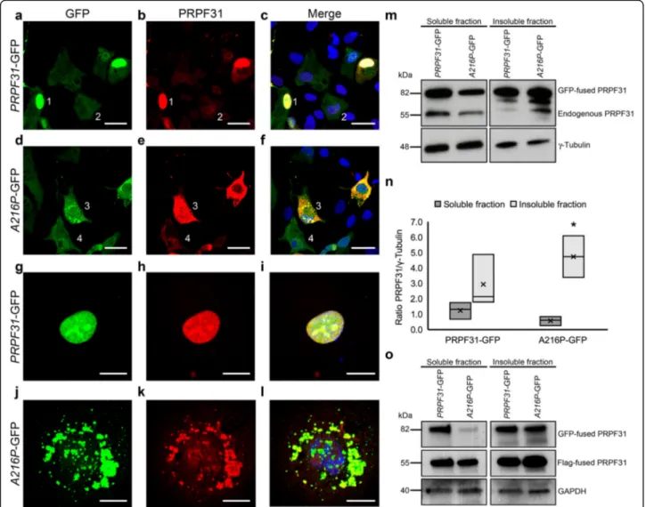
To better understand the role of p.A216P mutation in human-derived RPE cells, we have cloned the human WT PRPF31 and the A216P mutant genes in pEGFP-N1 plasmid and the GFP-tagged PRPF31 proteins were over-expressed in ARPE-19 cells. We observed that WT PRPF31-GFP was mainly found in the nucleus of the transfected cells (Fig. 6a-c and g-i), while the mutant A216P-GFP was mostly aggregated in the cytoplasm of the transfected cells (Fig. 6d-f and j-l), similar to our
previous observation in the RPE of the WT and Prpf31A216P/+mice, as shown in Fig.3.
Western blot analysis of soluble and detergent-insoluble fractions of the cell lysates was conducted to esti-mate the concentration of PRPF31 in both fractions. Western blotting and densitometry analysis of the immu-noblots (Fig.6m, n) show less mutant protein A216P-GFP in the soluble fraction compared to PRPF31-GFP protein, and the opposite in the detergent-insoluble fraction (Fig.6m, n). Additionally, it is possible to observe a deple-tion of the endogenous PRPF31 protein in the soluble frac-tion and highest levels of expression in the insoluble fraction (Fig.6m), suggesting that the cytoplasmic insoluble aggregates of A216P-GFP protein recruit the endogenous WT PRPF31 protein. To evaluate whether the WT PRPF31 protein increased in the insoluble fraction in cells co-transfected with the mutant protein PRPF31 (A216P), ARPE-19 cells were co-transfected with WT PRPF31 tagged to Flag (PRPF31-Flag) and PRPF31-GFP or A216P-GFP. After 24 h of incubation, the protein extracts of sol-uble and insolsol-uble fractions were analyzed by Western blot-ting (Fig.6o). It was observed that the WT protein tagged to GFP was expressed in both the soluble and insoluble fraction, while the mutant protein was expressed mostly in the insoluble fraction (Fig. 6o). Besides, it was found that the WT protein tagged to Flag decreased in the soluble fraction and increased in the insoluble fraction only in those cells expressing the PRPF31 protein carrying the p.A216P mutation. In this way, we confirm our hypothesis that the mutant PRPF31 protein recruits the WT protein in the insoluble fraction.
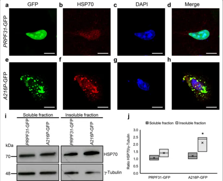
HSP70 plays an important role in retinal dystrophies, including RP (Furukawa & Koriyama, 2016) so we have also evaluated the distribution and expression of HSP70 protein in the ARPE-19 cells overexpressing either PRPF31-GFP or A216P-GFP by immunofluorescence and Western blotting. In the immunofluorescence stain-ing, PRPF31-GFP transfected cells show a weak labeling of HSP70 in the nucleus (Fig.7a-d). However, in A216P-GFP transfected cells, HSP70 staining is increased, and its signal colocalizes with the cytoplasmic aggregates of mutant PRPF31 protein (Fig.7e-h), similar to the
distri-bution observed for Hspa4l in the RPE of Prpf31A216P/+
mice. Additionally, Western blot analysis of the soluble
and insoluble fractions (Fig. 7i) and its densitometry
analysis (Fig.7j) show an increase of HSP70 in the insol-uble fraction of the cells transfected with A216P-GFP. Therefore, protein aggregation of mutant PRPF31 seems to activate the response of chaperones.
P.A216P mutation affects the mobility of PRPF31 protein to the nucleus in human RPE derived live cells
FRAP experiments provide qualitative and quantitative information about the mobility of a fluorescently tagged
protein in a defined compartment (Phair & Misteli, 2001; Reits & Neefjes, 2001). Different parameters can be evaluated by FRAP, such as the mobile fraction (mf) of a fluorescent-tagged protein and its half-life (τ1/2). The mf can be affected by different circumstances, such as interaction of the fluorescent-tagged protein with other proteins, cytoplasmic organelles or membranes. Considering that p.A216P mutation induces protein ag-gregation and expression of HSP70, which colocalizes with the cytoplasmic aggregates of PRPF31 protein, we decided to investigate whether the translocation of PRPF31 protein from the cytoplasm to the nucleus was affected in living cells. To test this hypothesis, we have
performed FRAP assay in ARPE-19 cells (Fig. 8). The
cell line was transfected with the plasmids pEGFP-N1, PRPF31-GFP and A216P-GFP and FRAP was performed as described in the Methods section of this manuscript. Pre- and post-photobleached cells are displayed in Fig.8a-l, showing that the amount of PRPF31 protein re-covered in the nucleus 200 s after bleaching is affected by the mutation (Fig.8l) but not in pEGFP-N1 (Fig.8d) or PRPF31-GFP (Fig. 8h) transfected cells. A slower re-covery curve for A216P-GFP were clearly observed when
compared to the control PRPF31-GFP (Fig.8m).
The changes produced in the recovery curve were due to changes in the size of mf and not due to
changes in the τ1/2, which is defined as the time
when the recovery of fluorescence intensity is half of
the plateau (τ1/2 PRPF31-GFP = 7.40 ± 3.08 s; τ1/2
A216P-GFP = 7.00 ± 2.66 s; p = 0.46). However, the mf significantly decreased in A216P-GFP transfected cells (mf PRPF31-GFP = 0.55 ± 0.10; mf A216P-GFP =
0.27 ± 0.15; p < 0.05) (Fig. 8n). The mf was
deter-mined by comparing the fluorescence in the bleached region after full recovery (F∞) with the fluorescence before bleaching (Fi) and just after bleaching (F0), be-ing defined as mf = (F∞ – F0)/(Fi – F0). The fluores-cence intensity was normalized to the Fi and F0 in
the Fig. 8m. The A216P-GFP transfected cells showed
less F∞ than the control (Fig. 8m). These results
sug-gest that p.A216P mutation decreases the amount of PRPF31 protein that moves to the nucleus but with-out affecting the diffusion time.
Discussion
PRPF31is a ubiquitously expressed gene encoding for a
component of the spliceosome complex involved in pre-mRNA processing. Mutations in this gene are associated with non-syndromic adRP, but the mechanism by which retinal degeneration occurs, is still unknown. Previously, two mutant mouse models (Prprf31+/−and Prpf31A216P/+) were generated to study the role of PRPF31 in the patho-genesis of adRP, but neither of these models presented ev-idences of RP-like photoreceptor degeneration, leading to conclude that the presence of one copy of WT-Prpf31 al-lele is sufficient to maintain the normal retina, and that the p.A216P mutation does not exert a dominant-negative effect (Bujakowska et al.,2009).
It has previously been described that the Prprf31+/−KO mice (Farkas et al.,2014; Graziotto et al.,2011), as well as other splicing-factor mouse models that do not present photoreceptor-degenerative phenotype, have an RPE-degenerative phenotype. In this study, a deep phenotypic characterization of 8 to 16 month-old Prpf31A216P/+ mice was performed to understand how the p.A216P mutation affects RPE function and we found evidence that supports a combination of haploinsufficiency and dominant-negative effect. Fundus analysis showed a severe RPE de-generation, with the presence of white-yellowish and auto-fluorescent spots in mutant mice. Concomitant functional impairment was detected in ERG c-wave. TEM images ac-cordingly showed some typical features of degenerative RPE such as vacuolization, atrophy of basal infoldings, thickening of Bruch’s membrane and accumulation of amorphous electrodense material within this membrane. Two previous studies (Farkas et al.,2014; Graziotto et al.,
2011) reported that two-year-old Prpf3T494M/+ and
Prpf8H2309P/+ and the one-year-old Prpf31+/− mice had
similar features to the ones that we have found in
8-month-old Prpf31A216P/+ mice. The earlier onset of RPE
degeneration in Prpf31A216P/+ might indicate a possible toxic effect of the p.A216P mutant protein.
In addition to the RPE atrophy, we have also observed accumulation of lipofuscin granules and drusen-like de-posits of free cholesterol between the RPE and the Bruch’s membrane or between both basal laminae. These deposits are similar to the basal linear deposits (See figure on previous page.)
Fig. 5Hspa4l is highly expressed in the RPE of Prpf31A216P/+mice. Analysis ofHspa4l expression by qPCR in the neuroretina and RPE samples show thatHspa4l mRNA is overexpressed in the RPE of Prpf31A216P/+mice (a). The boxplot a represents the fold change ofHspa4l expression in the neuroretina and RPE of WT andPrpf31A216P/+mice (WTn = 3 and Prpf31A216P/+n = 6). Statistically significant differences were determined by Mann-WhitneyU-test (*p < 0.05). Western blot of RPE samples showed higher expression of Hspa4l protein in Prpf31A216P/+mice compared to WT (b). Anti-Gapdh antibodies were used as loading control (b). Whole-mount of the RPE obtained from WT (c-h) andPrpf31A216P/+mice (i-n) were immunostained with anti-Prpf31 (c, f, i, l) and anti-Hspa4l antibodies (d, g, j, m). TRITC-phalloidin was used to stain F-actin microfilaments (c-n; blue). Magnified images are (f-h and l-n) and merged are shown (e, h, k, n) Prpf31 signal was mainly distributed in the nuclei of RPE cells in WT mice (c, f; arrowhead), while Prpf31 protein aggregates were observed in the cytoplasm (i, l) colocalizing with Hspa4l signal in mutant
Prpf31A216P/+mice as shown in the merged images (k, n). Prpf31 signal was very low in the nuclei ofPrpf31A216P/+mice (l; arrow). Scale bars
and basal lamellar deposits described in AMD patients (Curcio & Millican,1999). RPE atrophy, lipofuscin accu-mulation and thickening of Bruch’s membrane are char-acteristic histological hallmarks found in human AMD patients, and these features have been also described in animal models of RPE degeneration (Curcio & Millican, 1999; Pennesi et al., 2012). Therefore, the Prpf31A216P/+ mice display a primary RPE degeneration phenotype
with drusen-like deposits. Although RP phenotype is predominantly associated with PRPF31 mutations in humans, some affected individuals developed juvenile macular degeneration apart from the typical RP pheno-type, as described in two Chinese families carrying dif-ferent mutations in the PRPF31 gene (Lu et al.,2013; Xi et al., 2005). These results suggest that mutations in the PRPF31 gene could produce not only a characteristic RP Fig. 6 Overexpression ofA216P-GFP induces aggregation of PRPF31 protein. The ARPE-19 cell line was transfected with PRPF31-GFP (a-c, g-i) and A216P-GFP (d-f, j-l). The GFP-tagged proteins (green) and immunostained cells with anti-PRPF31 antibodies (red) are shown. PRPF31-GFP was found mainly in the cell nucleus (a-c; 1, g-i) and to a lesser extent, in the cytoplasm (a-c; 2).A216P-GFP transfected cells present PRPF31 aggregation in the cytoplasm (d-f; 3, j-l), and a very minor signal in the nucleus (d-f; 4). Images correspond to a maximum projection of a Z-stack. Western blot analysis of soluble and insoluble fractions of the transfected cells show a decrease in the concentration of endogenous PRPF31 in the detergent-soluble fraction and an increase in the detergent-insoluble fraction of the cells transfected withA216P-GFP. Anti-γ-tubulin antibody was used as loading control (m). Densitometry quantification of the blots (n) shows a significant increment ofA216P-GFP protein in the detergent-insoluble fraction when compare with the among ofA216P-GFP protein present in the soluble fraction (n). The boxplot n represents the ratio PRPF31/γ-tubulin in the soluble and insoluble fractions of PRPF31-GFP and A216P-GFP transfected ARPE-19 cells (n = 3 in each group). Statistically significant differences were determined by Mann-WhitneyU-test (*p < 0.05). Western blot analysis of soluble and insoluble fractions ofPRPF31-GFP and A216P-GFP transfected ARPE-19 cells and co-transfected with WT PRPF31 tagged to Flag showed a decrease in the concentration of WT PRPF31-Flag in the detergent-soluble fraction and an increase in the detergent-insoluble fraction of the cells co-expressing the mutantA216P-GFP protein. Anti-GAPDH antibody was used as loading control (o). Scale bars (a-f) represent 25 μm and (g-l) 10μm
phenotype in humans but also a phenotype with an early macular degeneration. Similar results have been ob-served with mutations in another gene such as RDS, which encodes the photoreceptor glycoprotein periph-erin. Mutations in RDS can produce both a clear RP phenotype and also macular dystrophy (Wells et al.,
1993). Although no single nucleotide polymorphism in
PRPF31 gene has been described in AMD, it has been
suggested that alterations in normal mRNA splicing could contribute to the pathophysiology of age-related
diseases such as AMD (Li et al., 2017). In the Prpf31
mutant mice, we observed that the splicing of several genes is compromised. Among these genes, the ABCA4
gene stands out. ABCA4 is a gene expressed mainly in photoreceptors but also RPE having a major function in retinol metabolism (Lenis et al., 2018). Mutations in this gene have been found to cause Stargardt’s disease, a her-editary juvenile macular degeneration, and AMD (Baum et al., 2003). Besides, it has been reported that ABCA4 splicing can be also affected by aging and in AMD cases (Li et al., 2017; Baum et al., 2003; Meshorer & Soreq,
2002). For these reasons, we suggest that primary RPE
degeneration phenotype with drusen-like deposits
present in these Prpf31 mutant mice could be due to splicing defects in genes that could be associated with macular degenerative diseases such as ABCA4.
Fig. 7 Overexpression of A216P-GFP induces HSP70 activation in ARPE-19 cells. Immunostaining of cultured ARPE-19 cells transfected with PRPF31-GFP (a-d) orA216P-GFP (e-h) displays PRPF31 aggregation in the cytoplasm of the cells overexpressing A216P-GFP (green) and colocalization of HSP70 (red) in the aggregates (e-h). Images correspond to a maximum projection of a Z-stack. Western blot analysis (i) and densitometry quantification (j) of the soluble and insoluble fraction of the transfected cells showing an increment of HSP70 concentration in the detergent-insoluble fraction of the cells transfected withA216P-GFP. Anti-γ-tubulin antibody was used as loading control (i). The boxplot j represents the ratio HSP70/γ-tubulin in soluble and insoluble fraction of PRPF31-GFP and A216P-GFP transfected ARPE-19 cells (n = 3 in each group). Statistically significant differences were determined by Mann-WhitneyU-test (*p < 0.05). Scale bars represent 10 μm
It is known that the RPE maintains structural integrity and function of photoreceptor cells and defends the ret-ina from free radicals and photo-oxidative damage (Simo et al., 2010; Strauss, 2005). A failure in any of the RPE roles might induce retinal degeneration and loss of vis-ual function. Two classical examples of RP due to muta-tions in RPE-specific genes are MERTK and RPE65, which cause RP (Gal et al.,2000) and Leber’s congenital amaurosis (Gu et al., 1997), respectively. Thus, p.A216P
PRPF31 mutation might affect photoreceptors due to a
primary defect in the RPE as MERTK or RPE65 muta-tions do.
As stated before, in humans, mutations in PRPF31 are associated with photoreceptor-cell degeneration and
loss. The reason why Prpf31A216P/+ mice have
RPE-degeneration rather than photoreceptor-cell loss is not clear, but we have found that gene expression level for
Prpf31 and the corresponding protein amount is much
higher in the RPE than in the neuroretina, although the nuclei in the neuroretina greatly outnumber the nuclei in the RPE monolayer. This was also observed in mouse, pig, cow and human samples. Yuan and co-workers have studied the expression of Prpf31 in different mouse tis-sues including the retina (Yuan et al., 2005). Although no differences were observed either in the expression level or pattern of Prpf31 protein among different tis-sues, they did not separate the neuroretina from the RPE as we have done in this study and in fact, their in situ hybridization results clearly show a much higher sion of Prpf31 in the RPE layer. The differential expres-sion of Prpf31 could give us clues to understand why the RPE specifically degenerates in these mice. The elevated requirement of Prpf31 protein level in the RPE could be related to high or specific splicing demands in an epithe-lium that fulfils many different roles: light absorption, transport of ions, water, and metabolic end-products from the subretinal space to blood, maintenance of pho-toreceptors and re-isomerization of all-trans-retinal into 11-cis-retinal. Thus, defects in splicing might have a great impact on the normal function and survival of RPE cells.
To explore the role of Prpf31 p.A216P mutation in the RPE, the distribution pattern of the protein was analysed by immunofluorescence staining of whole-mount RPE samples and Western blot of cytosolic and nuclear
frac-tions from WT and Prpf31A216P/+ mutant mice. As
expected, we found Prpf31 preferentially localized in the nucleus of RPE cells in WT mice, although it was also possible to observe a cytoplasmic fraction. On the other hand, in the mutant mice almost no Prpf31 was found within the nucleus, and most of the Prpf31 staining was present in the cytoplasm forming rounded clumps of Prpf31 protein resembling aggregates. The distribution of RPE cells having Prpf31 aggregates was not uniform, and we do not have an explanation for this finding. Nevertheless, our results are in accordance with the ones published by Deery and co-workers (Deery et al.,2002), in which they found that overexpression of A194E and A216P mutations in COS-7 cells induced mislocalization of the mutant PRPF31 proteins, distributed throughout the cytoplasm and with less intense staining in the nu-cleus when compared with WT-PRPF31 transfected cells. Also, Huranová and colleagues described similar results overexpressing A216P in HeLa cells. They found mislocalization of the mutant protein causing depletion of PRPF31 from Cajal bodies, where splicing takes place, and described a possible negative effect due to an abnor-mal interaction of A216P with its partner, PRPF6 (Hura-nova et al.,2009). It is important to note, that the Prpf31 protein detected in these mice corresponds to the en-dogenous WT and mutant forms of the protein. Thus, the aggregation that is found in the RPE of these mutant mice is not an artefact resulting from Prpf31
overexpres-sion. In Prpf31A216P/+ mice, aggregation of the mutant
protein completely depletes Prpf31 protein in the nu-cleus and, in this way, a combination of a dominant-negative effect and haploinsufficiency might contribute to RPE degeneration.
Although the exact mechanism underlying RPE degen-eration in the mutant mice is not well understood yet, our transcriptomic analysis show that 1.6% of evaluated genes are differentially expressed in the mutant RPE when compared to WT controls. As expected, alterna-tive splicing was also affected in the RPE of these mutant mice. Around 11% of evaluated genes have, at least, one differentially expressed probe selection region or junc-tion, indicative of alternative splicing. From the pool of affected genes, we selected Hspa4l for a detailed study, a member of the HSP70 family of chaperons that was up-regulated in the mutant RPE. The HSP70 family is a ubi-quitous and conserved family of molecular chaperons, (See figure on previous page.)
Fig. 8 Mobility of PRPF31 protein to the nucleus is affected by the p.A216P mutation. ARPE-19 cells transfected with the pEGFP-N1 plasmid alone or carrying the open reading frame of human WTPRPF31 (PRPF31-GFP) or carrying the point mutation p.A216P (A216P-GFP) were imaged − 3 s pre-photobleaching (a, e, i) and 0 s (b, f, j), 100 s (c, g, k) and 200 s (d, h, l) post-photobleaching. The recovery curve (m) indicates that the cells transfected with mutantA216P-GFP have a smaller mf than those transfected with PRPF31-GFP (n). The boxplot n represents the normalized fluorescence intensity and mobile fraction inPRPF31-GFP and A216P-GFP transfected ARPE-19 cells (n = 5 in each group). Statistically significant differences were determined by Mann-WhitneyU-test (*p < 0.05). Fi = fluorescence before bleaching, F0 = fluorescence just after bleaching, F∞ = fluorescence in the bleached region after full recovery. Scale bars represent 10μm
part of the cellular machinery for protein folding that prevents aggregation and protects cells from stress (Mashaghi et al., 2016; Furukawa & Koriyama, 2016). Several reports have shown that HSP70 and also small heat shock proteins play an important role in retinal dystrophies, including RP (Saliba et al.,2002), glaucoma (Nagashima et al., 2011; Park et al., 2001; Schallenberg et al.,2012) and AMD (Alge et al.,2002; Lee et al.,2011;
Nakata et al., 2005). Our qPCR analysis showed
in-creased expression of Hspa4l in the mutant RPE when compared with its WT control. We further confirmed this finding by Western blot analysis depicting a higher amount of protein in the mutant RPE. Moreover, im-munofluorescence staining showed colocalization of Hspa4l signal with aggregates of Prpf31 protein in the RPE cytoplasm of mutant mice. This suggests that Hspa4l can be acting as a chaperone for the mutant pro-tein in response to its aggregation. However, the aggre-gation of Prpf31 in the RPE of mutant mice could also activate other chaperones belonging to the family of small heat shock proteins such as Hsp27.
In addition, several other genes and signalling path-ways were found to have an altered splicing in the RPE of mutant mice, including those associated with inflammation, oxidative stress, retinol metabolism and cellular apoptosis Most of these pathways are com-monly affected in RPE degenerative diseases such as AMD and mutations of some genes including in this pathways such as ABCA4 cause macular degeneration (Lenis et al., 2018; Makarev et al., 2014). In line with these results, our histological findings in the mutant RPE correlate with some macular degenerative fea-tures. Apart from that, another group of genes af-fected by the A216P mutation in RPE are involved in ciliogenesis. This result is in agreement with the most recent results of Buskin and co-workers in which they show that iPS-derived RPE cells from RP11 patients present shorter Mv and primary cilia, loss of polarity, reduced barrier function and defective phagocytic cap-acity, when compared to iPS-derived RPE cells from healthy donors, suggesting that these distorted cellular characteristics result from alternative splicing in RP11
(Buskin et al., 2018). We observed that one group of
proteins affected by differential splicing in
Prpf31A216P/+mice are some splicing factors, including Prpf31. Four main causes are known to induce pro-tein aggregation: mutations, errors in propro-tein synthe-sis including splicing defects, environmental factors such as oxidative stress and aging. Therefore, if the Prpf31-protein level in the RPE nucleus is beneath its threshold for normal function, the mRNA splicing of
Prpf31 gene could be affected producing an aberrant
protein prone to aggregation, reinforcing the
dominant-negative effect and haploinsufficiency.
To explore the role of Hsp70 family in the RPE affected by a p.A216P mutation, we overexpressed both WT
PRPF31 and mutant A216P tagged to GFP in a
human-derived RPE cell line (ARPE-19). Immunofluorescence staining results are in line with the in vivo results, with WT PRPF31-GFP protein being located mainly in the nu-cleus, and mutant A216P-GFP protein being mostly aggre-gated in the cytoplasm. By Western blot analysis we found that cells overexpressing the mutant variant have a signifi-cant decrease in the soluble fraction of PRPF31 and an in-crease in the insoluble PRPF31 protein. Moreover, the overexpression of the mutant protein leads to a depletion of the soluble endogenous PRPF31 protein and WT PRPF31 tagged to Flag as well, suggesting a dominant-negative effect. These results are in accordance with one of the mechanisms proposed by Yin and coworkers in which they report that mutations in PRPF31 can induce
retinal degeneration (Yin et al., 2011). They proposed
three mechanisms: i) haploinsufficiency due to loss of function of the mutant protein or degradation of mutant mRNA by nonsense-mediated mRNA decay, thus com-promising the splicing machinery; ii) mutant proteins with dominant-negative activity that may interfere with splicing and potentially with other cellular activities, leading to de-generation of the affected tissue; and iii) mutations might promote proteins forming insoluble and cytotoxic aggre-gates that can affect the tissue by loss-of-function and dominant-negative effects (Yin et al., 2011). We also ob-served that Hsp70 was upregulated in the cells overex-pressing A216P-GFP and colocalized with PRPF31 mutant protein. These results corroborate our in vivo findings of Hspa4l upregulation in the RPE of Prpf31A216P/+mice and its colocalization with PRPF31 protein aggregates.
Considering that PRPF31 plays its role in the nucleus and p.A216P induces PRPF31 aggregation in the cyto-plasm of RPE cells, we decided to analyze how PRPF31 translocation to the nucleus was affected using FRAP assay. PRPF31 contains a classical nuclear localization se-quence (NLS) between residues 351 and 364. In a previous study published by Wilkie, et al. (Wilkie et al.,2006) dem-onstrated that p.A216P mutations did no affect the
inter-action with importin β1. However, FRAP studies in the
green monkey kidney fibroblast-like COS-7 cells of WT
PRPF31-GFPand mutant A216P-GFP transfected cells
in-dicated the presence of two-component recovery pro-cesses, a fast component for free diffusion within an unbounded compartment and a slow component for pas-sive diffusion through the nuclear pores. The kinetics of both components were not affected by the p.A216P muta-tion (Wilkie et al.,2006). Therefore, seems that the muta-tion p.A216P did not affect the NLS and its interacmuta-tion
with importinβ1 but the total amount of nuclear PRPF31
is affected probably because there is lest amount of cyto-plasmic PRPF31 that can cross the nuclear pores through