Context-dependent roles for lymphotoxin-β receptor signaling in
cancer development
Mónica T. Fernandes
a,b, Emmanuel Dejardin
cand Nuno R. dos Santos
a,d,e a Centre for Biomedical Research (CBMR), University of Algarve, 8005-139 Faro, Portugal;b PhD Program in Biomedical Sciences, Department of Biomedical Sciences and Medicine, University of Algarve, 8005-139 Faro, Portugal;
c Laboratory of Molecular Immunology and Signal Transduction, GIGA-Research, Molecular Biology of Diseases, University of Liège, 4000 Liège, Belgium;
d Instituto de Investigação e Inovação em Saúde (I3S), Universidade do Porto, 4200 Porto, Portugal; e Institute of Pathology and Molecular Immunology of the University of Porto (IPATIMUP), 4200 Porto, Portugal.
Corresponding author at: Centre for Biomedical Research (CBMR), University of Algarve, Campus de Gambelas, 8005-139 Faro, Portugal; Tel.: +351 289 800 900 (+7136). E-mail address: [email protected] (N.R. dos Santos).
Abstract
The LTα1β2 and LIGHT TNF superfamily cytokines exert pleiotropic physiological functions through activation of their cognate lymphotoxin-β receptor (LTβR). Interestingly, since the discovery of these proteins accumulating evidence has pinpointed a role for LTβR signaling in carcinogenesis. Early studies have shown a potential anti-tumoral role in a subset of solid cancers either by triggering apoptosis in malignant cells or by eliciting an anti-tumor immune response. However, more recent studies provided robust evidence that LTβR signaling is also involved in diverse cell-intrinsic and microenvironment-dependent pro-oncogenic mechanisms, affecting several solid and hematological malignancies. Consequently, the usefulness of LTβR signaling axis blockade has been investigated as a potential therapeutic approach for cancer. Considering the seemingly opposite roles of LTβR signaling in diverse cancer types and their key implications for therapy, we here extensively review the different mechanisms by which LTβR activation affects carcinogenesis, focusing on the diverse contexts and different models assessed.
Keywords: Lymphotoxin-β receptor; cell signaling; lymphotoxin; LIGHT; oncogenesis; tumor microenvironment.
1. Introduction
Lymphotoxin-β receptor (LTβR) is a member of the tumor necrosis factor receptor superfamily (TNFRSF) identified as a key mediator controlling the development, organization and homeostasis of lymphoid tissues and organs [1–3]. Moreover, it was reported to play a role in the adaptive immune response against pathogens [1], thymic medullary epithelial cell differentiation and central tolerance induction [4]. Currently it is known that LTβR is involved in many other biological processes such as liver regeneration [5], lipid homeostasis [6], high endothelial venule (HEV) differentiation and function [7], and protection against atherosclerosis [8]. Considering the immune system functions of LTβR signaling it is not unexpected that its deregulation leads to autoimmune and inflammatory diseases, including rheumatoid arthritis [9,10], Sjögren´s syndrome [11], autoimmune pancreatitis [12], hepatitis [13], and colitis [14]. Importantly, LTβR signaling has also been reported to be involved in cancer [15,16], albeit with contrasting, context-dependent effects. These effects and the current understanding of the LTβR signaling role in cancer development are the main focus of this review.
2. LTβR and its ligands: lymphotoxin and LIGHT
The human LTβR gene (LTBR or TNFRSF3) is located on chromosome (Chr) 12 (Figure 1A), in proximity to genes encoding other TNFRSF members, namely TNFR1 (TNFRSF1A) and CD27 (TNFRSF7) [17,18]. The LTBR full-length transcript encodes a 435-amino acid type I glycosylated protein consisting of three main domains: extracellular (ECD), transmembrane (TMD), and intracellular domain (ICD), also known as cytoplasmic domain (CD) (Figure 1B). Like other TNFRSF receptors, LTβR displays four cysteine-rich domains (CRD) in the ECD, which confer receptor specificity and affinity for the cognate ligands [17], but it does not contain a death domain in the cytoplasmic tail. It rather harbors here a proline-rich membrane proximal region [17] and two binding sites for members of the TNF receptor-associated factor (TRAF) family of zinc RING finger proteins [19]. Indeed, TRAF2 [20], TRAF3 [21], TRAF4 [22] and TRAF5 [23] have been reported to associate with LTβR. Moreover, within the TRAF-binding domain, distinct regions mediate self-interaction, receptor intracellular trafficking, and the activation of downstream signaling pathways like those activating NF-κB and those leading to cell death [24].
LTβR has been shown to be constitutively expressed by a wide variety of cells in lymphoid and visceral tissues such as epithelial and endothelial cells, follicular dendritic cells (FDCs), fibroblasts, and myeloid lineage cells (e.g., monocytes, dendritic cells (DCs), and
mast cells), but not on lymphocytes [14,18,25,26]. Since the only two known ligands for LTβR, lymphotoxin (LT) α1β2 heterotrimers and LIGHT/TNFSF14 homotrimers, are physiologically expressed in lymphocytes [27–29], this pattern of expression suggests that most if not all signals mediating LTβR activation are paracrine or juxtacrine in nature.
The genes encoding LTα, the TNF superfamily (TNFSF) member 1 (TNFSF1 or LTA) and LTβ, the TNFSF member 3 (TNFSF3 or LTB), reside in a tightly linked locus within the MHC class III region in human Chr 6, flanking the gene encoding TNFα (TNFSF2 or TNFA) (Figure 1A) [30–33]. The human full-length LTα mRNA encodes a 205-amino acid type II glycosylated protein, also known as TNFβ [33], while the full-length LTβ mRNA encodes a 244-amino acid type II glycosylated protein [30]. In contrast to the LTβ protein, which comprises a short N-terminal CD, a TMD and a C-terminal ECD [30], LTα lacks a TMD (Figure 1B). Therefore, when expressed in the absence of LTβ, LTα forms soluble LTα3 homotrimers stabilized primarily by interactions between hydrophobic and aromatic side chains [18,34,35]. When LTα is expressed together with LTβ, these proteins oligomerize generating cell-surface LTα1β2 heterotrimers [18,36,37]. LTα2β1 heterotrimers can also form, but these are a minor form detectable only in vitro and representing less than 10% of total LTαβ heterotrimers [37].The LTα subunit contributes primarily to the conformation of the heterotrimer [35], while the LTβ subunit provides the membrane anchor for LTα1β2 and confers specificity for LTβR binding [17]. LTB but not LTA expression in lymphocytes is constitutive but both are induced by cell stimulation [30]. The reported basal levels of LTB mRNA in lymphoid cells may be important to interact with and transport LTα to the cell surface as an LTα1β2 heterotrimer (instead of LTα2β1 or even soluble LTα3). Being inducible, LTα production is probably the rate-limiting step in this process [30,38,39].
The LTα- and LTβ-encoding genes display a restricted and similar pattern of expression, being mainly expressed in hematopoietic cells including activated T and B cells, natural killer (NK) cells [27,29], DCs [40] and lymphoid-tissue inducer (LTi) cells [41]. Cell-surface LTα1β2 heterotrimers are upregulated through lymphocyte activation, but also by cytokine and chemokine induction. For example, LTα1β2 is induced by IL-2 on human peripheral blood T cells [29], and IL-4, IL-7, CCL19 and CCL21 in murine splenic T cells [42]. Induction of LTα1β2 expression by viral proteins in infected hepatocytes [13,43] and cervical epithelium [44] was also reported.
The other known LTβR ligand is encoded by the human TNFSF member 14 (TNFSF14) or LIGHT gene and is located within an MHC paralog region on Chr 19, in close proximity to other TNFSF genes such as those encoding CD27L/CD70 (TNFSF7) and CD137L/4-1BB (TNFSF9) (Figure 1A) [45,46]. The LIGHT full-length transcript is translated into a 240-amino acid glycosylated type II transmembrane protein (Figure 1B) [45]. LIGHT monomers form homotrimers at the cell surface of activated lymphocytes [28], which can be
shed upon proteolytic cleavage [45]. Similarly to lymphotoxin genes, the LIGHT-encoding gene displays a restricted expression pattern being mainly expressed on activated peripheral blood T lymphocytes [28], monocytes, granulocytes and immature DCs [40,47,48], and also on mucosal tissue-derived CD4+ T and NK cells [49]. LIGHT was shown to be expressed also in thymic stromal cells such as DCs, fibroblasts, and endothelial and epithelial cells [26].
Some TNFSF members can bind the same receptor, as is the case for LTα/LTβ-containing ligands and LIGHT. LTα3 binds TNFR1, TNFR2 [50,51], and herpes virus entry mediator (HVEM) [28,50], a receptor expressed by T and B cells, NK cells, DCs and monocytes [52]. Since the LTβR discovery, no other receptor for LTα1β2 has been found [37]. In contrast, LTα2β1 heterotrimers may bind not only LTβR, albeit with low affinity [17,35,37], but also TNFR1 and TNFR2 [17,37]. LIGHT forms only homotrimers, which can bind and activate LTβR and HVEM [28], and the soluble decoy receptor 3 (DcR3), which acts as a negative regulator [53]. Although several of these interactions were reported in vitro (e.g. LTα2β1 binding to TNFR1/2), their physiological relevance in vivo remains questionable.
Even though both LTα1β2 heterotrimers and LIGHT homotrimers are often found at the cell membrane, in certain contexts they can be shed from the cell surface. For instance, LTα1β2 can be shed from human activated T cells, upon proteolysis mediated by matrix metalloproteinase (MMP)-8 and ADAM17/TNFα converting enzyme (TACE), to induce the expression of pro-inflammatory genes on synovial fibroblasts from rheumatoid arthritis patients [10]. LIGHT can also be actively shed from the cell surface of CD4+ T lymphocytes by MMPs in rheumatoid arthritis [54] but also in the context of immune cell regulation [55]. Although the soluble form of LIGHT binds and activates HVEM, the membrane-bound homotrimer shows enhanced activation of this receptor [45,56,57]. Interestingly, the membrane-bound form of LIGHT expressed in T lymphocytes has been shown to act as a T-cell receptor (TCR) costimulatory signal when bound either to an agonistic antibody or to its receptor DcR3, a phenomenon denominated reverse signaling [58,59]. Nevertheless, the biological significance of these different LIGHT forms is still not fully understood especially regarding LTβR activation. In certain contexts LIGHT shedding may induce distal functional effects on LTβR activation or may serve as a mechanism of self-inactivation [45].
3. LTβR activation, NF-κB signal transduction and target gene regulation
The TNFRSF members are typically activated by ligand-induced trimerization or even higher order oligomerization through the interaction of receptor CRD domains with each monomer-monomer interface groove [60]. As no exception to this notion, the central initiating event for
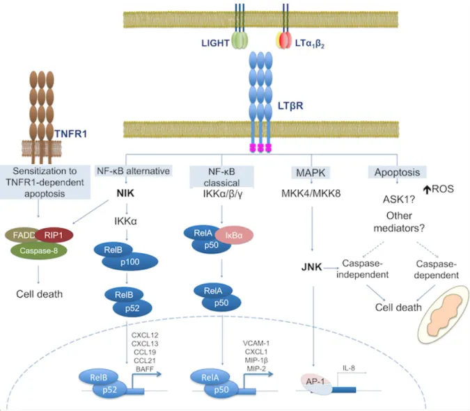
LTβR signaling is receptor aggregation. However, unlike other TNF receptors, each LTβR subunit can bind only two sites in the LTα1β2 heterotrimer, the LTα-LTβ (higher affinity) and LTβ-LTβ’ (lower affinity) interfaces for productive receptor activation [61]. Similarly, LIGHT has been shown to present only two high-affinity binding sites for LTβR [62]. Thus, the binding of LTα1β2 or LIGHT to LTβR brings two receptor molecules in close proximity (Figure 1C) [61,62] and the LTβR self-interaction region in the cytoplasmic domain promotes receptor aggregation and consequent conformational changes [24]. These events lead to the sequential recruitment of cytosolic adaptor proteins to the cytoplasmic region of LTβR, mainly TRAF proteins. These proteins may activate or repress signaling initiation leading to gene transcription through different signaling pathways such as the classical and the alternative NF-κB pathways, the c-Jun N-terminal kinase (JNK) MAP kinase pathway, and other signaling pathways leading to cell death [63] (Figure 2). LTβR-dependent downstream signaling can also be initiated independently of ligand binding either artificially by anti-LTβR agonistic antibodies that induce receptor aggregation [21,64], or pathologically by receptor overexpression leading to self-association [19,65,66].
Although LTβR activation has been reported to induce gene expression through ASK-MKK-JNK-dependent AP-1 activation [67,68] and LTβR interacts with the AP2 adaptor/clathrin complex to mediate unknown NF-κB-independent functions [19], cell death induction and NF-κB activation are the most studied events downstream LTβR. Despite lacking a cell death domain in its cytosolic domain, LTβR has been shown to induce death of cancer cell lines (e.g., HT-29, WiDr, Hep3BT2, and MCF-7) and to arrest tumor growth in cell line-derived xenograft models [69,70]. LTβR activation was shown to lead to cell death in the presence of IFN-γ [69] by either dependent (apoptosis) and/or caspase-independent (necroptosis/necrosis) mechanisms [20,71–74]. In addition, LTβR activation in combination with TNFR1 was proven essential to sensitize cortical thymic epithelial cells (cTECs) to TNFR1-mediated cell death [75,76]. The mechanism was shown to rely on NIK activation and on assembly of the RIP1/FADD/caspase8 death complex (Figure 2), but not on processing of p100 to p52, an essential step in the NF-κB alternative pathway [77]. Despite these findings, further research is warranted to fully understand the mechanisms of cell death induced by LTβR, which may depend on cell type, nature of the LTβR-activating stimulus and co-activation of other receptors.
Unlike the prototypical TNF receptors, which activate the classical but not the alternative NF-κB pathway (i.e., TNFR1), but like other TNFRSF members (e.g., BAFFR, CD40, CD27, Tweak, and CD30), LTβR binding by its ligands leads to both classical and alternative NF-κB pathway activation [19,78]. The activation of one or the other NF-κB signaling pathway is spatially and temporally regulated by LTβR trafficking [19] and varying levels of receptor cross-linking may be required for distinct conformational changes and
activation of different signal transduction pathways. Furthermore, the classical and the alternative NF-κB signaling pathways control distinct patterns of gene expression [78] and are therefore differentially involved in various functions attributed to LTβR signaling (Figure 2).
To activate the classical NF-κB signaling pathway, LTβR engagement leads to TRAF2 recruitment to its CD and subsequent IKK-mediated IκBα phosphorylation and degradation by the proteasome [79]. These events lead to p50-RelA heterodimer activation [78,80]. When upregulated, TRAF3 was shown to inhibit TRAF2 recruitment to LTβR, thus negatively regulating NF-κB activation [79]. When LIGHT or LTα1β2 accumulates at the surface of LTβR-inducing cells, higher-order clusters of LTβR may form on the target cell that seemingly trigger dynamin-2-dependent endocytosis of the receptor [19]. During this process, the LTβR CD was shown to remain exposed towards the cytosol and to compete with NIK for the binding of its inhibitory complex composed by TRAF3/TRAF2/cIAP1/cIAP2 [81,82]. As a consequence, the constitutive proteasomal degradation of NIK is alleviated, leading to NIK accumulation and activation of IKKα. These events lead to p100 processing to p52 and the translocation of p52/RelB dimers to the nucleus (Figure 2) [19,83]. LTβR-mediated activation of alternative NF-κB signaling is terminated by a mechanism of negative feedback control relying on IKKα-dependent destabilization of NIK [84]. Thus, TRAF3 inhibits NF-κB signaling by being part of a complex that mediates NIK targeting to proteasome degradation and, thus inhibits the processing of p100 to p52 [79,85]. Regarding kinetics, ligand binding to LTβR can induce a rapid and transient activation of the classical NF-κB pathway, followed by a delayed but sustained activation of the alternative pathway [78,80]. The delayed activation of the alternative pathway may be at least partially due to the requirement for increased Nfkb2 gene transcription (encoding p100), which is mediated by the IKKβ-dependent classical pathway [78,80]. Alternatively, it was proposed that LTβR activation induces the IKKα-dependent alternative pathway alone, resulting in p100 degradation and eventually activating RelA-containing and RelB-containing dimers [86]. Through the activation of p50/RelA heterodimers, LTβR signaling promotes for instance the upregulation of proinflammatory molecules including the CCL4/macrophage inflammatory protein (MIP)-1β, CXCL2/MIP-2 and vascular-cell adhesion molecule 1 (VCAM-1) in mouse embryonic fibroblasts (MEFs) [78], and CXCL1, CXCL2, intercellular adhesion molecule 1 (ICAM-1), VCAM-1, and E-selectin in endothelial cells [87]. Conversely, LTβR-mediated activation of p52/RelB heterodimers results in the production of lymphoid chemokines such as the CCL19/EBl1-ligand chemokine (ELC), CCL21/secondary lymphoid tissue chemokine (SLC), CXCL12/stromal cell-derived factor-1α (SDF-1α), CXCL13/B lymphocyte chemoattractant (BLC) and the cytokine B cell activation factor (BAFF), being all involved in lymphoid organogenesis and homeostasis [26,78].
4. Physiological roles of lymphotoxin signaling
LTα1β2/LIGHT-induced LTβR signaling is critically involved in lymphoid organogenesis and maintenance of secondary lymphoid structures, in addition to its roles in regulation of innate and adaptive immune response, inflammation, and tissue homeostasis.
Lymphoid organogenesis is largely associated with LTβR signaling induced by the LTα1β2 heterotrimer, as shown by studies blocking ligand-receptor interaction [88,89] or using LTβR, LTα, LTβ or LIGHT knockout mice [1–3,90]. LTβRknockout mice lack several secondary lymphoid organs, including peripheral and mesenteric lymph nodes (LNs), Peyer´s patches and gut-associated lymphoid tissues (GALT) [1]. LTα knockout mice generally lack peripheral and mesenteric LNs and Peyer´s patches, although mesenteric lymphoid aggregates were observed in a few mice [3,91]. LTβ knockout mice lack most LNs, but in contrast to LTα and LTβR-deficient mice conserved fully organized mesenteric LNs and cervical lymph node-like structures [2,92]. Mesenteric LN development was impaired by simultaneous LTβ and LIGHT inactivation, meaning that LIGHT can compensate for LTβ absence in mesenteric LN development [90]. In addition, LTα-, LTβ- and LTβR-deficient mice, but not LIGHT-deficient mice presented splenic structural defects. Discrepancies in the effects of ligand-receptor gene inactivation led to the supposition, yet to be confirmed, that either an alternative unknown ligand for LTβR or other nonspecific interactions could account for such phenotypic differences [1–3].
In the adult, LTβR signaling was reported to be critically involved in the adaptive immune response against pathogens due to its intervention in processes such as DC homeostasis and expansion [93,94], and lymphocyte maturation and survival [95–98]. Furthermore, its activation is continuously required for the maintenance of the integrity and organization of microenvironments from secondary lymphoid organs [1,88,89]. For example, LTβR is important for the development and structural maintenance of fibroblastic reticular cells (FRCs) in LNs and spleen [99,100]. In the spleen, LTβR activation was also shown to be essential for FDC differentiation [101]. Accordingly, LTβR-deficient mice present disrupted FDC and germinal center formation and, consequently deficient B cell affinity maturation [1]. LTβR signaling is also important for the trafficking of lymphoid and other hematopoietic cells, namely the recruitment, migration and organization inside organs, and the migration to other tissues [4,26,42,102]. Moreover, it is involved in the regulation of acute inflammatory reactions and in the development of inflammation-associated ectopic lymphoid structures [41,103]. In the latter process, LTβR-dependent stromal cell differentiation into reticular networks and induction of chemokines, cytokines and adhesion molecules play a
critical role. Finally, LTβR activation favors the recruitment of hematopoietic cells to lymphoid compartments by instructing the development and function of high endothelial venules (HEVs) [7,104].
Importantly, LTβR signaling leading to NIK/IKKα-dependent alternative NF-κB activation has been shown to be a key player for thymic medullary epithelial cell differentiation [105] and the maintenance of the thymic structure [4], considered essential for central tolerance induction. In this context, T cell development and selection, and the maintenance of the thymic microenvironments require reciprocal interactions between thymocytes and stromal cells where LTβR signaling is a critical mediator of this thymic crosstalk [4]. In addition, cTEC cell death mediated by LTβR and TNFR1 combined and NIK activation was proven essential for thymic involution in pathological conditions [75–77].
Although LTβR and its ligands are widely recognized as key players in immunity, they are also involved in many other biological processes such as liver regeneration [5,106], hepatic lipid metabolism [6], and adipocyte differentiation [107]. Importantly, LTβR signaling has also been reported to be involved not only in cell death and tumor growth inhibition, but also in cancer development and progression [15,16].
5. LTβR suppressor functions in solid tumors
5.1. LTβR activation leading to cancer cell deathThe lymphotoxin designation was first attributed upon LTα identification as a cytokine similar to TNFα that presented cytolytic/cytostatic effects on target cells [108]. Indeed, LTβR activation was first shown to mediate cytotoxic effects in tumors, thus pointing to a potential anti-cancer therapy, especially because this receptor was found to be expressed in a wide range of tumor types [65,70,73].
The direct anti-cell growth role of LTβR has been demonstrated in a subset of human epithelial cancer cell lines (e.g., HT-29, WiDr, Hep3BT2, MCF-7, and HeLa), where LTβR activation was shown to induce death with slow kinetics (36-72 h) either in the presence of IFN-γ [69,71] or through LTβR ligand-independent self-association caused by overexpression [66]. Furthermore, LTβR activation was reported to arrest tumor growth in mice xenografted with colorectal cancer cell lines and patient samples [69,70]. The molecular mechanism by which LTβR contributes to cancer cell line death has however remained elusive.
To study LTβR-induced anti-growth effects in cancer, Hu and coworkers used a lung experimental metastasis model in which mouse colon carcinoma cells were injected i.v. into BALB/c mice and found that CD11b+ myeloid cells, NK cells, and CD8+ and CD4+ T cells
collected from lung metastases expressed LTα1β2 and LIGHT [73]. This observation supported a previous report indicating that monoclonal antibody (mAb)-mediated LTβR activation in established CT26 cell line-derived subcutaneous tumors led to both T cell infiltration, probably mediated by pro-inflammatory chemokines, and tumor necrosis [70]. Supporting the notion that immune cells interact with tumor cells through LTβR to suppress spontaneous tumor development, recombinant LTα1β2 and LIGHT proteins or an agonist LTβR mAb could inhibit in vitro growth of human colon carcinoma and soft tissue sarcoma cell lines [73]. Likewise, using a syngeneic mouse model of sarcoma metastasis to the lung together with adoptive transfer of tumor-specific cytotoxic T lymphocytes (CTL), Yang and co-workers previously showed that LTβR was a direct effector of CTL-mediated tumor rejection in vivo [109]. Regarding the mechanism, LTβR stimulation by an agonistic mAb induced caspase- and mitochondrial-dependent apoptosis and activated classical and alternative NF-κB pathways in human cancer cell lines [73]. Furthermore, NF-κB inhibition promoted CT26 colon cancer cell metastatic potential in vivo, suggesting that in this context LTβR-mediated apoptosis and activation of the NF-κB signaling pathway might act in concert to suppress tumor development [73].
It has been suggested that LTα1β2 and LIGHT ligand expression by immune cells such as T cells [109], NK cells [110] or DCs [111] may engage LTβR on tumor cells and thus trigger antitumor cytotoxicity. Yet, tumor cell death in these studies was induced by recombinant ligands and/or LTβR agonistic antibodies, which may not reflect the physiological levels and activity of ligands expressed at the surface of immune cells. This caveat is underscored by results showing that LTβR activation and downstream signaling pathways induced in vitro by recombinant ligands or agonistic antibodies may depend on the duration and degree of receptor oligomerization [64]. Nevertheless, LTβR-mediated tumor suppression by either agonistic mAbs [69,70] or adoptively transferred tumor-specific CTLs [109] was put forward as a therapeutic approach to halt tumor growth and to override colon carcinoma and soft tissue sarcoma chemo- and radiotherapy resistance [70,73].
5.2. LTβR or HVEM activation leading to immune-mediated tumor rejection
5.2.1. LIGHT-induced recruitment and activation of anti-tumoral lymphocytes
Rather than identifying a direct effect of LTβR signaling in tumor regression, Winter and coworkers found that LTβR-mediated tumor regression could occur through an indirect pathway [112]. These authors used an experimental pulmonary metastasis model generated by intravenous injection of the D5 melanoma cell line (a B16 cell line subclone) in syngeneic mice, and found that infiltrating effector T cells, which expressed LTβR ligands, activated LTβR but did not induce apoptosis of D5 tumor cells in vitro. Instead, LTβR activation in D5 melanoma cells induced the secretion of chemokines that mediate macrophage migration
[112]. Although, direct anti-tumor effects could not be excluded, this report indicates that LTβR activation by LTα1β2 and/or LIGHT is involved in the induction of chemotactic molecules that create a tumor microenvironment favorable for lymphocyte homing, which in turn may boost anti-tumor immunity and contribute to tumor suppression. Also in this context, Yu and colleagues disclosed a role for LTβR signaling in tumor immune rejection [113]. LIGHT overexpression in a fibrosarcoma cell line that was then subcutaneously inoculated in C3B6F1 mice induced LTβR-mediated CCL21 and MAdCAM-1 expression in tumor microenvironmental cells. This in turn led to CD8 naïve T cell infiltration and activation, leading to the rejection of the established tumor. Furthermore, direct inoculation of LIGHT-expressing tumor cells in established non-LIGHT-expressing primary tumors led to their regression. Primary tumor rejection was also achieved when LIGHT-expressing tumor cells were inoculated in another subcutaneous site, indicating that LIGHT can generate a systemic immune response against distal tumors. These data support the rationale of using LIGHT-expressing tumor vaccines as a therapeutic tool [113]. In this line, other researchers genetically engineered attenuated Salmonella to express LIGHT and used it as a targeting vehicle for local expression of LIGHT in tumors. This approach led to LTβR and HVEM-dependent inhibition of both primary and metastatic tumor growth in subcutaneously injected syngeneic immunocompetent mice [114]. LIGHT expression induced both T and B lymphocyte infiltration and production of the CXCL9 chemoattractant in subcutaneous tumors, but it remained to be established whether these two effects were causatively linked [114]. LIGHT expression was also found to be frequent in patient-derived metastatic melanoma cells and in melanoma cell line-derived microvesicles, and to be correlated with T-cell infiltration [115]. In addition, another approach based on LIGHT-expressing adenovirus was tested for local tumor treatment. These viruses initiated priming of tumor-specific CD8+ T cells directly in the primary tumor, followed by the exit of CTLs, which homed to distal tumors to elicit immune-mediated eradication of spontaneous metastases [116]. Several studies therefore indicate that LIGHT is a potent primer of T-cell responses that can counter tumor growth and that it can be used as a therapeutic tool.
5.2.2. LTβR-mediated HEV differentiation and recruitment of anti-tumoral lymphocytes In addition to its role in chemokine production and chemoattraction, LTβR activation was shown to correlate with lymphocyte extravasation through HEVs and tumor infiltration, thus leading to tumor regression [117,118]. HEVs are specialized postcapillary vessels of secondary lymphoid organs, also found in chronically inflamed non-lymphoid tissues [119] and tumors [120]. These vessels mediate the extravasation of naïve and central memory lymphocytes from the peripheral blood to lymphoid tissues to initiate immune responses [121], and express LTβR, which is required for HEV differentiation and function [7]. In this
context, Martinet and coworkers have recently found that in human breast cancer, higher numbers of LTα1β2-expressing DCs were correlated with increased HEV density and T and B lymphocyte infiltration. Moreover, LTβ expression correlated with expression of chemokines associated with HEV-mediated lymphocyte extravasation (CCL19, CCL21 and CXCL13) [118]. Interestingly, these authors showed that the tumor HEV density was inversely correlated with breast cancer progression, from in situ ductal carcinoma to invasive ductal carcinoma, and found that high density of HEVs in breast tumors was correlated with a favorable prognosis [118]. These findings contradict the generally accepted assumption that tumor angiogenesis correlates with tumor progression and worse prognosis, and highlight the notion that different types of tumor blood vessels play distinct roles. A similar mechanism was also found in a mouse model of methylcholanthrene-induced fibrosarcoma, in which depletion of T regulatory cells (Tregs) led to HEV development, T-cell infiltration, LTα and LTβ upregulation and decreased tumor growth [117].
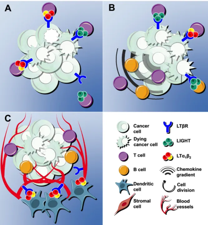
In summary, LTβR can mediate anti-tumor effects by direct cytotoxicity (Figure 3A) but also by other indirect mechanisms, like tumor cell sensitization to chemotherapeutic agents and radiation [70]. Furthermore, LTβR can stimulate host-mediated anti-tumor immune responses either by inducing the expression of pro-inflammatory cytokines and chemokines that chemoattract and activate lymphocytes [70,112] (Figure 3B), or by inducing the differentiation of HEVs that mediate lymphocyte trafficking to both normal organs and tumors [7,118] (Figure 3C) .
6. LTβR-mediated promotion of solid tumors
In contrast to the previously discussed anti-cancer roles of LTβR, a tumor-promoting role for this receptor has been disclosed in a wide variety of contexts. Cancer cells from different origins express LTβR [65,70,73], being often this expression increasingly more prevalent with cancer progression and metastasis [65,73,122]. Furthermore, LTBR gene upregulation or structural alterations leading to LTβR constitutive activation were reported to correlate with carcinogenesis [65,122–124] (Figure 4A). As shown below, LTβR is thought to promote oncogenesis either by directly fostering survival and/or proliferation of malignant cells or by generating a pro-tumorigenic inflammatory microenvironment.
6.1. LTBR genetic alterations leading toLTβR constitutive activation
An early study reporting an LTβR pro-tumorigenic role identified an NH2-terminally truncated form of LTβR in a pancreatic ductal carcinoma cell line. This truncated receptor as well as the full-length LTβR protein were shown to have fibroblast transforming activity in vitro and in
region, including the LTBR locus, was found to be in higher copy number in 51% and amplified in 7% of nasopharyngeal carcinoma (NPC) cases [122]. Additionally, LTβR protein was found to be frequently overexpressed in NPC tumors. Subsequently, LTβR overexpression in an immortalized nasopharyngeal epithelial cell line was shown to contribute to ligand-independent cell proliferation. Importantly, LTβR knockdown inhibited in
vivo tumor growth in an NPC xenograft mouse model [122]. Since LTβR stimulation
activated NF-κB in nasopharyngeal cells [122], the same authors showed that in cases without evident LTBR amplification genetic alterations affecting other NF-κB signaling regulators (TRAF3, TRAF2, NFKBIA, and A20/TNFAIP3) were present [125]. These results therefore support a role for LTβR-mediated NF-κB activation in NPC development.
The oncogenic potential of LTβR has also been reported in melanoma. Dhawan and co-workers have shown that LTβR expression is upregulated in human metastatic melanoma samples when compared to normal melanocytes and other melanoma lesions. In melanoma cell lines, LTβR activates the NF-κB pathway and induces cell proliferation and invasiveness, all in a ligand-independent manner [65]. These findings suggest that, like in pancreatic cancer, the elevated expression of LTβR in melanoma is by itself sufficient to drive cancer progression.
6.2. Ligand-dependent activation of LTβR in cancer development
Despite reports indicating that LTβR signaling can be activated in the absence of ligands, other studies have shown that these may play important roles in promoting cancer. Genetic studies in humans identified single nucleotide polymorphisms (SNPs) in the LTα gene that may be either cancer-protective or lead to an increased cancer risk. For example, one common SNP, LTA +252A>G or rs909253, was described in meta-analysis studies to be positively associated with cancer susceptibility to different types of cancer [126,127]. Such susceptibility was also found for specific cancer types, such as non-Hodgkin lymphoma [128,129], breast cancer [130,131] and gastric cancer [132,133]. Despite conflicting data on the association between LTA gene polymorphisms and risk for different types of cancer in different ethnic populations, and on whether the polymorphic allele is present in homozygosity or heterozygosity, it was reported that different LTA alleles may result in differential gene transcription and protein expression [134,135]. Since LTα plays a key role in immunity and inflammation [136], alterations in its production may affect anti-cancer immunity and inflammation-induced cancer. Yet, the exact mechanism by which it affects cancer risk in each context remains to be defined. Furthermore, the involved LTα-containing ligand, either LTα3 homotrimer or LTα1β2 heterotrimer, was not determined by these studies.
6.3. LTβR pro-oncogenic roles mediated by interactions with the tumor microenvironment
Immune cells are the main source of LTβR ligands and the interaction of these cells with tumor cells can either restrain, as discussed above, or promote tumor progression. Tumor and/or stromal cells respond to injury, infection and tissue stress by producing cytokines and chemokines that attract immune cells [137]. As a result, these cells migrate to the tumor microenvironment where they secrete inflammatory, pro-angiogenic and pro-tumorigenic factors that may affect tumor progression and metastasis. Thus, depending on the tumor microenvironment chemokine milieu, tumor-infiltrating immune cells can stimulate the immune response against tumor cells or rather help these to subvert the immune response and promote oncogenesis. As a signaling axis involved in immune cell communication, in addition to its involvement in the induction of tumor-suppressive microenvironments, as discussed above, LTβR signaling can also contribute for the induction of pro-oncogenic, inflammatory microenvironments. A wide range of studies have shown that inflammation can promote tumorigenesis by promoting angiogenesis, release of growth and survival factors, invasiveness, metastasis and evasion of host defense mechanisms [138].
6.3.1. LTβR-induced angiogenesis
The importance of angiogenesis for the growth of solid tumors has since long been recognized. As tumor growth and metastasis require persistent new blood vessel formation, a developing tumor shifts from the avascular phase to the angiogenic phase, the so-called angiogenic switch [139]. This switch is controlled by a balance between pro- and anti-angiogenic factors, which are secreted by the tumor cells themselves or by cells in the tumor microenvironment, in particular resident stromal cells and immune cells. It is known that the expression of pro- and anti-angiogenic factors by cancer cells can be controlled either directly by oncogenes, tumor suppressor genes and transcription factors or indirectly by extrinsic factors. Yet, the roles and the interplay among the various inflammatory cytokines and chemokines in the angiogenic switch are still poorly understood.
In this context, Hehlgans and co-workers have shown that inhibition of LTβR signaling can block angiogenesis and tumor growth [140,141]. Using methylcholanthrene-induced murine fibrosarcoma BFS-1 cells, these authors have shown that LTβR activation by LTα1β2- or LIGHT-expressing T and B lymphocytes induced the expression of the angiogenic mediator CXCL2 [140]. CXCL2 induction in BFS-1 cells depended on NF-κB activation and contributed for solid tumor growth in vivo. The described pro-tumorigenic effect was assumed to be due to the modulation of the tumor microenvironment through LTβR-mediated angiogenesis induction (Figure 4B) because LTβR inhibition blocked BFS-1 tumor angiogenesis while direct LTβR stimulation (with an agonistic anti-LTβR monoclonal antibody) did not increase proliferation or survival of fibrosarcoma cells [141].
6.3.2. LTβR-induced chronic inflammation
Tumors often arise in sites of chronic inflammation [142], which provide a microenvironment containing various mediators (e.g., cytokines, chemokines, and prostaglandins) with tumor-promoting properties, including enhanced cell proliferation, survival, angiogenesis and migration. In this context, Haybaeck and co-workers have found the involvement of LTβR signaling in the development of virus-induced chronic hepatitis and hepatocellular carcinoma (HCC) [13]. In hepatic primary tissue from hepatitis B or C (HBV- or HCV)-induced chronic hepatitis and HCC patients, these authors found upregulation of not only LTβR and its ligands (LTα, LTβ and LIGHT) but also pro-inflammatory chemokines (CCL2, CCL3 and CXCL10). LTBR was highly expressed in liver cell populations depleted of hematopoietic (CD45-positive) cells, while LTA, LTB and LIGHT were expressed both in hematopoietic and non-hematopoietic HCV-induced hepatitis and HCC liver cell fractions. Furthermore, expression of LTBR, LTA, LTB, LIGHT and inflammatory chemokines in a human hepatocyte cell line Huh-7.5 was shown to be directly linked to the presence of HCV infection. In transgenic mice expressing high levels of LTα and LTβ in a liver-specific manner, LTβR signaling induced chronic hepatitis characterized by inflammation, T and B lymphocytic infiltrates and hepatocyte apoptosis. Further experiments demonstrated that T and B cells, which express LTβR ligands, and LTβR-mediated canonical NF-κB signaling activation in hepatocytes were both required for LTβR-induced chronic hepatitis and HCC development [13]. These findings indicate that persistent lymphocyte-derived LTα1β2 and LTβR-induced NF-κB activation are tumor-promoting, and that rather than having direct oncogenic properties, LTβR signaling reshapes and generates an inflammatory, oncogenic hepatic microenvironment (Figure 4C). Interestingly, it was recently reported that short-term LTβR stimulation led to degradation of HBV-derived covalently closed circular DNA (cccDNA) in infected hepatocytes [143]. This anti-HBV effect was shown to be mediated by LTβR-induced APOBEC3B deaminase expression and indicates that LTβR agonists could be incorporated in anti-HBV combined therapeutic regimens [143]. Importantly, these data suggest that in contrast to the HCC-causing inflammation-related persistent LTβR stimulation, transient stimulation may actually prevent HBV-induced HCC.
Supporting the aforementioned studies on hepatitis and HCC [13], Simonin et al. (2013) have shown in a recent report that LTβ expression can be induced by the HCV NS5B polymerase in a human hepatoma cell line. Using transgenic mice with hepatocyte-targeted expression of the entire ORF of the genotype 1b HCV, Simonin and co-workers have also shown that LTβ hepatocyte expression in HCV transgenic liver tumors was associated with NF-κB activation, chemokine synthesis and intra-tumoral recruitment of macrophages and T and B lymphocytes [43]. In addition to these studies on viral-induced HCC, LTβR was shown
to be also involved in the pathogenesis of non-viral HCC. Using a mouse model of long-term choline-deficient high-fat diet, Wolf et al. (2014) identified CD8+ T cells and NKT cells recruited to the liver as key players in the development of steatosis and HCC. These cells were shown to interact with hepatocytes leading to their activation and to the release of soluble factors such as LIGHT and lymphotoxin. In addition, LTβR and classical NF-κB signaling were shown to be activated in hepatocytes, thus facilitating liver tumorigenesis [144]. More recently, LTβR signaling was found to participate in oncogene-driven HCC progression [145]. In an HCC mouse model initiated by constitutively active Akt (in combination with mutated β-catenin or Notch1), LTβ and LTβR expression were found to be upregulated in liver tumors. More importantly, blockade of LTβR signaling reduced tumor progression and prolonged mouse survival [145]. Together, these reports demonstrate that independently of the causing agent, LTβR persistent signaling in the context of chronic inflammation promotes HCC progression, and may be a potential therapeutic target.
Cancer therapy-induced cell death can also elicit an inflammatory response that may contribute to therapeutic resistance. This is the case of castration-resistant metastatic prostate carcinoma, the emergence of which constitutes a major complication limiting the success of androgen ablation therapy and underlying most prostate cancer-associated mortality. Using two animal models, the SV40 large T antigen-driven transgenic adenocarcinoma mouse prostate (TRAMP) cancer model and the mouse androgen-dependent CaP prostate cancer cell line subcutaneously allografted in castrated FVB mice, Ammirante et al. (2010) unveiled a mechanism underlying the emergence of castration-resistant prostate cancer. These researchers found that following androgen ablation therapies, the death of androgen-deprived primary cancer cells induced an inflammatory response with concomitant production of CXCL13 and other inflammatory chemokines, and recruitment of leukocytes, mostly B cells, into the regressing tumor. IKKβ activation in B cells, presumably by inflammatory cytokines, induced the expression of surface LTα1β2 in these cells. These LTα1β2-expressing B cells led to LTβR activation and IKKα nuclear translocation in prostate cancer cells to promote androgen-independent growth and survival [146].
A more recent study has shown that the endogenous “danger signal” HMGB1 protein was induced during prostate tumor progression in TRAMP mice, and that it was required for the infiltration and activation of T cells (but not B cells) within the tumor [147]. Prostate tumor-infiltrating T cells were shown to express LTα1β2 and, through LTβR activation in stromal cells, to promote the recruitment of tumor macrophages, presumably by inducing CCL2 expression. More importantly, LTβR signaling was shown to facilitate progression from hyperplasia to invasive prostate carcinoma [147]. Considering these findings with those obtained by Ammirante et al. (2010), it can be concluded that LTβR signaling may contribute
to several phases of prostate oncogenesis, through different molecular mechanisms (CXCL13 or CCL2 production) and involving different cellular players (T or B lymphocytes), and may therefore be of therapeutic value.
6.3.3. Induction of a pro-tumorigenic niche supported by LTβR-expressing stromal cells LTβR signaling has been implicated in other epithelial cancers, as for example ovarian cancer [148]. Lau and co-workers detected LTA and LTB overexpression in ovarian cancer cells and demonstrated that LTα1β2-expressing human ovarian primary cancer cells induce LTβR-expressing cancer-associated fibroblasts (CAFs) to express chemokines through NF-κB signaling. One of the chemokines identified as being induced in CAFs was CXCL11, which was able to promote proliferation and migration of CXCR3-expressing ovarian cancer cells [148]. Thus, in this setting cancer cells generate a pro-tumorigenic microenvironment through increased lymphotoxin expression and LTβR activation in stromal cells.
6.3.4. Immune evasion mediated by LTβR
Another way by which LTβR signaling is involved in cancer promotion is by dampening the host adaptive immune response to cancer. Because LTα1β2-LTβR signaling plays a role in immune self-tolerance due to its key role in medullary thymic epithelial cell development and function [4], blocking this signaling axis may rescue tumor-reactive effector T cells from thymic clonal deletion and thus counter cancer development [149]. To test this hypothesis, Zhou et al. (2009) used the TRAMP animal model co-expressing a TCR specific for SV40 large T antigen. Targeted mutation of the Lta gene was found to impair thymic negative selection of tumor-reactive T cells, resulting in decreased prostate cancer incidence and in milder malignant phenotype. Confirming the impact of LTβR signaling in prostate oncogenesis, short-term LTβR blockade in TRAMP mice rescued T cells from clonal deletion, reduced the progression of primary prostate cancer and prevented metastasis [150]. This study thus suggests that LTβR signaling may constitute a non-antigen-based strategy of immune cancer prevention potentially useful for patients with high genetic risk for prostate cancer. Another report has highlighted an alternative role for LTβR in tumor immunoevasion. Kim and co-workers showed that the human papillomavirus 16 (HPV16) E6 oncogene induced LTα, LTβ and LTβR expression in cervical cancer cell lines [44]. More importantly, LTβR signaling led to MHC class I downregulation in these cells and to resistance to cytotoxic T lymphocyte-mediated lytic activity [44]. Whether such mechanism of cancer cell evasion from the host immune system takes place in vivo and results in tumor progression remains to be determined.
7. LTβR role in hematological malignancies
Several reports indicate that hematological malignancies are fostered by LTβR signaling, either intrinsically to cancer cells or indirectly through microenvironmental cells. Studies aiming to identify genetic abnormalities underlying multiple myeloma pathogenesis identified alterations (e.g., deletions, amplifications, and point mutations) in several NF-κB regulators, in about 15% of patient samples and 30-40% of cell lines [124,151]. Such alterations included LTBR amplification in one patient sample and one cell line [124]. Despite the low frequency of abnormalities in LTBR and other functionally related genes, these studies indicated that constitutive activation of the LTβR-activated noncanonical NF-κB pathway promotes multiple myeloma oncogenesis [124].
LTα1β2-LTβR signaling has also been shown to mediate paracrine or juxtacrine tumor-stroma interactions leading to microenvironment modulation and establishment of chemoattractive tumor-permissive niches in secondary lymphoid organs (Figure 4D). Rehm and co-workers identified the homeostatic chemokine receptor CCR7 as a determinant factor in dictating the location and survival of B-cell lymphoma cells within secondary lymphoid organs [152]. Using the Eµ-Myc transgenic mouse model of aggressive human B-cell lymphoma, these researchers found that CCR7 controls lymphoma B-cell dissemination to LNs and to the splenic T-cell zone where, through LTα1β2 expression, cancer cells stimulate LTβR in gp38+ FRCs. This molecular crosstalk results in the expansion of stromal FRC networks and release of chemoattractant homeostatic chemokines (e.g., the CCR7 ligands, CCL19 and CCL21) and trophic factors (e.g., IHH/indian hedgehog) that confer a survival advantage to lymphoma cells [152]. More recently, these authors used the murine Eµ-Tcl1 model of B-cell chronic lymphocytic leukemia to show that the CXCL13-CXCR5 signaling axis mediates leukemic B cell access to a stromal compartment enriched with FDCs in splenic B cell follicles [153]. Here, leukemic B cells and FDCs engage in a reciprocal crosstalk in which LTα1β2-expressing leukemic cells activate LTβR and thus stimulate the differentiation of FDC networks and the production of CXCL13, CCL21, and other pro-proliferative and pro-survival growth factors [153]. In both studies, the inhibition of LTβR-mediated interactions between malignant and microenvironmental cells impaired disease progression and was therefore pointed as a possible strategy to complement standard cytotoxic therapies [152,153]. Recently, high expression of LTα and LTβ-encoding genes was identified in human primary T-cell acute lymphoblastic leukemia expressing TAL or LMO oncogenes (TAL/LMO molecular subtype) [154]. Highlighting the relevance of these findings, LTβR activation in thymic stromal cells was shown to promote T-cell leukemogenesis in a mouse model of T-cell acute leukemia/lymphoma [154]. Leukemic cells from these mice
were shown to express high levels of LTα and LTβ, from an early stage, and importantly, both early appearance of malignant cells and mouse survival were delayed in the absence of stromal LTβR. Since stromal cells dependent on RelB expression were shown be involved in mouse leukemogenesis [155], these studies support the notion that LTβR activation in stromal cells promotes T-cell leukemogenesis through NF-κB activation.
8. Signaling pathways mediating LTβR activity in cancer
As highlighted in the above sections, the classical or alternative NF-κB pathways appear to be the main mediators of most cellular events stemming from LTβR signaling that contribute to its pro- and anti-oncogenic effects. However, a number of reports suggest that this is not always the case. In fact, some anti-oncogenic effects of LTβR signaling leading to cancer cell death were reported to be mediated by other downstream components such as the reactive oxygen species-induced apoptosis signal-regulating kinase (ASK1) [72] and caspases (e.g., caspase 3 and 8) [66,71,73]. On the other hand, only few pro-tumorigenic effects of LTβR signaling were found to result from activation of mediators other than NF-κB. Ammirante et al. (2010) reported that LTβR activation in prostate carcinoma cells by lymphotoxin expressed on B cells infiltrating regressing tumors after castration was required for IKKα translocation to the nucleus and STAT3 activation, nevertheless a collaboration with another unidentified critical cytokine/receptor activating STAT3 was predicted [146]. Although JNK has been shown to be activated by LTβR (Figure 2) and to be implicated in cancer, in promoting or suppressing it [156], no report has so far addressed whether this kinase is involved in cancer-related LTβR activity.
9. Signaling pathways with context-dependent outcomes in carcinogenesis
Taken together, the aforementioned reports demonstrate the dual role of LTα1β2 /LIGHT-LTβR signaling axis in cancer development. These proteins are not unique in that, other signaling proteins, such as tumor necrosis factor alpha (TNFα), transforming growth factor beta (TGFβ), NOTCH1 and NF-κB, share this context-dependent role in oncogenesis.
In accordance with its designation, TNF has been shown to induce apoptosis or necrosis in a variety of cancer cell types. TNF was shown to kill directly cancer cells [157], but its anti-oncogenic effects seem to involve mainly damage to the tumor vasculature through endothelial cell apoptosis [158,159] and the stimulation of anti-tumoral immune responses [160–162]. In contrast to these findings, higher levels of TNFα were detected in
the serum of cancer patients and in pre-neoplastic and tumor tissues, being associated with tumor progression [163–165]. Accordingly, in many studies, TNF was reported to prompt a broad range of pro-carcinogenic signaling mechanisms leading to tumor initiation and promotion (often in the context of chronic inflammation) including survival, proliferation, angiogenesis, invasion, and metastatic dissemination of cancer cells [166–170]. These contradictory roles in carcinogenesis seem to be associated with different tumor types and cellular contexts, and can be partly explained by levels of TNF production, chronic low doses leading to cancer development and progression and acute high doses leading to tumor regression [171].
TGFβ signaling is known to play dual roles in cancer [172–174]. In early stages of carcinogenesis, TGFβ mediates tumor-suppressing effects through cell-autonomous mechanisms, including suppression of cell proliferation and induction of apoptosis [173,175]. Supporting this tumor suppressive role of TGFβ signaling, genetic and epigenetic alterations attenuating or inactivating TGFβ receptors and downstream signaling components were reported in diverse types of cancer (reviewed in [175]). TGFβ was also shown to suppress oncogenesis indirectly by preventing the molecular crosstalk between TGFβ receptor-expressing stromal cells and cancer cells [176]. On the other hand, TGFβ can also promote tumor cell growth, invasiveness and metastasis in advanced tumors. Throughout tumor progression cancer cells dampen the growth-inhibitory TGFβ response, while its production increases in the tumor microenvironment [177]. As a consequence, by mechanisms such as increased chemokine expression and inflammation, immune response evasion, sustained angiogenesis, and epithelial-mesenchymal transitions (EMT) TGFβ leads to enhanced invasiveness and metastasis [177–180]. Therefore, the role played by TGFβ signaling likely depends on cancer type and cellular context. However, unlike LTβR signaling, TGFβ tumor-suppressing or -promoting effects appear to relyon the stage of tumor development.
Notch signaling was also reported to mediate contradictory effects on oncogenesis. Activating mutations were identified in NOTCH1 and NOTCH2 genes in hematological malignancies (T-cell acute lymphoblastic leukemia, chronic lymphocytic leukemia, mantle cell lymphoma, and marginal cell lymphoma) and in breast adenocarcinoma [181–186]. Although the mechanisms are not fully understood, Myc induction seems to be a common downstream target in these different tumor contexts [187]. More recently, evidence was gathered indicating that NOTCH1 and NOTCH2 can also act as a tumor-suppressor gene in malignancies where inactivating mutations were detected. These included squamous cell carcinomas from skin, head and neck and lung [188–190] and chronic myelomonocytic leukemia [191]. In addition, NOTCH1 protein expression was found to mediate acute myeloid leukemia growth arrest and apoptosis [192,193]. The mechanisms remain to be identified but likely involve the resulting impaired activation of targets mediating
pro-differentiation and anti-growth effects, and the promotion of an inflammatory state caused by Notch loss-of-function [187,194].
Interestingly, the main signaling pathway downstream LTβR activation, that leading to NF-κB activation, has also been recognized to have opposing effects in cancer development. Although mutations affecting NF-κB and inhibitors of IκB kinase β (IKK) are rarely found in human cancer, NF-κB subunits are frequently activated, resulting from either the induction of upstream pathways or loss of negative feedback mechanisms. Regardless of the causes of NF-κB aberrant activation, these transcription factors play prominent tumor-promoting roles, intrinsic, by rendering cancer cells resistant to apoptosis and/or highly proliferative, and extrinsic, by stimulating neoangiogenesis and inducing pro-invasive/pro-metastatic inflammatory microenvironments [195]. Contrasting with a large body evidence supporting their pro-oncogenic action, some reports have revealed an unexpected tumor suppressor role for NF-κB proteins in essentially two types of scenario. First, NF-κB exhibits tumor suppressor activity when acting in concert with well-characterized tumor suppressors, like p53 and ARF. These tumor suppressors bind NF-κB subunits to repress the potentially tumorigenic genes normally induced by NF-κB activation, most likely in an early stage of cancer development before cancer cells undergo loss of the implicated tumor-suppressor genes [196,197]. Second, in contexts where pro-survival signals derive from other oncogenes, NF-κB activation may enhance cytotoxic drug-mediated senescence in tumors, thereby exerting a tumor suppressor function [198,199]. Therefore, the NF-κB role in carcinogenesis is highly dependent on the tumor stage, tumor type, and presence of specific genetic alterations.
10. Conclusions
Since the discovery of the lymphotoxin signaling system, several researchers have investigated its role in cancer, including solid and hematological malignancies. As discussed in this review, early studies have uncovered a potential anti-tumoral role in several cancer types (Table 1). LTα1β2- and/or LIGHT-induced activation of LTβR in a subset of solid cancers was reported to promote direct cytotoxic effects (Figure 3A) and/or indirect effects involving alterations in the tumor microenvironment (e.g., induction of chemokine expression and development of HEV), which lead to increased anti-tumoral immune response (Figure 3B,C). These reports disclosed a role for acute LTβR activation in anti-cancer immunity and so this was suggested as a potential therapeutic approach. Conversely, during the last decade, several studies provided firm evidence that LTβR signaling can promote both solid
and hematological malignancy carcinogenesis. In some instances pro-oncogenic LTβR signaling is intrinsic to cancer cells, in others it acts in tumor-promoting microenvironmental cells (Table 2). In the first setting, LTβR signaling can be activated either independently of ligand binding due to LTBR gene amplification or other molecular events leading to LTβR overexpression (Figure 4A), or by increased expression of LTα1β2 and/or LIGHT in the microenvironment (Figure 4B and C). In the latter situation, LTβR signaling in cancer cells leads to the secretion of factors (e.g., homeostatic chemokines and cytokines) that stimulate angiogenesis (Figure 4B) and/or attract infiltrating tumor-promoting immune cells (Figure 4C) thus stimulating cancer progression. Finally, in the setting where LTα1β2-expressing cancer cells activate LTβR in stromal cells, the latter can secrete chemokines or potentially other factors that favor cancer progression (Figure 4D). The role of infiltrating immune cells is rather complex, since in some contexts these can impair tumor progression through induction of host-mediated immunological responses as discussed above, while in other contexts they support tumor development by upregulating pro-inflammatory cytokines and by modulating the microenvironment. The balance between suppressing and tumor-promoting immune cell activity most likely depends on tumor stage, on the nature of recruited cells and on the type of factors produced by the tumor microenvironment.
Altogether, the reports previously cited have disclosed several factors influencing the pro- or anti-oncogenic activities of LTβR signaling. Several variables such as the tumor type, the progression stage, the cancer-intrinsic genetic and epigenetic alterations, the status of activated signaling pathways, the microenvironmental factors, and the experimental model used may ultimately determine if the overall effect of LTβR activation is pro- or anti-tumorigenic. Moreover, the mechanisms by which LTβR may foster or counter tumor progression are not completely understood. Nevertheless, the classical and alternative NF-κB signaling pathways are both activated by LTβR in all scenarios, which corroborates the dual role of NF-κB signaling observed in different cancer contexts [195].
Another important issue to consider when studying LTβR role in carcinogenesis is the mechanism of activation. It may be constitutively activated due to overexpression and self-oligomerization, or it may be activated only in the presence of its ligands. In the latter case, heterotypic interactions with cells present in the tumor microenvironment are usually involved. Furthermore, it is important to determine which LTβR ligand is involved, if LTα1β2, LIGHT, or both. Importantly, how ligand-induced activation of LTβR is achieved (e.g., membrane-bound or soluble ligand) or experimentally mimicked (e.g., lymphoid cells expressing the ligand, recombinant soluble ligand, or soluble or immobilized agonistic LTβR antibody) should be carefully considered, since they may lead to different cellular outcomes. For instance, it was reported that the degree of receptor clustering and the varying lifetime of the oligomerized states may lead to diverse cellular responses following receptor activation
[21,64,69]. Moreover, during the course of LTβR stimulation, which may be short or prolonged, different NF-κB complexes are activated and may result in the expression of different sets of target genes [78,80].
Considering the described LTβR pro-oncogenic functions and the notion that this receptor is most often activated by ligand binding, blockade of LTβR signaling and interruption of crosstalk between tumor and microenvironmental cells has been proposed as a therapeutic approach [200]. Because of the dual functions of LTβR in cancer development and progression, it is imperative to learn more about the mechanisms and contexts in which LTβR may exert pro-oncogenic effects, and thus pave the way for the development of rational and more effective cancer therapies.
Acknowledgments
The authors thank Anna Hupalowska for assistance with illustrations. This work was supported by grants from Fundação para a Ciência e a Tecnologia (PTDC/SAU-OBD/103336/2008 and PEst-OE/EQB/LA0023/2013), Núcleo Regional Sul da Liga Portuguesa Contra o Cancro (NRS/LPCC-Terry Fox), and Fundação MSD to N.R.d.S; Plan Cancer Action 29 and IAP7/32 (Belgium) to E.D.. M.T.F. is a recipient of a FCT PhD fellowship (SFRH/BD/75137/2010). N.R.d.S. has been supported by FCT Ciência 2007 and FCT Investigator contracts.
References
[1] A. Fütterer, K. Mink, A. Luz, M.H. Kosco-Vilbois, K. Pfeffer, The lymphotoxin beta receptor controls organogenesis and affinity maturation in peripheral lymphoid tissues, Immunity. 9 (1998) 59–70.
[2] P.A. Koni, R. Sacca, P. Lawton, J.L. Browning, N.H. Ruddle, R.A. Flavell, Distinct roles in lymphoid organogenesis for lymphotoxins alpha and beta revealed in lymphotoxin beta-deficient mice, Immunity. 6 (1997) 491–500.
[3] P. De Togni, J. Goellner, N.H. Ruddle, P.R. Streeter, A. Fick, S. Mariathasan, et al., Abnormal development of peripheral lymphoid organs in mice deficient in lymphotoxin, Science. 264 (1994) 703–707.
[4] T. Boehm, S. Scheu, K. Pfeffer, C.C. Bleul, Thymic medullary epithelial cell differentiation, thymocyte emigration, and the control of autoimmunity require lympho-epithelial cross talk via LTbetaR, J. Exp. Med. 198 (2003) 757–769.
[5] A.V. Tumanov, E.P. Koroleva, P.A. Christiansen, M.A. Khan, M.J. Ruddy, B. Burnette, et al., T cell-derived lymphotoxin regulates liver regeneration, Gastroenterology. 136 (2009) 694–704.e4.
[6] J.C. Lo, Y. Wang, A.V. Tumanov, M. Bamji, Z. Yao, C.A. Reardon, et al., Lymphotoxin beta receptor-dependent control of lipid homeostasis, Science. 316 (2007) 285–288. [7] J.L. Browning, N. Allaire, A. Ngam-Ek, E. Notidis, J. Hunt, S. Perrin, et al.,
Lymphotoxin-beta receptor signaling is required for the homeostatic control of HEV differentiation and function, Immunity. 23 (2005) 539–550.
[8] D. Hu, S.K. Mohanta, C. Yin, L. Peng, Z. Ma, P. Srikakulapu, et al., Artery Tertiary Lymphoid Organs Control Aorta Immunity and Protect against Atherosclerosis via Vascular Smooth Muscle Cell Lymphotoxin β Receptors, Immunity. 42 (2015) 1100– 1115.
[9] V. Bekiaris, J.R. Šedy, M. Rossetti, R. Spreafico, S. Sharma, A. Rhode-Kurnow, et al., Human CD4+CD3- innate-like T cells provide a source of TNF and lymphotoxin-αβ and are elevated in rheumatoid arthritis, J. Immunol. 191 (2013) 4611–4618.
[10] J. Young, X. Yu, K. Wolslegel, A. Nguyen, C. Kung, E. Chiang, et al., Lymphotoxin-alphabeta heterotrimers are cleaved by metalloproteinases and contribute to synovitis in rheumatoid arthritis, Cytokine. 51 (2010) 78–86.
[11] M.K. Gatumu, K. Skarstein, A. Papandile, J.L. Browning, R.A. Fava, A.I. Bolstad, Blockade of lymphotoxin-beta receptor signaling reduces aspects of Sjögren’s syndrome in salivary glands of non-obese diabetic mice, Arthritis Res. Ther. 11 (2009) R24.
[12] G.M. Seleznik, J. Zoller, T. O’Connor, R. Graf, M. Heikenwalder, The role of lymphotoxin signaling in the development of autoimmune pancreatitis and associated secondary extra-pancreatic pathologies, Cytokine Growth Factor Rev. 25 (2014) 125– 137.
[13] J. Haybaeck, N. Zeller, M.J. Wolf, A. Weber, U. Wagner, M.O. Kurrer, et al., A lymphotoxin-driven pathway to hepatocellular carcinoma, Cancer Cell. 16 (2009) 295– 308.
[14] P. Stopfer, D.N. Männel, T. Hehlgans, Lymphotoxin-beta receptor activation by activated T cells induces cytokine release from mouse bone marrow-derived mast cells, J. Immunol. 172 (2004) 7459–7465.
[15] M.S. Drutskaya, G.A. Efimov, A.A. Kruglov, D.V. Kuprash, S.A. Nedospasov, Tumor necrosis factor, lymphotoxin and cancer, IUBMB Life. 62 (2010) 283–289.
[16] M.J. Wolf, G.M. Seleznik, N. Zeller, M. Heikenwalder, The unexpected role of lymphotoxin beta receptor signaling in carcinogenesis: from lymphoid tissue formation to liver and prostate cancer development, Oncogene. 29 (2010) 5006–5018.
[17] P.D. Crowe, T.L. VanArsdale, B.N. Walter, C.F. Ware, C. Hession, B. Ehrenfels, et al., A lymphotoxin-beta-specific receptor, Science. 264 (1994) 707–710.