Article
*e-mail: [email protected]
Preparation of Silver-Montmorillonite Nanocomposites by Reduction
with Formaldehyde and Borohydride
Petr Praus,*
,aMartina Turicová
aand Mariana Klementová
baDepartment of Analytical Chemistry and Material Testing, VŠB-Technical University of Ostrava, 17,
listopadu 15, 708 33 Ostrava-Poruba, Czech Republic
bInstitute of Inorganic Chemistry of the ASCR, v.v.i., 250 68 Husinec-ěež 1001, Czech Republic
Montmorilonita rica em sódio (MMT) foi intercalada com cátions prata, os quais foram subsequentemente reduzidos com formaldeído ou boroidreto de sódio. Os nanocompostos prata-montmorilonita foram estudados por espectrometria UV-Vis, microscopia eletrônica de transmissão (TEM) e difratometria de raios-X (XRD). A influência de diferentes agentes redutores na dispersão e no tamanho das partículas de prata depositadas na superfície da montmorilonita foi investigada. A redução com boroidreto produziu partículas de prata dispersas uniformemente, com tamanhos limitados entre 3 e 13 nm. A redução com formaldeído gerou partículas dispersas irregularmente com uma distribuição de tamanho muito maior, variando entre 3 e 100 nm. Além disso, entre as partículas de prata preparadas por redução com formaldeído, algumas partículas de Ag2O foram obtidas como resultado de redução incompleta. Os compósitos de prata-montmorilonita preparados por redução com boroidreto e formaldeído contêm 2,4% e 5,3% em massa de Ag, respectivamente.
Na-rich montmorillonite (MMT) was intercalated with silver cations, which were subsequently reduced with formaldehyde or sodium borohydride. The silver-montmorillonite nanocomposites were studied by UV-visible spectrometry, transmission electron microscopy (TEM) and X-ray diffractometry (XRD). The influence of different reducing agents on the dispersity and size of silver particles deposited on montmorillonite surface was investigated. The reduction with borohydride gives rise to uniformly dispersed silver particles with a narrow particle size distribution from 3 nm to 13 nm. Formaldehyde reduction forms unevenly dispersed particles with a much wider size distribution, ranging from 3 to 100 nm. In addition, some Ag2O particles were found among silver particles prepared with formaldehyde, likely as a result of incomplete reduction. Silver-montmorillonite composites prepared by reduction with borohydride and formaldehyde contain 2.4 wt% and 5.3 wt% of Ag, respectively.
Keywords: silver, nanocomposites, montmorillonite, reduction, formaldehyde, borohydride
Introduction
Silver nanocomposites are used for many industrial applications, such as resonant and nonlinear optical elements, high-dielectric strength media for capacitors, enhanced electrical conductivity of ceramic media, pattern etching using HF, augmentation of electrode response, magnetic spin glasses, etc. A variety of conventional sol-gel preparation methods exists for Ag-nanocomposites fabrication, either as bulk powder or thin films, using
organically modified gels or gel precursors.1,2 Another
type of Ag-nacomposites are silver nanoparticles deposited
on inorganic platforms, such as Al2O3, SiO2, SiO2/B2O3,
SiO2/TiO2, TiO2, BaTiO3, etc.1 Phyllosilicates, such as
montmorillonite2-4 and kaolinite,5 have been also used as
carriers for silver particles.
For the preparation of Ag-nanocomposites, silver nitrate is often used as the primary source of silver ions. There
are several ways of Ag+ reduction: use of gamma,6UV,7 or
femtosecond laser irradiations,8 heating,9 electrochemical
reduction,10 application of reducing chemicals, such
as hydrazine,11 sodium borohydride,5 glycerol,3,12
N,N-dimethylformamide,13 glucose,14 ethylene glycol,4
addition, reduction of [Ag(NH3)2]+ by various saccharides
was described by Panáþeket al.18
The aim of this work was to prepare a temperature-stable silver nanocomposite consisting of silver nanoparticles deposited on an inorganic carrier. For this purpose, silver ions were adsorbed on monmorillonite (MMT) and reduced by borohydride or formaldehyde. The size and dispersity of silver particles located on the phyllosilicate surface were studied. The development of methods for the preparation of silver nanoparticles of required diameter ranges is desired.
Experimental
Nanocomposite preparation
Reagents
All chemicals were of analytical reagent grade: formalin (37%), sodium hydroxide, triethanolamine (TEA), sodium bicarbonate (Lachema, Czech Republic) and sodium
borohydride (NaBH4, Merck, Germany). Water used for
the preparation of solutions was deionised by reverse osmosis.
Adsorption procedure
Na+-rich montmorillonite SWy 2 (Crook County,
Wyoming) with exchange capacity of 1.2 mequiv g-1,
determined by saturation with NH4+ 19 and by analysis of
released metals (Na+, K+, Ca2+, Mg2+), was used for the
adsorption of silver cations. The MMT formula is Na0.38
K0.04(Ca0.12Mg0.50Fe3+
0.41Al2.90Ti0.01Mn0.01)Si8O20(OH)4, as
calculated from the results of X-ray fluorescence analysis. A fraction of MMT (particle size < 5 µm) separated by sedimentation was used.
The adsorption procedure was adopted from our recent
work.2 A portion of MMT (about 0.1 g) was added into
100 mL of 0.010 mol L–1 AgNO
3 solution and this suspension
was shaken vigorously for 24 h. The suspension was then
centrifuged (4500 min-1) for 20 min, filtered through 1.2 µm
glass fibre filters (Whatman), and dried gently at 50 oC for
24 h. All experiments were performed at room temperature
varied from 20 oC to 24 oC. Montmorillonite containing silver
ions is labelled Ag+-MMT.
Reduction procedure
Two procedures for silver reduction were tested: the reduction with formaldehyde and the reduction with
NaBH4. Formaldehyde reduction consisted of the following
steps: (i) 10 mg of Ag+-MMT were placed into a test tube,
(ii) 5 mL of 0.37% formaldehyde solution and (iii) 0.5 mL
of alkaline reagent (NaOH, NaHCO3, TEA) were added,
and the suspension was mixed and shaken. For the reduction
with NaBH4, 10 mg of Ag+-MMT were placed into a test
tube and 5 mL of NaBH4 solution (0.010 mol L-1) were
added and shaken.2 This reaction is vigorous and takes
only several min. Ag-MMT suspensions were analysed by UV-visible spectrometry.
After reduction, Ag-MMT was filtered out through 1.2 µm glass fibre filters and washed several times with
water to remove residual Ag+ ions, which were then
analysed in the filtrates by silver electrode (Theta 90, Czech Republic). Silver was also analysed in the filtrates, after
dissolution with HNO3, by atomic absorption spectrometry
(see below). Silver particles adsorbed on MMT were further studied by transmission electron microscopy and X-ray powder diffraction.
Ag-montmorillonite composites prepared with
borohydride were labelled as Ag-MMT/NaBH4, while those
prepared with formaldehyde were labelled as Ag-MMT/F. The products of reduction with formaldehyde were also
labelled as F-NaOH, F-NaHCO3 and F-TEA, depending
on the alkaline reagent used.
Characterization methods
UV-visible absorption spectrometry
UV-visible absorption spectra were measured with a Lambda 25 double-beam spectrometer (Perkin Elmer, USA). All spectra were recorded using 1 cm quartz cuvettes within the range of 200 nm to 800 nm. The cuvettes were filled with shaken suspensions and the spectra of MMT,
Ag+-MMT and Ag-MMT were recorded immediately
against distilled water.
Atomic absorption spectrometry
An atomic absorption spectrometer Spectra AA30 (Varian Inc., USA) was used for the determination of silver
in the filtrates using a standardised method.20 Air-acetylene
flame was employed as the atomisation technique.
Transmission electron microscopy
Transmission electron microscopy analysis was carried out
on a JEOL JEM 3010 microscope operated at 300 kV (LaB6
cathode, point resolution 0.17 nm) with an EDX (Energy Dispersive X-ray) detector attached. Images were recorded on a CCD camera with the resolution of 1024 × 1024 pixels using the Digital Micrograph software package.
(SAED) patterns were analyzed using the Process
Diffraction software package.21
X-ray powder diffraction
The X-ray powder diffraction study was performed using a powder diffractometer INEL CPS 120 (INEL, France) equipped with a curved position-sensitive detector PSD 120
MB/11 (reflection mode, Ge-monochromatized, Cu-Kα1
radiation), which allows simultaneous data collection over 0° to 120° with steps of 0.031° in 2-theta. The diffraction patterns were acquired in ambient atmosphere under constant conditions (2000 s, 35 kV, 20 mA).
Statistical software
All statistical calculations were made at α = 0.05
significance level by the program QC.Expert 2.5 (Trilobyte Statistical Software, Ltd., Czech Republic).
Results and Discussion
In the first step of nanocomposite preparation, silver
cations were adsorbed on montmorillonite (Ag+-MMT)
by shaking in AgNO3 solution for 24 h. Thereafter,
Ag+-MMT was filtered out and dried, and silver cations
were reduced by addition of aqueous solutions of formaldehyde or sodium borohydride.
Reduction of silver ions
The reduction with formaldehyde is described by the
following reactions:14,22
2 Ag+ + 2 OH− Ag
2O + H2O (1)
Ag2O + HCHO 2 Ag + HCOOH (2)
Alkaline conditions, which are essential for reaction 1, were provided by addition of sodium hydroxide, sodium bicarbonate or triethanolamine (TEA).
The reduction with borohydride consists of (i)in-situ
generation of hydrogen by the decomposition of NaBH4
and (ii) reaction of hydrogen and silver ions:2
2 H2O + BH4− 4 H
2 + BO2− (3)
2 Ag++ H
2 2 Ag + 2H+ (4)
No other additives are necessary. Ag-MMT composites were separated from the dispersions by filtration (see
Experimental). The efficiency of the reduction was determined by electrochemical analysis of residual silver cations in the filtrates with a silver electrode. The limit of quantification (LOQ) of the method was estimated at
1×10⁻⁶ mol L-1 of Ag+, which corresponds to 0.02 wt% of
silver in Ag-MMT. The concentrations of residual Ag+ were
lower than LOQ in all filtrates. It corresponds to a reduction
efficiency ≥ 99.98%.
The content of silver in each Ag-MMT composite was determined by AAS after its dissolution in a mixture
of HF, HNO3 and HClO4 (5:4:4).23 The reproducibility
of the Ag-MMT preparation was estimated at 2.6%. The amount of Ag in Ag-MMT significantly depends on the reducing reagent used. While using borohydride, 2.4 wt%
Ag (0.22 mmol g-1) was found. The content of Ag formed
by formaldehyde was similar in all Ag-MMT samples
regardless of the employed alkaline agent, i.e., 5.3 wt%
(0.49 mmol g-1). This difference is given by the size of
silver particles. Small Ag particles of Ag-MMT/NaBH4
probably passed through the filter pores into the filtrates in higher amounts than those of Ag-MMT/F. This was also confirmed by TEM (see below) and by analysis of Ag in the filtrates. A higher content of Ag in Ag-MMT should be achieved by filtration through denser filters.
UV-visible absorption study
It is well known that the surface plasmon of silver nanosized particles exhibits a characteristic adsorption band around 410 nm. Therefore, the optimal reaction time and concentrations of all reagents could be tuned by
recording the UV-visible absorption spectra of Ag+-MMT
suspensions.
The initial concentrations of NaOH and NaHCO3 were
0.010, 0.050, 0.10, and 0.20 mol L-1. Solutions of TEA had
concentrations of 0.010, 0.050 and 0.10 mol L-1. UV-visible
spectra were measured after 0.5, 1, 2, and 4 h. Absorbance
was measured at the absorption band maxima (λ
max).
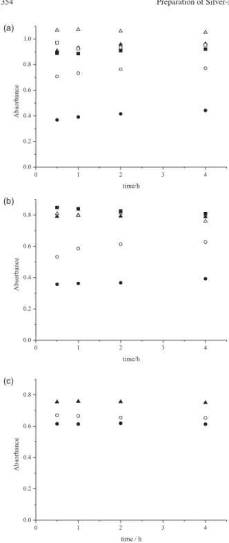
Graphs showing the changes of absorbance versus
reaction time are shown in Figures 1a-c. In the case of formaldehyde, the absorbance increases during the first two hours and then stays nearly constant. Therefore, two hours were selected as the sufficient reaction time for the quantitative reduction. The optimum concentrations of the bases were chosen so that the highest reduction efficiency, expressed by the highest absorbance around 410 nm,
was reached: 0.10 mol L-1 for both NaOH and NaHCO
3,
and 0.50 mol L-1 for TEA. At the higher concentrations,
the absorption band becomes lower and broader. The optimum conditions for the reduction with borohydride
λ
max ca. 400 nm. The absorption maxima related to
formaldehyde reduction are shifted to longer wavelengths
depending on the base employed: λ
maxca.412 nm for NaOH
and TEA, λ
maxca.425 nm for NaHCO3. The shifts in the
λ
max of the silver plasmon to longer (red shift) and shorter
wavelengths (blue shift) have already been described in
the literature.22,24,25 There are several reasons for these
effects: electron density on the surface of nanoparticles, irregular shape and size of nanoparticles, media constants of solutions, refraction coefficients, etc.
Besides the shifts, broadening of the plasmon peak was also observed, which is in agreement with the findings of
other authors.11,16 Such broadening might be caused by
the aggregation of silver particles. The narrowest peak
belongs to silver particles reduced by NaBH4. The other
silver peaks become broader in accordance with the alkaline agents used in combination with formaldehyde: NaOH <
TEA < NaHCO3.
In addition, there is a band at about λ
max = 250 nm in the
absorption spectra (Figure 2), which most likely originates from light scattering on montmorillonite particles. The absorption band at around 300 nm, which has been
attributed to residual Ag+ ions14 or to the existence of Ag
nanoparticles smaller than 2 nm,25 was not found at all.
Based on the data mentioned above, the following can be said. Borohydride reduction is much faster than that by formaldehyde, and thus silver particles are expected to be smaller, as indicated by the narrower absorption peak and
the shorter λ
max. As the concentration of formaldehyde was
identical in all experiments, the concentration of hydroxide ions should be a key parameter for the formaldehyde reduction rate (see reactions 1 and 2). Taking into account
the strength of the bases (TEA with pKb = 6.24 and NaHCO3
Figure 1.(a) Time dependence of silver plasmon absorbance after
the reduction with formaldehyde using NaOH: (z) 0.010 mol L-1,
({) 0.050 mol L-1, (S) 0.10 mol L-1, (U) 0.20 mol L-1, () 0.30 mol L-1,
and ( ) 0.40 mol L-1; (b) time dependence of silver plasmon absorbance
after the reduction with formaldehyde using NaHCO3: (z) 0.010 mol L-1,
({) 0.050 mol L-1, (S) 0.10 mol L-1, (U) 0.20 mol L-1, () 0.50 mol L-1;
(c) time dependence of silver plasmon absorbance after the reduction with formaldehyde using TEA: (z) 0.05 mol L-1, ({) 0.10 mol L-1,
(S) 0.50 mol L-1.
UV-visible absorption spectra
The absorption spectra of silver particles are presented in Figure 2. The absorption maximum corresponding to borohydride reduction occurs at wavelength
Figure 2. UV-visible absorption spectra of silver particles produced under
with pKb = 7.65), the concentration of hydroxide ions in the aqueous solutions of the weak bases can be estimated from
the well-known equation: pOH≈0.5 (pK
b – log cB), where
cB is the concentration of the weak base. The calculated
concentration of OH– ions, c
OH, decreases in the order:
NaOH (cOH≈10-1 mol L-1) > TEA (c
OHca. 10-3.3 mol L-1) >
NaHCO3 (cOH≈10-4.3 mol L-1), which is consistent with
the order of absorption band broadening. For comparison,
λ
max values increase in the order: NaBH4 < NaOH ≈TEA <
NaHCO3. Therefore, it can be assumed that the size of silver
particles increases in the order NaBH4 < NaOH < TEA <
NaHCO3. The particles diameter and their dispersity were
further investigated by TEM.
TEM study
TEM micrographs of silver particles deposited on montmorillonite surface are shown in Figure 3, together with the corresponding SAED patterns as insets. In the SAED patterns, which are compared to the XRD diffraction patterns in Figure 4, crystalline cubic Ag and montmorillonite were detected. The apparent miss of non-basal peaks of montmorillonite in SAED against XRD is due to the strongly preferred orientation of montmorillonite in TEM samples, which were prepared by sedimentation from suspension. The diffraction peaks of Ag are not well defined, which suggests a number of structural defects.
In addition, a diffraction peak corresponding to AgO/
Ag2O was observed in the sample Ag-MMT/F-NaHCO3.
This peak very likely indicates the presence of Ag2O as
a result of incomplete reduction according to reaction 2. The fact that the oxide is not found in the SAED patterns
of Ag-MMT/NaBH4 (Figure 4) suggests that
silver-montmorillonite composites are stable against oxidation. The composites were repeatedly analyzed by TEM during five months after their preparation. They were stored in common Eppendorf tubes, in the dark, at room temperature.
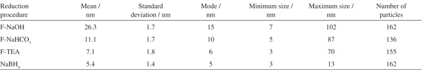
The size distribution of silver nanoparticles was quantified using images in the 100 nm scale. About 150 particles were analysed for each sample. As the particles are more or less isometric (Figure 3), their size can be represented by the diameter of a circle with the same area. The sizes were plotted into a histogram with a 1 nm interval to characterize the size distribution of silver nanoparticles (Figure 5). Statistics was calculated on the diameters as well as their logarithms. Generally, if the distribution is plotted against a logarithmic scale, the maxima are closer to a Gaussian curve and the statistical results (Table 1) appear more meaningful than those based on a linear scale.
The reduction with formaldehyde leads to a wide particle size distribution (3-102 nm) and a lower dispersion on the MMT surface (Figure 3a). The reduction with
Figure 3. TEM micrographs of silver particles on montmorillonite. (a) Ag
particles prepared by reduction with F-NaOH, (b) NaBH4, (c) twinning of Ag particles prepared by reduction with F- NaOH, (d) Ag crystal along [101] in Ag-MMT/F-NaHCO3.
Figure 4. XRD and SAED patterns of Ag-MMT nanocomposites.
Table 1. Basal d001 spacings of MMT in Ag-MMT nanocomposites
MMT sample Reduction procedure d001 / nm
MMT none 1.22
Ag+-MMT none 1.30
Ag-MMT F-NaOH 1.26
Ag-MMT F-NaHCO3 1.26
Ag-MMT F-TEA 1.39
Ag-MMT NaBH4 1.25
Table 2. Statistics of silver particle size distribution based on logarithms of particle diameter Reduction
procedure
Mean / nm
Standard deviation / nm
Mode / nm
Minimum size / nm
Maximum size / nm
Number of particles
F-NaOH 26.3 1.7 15 7 102 162
F-NaHCO3 11.1 1.7 10 5 87 136
F-TEA 7.1 1.8 6 3 70 155
NaBH4 5.4 1.4 5 3 13 162
NaBH4 produces silver nanoparticles with sizes from 3 nm
to 13 nm (the mean size is 5.4 nm), highly concentrated and evenly dispersed on MMT (Figure 3b). The statistical data are summarised in Table 2. In comparison with other
authors,4,14,18 the particle size distribution is quite narrow.
For the production of particles with a diameter range of
several nanometers,11 the method of NaBH
4 has to be
further optimised.
The particle size distributions correspond well with the spectral band broadening (Figure 2), except of the sample prepared by reduction with formaldehyde and NaOH.
Figure 5. Size distribution histograms of silver particles in silver-montmorillonite nanocomposites prepared by the reduction with: (a) formaldehyde +
NaOH, (b) formaldehyde + NaHCO3, (c) formaldehyde + TEA, and (d) NaBH4.
According to the absorption spectra of this sample, the formation of much smaller particles than those observed by TEM was expected. This discrepancy could be explained by the defect microstructure of silver particles, such as twining (Figure 3c), likely originated by their fast formation due to the high hydroxide concentration. The existence of particles
composed from Ag and Ag2O should be also expected.
XRD study
The XRD study of MMT, Ag+-MMT, and Ag-MMT
nanocomposites was carried out to determine the location of silver particles on montmorillonite. From XRD patterns,
montmorillonite d001-basal spacings were calculated
(Table 1). The initial increase of MMT d001 values from
1.22 nm for MMT to 1.30 nm for Ag+-MMT confirms
the intercalation of silver ions into the interlayer space of montmorillonite. Silver ions intercalation into MMT has
been already observed by the increase of d001 values as well
as by the release of interlayer cations.2
The presence of montmorillonite 001 basal diffractions (Figure 4) indicates that MMT retains its layered structure after the reduction. Silver nanoparticles, due to their
size ≥ 3 nm (see Table 2), cannot be located in the interlayer
space and, therefore, are placed on the montmorillonite outer surface as shown on TEM micrographs. The MMT interlayer spacing is occupied by sodium or TEA cations compensating the negative layer charge, and this is documented by montmorillonite 001 diffractions (Figure 4)
and d001 values (Table 1).
Conclusions
In this study, silver-montmorillonite nanocomposites were prepared. The effect of the different reducing agents, such as borohydride and formaldehyde with several alkaline reagents, on the dispersity and size of silver particles supported on montmorillonite surface was demonstrated. The reduction with borohydride produced evenly dispersed silver nanoparticles with sizes ranging from 3 nm to 13 nm, while formaldehyde gave rise to particles from 3 nm to 100 nm. The contents of Ag in Ag-MMT were
equal to 2.4 wt% and 5.3 wt% by the use of NaBH4 and
formaldehyde, respectively, depending on Ag particle size.
In addition, Ag2O particles were also found due to the
incomplete reduction of Ag2O by formaldehyde.
In future studies, we intend to improve the reduction procedure, by which we could prepare silver particles with the size on demand. Silver-montmorillonite nanocomposites will be further tested as catalysts and luminescent materials possibly applicable as nanosensors.
Acknowledgments
The authors would like to thank Dr. M. Valášková (Centrum of Nanotechnology, VŠB-Technical University of Ostrava) for the measurement of XRD patterns. This work was supported by the Ministry of Education, Youth and Sport of the Czech Republic (MSM 6198910016).
References
1. Kwiatkowski, K. C.; Lukehart, C. M.; Nanostructured Materials and Technology, Nalwa, H. S. ed., Academic Press: London, 2002.
2. Praus, P.; Turicová, M.; Valášková, M.; J. Braz. Chem. Soc.
2008,19, 549.
3. Valášková, M.; Martynková, G. S.; Lešková, J.; ýapková, P.; Klemm, V.; Rafaja, D.; J. Nanosci. Nanotechnol. 2008, 8,
3050.
4. Ayyappan, S.; Subbanna, G. N.; Gopalan, R. S.; Rao, C. N. R.; Solid State Ionics1996,84, 271.
5. Patakfalvi, R.; Oszkó, A.; Dékány, I.; Colloids Surf., A 2003,
220, 45.
6. Temgire, M. K.; Joshi, S. S.; Radiat. Phys. Chem.2004,71,
1039.
7. Kapoor, S.; Langmuir1998,14, 1021.
8. Hua, B.; Qiu, J.; Shimotsuma, Z.; Fujita, K.; Hirao, K.; J. Mater. Sci.2005,20, 644.
9. Sun, L.; Zhang, Y.; Dang, H.; Mater. Lett.2003,57, 3874. 10. Plieth, W., Dietz, H.; Anders, A.; Sandmann, G.; Meixner A.;
Weber M., Kneppe H; Surf. Sci.2005,597, 119.
11. Zhang, W.; Qiao, X.; Chen, J.; Wang, H.; J. Colloid Interface Sci.2006,302, 370.
12. Ullah, M. H.; Il, K.; Ha, C. S.; Mater. Lett.2006,60, 1496. 13. Pastoriza-Santos, I.; Liz-Marzán, L. M.; Pure Appl. Chem. 2000,
72, 83.
14. Wang, H.; Qiao, X.; Chen, J.; Wang, X.; Ding, S.; Colloids Surf., A2005,256, 111.
15. Chou, K. S.; Chou, Ch. Y.; Mater. Chem. Phys. 2000, 64,
241.
16. Hsu, S. L.; Wu, R. T.; Mater. Lett.2007,61, 3719.
17. Sun, X.; Luo, Y.; Mater. Lett.2005,59, 3847.
18. Panáþek, A.; Kvítek, L.; Prucek, R.; KoláĜ, M.; VeþeĜová, R.; Pizúrová, N.; Sharma, V. K.; NevČþná, T.; ZboĜil, R.; J. Phys. Chem. B 2006,110, 16248.
19. Madejová, J.; Arvaiová, B.; Komadel, P.; Spectrochim. Acta, Part A1999,55, 2467.
20. Varian Techtron Pty. Ltd.; Analytical Methods for Flame Spectrometry. Varian: Springvale, 1979.
21. Lábár, J. L.; Microsc. Microanal.2002,16, 21.
22. Wilcoxon, J. P.; Martin, J. E.; Provencio, P.; J. Chem. Phys.
2001,115, 998.
23. Czechoslovakia State Standard; ýSN 720101, Basic Analysis of Silicates. Decomposition. Prague, 1974.
24. Jiang, Z. J.; Liu, Ch. Y.; Liu, Y.; Appl. Surf. Sci.2004,233,
135.
25. Zhao, K.; Yao, J.; Mater. Lett.2006,60, 3826.
![Figure 3. TEM micrographs of silver particles on montmorillonite. (a) Ag particles prepared by reduction with F-NaOH, (b) NaBH 4 , (c) twinning of Ag particles prepared by reduction with F- NaOH, (d) Ag crystal along [101] in Ag-MMT/F-NaHCO 3 .](https://thumb-eu.123doks.com/thumbv2/123dok_br/18993364.461427/5.892.482.824.146.383/micrographs-particles-montmorillonite-particles-prepared-reduction-particles-reduction.webp)