Case Repor
t
Study conducted at the Unidade Otorrinolaringológica do Recife – Recife (PE), Brazil.
Conflict of interests: None
(1) School of Medical Sciences, Universidade de Pernambuco – UPE – Recife (PE), Brazil.
(2) Research Group in Physiopathology of the Stomathognathic System, Universidade Federal de Pernambuco – UFPE – Recife (PE), Brazil. (3) Graduate Program (Masters degree) in Production Engineering, Univer-sidade Federal de Pernambuco – UFPE – Recife (PE), Brazil.
(4) Undergraduate Program in Medicine, Faculdade Pernambucana de Saúde – FPS – Recife (PE), Brazil.
Correspondence address: Luiz Alberto Alves Mota. R. Venezue-la, 182, Espinheiro, Recife (PE), Brasil, CEP: 52020-170. E-mail: [email protected]
Received: 12/23/2010; Accepted: 2/13/2012
female patient with adductor spasmodic dysphonia: a case report
Aplicação da técnica de emissão em tempo máximo de fonação
em paciente com disfonia espasmódica adutora: relato de caso
Luiz Alberto Alves Mota1, Catarina Matos Brito Santos2, Jamile Meira de Vasconcelos3, Bruno Calife Mota4, Henrique de Sá Carneiro Mota4
ABSTRACT
Adductor Spasmodic Dysphonia (ASD) is a neurological disorder of central motor processing, characterized by involuntary and inappropriate contractions of the phonatory muscles, producing hyperadduction of the vocal folds, which causes a tremulous, falte-ring and strained-strangled voice. The aim of this study was to describe the vocal, acoustic and laryngeal parameters measured for a female patient with ADS pre and post speech therapy using the Technique of Sustained Maximum Phonation Time (SMPT). This technique aims to promote increase in glottal resistance, improve phonatory stability, and enhance glottal coaptation. A 66-year-old female patient with ASD took part in this study. She was submitted to otorhinolaryngologic and speech-language assessment before and after the application of the SMPT technique. The results showed modification of vocal, acoustic and laryngeal parameters, such as re-classifying her dysphonia from G3R1B1A0S3I3 to G2R1B1A0S2I2, her pitch from severe to adequate, her spectrographic trace from
unstable to more stable, and an expressive increase in mean fundamental frequency and mean vocal intensity, besides improvement of her glottal efficiency, with closure of the anteroposterior glottal opening. Speech therapy using the SMPT technique was considered a suitable treatment option for this case, given the good results obtained, especially the improvements in vocal quality and phonatory stability. The importance of further studies with the aim to provide greater scientific evidence for the effectiveness of the technique when treating ASD is emphasized.
Keywords: Voice; Dysphonia; Voice disorders; Neurology; Speech therapy
INTRODUCTION
Spasmodic dysphonia, also called spastic dysphonia or laryngeal dystonia, is characterized by involuntary and inap-propriate contractions of vocal muscles, the adductor type be-ing the most common one(1). Adduction Spasmodic Dysphonia
(ASD) is a vocal disorder which is recognized as spasms of the laryngeal muscles taking place during phonation, thus prompt-ing tight closure of the larynx and therefore producprompt-ing a tense,
strained and strangled voice, with stoppages and breaks(2,3). In
terms of incidence, it occurs most in women aged over 30(2,4).
In this type of dysphonia, sound breaks are usually more evident in words that begin with vowels. In addition, vocal tremor may occur due to involuntary contractions of the mus-cles responsible for vocal fold adduction. Constant tremor may be restricted to the larynx or involve the pharyngeal muscles, this only being observed during speech(1). Diagnosis is mainly
clinical and may be aided by laryngostroboscopy and elec-tromyography, which reveal irregular spasms of the adductor muscles during phonation, and increased and uncoordinated activity of the thyroarytenoid muscles(1).
There are three possible treatments for spasmodic dyspho-nia, and these may be considered in isolation or in combination: to inject very small amounts of botulinum toxin (Botox®) into
the affected muscles of the larynx, surgery, and voice therapy(5).
A study that associated the application of botulinum toxin (Botox®) with speech therapy showed this was effective in
treating adductor spasmodic dysphonia, since better results were obtained than those resulting from using one of these two treatments in isolation from the other(5). On the other hand,
effective-ness of voice therapy in bringing about improvement in patients with ASD(6), and the authors were reluctant to recommend such
treatment, especially in moderate and severe cases(2).
In general, speech therapy for spasmodic dysphonia involves using the therapeutic techniques proposed for hyperkinetic dysphonia, with exercises to relax the body and larynx and for respiratory control; inspiratory phonation to reduce supraglottal constriction; resonance exercises and others on emitting sounds smoothly(5). Although the literature suggests treating ASD
with techniques recommended for hyperkinetic dysphonias in general, this type of therapy on its own has not proved to be effective(5). Therefore, in an attempt to obtain more promising
results, a therapeutic approach was chosen which focuses on the technique of forming utterances in Maximum Phonation Time (MPT), which belongs to the Method of Glottal Competence, the objective of which is to foster increased glottal resistance, improve phonatory stability, and enhance glottal closure(7).
In this case study, the vowel /a/ was used at different fre-quencies and at high intensities(7). The choice of this technique
in the way it has been described resembles how to carry out the Lee Silverman Voice Treatment (LSVT®) method. Studies on
this method have demonstrated there have been good results not only in patients with Parkinson›s disease (PD), but also in patients with other neurological dysphonias(8). In a study that
examined the voice and speech production of a patient with PD, after one month, six months and 12 months of treatment with LSVT®, the results showed an increase in intensity and
vocal stability(9).
The aim of this study was to describe the vocal, acoustic and laryngeal parameters pre and post speech therapy using MPT in a female patient with ASD.
CLINICAL CASE PRESENTATION
The patient with adductor spasmodic dysphonia who took part in this case study was a retired 66-year-old woman, who is a singer and conductor of classical music. The evaluation and therapy procedures were conducted in a private medical practice in Recife. This study was analyzed and approved by the Committee for Ethics in Research on Human Beings of the Hospital Universitário Oswaldo Cruz (HUOC/PROCAPE), under protocol number 100/2009. The patient consented to this study and to the results being published by signing a Free and Informed Consent Term, in accordance to Resolution 196/96.
The patient made an appointment in an Otorhinolaryngol-ogy outpatient clinic where, initially, a doctor interviewed her for a detailed record of her medical history, comorbidities, complaints about her ability to communicate, her voice, her speech and aspects of her social life. At that moment, the
patient reported that she had been feeling a tremor in her spoken voice for about five years and that this condition had worsened within the past six months, when these symptoms also started to occur when she was singing. The patient said she did not smoke, did not drink alcohol, was not allergic to any medication and did not have diabetes mellitus. She did suffer from systemic high blood pressure, although this was being controlled by medications. An otorhinolaryngologic physical examination showed that some of her teeth were missing. Anterior rhinoscopy and otoscopy examinations of her face and neck did not reveal any alterations.
Subsequently she underwent a videolaryngoscopy ex-amination, with the following equipment being used for this procedure: an Endoview videolaryngoscope with a 7.0 mm, 70° rigid videoscope of the larynx; a Hi-light 250-watt light source (Endoview®); a micro-camera (Toshiba®); a VCR
(Panasonic®); a video monitor (Panasonic®) and a microphone.
Videolaryngoscopy was conducted with a rigid 70° scope and a flexible 2.4 mm scope.
On initial examination, it was observed there was extreme supraglottal compression to phonation, anatomical integrity of the structures of the hypopharynx, the pyriform sinuses were free and there was anatomical alignment of the arytenoids. In the glottal region, mucosa was observed with intense hy-peremia and edema, as well as in regions of the arytenoids and posterior commissure. The anterior-posterior glottis was present during emission of the vowels /e/ and /i/, and there was extreme laryngeal tremor in the vocal cords associated with extreme laryngeal hyperconstriction, which made it difficult to conduct the exam. No abnormalities were detected in the infraglottic region (Chart 1).
To complement the diagnosis and rule out other associated disorders, an audiological evaluation was performed with tonal and vocal audiometry and imitaciometry. Hearing thresholds within normal limits were obtained as well as type A tympa-nograms and bilaterally present contralateral stapedial reflexes. In addition, the neurologist conducted an evaluation, the result of which was normal.
As the results of both evaluations did not indicate any alte-rations, it was found there were no impediments to the auditory monitoring of voice production. In addition, neurodegenerative diseases such as PD were discarded and the diagnostic hypo-thesis was Adductor Spasmodic Dysphonia (ASD).
The patient was advised to return to the outpatient clinic after evaluations were complete, which occurred two months later. Even without any treatment she reported an improvement in her vocal quality, although this evolved with a vocal tremor at certain times, including on the soft palate. At that point, she was referred for clinical assessment in order to verify the
pos-Chart 1. Videolaryngoscopic findings pre and post speech therapy
Parameters Pre-speech therapy Post-speech therapy
Hyperemia LP Extreme Slight
Edema LP Extreme Slight
Hyperconstriction L Present and extreme, which made it difficult to conduct the exam Present and slight Vocal tremor Present and extreme, which made it difficult to conduct the exam Present and slight
Glottal cleft AP Present Absent
sibility of voice therapy, since she had refused the therapeutic options initially proposed: applying botulinum toxin (Botox®)
or surgical intervention.
The data collected in the initial speech-language pathology assessment showed severe impairment of the intelligibility of her speech due to her difficulty in initiating sonorization and controlling vocal frequency and intensity. This occurred due to involuntary and inappropriate contractions of the phonatory muscles, which lead to trembling, tense and strained voice, which are characteristic of ASD. Facial distortions were also identified, especially in the upper face, due to the great vocal effort and tension in the vocal tract. After this evaluation, in a joint decision with the multidisciplinary team involved, it was decided to attempt speech therapy using the MPT technique.
Vocal, auditory-perceptive and acoustic evaluations were conducted before and after treatment, with the aim to analyze the following parameters: vocal quality, loudness, pitch, resonance, vocal attack, spectrographic trace, intensity, mean fundamental frequency, jitter, shimmer, maximum phonation time of the vowel /E/, the s/z ratio, and classification of the dysphonia using the GRBASI scale. This scale was proposed to describe deviations in voice quality, including the parameter of /i/ (instability) in the classification of dysphonia. The (G) indicates the overall degree of dysphonia, the (R) roughness,
the (B) breathiness, the (A) asthenia, the (S) strain and the (I) instability. The following gradations are considered: (0) no deviation (normal); (1) mild deviation; (2) moderate deviation; and (3) severe deviation. Vocal instability is a parameter fre-quently found in cases of spasmodic dysphonias, this being the reason for choosing the GRBASI scale for auditory-perceptual assessment of voice quality.
The following phonatory tasks were performed: sustained emission of the vowels /a/ and /E/ and the fricatives /s/ and /z/ under usual conditions of speech, counting from 1 to 10, spontaneous speech, reading a text aloud, and singing the song “Happy Birthday”. The counting of numbers, spontaneous speech, reading a text aloud and the song were used to assist in the perceptual and auditory evaluation of parameters such as vocal quality, resonance and vocal attack. The two evaluations, pre- and post-treatment, were recorded using VoxMetria (CTS) and 14.1 GRAM programs, with the patient seated and maintain-ing the same posture, with a distance of 3 cm between her mouth and the Multilaser model PH-020 microphone for all records.
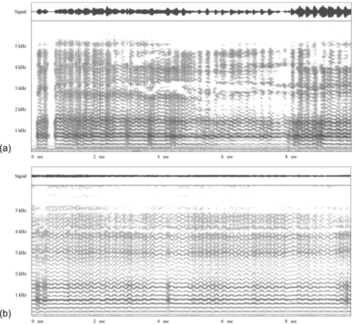
Data were collected from speech therapy assessments pre- and post-treatment (Chart 2), as were their normal values. In addition, spectrograms were obtained from the patient’s emis-sions of the vowel /a/, also pre- and post- treatment (Figures 1a and 1b).
Chart 2. Findings of the auditory-perceptual and acoustic evaluation of the voice during the speech therapy pre and post treatment periods
Parameters Pre-speech therapy Post-speech therapy
Auditory-perceptual analysis
Vocal quality Hoarse, harsh, broken and
tense-strangled
Hoarse, harsh, broken and tense-strangled
GRBASI scale G3R1B0A0S3I3 G2R1B0A0S2I2
Loudness Adequate Adequate
Pitch Deep Adequate
Resonance Laryngopharyngeal Laryngopharyngeal, with reduction of the
local focus of resonance
Vocal attack Sudden Sudden
Acoustic analysis
Spectrographic trace Unstable, with interruption of
the trace of the harmonics
Unstable, with reduction of the interruption of the trace of the harmonics
Mean vocal intensity VN: mean 64 dBn(12)
61.20 dB (N) 69.72 dB (N↑)
Standard-deviation from vocal intensity 2.92 dB 3.30 dB (↑)
Coefficient of the variation of the standard-deviation of vocal intensity
4.77% 4.73% (↓)
Mean fundamental frequency VN: mean 192.2 Hz(10)
166.98 Hz (N) 220.29 Hz (N↑)
Standard-deviation from fundamental frequency 35.08 Hz 15.60 Hz (↓) Coefficient of the variation of the standard-deviation of
the fundamental frequency
21% 7.08% (↓)
Jitter
VN: mean 4.21%(13)
6.17% (A) 0.99% (N↓)
Shimmer
VN: mean 3.17%(13)
27.98% (A) 12.11% (A↓)
Maximum time of the phonation of the vowel /E/ VN: mean 15.2s(7)
12.15s (A) 15.79s (N↑)
s/z Ratio VN: 0.8 - 1.2(7)
1.29 (A) 0.97 (N↓)
Throughout the therapeutic process, the patient demons-trated her willingness and commitment to undergoing the treatment proposed. There were weekly 40-minute sessions for a period of four months; a total of 16 sessions. She was asked to carry out a sequence of exercises at home once a day when she had had a session, and twice on the days when she had not. We emphasized the fact that, because the patient is a singer and music conductor, this may have been a determinant in the treatment and may well have had a direct implication with regards to her dedication and adherence to the treatment.
During the emissions in maximum phonation time, the vowel /a/ was used at different frequencies and with strong intensity. The patient was advised to keep her mouth wide open, without undue muscular effort, and to control the quality of her voice.
After four months of speech therapy, the patient was once again submitted to auditory-perceptive and acoustic evaluation of her voice, and videolaryngoscopy. The videolaryngoscopy found changes in her glottal region: mucosa with hyperemia and edema to a minor degree, as well as in regions of the
aryte-noids and posterior commissure. On evaluating the mobility of her vocal folds, laryngeal tremor and hyperconstriction were seen to a slight degree, apart from absence of the anterior--posterior glottal cleft (Table 1).
The findings indicated an improvement in vocal quality and oral communication that enabled patient to take up her singing activities again, which until then had been suspended.
DISCUSSION
As found in this study, literature states that in cases of ASD there is a predominance of altered vocal characteristics, such as a hoarse, rough and strained-strangled voice quality, with difficult initiation and blocks in the larynx, sudden vocal attacks, and deviation of pitch to bass sounds(10). It is also
said that it is possible to observe an excessive muscular effort in the neck and head regions, and great tension in the vocal tract, which may explain the occurrence of laryngopharyngeal resonance in this case(11) .
After speech therapy, the patient showed significant
provement in vocal quality, with reduced phonation instability, appropriateness of pitch, and greater efficiency of the vibra-tory pattern of her vocal folds. There was also evidence of a decrease in the degree of deviation of dysphonia, this being modified from G3R1B1A0S3I3 to G2R1B1A0S2I2.
The spectrographic trace furnishes data on the stability of the sounds emitted, which requires the central nervous system to be under accurate control(12). The instability of the patient’s
emitted sounds continued to be observed after treatment (Figu-re 1), but with a (Figu-reduction in the interruption of the trace of the harmonics. The alteration in this parameter is possibly related to the non-coordination of the laryngeal muscles which can be perceived in the voice, commonly present in neurological dysphonias(13).
With regards to the fundamental frequency, this rose from 166.98 Hz to 220.29 Hz, a value within the normal range set for women. The significant reduction from 21% to 7% in the coefficient of variation of the standard deviation from the fundamental frequency may be related to the greater stability of the vibratory pattern of the vocal folds when a vowel is being produced.
As to intensity, an increase from 61.20 dB to 69.72 dB was observed. Despite this increase, which may have a sig-nificant impact on improving the intelligibility of speech, the coefficient of variation of the standard deviation from vocal intensity dropped slightly, which may be related to the natural variation of her voice.
The jitter indicates the variability of the frequency in the short term, measured between neighboring glottal cycles. The shimmer indicates the variability of the amplitude of the sound wave also in the short term and is a measure of phona-tory stability(14). The values of jitter and shimmer were greatly
reduced after speech therapy, with jitter falling from 6.17% to 0.99% and shimmer from 27.98% to 12.11%. These decreases confer a value that is considered normal on jitter and causes a significant reduction in shimmer, which represents stability in the emission and intelligibility of speech. Such measures tend to be high both in old age and also in neurological voice disorders, since they are related to the regularity of vibration of the vocal folds, possibly a reflex of the reduction in the neuromuscular control of the adductor and abductor laryn-geal mechanism(13). It is worth pointing out that this patient is
elderly, and that the normal parameters described for this age group were used.
The maximum phonation time of vowel /E/ increased from 12.15 seconds to 15.79 seconds, thus reaching the normal
range, and the s/z ratio dropped from 1.29 to 0.97, which sug-gests that glottal coaptation was more efficient (these being tests that assess glottal control)(12).
The main modifications evidenced by the videolaryngo-scopic tests were closure of the anteroposterior glottal cleft and a reduction in laryngeal tremors. This finding is probably related to the gain in vocal intensity, which depends on raising the subglottal air pressure and improving adduction of the vocal fold(15). It is stressed that the increase in maximum phonation
time and the improvement in the s/z ratio also reflect greater enhancement of glottal competence.
Our findings are consistent with another study that used LSVT® on patients with PD(9). In that study, an increase in the
sound pressure level vowels was found to have increased by 20 dB and the mean fundamental frequency by 15-20 Hz. In addi-tion, there was greater stability in the measures of disturbance of amplitude and frequency, and an increase in the energy observed in the spectrum, as well as reduction in noise(9).
FINAL COMMENTS
This article enables consideration to be given to vocal therapy being conducted by the SMPT technique with the vowel /a/ for variations in frequency and at high intensity, despite the important vocal alteration inherent in spasmodic dysphonia cases. This study showed that this technique is worth recommending in order to minimize voice disorders in this case, bearing in mind the good results obtained. Therefore, speech therapy based on these procedures can be considered not only as an adjunct to other forms of treatment, but also as the preferred option for selected cases, since vocal and laryngeal changes were minimized by using the technique described in this study. Certain parameters were modified, such as improving the degree of dysphonia, adjusting pitch, decreasing the disturbance in the stability of vocal intensity and fundamental frequency, reducing jitter and shimmer values and the coefficient of variation, besides enhancing glottal closure and reducing vocal tremor.
RESUMO
A Disfonia Espasmódica Adutora é uma desordem neurológica do processamento motor central, caracterizada por contrações involuntárias e inapropriadas da musculatura fonatória, produzindo uma hiperadução das pregas vocais, o que promove uma voz trêmula, entrecortada e tensa-estrangulada. O objetivo deste estudo foi descrever os parâmetros vocais, acústicos e laríngeos pré e pós-tratamento fonoaudiológico realizado por meio da aplicação da Técnica de Emissão em Tempo Máximo de Fonação (TETMF) em paciente com Disfonia Espasmódica de Adução. Esta técnica tem como objetivo promover o aumento da resistência glótica, melhorar a estabilidade fonatória e adequar a coaptação glótica. Participou deste estudo de caso uma paciente de 66 anos de idade, gênero feminino, com Disfonia Espasmódica Adutora. A paciente foi submetida à avaliação otorrinolaringológica e fonoaudiológica pré e pós-aplicação da TETMF. Foi verificada modificação de parâmetros vocais, acústicos e laríngeos, tais como a classificação da disfonia de G3R1B1A0S3I3 para G2R1B1A0S2I2, pitch de grave a adequado, traçado espectrográfico instável para mais estável e expressivo
aumento da frequência fundamental média e da intensidade vocal média, além da melhora da eficiência glótica com o fechamento da fenda glótica anteroposterior. A terapia fonoaudiológica com a aplicação da TETMF foi considerada uma adequada opção de tratamento para o caso, tendo em vista os resultados alcançados, com destaque para a qualidade vocal e estabilidade fonatória. Ressalta-se a importância de novos estudos para comprovação da eficácia da técnica no tratamento da Disfonia Espasmódica Adutora.
Descritores: Voz; Disfonia; Distúrbios da voz; Neurologia; Fonoterapia
REFERENCES
1. Santos VJ, Mattioli FM, Mattioli WM, Daniel RJ, Cruz VP. Distonia laringea: relato de caso e tratamento com toxina botulínica. Braz J Otorhinolaryngol. 2006;72(3):425-7.
2. Tsuji DH, Chrispim FS, Imamura R, Sennes LU, Hachiya A. Impacto na qualidade vocal da miectomia parcial e neurectomia endoscópica do músculo tireoaritenoideo em paciente com disfonia espasmódica de adução. Braz J Otorhinolaryngol. 2006;72(2):261-6.
3. Ludlow CL, Adler CH, Berke GS, Bielamowicz SA, Blitzer A, Bressman SB et al. Research priorities in spasmodic dysphonia. Otolaryngol Head Neck Surg. 2008;139(4):495-505.
4. Sulica L. Contemporary management of spasmodic dysphonia. Curr Opin Otolaryngol Head Neck Surg. 2004;12(6):543-8.
5. Pinto JA, Godoy LBM, Sonego TB, Costa PJBM. Efeitos da aplicação de Botox® associada à fonoterapia em disfonia espasmódica: estudo de caso. Acta ORL. 2011;29(1):23-7.
6. Murry T, Woodson GE. Combined-modality treatment of adductor spasmodic dysphonia with botulinum toxin and voice therapy. J Voice. 1995;9(4):460-5.
7. Behlau M, Madazio G, Feijó D, Gielow I, Rehder MI. Aperfeiçoamento vocal e tratamento fonoaudiológico das disfonias. In: Behlau M (org). Voz: o livro do especialista II. Rio de Janeiro: Revinter; 2005. p.410-529.
8. Smith M, Ramig L. Neurological disorders and the voice. In: Rubin JS, Korovin G, Sataloff R, Gould WJ. Diagnosis and treatment of voice disorders. New York: Tokyo: Igaku-Shoin; 1995. p. 204-9.
9. Dromey C, Ramig LO, Johnson AB. Phonatory and articulatory changes associated with increased vocal intensity in Parkinson disease: a case study. J Speech Hear Res. 1995;38(4):751-64.
10. Behlau M, Pontes P. Avaliação e tratamento das disfonias. São Paulo: Lovise; 1995. p.20-186.
11. Boone DR, MCFarlane SC. Distúrbios da voz. In: Boone DR, MCFarlane SC. A voz e a terapia vocal. Porto Alegre: Artes Médicas; 1994. p.61-98.
12. Behlau M, Madázio G, Feijó D, Pontes P. Avaliação de voz. In: Behlau M (org). Voz: o livro do especialista I. 1a ed. Rio de Janeiro: Revinter; 2005. p. 85-180.
13. Carrillo L, Ortiz KZ. Análise vocal (auditiva e acústica) nas disartrias. Pró-Fono. 2007;19(4):381-6.
14. Pontes PA, Vieira VP, Gonçalves MI, Pontes AA. Características das vozes roucas, ásperas e normais: análise acústica espectrográfica comparativa. Rev Bras Otorrinolaringol. 2002;68(2):182-8.