Yeasts from the oral cavity of children with AIDS: exoenzyme
production and antifungal resistance
Leveduras da cavidade oral de crianças com AIDS: produção de
exoenzimas e resistência antifúngica
Vera Lúcia Bosco*
Esther Goldenberg Birman** Arlete Emily Cury***
Claudete Rodrigues Paula****
ABSTRACT:The oral fungal microbiota of 30 children with AIDS, of both genders, aged from two to six years, receiving outpatient treatment, was evaluated and compared with that of a control group composed of 30 healthy subjects with matching ages and genders. Virulence factors, such as exoenzyme production, and susceptibility to five antifungal agents using an E-Test kit were evaluated.C. albicanspredominated over other species in the AIDS group, showing a higher production of proteinase and phospholipase when compared with that observed in the control group. In this study few clinical manifestations of and low selectivity forC. albicans(23.3%) were observed in the AIDS group. The enzymatic studies showed that 53.8% of the AIDS strains were strongly positive whereas only 33.3% of the non-AIDS strains were positive. Amphotericin B was the most effective drug among the antifungal agents tested againstC. albi-cans. The frequency, selectivity and level of exoenzyme production byC. albicanssuggest a higher pathogenicity in the AIDS children than in the control children.
DESCRIPTORS:Acquired immunodeficiency syndrome; Child; Antifungal agents.
RESUMO:A microbiota fúngica bucal de 30 crianças com AIDS, de ambos os gêneros, com idades entre dois e seis anos, recebendo atendimento ambulatorial, foi avaliada e comparada com a de um grupo controle constituído de 30 indivíduos saudáveis com idades e gêneros equivalentes. Fatores de virulência, tais como a produção de exoenzimas, e a suscetibilidade dos microorganismos a cinco agentes antifúngicos, medida por meio de um kit E-Test, foram avaliados. O microorganismoC. albicanspredominou sobre outras espécies no grupo com AIDS, demonstrando uma maior produção de proteinase e fosfolipase quando comparada com a produção observada no grupo controle. No grupo com AIDS, foram observadas poucas manifestações clínicas deC. albicans, além de uma baixa seletividade para o microorganismo (23,3%). Os estudos enzimáticos mostraram que 53,8% das linhagens no grupo com AIDS foram fortemente positivas, ao passo que apenas 33,3% o foram no grupo controle. Dentre os agentes antifúngicos testados, a anfotericina B foi a droga mais efetiva contraC. albicans. A freqüência, a seletividade e o nível de produção de exoenzimas pelo microorganismo sugerem a ocorrência de uma maior patogenicidade nas crianças com AIDS do que nas crianças do grupo controle.
DESCRITORES:Síndrome de imunodeficiência adquirida; Criança; Antimicóticos.
INTRODUCTION
The infection caused by HIV in children leads to deep immunosuppression, has a shorter incuba-tion period than in adults, and produces various manifestations that include oral symptoms. These occur earlier in children and facilitate the develop-ment of opportunistic infections like fungal infec-tions. The disease progression is faster and more severe in children, as they possess an immature immune system and are still undergoing develop-ment5,8,9,13,23. The early diagnosis of lesions and
their complications is an important component in the treatment of children, particularly considering that candidiasis is one of the first signs of viral di-sease progression4,11,15
. Pseudomembranous candi-diasis has a close correlation with the immune sta-tus of the patient, especially in symptomatic children, in whom reduced CD4+ leukocyte counts lead to an increase in the frequency and severity of AIDS-associated manifestations1,2,6,10
.
Among the first reports concerning AIDS are ca-ses of children exhibiting early and unexplained immunodeficiencies associated with opportunistic
*PhD, Pediatric Dentistry, School of Dentistry, Federal University of Santa Catarina.
infections, favored by intrauterine or post-birth transmission10,16
. Oral candidiasis and parotid en-largement are considered to be important signs of disease progression, guiding decisions during the treatment of children with AIDS2,3,8,9,24
.
In the present study, the frequency of yeasts, their characteristics and levels of proteinase and phospholipase production in children with AIDS were evaluated and compared to those of 30 clini-cally healthy control individuals without associa-ted risk factors. The susceptibility ofC. albicansto five antifungal agents was also analyzed using a commercial kit (E-Test).
MATERIAL AND METHODS
Patients
Among the patients treated and followed up by the Section of Infectology, Santa Casa de Miseri-córdia, São Paulo, SP, Brazil, we evaluated male and female children aged between two and six ye-ars, born to mothers with AIDS. The children exhi-bited anti-HIV antibodies, which were detected using the ELISA and Western Blot tests at 15 months of age. The results of recent laboratory exams as well as past and present medical histori-es were obtained from the hospital records. Using these criteria, 30 symptomatic children with AIDS receiving regular treatment with drugs like sulfa-methoxazol + trimethoprim (Bactrin), and antire-troviral agents such as zidovudine (AZT), dideoxy-nosine (ddI), and/or 2-deoxy-3-tiacitidine (3TC or Epivir) were selected. A control group composed of 30 children, healthy at examination and with mat-ching ages and genders, was evaluated and com-pared as to intraoral lesions. These children had not received any blood transfusion and had no his-tory of associated risk factors. The ELISA and Wes-tern Blot tests were negative.
The Ethics Committee of the Santa Casa de Mi-sericórdia of São Paulo, and that of the University of São Paulo approved the procedures adopted in the present study, which was initiated only after the agreement and informed consent was obtained from the parents and/or guardians of both groups of children. They were examined under artificial il-lumination. The extraoral examination consisted of visual inspection and cervico-facial palpation, performed initially to detect lymphadenitis and/or enlarged parotid glands, as well as any facial lesi-ons. The intraoral examination was carried out using wooden spatulas for lip retraction. The cha-racteristics and probable clinical diagnoses of all
oral manifestations were recorded. Protective mea-sures were taken against infection dissemination between patients.
Sampling of biological material
Sterile, alginated swabs (Cefar, Brazil), previ-ously moistened in saline solution were used to perform circular and rotatory movements at the base of the upper and lower vestibules, on the floor of the mouth, and/or on the tongue of each child to collect material for yeast analysis. The material was inoculated onto Petri dishes containing Sa-bouraud-dextrose agar (Difco, Detroit, USA) and 100µg/ml chloramphenicol (Park Davis, São Pau-lo, Brazil).
Yeast isolation and identification
Inoculated plates were incubated at 25ºC up to 15 days. Subsequently, colonies were selected by color, size, consistency, surface texture, borders, brightness and topography, and the strains were subcultured in test tubes containing the same cul-ture medium. Isolated colonies were identified em-ploying germ tube test, clamidoconidia formation, micromorphological aspects, carbon and nitrogen assimilation and sugar fermentation12.
Exoenzyme production
Exoenzyme (proteinase and phospholipase) production tests and determination of their levels were performed according to Price et al.17
and Ru-chel et al.21
The standard strains (C. albicans
ICB-12A) were also used for comparison.
Susceptibility to antifungal agents
Candida strains were analyzed with regard to
their susceptibility to five antifungal agents, based on minimum inhibitory concentrations (MIC) in the E-Test7
(Biodesk, Solna, Sweden) performed according to the manufacturers’ instructions. The susceptibility to antifungal agents was based on the criteria of Thornsberry, Sabath22
and Richard-son, Warnock20
(seric levels). The five agents used were: amphfotericin B, 5-fluorocytosine, flucona-zole, itraconazole and ketoconazole. Standard strains CBS (Central Bureau voor Schimmelcultu-res) were used as controls.
Statistical analysis
phospholi-pases, and the Kruskal-Wallis test for antifungal evaluation.
RESULTS
Table 1 shows the distribution of children with AIDS according to age and CD4+ cell count. There were no children included in the range of no sup-pression (³1,000 cell/mm³); a percentage of 33.3% (10/30) showed a moderate suppression (500-999 cell/mm³) and 66.6% (20/30) presented a severe suppression (< 500 cell/mm³), although differen-ces were not statistically significant (χ2
= 2.11). The cultures were positive in all of the children (7/30) with confirmed symptoms.
There was an association between candidiasis and moderate suppression in only two of the seven children (28.7%), while candidiasis and severe suppression were associated in five out of the se-ven children (71.4%).
With regard to yeast isolation, 83.3% (25/30) of the samples collected from children with AIDS gave positive results. Twenty-four samples were positive forCandida albicans, one forCandida
pa-rapsilosis and one for Candida tropicalis, which
was associated withC. albicans(Graph 1).
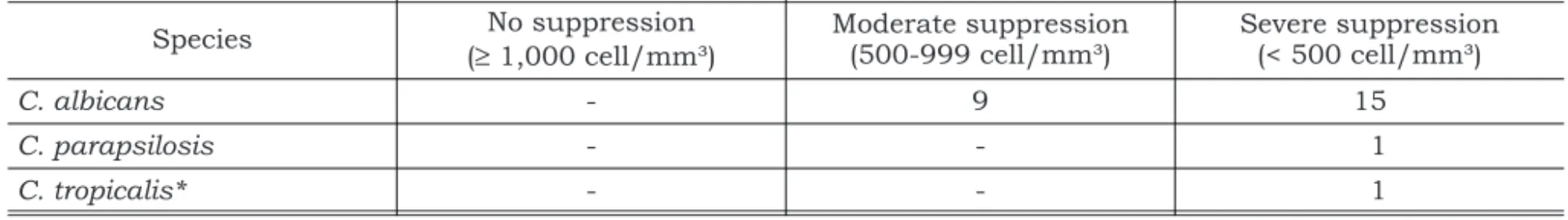
The distribution ofCandidaspecies in children with AIDS according to CD4+ cell count is shown on Table 2. Positive cultures for Candida were found in 34.6% (9/26) of the children with mode-rate suppression, and in 57.6% (15/26) of those with severe suppression. Among severely immuno-suppressed children, 3.85% (1/26) showed a posi-tive culture for Candida parapsilosis, and 3.85% (1/26) forCandida tropicalisassociated withC.
al-bicans. The AIDS group presented fungal
selecti-vity species among the positive samples. There was a strong correlation between the presence of AIDS and yeasts (χ2= 7.89, p = 0.005).
TABLE 1 -Distribution of children with AIDS according to age and CD4+ cell count.
Age No suppression
(³1,000 cell/mm³)
Moderate suppression (500-999 cell/mm³)
Severe suppression
(< 500 cell/mm³) Total
2 to 4 years old − 4 2 6
4 to 6 years old − 6 18 24
Total 0 10 20 30
TABLE 2 -Candidaspecies isolated from the oral cavity of children with AIDS, evaluated by CD4+ cell count.
Species No suppression
(³1,000 cell/mm³)
Moderate suppression (500-999 cell/mm³)
Severe suppression (< 500 cell/mm³)
C. albicans - 9 15
C. parapsilosis - - 1
C. tropicalis* - - 1
*Associated withC. albicans.
In the control children, 10 samples were positi-ve for yeasts: two forC. albicans, four forCandida
guilliermondii, one for Candida lusitaniae, two for
Rhodotorula rubra,and one forAureobasidium
pul-lulans (Graph 1). The enzyme activity study
sho-wed that all yeasts isolated from the AIDS group possessed proteinase activity. Fourteen samples (53.8%) were strongly positive while 12 (46.1%) ex-hibited positive activity. Nearly all samples 96.1% (25/26) exhibited a strongly positive phospholipa-se activity.
AmongCandida strains isolated from the con-trol group, 33.3% (3/10) presented a strongly posi-tive proteinase activity, 44.4% (4/10) presented positive activity, and 22.2% (2/10) were proteina-se-negative. Phospholipase production, according to the samples in this group, was strongly positive in 66.6%, and only positive in 33.3%.
The Mann-Whitney test detected no statistically significant differences in proteinase activity betwe-en the control and the AIDS groups (U = 149.5; U’ = 8; p = 0.2275), but revealed higher phospholi-pase levels in the control group (U = 205; U’ =−47.5; p = 0.0004).
The antifungal susceptibility analysis demons-trated that some strains were resistant as the cha-racteristic formation of the inhibition was not ob-served. The minimum inhibitory concentration test was 0.38 to 2.0µg/ml for amphotericin B; 0.5 to 12.0µg/ml for 5-fluorocytosine; 0.019 to 0.064 µg/ml for ketoconazole; 2.0 to 4.80 µg/ml for flu-conazole and 0.012 to 8.0µg/ml for itraconazole. Considering that the main objective of this study was to evaluate the antifungal susceptibility of
Candidaspecies isolated from children with AIDS,
the E-Test was not performed with samples from the control group.
Candida was more resistant to fluconazole
(80.76%), followed by itraconazole (42.30%),
5-flu-orocytosine and ketoconazole (38.46%), and amphotericin B (23.07%), with only six samples showing resistance to the latter (Table 3).
Statistically significant differences were found using the Kruskal-Wallis test when individual comparisons were made among the antifungal agents (t= 19.33; p = 0.0007). In these individual comparisons (p < 0.01), the error rate was less than 1%, indicating activity differences between the antifungal agents.
DISCUSSION
The known number of pediatric AIDS cases does not reflect the real impact of the epidemic. The disease continues to grow and will soon be one of the five major causes of child death, although new drugs may revert this prediction. Since the mouth is easily accessible for clinical examination, it is one of the first areas to show signs of AIDS. Clinical manifestations and various other aspects must be examined, not only for diagnostic purpo-ses, but also to delineate an adequate treatment plan and prognosis1,3,5.
In our subjects, candidiasis was present with a frequency similar to that reported by other resear-chers6,18
. Pseudomembraneous candidiasis predo-minated over the erythematous type, both affec-ting the children with moderate and severe suppression. Candidiasis is considered to be a prognostic factor and may indicate rapid disease progression in children, differently from parotid enlargement which is associated with a slower pro-gression10
.
Pseudomembraneous and erythematous candi-diasis were detected on the tongue and/or palate, with a higher frequency of the former, corrobora-ting literature data5,18
. Although some hypotheses associate the presence of linear gingivitis with ery-thematous candidiasis, this was not a finding in TABLE 3 -Susceptibility to antifungal agents ofCandida albicansstrains isolated from the oral cavity of children with AIDS.
Antifungal agent Sensitive Resistant
n % n %
Amphotericin B 20 76.9 6 23.1
5-Fluorocytosine 16 61.5 10 38.5
Ketoconazole 16 61.5 10 38.5
Fluconazole 5 19.2 21 80.8
the group studied here25
. The frequency of candidi-asis has an important relationship with extrinsic factors such as treatment and hospitalization. Both may lead to low rates of clinical manifestati-ons, which was demonstrated by the treatment gi-ven to our patients and their subsequent outpati-ent control.
Candida albicans was the predominant yeast
isolated from the oral cavity of the children with AIDS. A statistically significant correlation was found between AIDS and the presence ofCandida, which is important despite the lack of relevant cli-nical candidiasis manifestations in most children since many yeast infections show no clinical mani-festations16.
A large number of C. albicans carriers have been reported in some studies with children, in whom this species is the most prevalent microor-ganism, both in the oral cavity and in the esopha-gus. However, a smaller number ofC. albicans car-riers among children, corroborated by our data, reveal the prevalence of erythematous candidia-sis14
.
The children with AIDS in the present study ex-hibitedC. albicans almost exclusively as the pre-dominant fungus, which may be attributed to the antiviral drugs employed during treatment19
. Ho-wever, considering the secondary effects of such drugs on the development ofC. albicans, the con-trol children showed a greater diversity of species.
Evaluation of the production of proteinase and phospholipase, exoenzymes considered to be viru-lence factors ofCandidaspecies, is relevant to the expression of pathogenicity considering thatC.
al-bicans is associated with various factors that
re-quire attention, especially when related to AIDS11
. High levels of phospholipase and proteinase, in ad-dition to the selectivity of species present in the AIDS group, provide conditions for greater adhe-rence, colonization and development, facilitating yeast multiplication. Proteinase expression seems to be more active in isolates from HIV-infected patients, a relevant aspect considering that anti-retroviral agents may reduce exoenzyme produc-tion13
, a fact not confirmed in this study.
Inhibitors present in the saliva, and certain an-tibodies (IgA) inhibit fungal adherence, and immu-nodepression seems to favor greater pathogenicity, especially ofC. albicans, thus allowing its clinical manifestation9,16
. Few studies have examined these aspects and they will not be discussed further in the present study since different time periods,
drugs and conditions seen in adult AIDS groups should also be considered.
Drugs effective againstC. albicansstrains were studied using a commercial E-Test kit and the cri-teria for susceptible resistance were followed ac-cording to Richardson, Warrnock20 and
Thorn-sberry, Sabath22
. In this study amphotericin B seems to be the most effective. However, we point out the secondary effects, contra-indications and other problems regarding intravenous use to be li-mitations to the use of this drug, in addition to the lack of a topical preparation for dental use. Among the azoles, itraconazol and ketoconazole are note-worthy; fluconazole was the least effective due to high resistance and the need for higher titers to inhibit fungal growth. In vitrotests are important markers, although in vivo studies, when perfor-med, deserve greater consideration. The clinical use of antifungal agents, in association within vi-trotests, may contribute to a more effective treat-ment of candidiasis, not always achieved with em-pirical treatments or even with those based on well controlled tests. The range of available drugs is still very small and so the use of certain agents cannot be avoided.
The development and use of new drugs and an early diagnosis, associated to more detailed know-ledge about infants with AIDS, have demonstrated the effectiveness of certain treatments which have resulted in a lower frequency of oral/facial and bacterial manifestations. Progress in studies on yeasts has been increasing, although new and more effective therapeutics must be developed.
Our results show that the yeasts found in the AIDS group were predominantlyCandida albicans
while the control group exhibited a more diversity of yeast species.
CONCLUSIONS
REFERENCES
1. Center for Diseases Control and Prevention. Diagnosing HIV infection in children. MMWR 1994;43:2-9.
2. Chain A, Milnes A, King SM, Read S. The relationships of oral manifestations to parameters of immune function and CDC stage in children born to HIV-positive women. Pediatr AIDS and HIV infection: fetus to adolescents 1994; 5:101-107.
3. Chigurupati R, Raghavan SS, Studen-Pavlovich D. Pediat-ric HIV infection and its oral manifestation: a review. Pediatr Dent 1996;18:106-13.
4. Costa LRRS, Villena RS, Birman EG. AIDS in children: an up to date review of stomatological aspects. Rev FOLA/Oral 1996;2(1):21-5.
5. Costa LRRS, Villena RS, Sucasas PSC, Birman EG. Oral finding in pediatric AIDS: a case control study in Brazilian children. J Dent Child 1998;65:186-90.
6. Del Toro A, Berkowitz RJ, Meyerowits C, Frenkel LM. Oral findings in asymptomatic (P-1) and symptomatic (P-2) HIV-infected children. Pediatr Dent 1996;18:114-6. 7. E-Test Technical Guide 4b. Antifungal susceptibility
test-ing of yeasts. AB Biodisk, Solna; 1994.
8. Flaitz CM, Hicks MJ. Oral candidiasis in children with im-mune suppression: clinical appearance and therapeutic considerations. ASDC J Dent Child 1999;66:161-6. 9. Howell RB, Jandinsky J, Palumbo P, Shey Z, Houpt M. Oral
soft tissue manifestations and CD4 lymphocyte count in HIV-infected children. Pediatr Dent 1996;18:117-20. 10. Katz MH, Mastrucci MT, Leggott PJ, Westenhouse J,
Greenspan JS, Scott GB. Prognostic significance of oral le-sions in children with perinatally acquired human immu-nodeficiency virus infection. Am J Dis Child 1993; 147:45-8.
11. Kline MV. Oral manifestation of pediatric human immuno-deficiency virus infection: a review of the literature. Pediat-rics 1996;97:380-8.
12. Kurtzman CP, Fell JW. The yeasts, a taxonomic study. 4th ed. Amsterdam: Elsevier; 1998.
13. Leggott PJ. Oral manifestations of HIV infection in chil-dren. Oral Surg Oral Med Pathol 1992;73:187-92. 14. Nicolatou O, Theodoridou M, Mostrou G, Velegraki A,
Legakis NJ. Oral lesions in children with perinatally
ac-quired human immunodeficiency virus infection. J Oral Pathol Med 1999;28:49-53.
15. Oleske J, Minnefor A, Cooper R Jr, Thomas K, dela Cruz A, Ahdieh H, et al. Immune deficiency syndrome in children. JAMA 1983;249:2345-9.
16. Unexplained immunodeficiency and opportunistic infec-tions in infants – New York, New Jersey, California. MMWR Morb Mortal Wkly Rep 1982;31:665-7.
17. Price MF, Wilkinson ID, Gentry LO. Plate method for detec-tion of phospholipase activity in Candida albicans. Sabouraudia 1992;20:7-14.
18. Ramos-Gomes FJ, Hilton JF, Canchola AJ, Greenspan D, Greenspan JS, Maldonado YA. Risk factors for HIV-related soft-tissue manifestations in children. Pediatr Dent 1996; 18:121-6.
19. Reichart PA. Clinical management of selected oral fungal and viral infection during HIV-disease. Int Dent J 1999; 49:251-9
20. Richardson MD, Warnock DW. Fungal infection: diagnosis and management. Boston: Blackwell Scientific Publica-tions; 1993.
21. Ruchel R, Tegeler R, Trost M. A comparison of secretory proteinases from different strains of Candida albicans. Sabouraudia 1982:20:233-44.
22. Thornsberry C, Sabath LD. Approximate concentration of antimicrobial agents achieved in blood. In: Lennette EH, Balows A, Auster Jr. WJ, Shadomy HJ (eds). Manual of Clinical Microbiology. 4thed. Washington: American Soci-ety for Microbiology; 1985. p. 1021-2.
23. Tovo PA, de Martino M, Gabiano C, Cappello N, D’Elia R, Loy A, et al. Prognostic factors and survival in children with perinatal HIV-1 infection. The Italian Register for HIV In-fection in Children. Lancet 1992;339:1249-53.
24. Valdez IH, Pizzo PA, Atkinson JC. Oral health of pediatric AIDS patients: a hospital-based study. ASDC J Dent Child 1994;61:114-8.
25. Velegraki A, Nicolatou O, Theodoridou M, Mostrou G, Legakis NJ. Paediatric AIDS – related linear gingival ery-thema: a form of erythematous candidiasis? J Oral Pathol Med 1999;28:178-82.
Recebido para publicação em 17/01/03 Enviado para reformulação em 23/07/03 Aceito para publicação em 08/09/03