Novel Function of Lysine Methyltransferase
G9a in the Regulation of Sox2 Protein
Stability
Jae-Young Lee1, Se-Hwan Lee1, Sun-Hee Heo1, Kwang-Soo Kim1, Changhoon Kim2, Dae-Kwan Kim1, Jeong-Jae Ko1, Kyung-Soon Park1*
1Department of Biomedical Science, College of Life Science, CHA University, Seoul, Korea,2Department of Biomedical Science, Graduate School of Biomedical Science & Engineering, Hanyang University, Seoul, Korea
*kspark@cha.ac.kr
Abstract
G9a is a lysine methyltransferase (KMTase) for histone H3 lysine 9 that plays critical roles in a number of biological processes. Emerging evidence suggests that aberrant expression of G9a contributes to tumor metastasis and maintenance of a malignant phenotype in can-cer by inducing epigenetic silencing of tumor suppressor genes. Here, we show that G9a regulates Sox2 protein stability in breast cancer cells. When G9a lysine methyltransferase activity was chemically inhibited in the ER(+) breast cancer cell line MCF7, Sox2 protein lev-els were decreased. In addition, ectopic overexpression of G9a induced accumulation of Sox2. Changes in cell migration, invasion, and mammosphere formation by MCF7 cells were correlated with the activity or expression level of G9a. Ectopic expression of G9a also increased Sox2 protein levels in another ER(+) breast cancer cell line, ZR-75-1, whereas it did not affect Sox2 expression in MDA-MB-231 cells, an ER(-) breast cancer cell line, or in glioblastoma cell lines. Furthermore, treatment of mouse embryonic stem cells with a KMT inhibitor, BIX-01294, resulted in a rapid reduction in Sox2 protein expression despite increased Sox2 transcript levels. This finding suggests that G9a has a novel function in the regulation of Sox2 protein stability in a cell type-dependent manner.
Introduction
Lysine methyltransferase G9a is ubiquitously expressed in most tissues, including bone mar-row, thymus, spleen, lymph node, and fetal liver [1]. G9a knock-out mice are embryonic lethal between embryonic (E) days E9.5–E12.5. Even though G9a-/- embryonic stem cells (ESCs) do not show abnormalities in culture, they exhibit severe differentiation defects, suggesting a role for G9a in lineage commitment and differentiation [2]. Consistent with this notion, there is considerable evidence that G9a represses Oct3/4, Nanog, and DNMT3L, which are required for maintenance of pluripotent differentiation potential in ESCs [2]. G9a is also implicated in OPEN ACCESS
Citation:Lee J-Y, Lee S-H, Heo S-H, Kim K-S, Kim C, Kim D-K, et al. (2015) Novel Function of Lysine Methyltransferase G9a in the Regulation of Sox2 Protein Stability. PLoS ONE 10(10): e0141118. doi:10.1371/journal.pone.0141118
Editor:Sue Cotterill, St. Georges University of London, UNITED KINGDOM
Received:March 6, 2015
Accepted:October 5, 2015
Published:October 22, 2015
Copyright:© 2015 Lee et al. This is an open access article distributed under the terms of theCreative Commons Attribution License, which permits unrestricted use, distribution, and reproduction in any medium, provided the original author and source are credited.
Data Availability Statement:All relevant data are within the paper and its Supporting Information files.
Funding:This work was carried out with the support of“Cooperative Research Program for Agriculture Science & Technology Development (PJ010033)”, Rural Development Administration, Republic of Korea.
genomic imprinting. G9a is recruited by the non-coding RNAsAirandKcnqlot1to target genes and stimulate the formation of heterochromatin in a lineage-specific manner [3,4].
G9a localizes to euchromatin in a heteromeric complex with a G9a-like protein (GLP), a highly homologous lysine methyltransferase, to repress gene transcription, especially during embryonic development. G9a-mediated gene repression is associated with its ability to mono-and dimethylate H3K9 mono-and H3K27 [2,5–10]. Additionally, G9a/GLP can directly recruit DNA methyltransferases to promoters, resulting in gene repression via the methylation of CpG islands [11,12]. In addition to the methylation of histone 3, G9a mediates methylation of vari-ous non-histone proteins, including p53 [6,13].
While substantial studies suggest that the function of G9a is associated with transcriptional repression, it switches from a repressor to an activator by changing its interacting partners. For example, G9a interacts with the H3K4 demethylase Jarid1a at the embryonic Eyglobin pro-moter to repress its expression, while it recruits Mediator to theβmajpromoter, resulting in its activation [14]. Similarly, G9a also acts with CARM1 and p300 as a co-activator of nuclear receptors, independent of its lysine methyltransferase activity [15].
A large number of studies have indicated that G9a is also highly expressed in a variety of human cancers, such as breast, lung, and hepatocellular carcinoma [5,16,17]. Knockdown of G9a in lung cancer cell lines causes apoptosis and growth arrest with an increase in the sub-G1 population [18]. In prostate cancer, downregulation of G9a results in centrosome disruption, inhibition of cell growth, and increased cellular senescence in cancer cells [19]. Studies in aggressive forms of lung cancer further revealed that G9a expression is correlated with poor prognosis, increased cell migration, invasion, and metastasis [5]. However, the molecular basis of G9a activity in cancer cells is not well understood.
Here, we identify an essential role for G9a in maintaining Sox2 protein stability in ER(+) breast cancer cell lines. We found that lysine methyltransferase activity is closely related to the accumulation of Sox2 protein, the protein level of which is closely related to cell migration, invasion, and mammosphere formation by ER(+) breast cancer cell lines. However, G9a did not affect Sox2 protein levels in MDA-MB-231 cells, an ER(-) breast cancer cell line, or in glio-blastoma cell lines. These findings provide evidence that G9a-mediated stabilization of the Sox2 protein depends on the subcellular context of each cell.
Materials and Methods
Cell culture
The cell lines MCF-7 and the mouse embryonic stem cell line J1 were obtained from the Amer-ican Type Culture Collection (ATCC, USA). MCF-7 cells were cultured with 10% fetal bovine serum and 1% penicillin/streptomycin in Dulbecco’s modified Eagle’s medium (DMEM). Mouse embryonic stem cells (mESCs) were maintained on 0.1% gelatin coated dishes in DMEM (Gibco Invitrogen, Carlsbad, CA,http://www.invitrogen.com) supplemented with 10% horse serum (Gibco Invitrogen), 2 mM glutamine, 100 U/ml penicillin, 100μg/ml
Plasmid, siRNA, and transfection
Human G9a, Sox2 expression plasmids were purchased from Addgene (Cambridge, MA) and human G9a siRNAs (L-006937-00-0010) and Sox2 siRNAs (L-011778-00-0005) were obtained from Dharmacon, Inc (Chicago, IL). Plasmids or siRNAs were transfected using Lipofectamine 2000 (Invitrogen) according to the manufacturer’s instructions. Cells were collected 24 hr after transfection for further analysis.
RNA isolation and real-time PCR
Total cellular RNA was extracted using TRIzol reagent (Invitrogen), according to the manufac-turer’s instructions. Total RNA (2μg) was used for single-stranded cDNA synthesis using
Omniscript Reverse Transcriptase (Qiagen, Hilden, Germany). Gene-specific primers were designed for Sox2, Oct4, G9a, and GAPDH, as described inS1 Table. Quantitative real-time PCR was performed with the CFX96 Touch™Real-Time PCR Detection System (Bio-Rad Labo-ratories, CA, USA) using SYBR Green I (Qiagen, Valencia, CA, USA). Cycling conditions were as follows: 40 cycles of 95°C for 30 s, 49°C for 30 s, and 72°C for 30 s. Results were calculated as relative expression and normalized to internal human GAPDH using theΔCtmethod.
Protein extraction and Western blotting
Total protein was isolated using cell lysis buffer (#9803, Cell Signaling Technology, Inc.) according to the manufacturer’s instructions. Proteins (20μg) were separated by 10%
SDS-PAGE and transferred to a polyvinylidene difluoride (PVDF) membrane (Millipore, Bed-ford, MA). The membrane was incubated with antibodies against Sox2 (sc-20088, Santa Cruz Biotechnology, Inc.), Oct4 (sc-5279, Santa Cruz Biotechnology, Inc.), G9a (#3306, Cell Signal-ing Technology, Inc.), andβ-actin (sc-47778, Santa Cruz Biotechnology, Inc.) overnight at 4°C, followed by incubation with secondary antibody for 1 h at room temperature. Immunoreactive proteins were detected using the WEST-ZOL1(plus) Western Blot Detection System
(iNtRON, Korea).
Migration and invasion assay
Migration of MCF7 cells was assessed by scratch/wound assays. Monolayers formed by cells were wounded using a micropipette tip when the cells were fully confluent. Detached cells were removed by washing with medium and plates were photographed at the indicated times. The Matrigel invasion assay was performed using 24-well Transwell inserts (6.5 mm diameter, 8μm pores, Corning, NY, USA) coated with 30μg of Matrigel (#356231, BD Biosciences).
Cells (1 x 10⁵) were seeded in serum-free medium in the upper chamber and DMEM contain-ing 10% FBS was added to the lower chamber. Cells were incubated to assess invasion through the membrane for 48 h. Invasive cells were fixed and stained with Crystal violet (Sigma). The intensity of the Crystal violet staining was measured using GelQuant software (biochemlabso-lutions.com) and expressed as the means ± SE of triplicate wells.
Co-immunoprecipitation (Co-IP)
Cells were lysed by sonication in Co-IP buffer (20 mM Tris-HCl, 137 mM NaCl, 1% Triton X-100, 2 mM EDTA) containing Protease and Phosphatase Inhibitor Cocktail (Pierce Biotechnol-ogy, Rockford, IL, USA), and then centrifuged at 12,000 × g for 15 min at 4°C. Following pro-tein isolation, 500μg of total protein per sample were diluted with Co-IP buffer, pre-cleared
Biotechnology, Inc.) or 4 ug of anti-G9a antibody (#3306, Cell Signaling Technology, Inc.) overnight at 4°C. After washing, lysate/beads were boiled in 20μL of SDS sample buffer, and a
10μL aliquot of the eluate was fractionated by 12% SDS—PAGE followed by Western blotting
to confirm physical interaction between Sox2 and G9a. Reactive bands were detected with an ECL system (Amersham, Amersham, UK) according to the manufacturer’s instructions.
Statistical analysis
The data are presented as the means ± SE. The statistical significance of the results was assessed using one-way ANOVA, followed by Student’s t-test.
Results
Chemical inhibition of G9a activity decreases Sox2 protein stability in
breast cancer cells
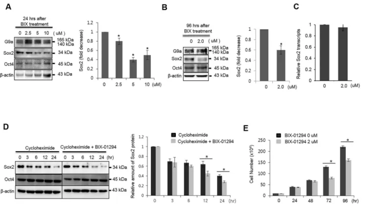
The expression of lysine methyltransferase G9a is highly correlated with metastasis of various cancers and a poor prognosis [20]. Since monomethylated Sox2 interacts with the E3 ligase WWP2 in ESCs, thereby causing its ubiquitination and degradation [21], we questioned whether the activity of G9a is related to the protein stability of Sox2 in breast cancer. As a first step, we examined whether Sox2 protein stability is regulated by G9a activity. Unexpectedly, chemical inhibition of G9a activity using the KMT inhibitor BIX-01294 resulted in reduced amounts of Sox2 protein in MCF7 breast cancer cells (Fig 1A). By contrast, the levels of Oct4 proteins were unchanged. Sox2 protein levels were also decreased when cells were treated with low concentrations of BIX-01294 for 96 h (Fig 1B). In contrast to the changes observed in pro-tein levels, transcript levels of Sox2 were not changed by treatment with BIX-01294 (Fig 1C). The transcription of Sox2 in MCF7 cells was not affected by higher concentrations of BIX-01294 (S1 Fig). When Sox2 protein biosynthesis was blocked by treatment with cycloheximide, BIX-01294 treatment induced degradation of Sox2 (Fig 1D). The proliferation rate of MCF7 cells also decreased upon BIX-01294 treatment (Fig 1E). When we treated MCF-7 cells with another inhibitor of G9a, UNC0638, we obtained similar results to those obtained with BIX-01294, i.e., the amount of Sox2 protein decreased upon treatment with 10μM UNC0638 (S2
Fig). However, chemical inhibition of G9a activity or siRNA-mediated inhibition of G9a expression had no effect on Sox2 protein stability in ER(-) MDA-MB-231 cells (S3 Fig). Nota-bly, G9a was expressed at high levels in ER(+) MCF7 and ZR-75-1 cells, but was hardly detect-able in ER(-) MDA-MB-231 cells (S4 Fig). These results indicate that the stability of the Sox2 protein was affected by G9a lysine methyltransferase activity only in ER(+) MCF7 cells.
Sox2 protein stability is affected by G9a expression
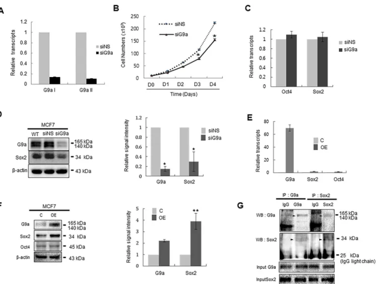
We next tested whether the level of G9a expression correlates with Sox2 protein stability. To examine this, we transiently knocked-down G9a expression by siRNA transfection (Fig 2A). Similar to the results observed following BIX-01294 treatment, the rate of cell proliferation of MCF7 cells was decreased by transfection of siG9a (Fig 2B).
G9a interacts with Sox2 in MCF7 cells, immunoprecipitation was performed using non-specific IgG or an anti-Sox2 antibody. Immunoblot analysis of the precipitates with antibody against G9a showed that G9a was recovered in the Sox2 immmunoprecipitates, and reciprocal immu-noprecipitation with anti-G9a antibodies confirmed this interaction (Fig 2G). These data indi-cate that G9a increases Sox2 protein stability in MCF7 breast cancer cells, possibly through direct interaction.
G9a regulates the oncogenic characteristics of MCF7 cells
Since Sox2 is a key player in the oncogenicity of breast cancer, we hypothesized that alteration of G9a activity would have an effect on the oncogenic characteristics of MCF7 cells, such as migration, invasion, and cancer stem cell characteristics. As expected, migration assay revealed that chemical inhibition of the lysine methyltransferase activity of G9a resulted in decreased cell migration compared with untreated cells (Fig 3A). Consistent with this, overex-pression of G9a led to a marked increase in the migration of MCF7 cells (Fig 3B). siRNA-mediated suppression of Sox2 abrogated the effect of G9a on cell migration, indicating that overexpression of G9a stimulates the migration of MCF7 cells in a Sox-dependent manner. Next, we determined whether G9a overexpression affected the invasive capacity of MCF7 cells Fig 1. Chemical inhibition of G9a activity reduces the stability of Sox2 protein in MCF7 cells.(A) MCF7 cells were treated with the indicated doses of BIX-01294 for 24 h. Whole cell lysates were harvested and analyzed by immunoblotting with G9a-, Sox2-, and Oct4-specific antibodies. (B) MCF7 cells were treated with 0.2μM of BIX-01294 for 96 h, and whole cell lysates were analyzed for the expression of Sox2 and Oct4. (C) MCF7 cells were treated with 2.0μM BIX-01294 for 96 h, and the levels of Sox2 transcripts were analyzed by real-time RT-PCR analysis. mRNA levels were normalized to those of GAPDH. (D) Sox2 and Oct4 protein levels in MCF7 cells were analyzed by immunoblotting after cells were treated with 2.0μM BIX-01294 in the presence of the protein synthesis inhibitor, cycloheximide (5μM). The graph shows band intensity data normalized according to the controls andβ-actin. (E) Cell growth was analyzed by counting cell numbers at the indicated times after treatment with 2.0μM BIX-01294. For quantitative analysis of the immunoblotting results, the mean density of the Sox2 band was measured with Multi Gauge V3.0 software, and the band density was divided by the density ofβ-actin to obtain the normalized band density.*P<0.05 compared with the control group. Data are expressed as the mean±SD and are representative of three independent experiments.
in a Matrigel assay. As expected, MCF7 cells transfected with G9a-expressing plasmids showed increased invasiveness; siSox2-mediated suppression of Sox2 abrogated this effect (Fig 3C). Because Sox2 plays a crucial role in promoting the stem cell characteristics of breast cancer cells, we examined whether G9a activity is related to the self-renewal of MCF7 cells by analyzing their ability to form mammospheres. When MCF7 cells were cultured under non-adherent conditions in the presence or absence of BIX-01294, cells treated with BIX-01294 Fig 2. The expression level of G9a affects the amount of Sox2 protein in MCF7 cells.(A) MCF7 cells were transfected with siNS or siG9a for 24 h, and G9a transcript levels were analyzed by real-time PCR by priming two positions of G9a ORF (G9aI and G9aII). mRNA levels were normalized to those of GAPDH. G9aI and G9a2 primer sets were designed to amplify 1299~1531 bp and 2326~2637 bp of G9a ORF, respectively. (B) The effect of G9a on the proliferation of MCF7 cells. MCF7 cells (1 × 10⁴) were transfected with siNS or siG9a and then counted at the indicated times post-transfection. (C) MCF-7
cells were transfected with siNS or siG9a for 24 h, and the effect of G9a knockdown on the transcription of Oct4 and Sox2 was quantitatively analyzed by real-time RT-PCR. (D) MCF-7 cells were transfected with siNS or siG9a for 24 h, and the effect of G9a knockdown on the amount of Oct4 and Sox2 protein was analyzed by immunoblot analysis. (E) MCF-7 cells were transfected with control or G9a expression plasmids for 24 h, and the effect of G9a
overexpression on the transcription of Sox2 and Oct4 was quantitatively analyzed by real-time RT-PCR. (F) MCF-7 cells were transfected with control or G9a expression plasmids for 24 h, and the effect of G9a overexpression on the amount of Sox2 and Oct4 was quantitatively analyzed by immunoblot analysis. (G) Co-immunoprecipitation of endogenous Sox2 and G9a in MCF7 cells. Total protein extract of MCF7 cells was immunoprecipitated with Sox2 and then immunoblotted with G9a antibody. The reciprocal experiment was also performed in which total protein of MCF7 cells was immunoprecipitated with G9a and then immunoblotted with Sox2 antibody. The specific bands corresponding to the G9a and Sox2 was arrowed. Abbreviations: siNS, non-specific siRNA; siG9a, siRNA targeting G9a; C, control plasmid; OE, G9a-expressing plasmid; IP, immunoprecipitation; IB: immunoblotting. For quantitative analysis of the immunoblotting results, the mean density of the Sox2 or G9a bands was measured with Multi Gauge V3.0 software. The band density was then divided by the density ofβ-actin to obtain the normalized band density.*P<0.05 and**P<0.01 compared with the control group. Data are expressed as the
mean±SDS and are representative of three independent experiments.
formed fewer mammospheres, and those that were formed were smaller than those formed by untreated cells (Fig 3D). These results suggest that G9a plays an essential role in the oncogenic characteristics of MCF7 cells.
G9a regulates Sox2 protein stability in ER(+) breast cancer cells and
mouse embryonic stem cells, but not in ER(-) breast cancer cells and
glioblastoma cells
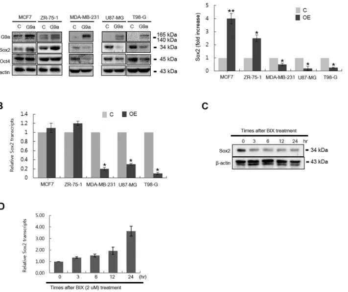
We next questioned whether G9a regulates Sox2 protein stability in other cell types. Interestingly, ectopic expression of G9a in ZR-75-1 cells, which like MCF7 cells are a representative ER(+) breast cancer cell line, increased Sox2 protein levels (Fig 4A). Contrary to the findings in MCF7 and ZR-75-1 cells, ectopic expression of G9a reduced Sox2 protein levels in MDA-MB-231 cells, Fig 3. Expression level of G9a is correlated with the migration and invasion of MCF7 cells.(A) Wound-healing assay examining the effect of chemical inhibition of G9a activity on the migration of MCF7 cells. MCF7 cells were treated with 0.2μM BIX-01294 and wounded using a micropipette tip when the cells were fully confluent. Cell migration was then monitored. (B) Wound-healing assay to examine the effect of ectopic expression of G9a on the migration of MCF7 cells. MCF7 cells were transfected with control or G9a expression plasmids and then re-seeded for the migration assay. When fully confluent, the cells were wounded using a micropipette tip, and cell migration was photographed at the indicated times. (C) The effect of G9a on the invasiveness of MCF7 cells was examined in a Transwell invasion assay. MCF7 cells were first transfected with control or G9a expression plasmids along with siNS or siSox2. Twenty-four hours later, cells were suspended in serum-free medium and seeded in 24-well Matrigel-coated Transwell chambers. Cells crossing the Matrigel-coated filter were fixed and stained with Crystal violet. The number of stained cells was then counted. Representative pictures are shown (left panel). The average number of stained cells in five random microscopic fields from three independent experiments is presented graphically (right panel). (D) The effect of G9a on the mammosphere-forming ability of MCF7 cells was examined in cells maintained under low-serum non-adherent culture conditions for 7 days in the presence or absence of 2μM BIX-01294. Abbreviations: NT, no treatment; Control (siNS), cells transfected with a control plasmid and non-specific siRNA; OE (siNS), cells transfected with a G9a-expressing plasmid and non-specific siRNA; OE (siSox2), cells transfected with a G9a-expressing plasmid and siSox2.*P<0.05 and**P<0.01 compared with the control group. Data are expressed as the mean±SD and representative of three independent
experiments.
an ER(-) breast cancer cell line, and in glioblastoma cells (U87-MG and T98-G;Fig 4A). Expres-sion of Sox2 mRNA in ZR-75-1 cells was not affected by overexpresExpres-sion of G9a, suggesting that the mechanism by which ZR-75-1 regulates Sox2 levels upon G9a expression is similar to that in MCF-7 cells (Fig 4B). However, the transcription of Sox2 in MDA-MB-231, U87-MG, and T98-G was decreased by ectopic expression of G9a (Fig 4B). These results indicate that G9a regu-lates Sox2 expression in MDA-MB-231, U87-MG, and T98-G via epigenetic regulation of tran-scriptional expression rather than by modulating protein stability. Since Sox2 plays an essential Fig 4. Effect of G9a on the transcription and protein expression of Sox2 in various cell types.(A) The effect of ectopic expression of G9a on Sox2 and Oct4 protein expression in three breast cancer cell lines (MCF7, ZR-75-1, and MDA-MB-231) and two glioblastoma cell lines (U87-MG and T98-G) was determined by immunoblot analysis. Each cell line was transfected with a G9a-expressing plasmid for 24 h and then harvested for analysis. For quantitative analysis of immunoblotting results, the mean density of the Sox2 band was measured with Multi Gauge V3.0 software, and the band density was divided by the density ofβ-actin to obtain the normalized band density. (B) Cells were transfected with control or G9a expressing plasmids for 24 h, and the effect of ectopic expression of G9a on the transcription of Sox2 was quantitatively analyzed by real-time RT-PCR. (C) The effect of G9a activity on Sox2 protein stability in mouse ESCs. Mouse ESCs were treated with 0.2μM BIX-01294 for the indicated times and then harvested for immunoblot analysis. (D) The effect of G9a activity on the transcription of Sox2 in mouse ESCs. Mouse ESCs were treated with 0.2μM BIX-01294 for the indicated times and then harvested for quantitative analysis of Sox2 transcription by real-time RT-PCR. Abbreviations: G9a, G9a-expressing plasmid; Sox2, Sox2-expressing plasmid; C, control plasmid.*P<0.05 compared with the control group. Data are expressed as the mean±SD and are representative of three independent experiments.
role in maintaining pluripotency in ESCs, we next questioned whether treating mouse ESCs with a chemical inhibitor would affect G9a lysine methyltransferase-mediated Sox2 protein stability in a manner similar to that observed in MCF7 cells.Fig 4Cshows that Sox2 protein levels in mouse ESCs decreased at 3 h after BIX-01249 treatment. We found it interesting that, in contrast to the changes observed in protein levels, Sox2 transcript levels were increased in a time-depen-dent manner (Fig 4D). Consistent with the increases of Sox2 transcript levels, we observed that the levels of H3K9me2 on the Sox2 promoter decreased significantly at 24 h after BIX-01294 treatment (data not shown). These results suggest that G9a regulates Sox2 levels in mouse ESCs through a dual mechanism: epigenetic regulation of transcription and regulation of protein sta-bility. Taken together, we concluded that the ability of G9a to increase Sox2 protein stability depends on the intracellular environment of each cell.
Discussion
Lysine methyltransferase G9a is a major histone modifier that links chromatin status to tran-scriptional gene regulation. Recent studies confirm that G9a methylates lysine residues within various non-histone proteins, including p53, Reptin, ACINUS, WIZ, CDYL1, and C/EBPβ
[22–25]. Here, we provide evidence that G9a regulates Sox2 protein stability in ER(+) breast cancer cells and mouse ESCs.
First, chemical inhibition of G9a activity resulted in a decrease in Sox2 protein levels with-out any changes in Sox2 transcript levels. Second, siRNA-mediated suppression of G9a coin-cided with decreased levels of Sox2 protein expression without parallel changes in Sox2 transcripts. Third, overexpression of G9a in MCF7 and ZR-75-1 cells induced marked accumu-lation of Sox2 protein, even though it had no effect on Sox2 transcript levels.
We found it interesting that the effect of G9a overexpression on the Sox2 protein was only observed in ER(+) breast cancer cell lines such as MCF7 and ZR-75-1, but not in ER(-) MDA-MB-231 cells. This suggests the possibility that the role played by G9a in maintaining Sox2 protein stability is specific to ER(+) breast cancer cells. However, it is not clear how G9a act as a Sox2 protein stabilizer in ER(+) cells, or whether the effect of G9a on Sox2 protein sta-bility depends on the ER signal. High expression of G9a in ER(+) MCF7 and ZR-75-1 cells is consistent with a previous report showing that G9a is a co-activator of nuclear receptors such as estrogen receptorα[26]. The expression pattern of G9a leads to the hypothesis that abun-dantly expressed G9a protein in ER(-) cells directly methylates Sox2 to protect it from ubiqui-tin-dependent degradation. Since G9a-mediated repression of E-cadherin in human breast cancer is mediated via interaction with Snail (which controls epithelial-mesenchymal transi-tion during embryogenesis and tumor progression) [27], it is possible that G9a stabilizes the Sox2 protein through coordinated interaction with unidentified co-factors that are exclusively expressed by ER(+) breast cancer cells. The detailed mechanism(s) underlying the cell type-specific activity of G9a on Sox2 protein stability requires further investigation.
amount of Sox2 protein in mouse ESCs by finely tuning the transcriptional and post-transla-tional mechanisms responsible for Sox2 expression.
We also found it interesting that, even though the effect of G9a on Sox2 protein expression in MCF7 and mouse ESCs is opposite to that in MDA-MB-231 cells, chemical inhibition of G9a activity led to a similar reduction in the proliferation of all three cell lines (S7 Fig). This suggests that the mechanism underlying G9a-mediated cell proliferation is independent of Sox2. The detailed mechanism by which G9a regulates cell proliferation requires further investigation.
The present study provides evidence that the amount of Sox2 protein in breast cancer cells is regulated by the expression and activity of G9a lysine methyltransferase. Therefore, it will be of particular importance to demonstrate that Sox2 is indeed methylated by G9a, and that meth-ylated Sox2 is resistant to proteolytic degradation.
Supporting Information
S1 Fig. MCF-7 cells were treated with the indicated concentrations of BIX-01294, and the effect of various concentrations of BIX-01294 on the transcription of G9a, Sox2, and Oct4 was quantitatively analyzed by real-time RT-PCR.
(TIF)
S2 Fig. The effect of chemical G9a inhibitor, UNC-0638, on Sox2 protein accumulation. MCF7 cells were treated with the indicated doses of UNC-0638 for 24 h. Whole cell lysates were then harvested and analyzed by immunoblotting with G9a- and Sox2-specific antibodies. (TIF)
S3 Fig. The effect of chemical inhibition (BIX-01294) (left) or siRNA-mediated knockdown of G9a expression (right) on the amount of Sox2 protein in MDA-MB-231 cells was ana-lyzed by immunoblotting.
(TIF)
S4 Fig. Immunoblot analysis of G9a expression levels in the MCF7, ZR-75-1, MDA-MB231, U-87MG, and T98G cells.β-actin was used as a loading control. (TIF)
S5 Fig. The transfection efficiency was analyzed by the detection of fluorescence 48 hrs after transfection of G9a-GFP expressing plasmid into MCF7 cells.
(TIF)
S6 Fig. The effect of G9a overexpression on the proliferation of MCF7 cells.Cell growth was analyzed by counting cell numbers at the indicated times after transfection with control or G9a plasmids. Abbreviations: Parent, wild-type MCF7 cells; C, MCF7 cells transfected with control plasmid; OE, MCF7 cells transfected with the G9a-expressing plasmid.P<0.05 compared with the control group. Data are expressed as the mean ± SD and are representative of three independent experiments.
(TIF)
S7 Fig. The effect of G9a overexpression on the proliferation of MDA-MB-231 (A) and mouse ESCs (B).Cell growth was analyzed by counting cells at the indicated times post-BIX-01294 treatment.P<0.05 compared with the control group. Data are expressed as the mean ± SD and are representative of three independent experiments.
S1 Table. Real-time RT-PCR primers. (DOCX)
Acknowledgments
This work was carried out with the support of“Cooperative Research Program for Agriculture Science & Technology Development (PJ010033)”, Rural Development Administration, Repub-lic of Korea.
Author Contributions
Conceived and designed the experiments: JYL SHH KSP. Performed the experiments: JYL SHL KSK CK. Analyzed the data: JJK DKK KSP. Wrote the paper: KSP.
References
1. Brown SE, Campbell RD, Sanderson CM (2001) Novel NG36/G9a gene products encoded within the human and mouse MHC class III regions. Mamm Genome 12: 916–924. PMID:11707778
2. Tachibana M, Sugimoto K, Nozaki M, Ueda J, Ohta T, Ohki T, et al. (2002) G9a histone methyltransfer-ase plays a dominant role in euchromatic histone H3 lysine 9 methylation and is essential for early embryogenesis. Genes Dev 16: 1779–1791. PMID:12130538
3. Pandey RR, Mondal T, Mohammad F, Enroth S, Redrup L, Komorowski J, et al. (2008) Kcnq1ot1 anti-sense noncoding RNA mediates lineage-specific transcriptional silencing through chromatin-level regu-lation. Mol Cell 32: 232–246. doi:10.1016/j.molcel.2008.08.022PMID:18951091
4. Nagano T, Mitchell JA, Sanz LA, Pauler FM, Ferguson-Smith AC, Feil R, et al. (2008) The Air noncod-ing RNA epigenetically silences transcription by targetnoncod-ing G9a to chromatin. Science 322: 1717–1720.
doi:10.1126/science.1163802PMID:18988810
5. Chen MW, Hua KT, Kao HJ, Chi CC, Wei LH, Johansson G, et al. (2010) H3K9 histone methyltransfer-ase G9a promotes lung cancer invasion and metastasis by silencing the cell adhesion molecule Ep-CAM. Cancer Res 70: 7830–7840. doi:10.1158/0008-5472.CAN-10-0833PMID:20940408
6. Huang J, Dorsey J, Chuikov S, Perez-Burgos L, Zhang X, Jenuwein T, et al. (2010) G9a and Glp meth-ylate lysine 373 in the tumor suppressor p53. J Biol Chem 285: 9636–9641. doi:10.1074/jbc.M109. 062588PMID:20118233
7. Kondo Y, Shen L, Ahmed S, Boumber Y, Sekido Y, Haddad BR et al. (2008) Downregulation of histone H3 lysine 9 methyltransferase G9a induces centrosome disruption and chromosome instability in can-cer cells. PLoS One 3: e2037. doi:10.1371/journal.pone.0002037PMID:18446223
8. Smallwood A, Esteve PO, Pradhan S, Carey M (2007) Functional cooperation between HP1 and DNMT1 mediates gene silencing. Genes Dev 21: 1169–1178. PMID:17470536
9. Mozzetta C, Pontis J, Fritsch L, Robin P, Portoso M, Proux C, et al. (2014) The histone H3 lysine 9 methyltransferases G9a and GLP regulate polycomb repressive complex 2-mediated gene silencing. Mol Cell 53: 277–289. doi:10.1016/j.molcel.2013.12.005PMID:24389103
10. Wu H, Chen X, Xiong J, Li Y, Li H, Ding X, et al. (2011) Histone methyltransferase G9a contributes to H3K27 methylation in vivo. Cell Res 21: 365–367. doi:10.1038/cr.2010.157PMID:21079650 11. Esteve PO, Chin HG, Smallwood A, Feehery GR, Gangisetty O, Karpf AR, et al. (2006) Direct
interac-tion between DNMT1 and G9a coordinates DNA and histone methylainterac-tion during replicainterac-tion. Genes Dev 20: 3089–3103. PMID:17085482
12. Zhang J, Gao Q, Li P, Liu X, Jia Y, Wu W, et al. (2011) S phase-dependent interaction with DNMT1 dic-tates the role of UHRF1 but not UHRF2 in DNA methylation maintenance. Cell Res 21: 1723–1739.
doi:10.1038/cr.2011.176PMID:22064703
13. Islam K, Chen Y, Wu H, Bothwell IR, Blum GJ, Zeng H, et al. (2013) Defining efficient enzyme-cofactor pairs for bioorthogonal profiling of protein methylation. Proc Natl Acad Sci U S A 110: 16778–16783.
doi:10.1073/pnas.1216365110PMID:24082136
14. Chaturvedi CP, Somasundaram B, Singh K, Carpenedo RL, Stanford WL, Dilworth WL, et al. (2012) Maintenance of gene silencing by the coordinate action of the H3K9 methyltransferase G9a/KMT1C and the H3K4 demethylase Jarid1a/KDM5A. Proc Natl Acad Sci U S A 109: 18845–18850. doi:10. 1073/pnas.1213951109PMID:23112189
target genes. Proc Natl Acad Sci U S A 109: 19673–19678. doi:10.1073/pnas.1211803109PMID: 23151507
16. Dong C, Wu Y, Yao J, Wang Y, Yu Y, Rychahou PG, et al. (2012) G9a interacts with Snail and is critical for Snail-mediated E-cadherin repression in human breast cancer. J Clin Invest 122: 1469–1486. doi: 10.1172/JCI57349PMID:22406531
17. Kondo Y, Shen L, Suzuki S, Kurokawa T, Masuko K, Tanaka Y, et al. (2007) Alterations of DNA methyl-ation and histone modificmethyl-ations contribute to gene silencing in hepatocellular carcinomas. Hepatol Res 37: 974–983. PMID:17584191
18. Cho HS, Kelly JD, Hayami S, Toyokawa G, Takawa M, Yoshimatsu M, et al. (2011) Enhanced expres-sion of EHMT2 is involved in the proliferation of cancer cells through negative regulation of SIAH1. Neo-plasia 13: 676–684. PMID:21847359
19. Chen H, Yan Y, Davidson TL, Shinkai Y, Costa M (2006) Hypoxic stress induces dimethylated histone H3 lysine 9 through histone methyltransferase G9a in mammalian cells. Cancer Res 66: 9009–9016.
PMID:16982742
20. Hua KT, Wang MY, Chen MW, Wei LH, Chen CK, Ko CH, et al. (2014) The H3K9 methyltransferase G9a is a marker of aggressive ovarian cancer that promotes peritoneal metastasis. Mol Cancer 13: 189. doi:10.1186/1476-4598-13-189PMID:25115793
21. Fang L, Zhang L, Wei W, Jin X, Wang P, Tong Y, et al. (2014) A methylation-phosphorylation switch determines Sox2 stability and function in ESC maintenance or differentiation. Mol Cell 55: 537–551.
doi:10.1016/j.molcel.2014.06.018PMID:25042802
22. Pless O, Kowenz-Leutz E, Knoblich M, Lausen J, Beyermann M, Walsh MJ, et al. (2008) G9a-mediated lysine methylation alters the function of CCAAT/enhancer-binding protein-beta. J Biol Chem 283: 26357–26363. doi:10.1074/jbc.M802132200PMID:18647749
23. Rathert P, Dhayalan A, Murakami M, Zhang X, Tamas R, Jurkowska R, et al. (2008) Protein lysine methyltransferase G9a acts on non-histone targets. Nat Chem Biol 4: 344–346. doi:10.1038/ nchembio.88PMID:18438403
24. Lee JS, Kim Y, Kim IS, Kim B, Choi HJ, Lee JM, et al. (2010) Negative regulation of hypoxic responses via induced Reptin methylation. Mol Cell 39: 71–85. doi:10.1016/j.molcel.2010.06.008PMID: 20603076
25. Huang J, Dorsey J, Chuikov S, Perez-Burgos L, Zhang X, Jenuwein T, et al. (2010) G9a and Glp meth-ylate lysine 373 in the tumor suppressor p53. J Biol Chem 285: 9636–9641. doi:10.1074/jbc.M109. 062588PMID:20118233
26. Lee DY, Northrop JP, Kuo MH, Stallcup MR (2006) Histone H3 lysine 9 methyltransferase G9a is a tran-scriptional coactivator for nuclear receptors. J Biol Chem 281: 8476–8485. PMID:16461774
27. Dong C, Wu Y, Yao J, Wang Y, Yu Y, Rychahou PG, et al. (2012) G9a interacts with Snail and is critical for Snail-mediated E-cadherin repression in human breast cancer. J Clin Invest 122: 1469–1486. doi: 10.1172/JCI57349PMID:22406531


