Nanomed. J., 3(3):155-158, Summer 2016
155
A short review on Ferrofluids surface modification by natural and
biocompatible polymers
Mahyar Ebrahimi*
Sharif University of Technology, Tehran, Iran
ABSTRACT
This paper provides an overview of how the surface properties of ferromagnetic nanoparticles dispersed in fluids is modified by natural and biocompatible polymers. Among common magnetic nanoparticles, magnetite (Fe3O4) and maghemite (-Fe203) are popular candidates because of their biocompatibility. Natural polymeric coating materials are the most commonly used biocompatible magnetic nanoparticle coatings. In this paper, recent progresses in the methods of ferrofluids surface modification by the common natural polymers consisting of dextran, chitosan, gelatin and starch are reviewed.
Keywords: Biocompatibility, Ferrofluid, Magnetic nanoparticle, Surface modification
*Corresponding Author Email:[email protected]
Tel: (+98)912-0315625
Note. This manuscript was submitted on March 2, 2016; approved on May 24, 2016
INTRODUCTION
A magnetic fluid is a fluid in which fine magnetic particles are suspended in a carrier liquid. There are two major types of magnetic fluids; magneto-rheological fluids and Ferrofluids. The main difference of these two types is the size of magnetic particle. The former is the suspension of micrometer sized particles and the latter is the suspension of nanometer sized single domain magnetic particles. Broadly when particles size is between 5-40 nm the fluid is called Ferrofluid. The most important special property of ferrofluids is that they exhibit magnetic field dependent viscosity which allows tuning of the viscosity by changing the field in a fraction of second. The second special property is that properties of ferrofluids can be controlled by external magnetic field. The third is that they can generate heat when exposed to an alternating magnetic field. The fourth remarkable property is tunable magnetic properties of nanoparticles. Coercivity, Curie temperature and magnetic susceptibility of magnetic nanoparticles
can be improved and tuned by nanoparticle size and synthesis method [1-3].
For biomedical applications a nanoparticle must has high saturation magnetization, stability, biocompatibility and interactive functions at the surface. For in/vivo applications magnetic iron nanoparticles are main candidates [1]. Among magnetic iron oxides, magnetite (Fe3O4) and maghemite (-fe203) are popular candidates because their biocompatibility has already been proven [1, 4]. Iron oxide nanoparticles have hydrophobic surfaces and tend to agglomerate if suitable surface modification is not applied [1, 5]. The clusters caused by agglomeration would show different behavior due to strong magnetic dipole-dipole interaction [5]. Therefore surface modification for prevention of agglomeration is imperative. Surface modification can also be done for attachment of bioactive molecules. This surface modification can be carried out by adding atomic layers of organic polymers, inorganic metallic or oxides [1]. The most commonly used biocompatible magnetic nanoparticle coatings are natural polymeric coating materials consisting Nanomed. J., 3(3):155-158, Summer 2016
DOI: 10.7508/nmj.2016.03.002
REVIEW PAPER
How to cite this article
Nanomed. J., 3(3):155-158, Summer 2016
156 M. Ebrahimi
of dextran, chitosan, gelatin and starch. Recent progresses in the surface modification of ferrofluids by natural polymers are reviewed in this paper.
Dextran
Dextran is a long chain polymer composed of glucose with mostly -1, 6 glycoside linkages. In alkaline solutions dextran interacts with hydroxyl groups present on iron oxide particles [6]. It has been shown that dextran coating is biocompatible [7] and stable in most tissues environments [8]. Dextran is also biodegradable, inexpensive, non-toxic and easily available [9] and it enhances the blood circulation time [10, 11].
In recent years the most common dextran coated iron oxide synthesis method has been co-precipitation. Easo and mahanan [12] synthesized superparamagnetic iron oxide nanoparticles by insitu co-precipitation of ferrous and ferric salts with base tethrametylammonium hydroxide in the presence of urea. The produced particles were found to have a narrow size distribution. They observed that synthesized particles have negligible effect on cell adhesion capacity and morphology of L929 cells.
One problem in application of dextran is that dextran with molecular weight below 60 kDa will filter through the glomerulus and cause problem [13]. Considering this factor surasuaty et al. [14] synthesized high molecular weight coated iron oxide nanoparticles by alkaline co-precipitation of ferrous and ferric salts in aqueous solution. They reported high degree of blood compatibility and high stability (about 4 months).
Anastasia et al. investigated the effect of co-precipitation type on properties of dextran coated iron oxide nanoparticle [15].
They found that timing of the addition of the dextran into the reaction mixture has strong effect on nanoparticle polymers. They reported that semi-two-step methods based on the simultaneous injection of recuing agent and the dextran solution into the reaction mixture of the synthesizing has the best performance. They also reported that many of the nanoparticle properties can be tuned by adjusting the timing of dextran addition to the reaction.
For gaining more stability double coating nanoparticles can be produced. For example Barroset al. [16] were formed magnetic nanoparticle double coated with dextran and chitosan by layer-by-layer deposition method and reported 60 days stability.
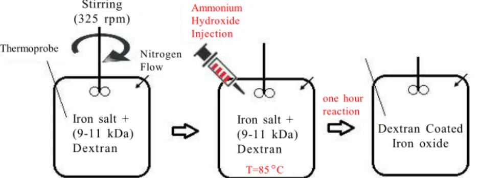
Fig. 1 shows a schematic of one step Dextran coated Iron oxide nanoparticle synthesis in which co-precipitation reaction is completed at 85oC for an hour
after addition of the reducing agent (ammonium hydroxide).
Chitosan
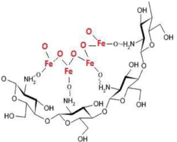
Chitosan is a biocompatible, biodegradable and bioactive polyamino-saccharide which has been widely used in biological applications [17, 18]. As it can be observed in Fig. 2 the amino group (NH2) of chitosan is bonded to the particle.
Various methods have been used to produce chitosan coated magnetic nanoparticles such as blending [19] and co-precipitation [20]. Mainly the chitosan particles are formed through the electrostatic interaction between positively charged chitosan and negatively charged tripolyphosphates (TPP). Li et al. [21] synthesized Fe3O4-chitosan nanoparticles by the covalent binding of chitosan onto the surface of hydrothermally prepared Fe3O4 nanoparticle. They reported that the coating did not
Iron salt + (9-11 kDa) Dextran
Iron salt + (9-11 kDa) Dextran
Dextran Coated Iron oxide Stirring
(325 rpm)
Thermoprobe
Nitrogen Flow
Ammonium Hydroxide Injection
T=85C
one hour reaction
Nanomed. J., 3(3):155-158, Summer 2016
157 change the spinel structure of Fe3O4and the produced nanoparticles were mono-disperse with spherical shape.
Songet al. [22] synthesized chitosan coated Fe3O4 nanoparticles by combining Fe3O4and chitosan chemically modified with PEG and lactobionicacid (LA) group in one step. They reported that the nanoparticles were stable, cytotoxic and non-tissue toxic.
Zamora Mora et al. [23] encapsulated Fe3O4 nanoparticles into chitosan cross linked with tripolyphosphades. They observed that size of chitosan shell is increased and the results of cytotoxic analyses proved its biocompatibility.
Gelatin
Gelatin is a mixture of peptide and proteins derived from collagen partial hydrolyses. It is low toxic bio degradable and non-immunogenic [24]. In the synthesis procedure of gelatin coated nanoparticles adjusting the cross-linking degree of the layer is so important and it affects many of particle properties [25].
Recently Che et al. [25] produced magnetic nanoparticle coated by gelatin layer. They used glutaraldehyde as cross- linking agent to adjust the amount of cross-linking.
Gelatin also can be used in Ferrogels. Helminger et al.[26] presented a synthesis method to produce biocompatible Ferrogels with gelatin, gel matrix and magnetic nanoparticles. They reported that different cross-linking degree of gel leads to different mechanical and magnetic properties of the gel.
Starch
Starch is a carbohydrate consisted of glucose units which are joined by glycosidic bonds and is synthesized by most of the green plants as energy storage medium.
Cross-link starch coated iron oxide nanoparticles have attracted attention for biomedical applications. Coleet al. [27] produced long circulation life magnetic nanoparticles of PEG modified cross-linked starch coated iron oxide nanoparticles. They reported good stability and performance of starch coated nanoparticles. Other researchers reported successful synthesis and application of iron oxide nanoparticle coated with starch [28, 29].
CONCLUSION
All in all, The best candidate for using as biocompatible magnetic nanoparticle coatings are natural polymeric coating materials including dextran, chitosan, gelatin and starch. This paper presented a general overview of these biocompatible and biodegradable coating materials in ferrofluids surface modification for in/vivo applications.
REFERENCES
1. Gupta AK, Gupta M. Synthesis and surface engineering of iron oxide nanoparticles for biomedical applications. Biomaterials. 2005; 26(18): 3995-4021.
2. Wu W, He Q, Jiang C. Magnetic iron oxide nanoparticles: synthesis and surface functionalization strategies. J Cheminform. 2009; 40(24): i.
3. Issa B, Obaidat IM, Albiss BA, Haik Y. Magnetic nanoparticles: surface effects and properties related to biomedicine applications. Int J Mol Sci. 2013; 14(11): 21266-305. 4. Schwertmann U, Cornell RM. Iron oxides in the laboratory:
preparation and characterization. John Wiley & Sons; 2008. 5. Hamley IW. Nanotechnology with soft materials. Angew Chem
Int Ed Engl. 2003; 42(15): 1692-712.
6. Sreeja V, Joy PA. Effect of inter–particle interactions on the magnetic properties of magnetite nanoparticles after coating with dextran. Int J Nanotechnol. 2011; 8(10-12): 907-15. 7. Berry CC, Wells’ S, Charles S, Curtis AS. Dextran and albumin
derivatised iron oxide nanoparticles: influence on fibroblasts in vitro. Biomaterials. 2003; 24(25): 4551-7.
8. Frazier RA, Davies MC, Matthijs G, Roberts CJ, Schacht E, Tendler SJ, Williams PM. In situ surface plasmon resonance analysis of dextran monolayer degradation by dextranase. Langmuir. 1997; 13(26): 7115-20.
9. Thomas JJ, Rekha MR, Sharma CP. Unraveling the intracellular efficacy of dextran-histidine polycation as an efficient nonviral gene delivery system. Mol Pharm. 2011; 9(1): 121-34. Fig. 2. Molecular representations of Iron oxide - chitosan
Nanomed. J., 3(3):155-158, Summer 2016
158
surface modification by natural polymers
10. Berry CC, Curtis AS. Functionalisation of magnetic nanoparticles for applications in biomedicine. J Phys D Appl Phys. 2003; 36(13):R198.
11. Berry CC, Wells’ S, Charles S, Curtis AS. Dextran and albumin derivatised iron oxide nanoparticles: influence on fibroblasts in vitro. Biomaterials. 2003; 24(25): 4551-7. 12. Easo SL, Mohanan PV. Dextran stabilized iron oxide
nanoparticles: synthesis, characterization and in vitro studies. Carbohydr Polym. 2013; 92(1): 726-32.
13. Feest TG. Low molecular weight dextran: a continuing cause of acute renal failure. Br Med J. 1976; 2(6047): 1300. 14. Saraswathy A, Nazeer SS, Nimi N, Arumugam S, Shenoy SJ,
Jayasree RS. Synthesis and characterization of dextran stabilized superparamagnetic iron oxide nanoparticles for in vivo MR imaging of liver fibrosis. Carbohydr Polym. 2014; 101: 760-8.
15. Hauser AK, Mathias R, Anderson KW, Hilt JZ. The effects of synthesis method on the physical and chemical properties of dextran coated iron oxide nanoparticles. Mater Chem Phys. 2015; 160: 177-86.
16. Barbosa-Barros L, García-Jimeno S, Estelrich J. Formation and characterization of biobased magnetic nanoparticles double coated with dextran and chitosan by layer-by-layer deposition. Colloids Surf A Physicochem Eng Asp. 2014; 450: 121-9.
17. Chang YC, Chen DH. Adsorption Kinetics and Thermodynamics of Acid Dyes on a Carboxymethylated Chitosan Conjugated Magnetic Nano Adsorbent. Macromol Biosci. 2005; 5(3): 254-61.
18. Chang YC, Chen DH. Preparation and adsorption properties of monodisperse chitosan-bound Fe 3 O 4 magnetic nanoparticles for removal of Cu (II) ions. J Colloid Interface Sci. 2005; 283(2): 446-51.
19. Denkbaº EB, Kiliçay E, Birlikseven C, Öztürk E. Magnetic chitosan microspheres: preparation and characterization. React Funct Polym. 2002; 50(3): 225-32.
20. Honda H, Kawabe A, Shinkai M, Kobayashi T. Development of chitosan-conjugated magnetite for magnetic cell separation. J. Ferment. Bioeng. 1998; 86(2): 191-6. 21. Li GY, Jiang YR, Huang KL, Ding P, Chen J. Preparation and
properties of magnetic Fe 3 O 4–chitosan nanoparticles. J Alloys Compd s. 2008; 466(1): 451-6.
22. Song X, Luo X, Zhang Q, Zhu A, Ji L, Yan C. Preparation and characterization of biofunctionalized chitosan/Fe3O4
magnetic nanoparticles for application in liver magnetic resonance imaging. J Magn Magn Mater. 2015; 388: 116-22. 23. Zamora-Mora V, Fernández-Gutiérrez M, San Román J, Goya G, Hernández R, Mijangos C. Magnetic core–shell chitosan nanoparticles: Rheological characterization and hyperthermia application. Carbohydr Polym. 2014; 102: 691-8.
24. Mody VV, Cox A, Shah S, Singh A, Bevins W, Parihar H. Magnetic nanoparticle drug delivery systems for targeting tumor. Appl Nanosci. 2014; 4(4): 385-92.
25. Che E, Gao Y, Wan L, Zhang Y, Han N, Bai J, Li J, Sha Z, Wang S. Paclitaxel/gelatin coated magnetic mesoporous silica nanoparticles: Preparation and antitumor efficacy in vivo. Microporous Mesoporous Mater. 2015; 204: 226-34. 26. Helminger M, Wu B, Kollmann T, Benke D, Schwahn D, Pipich
V, Faivre D, Zahn D, Cölfen H. Synthesis and Characterization of Gelatin Based Magnetic Hydrogels. Adv Funct Mater. 2014; 24(21): 3187-96.
27. Cole AJ, David AE, Wang J, Galbán CJ, Yang VC. Magnetic brain tumor targeting and biodistribution of long-circulating PEG-modified, cross-linked starch-coated iron oxide nanoparticles. Biomaterials. 2011; 32(26): 6291-301. 28. Cole AJ, David AE, Wang J, Galbán CJ, Hill HL, Yang VC.
Polyethylene glycol modified, cross-linked starch-coated iron oxide nanoparticles for enhanced magnetic tumor targeting. Biomaterials. 2011; 32(8): 2183-93.