Tat–Dependent Translocation of an F
420
–Binding Protein
of
Mycobacterium tuberculosis
Ghader Bashiri1, Ellen F. Perkowski2, Adrian P. Turner3, Meghan E. Feltcher2, Miriam Braunstein2, Edward N. Baker1*
1School of Biological Sciences,Maurice Wilkins Centre for Molecular Biodiscovery, The University of Auckland, Auckland, New Zealand,2Department of Microbiology and Immunology, University of North Carolina School of Medicine, Chapel Hill, North Carolina United State of America,3Microscopy and Graphics Unit, School of Biological Sciences, The University of Auckland, Auckland, New Zealand
Abstract
F420is a unique cofactor present in a restricted range of microorganisms, including mycobacteria. It has been proposed that
F420has an important role in the oxidoreductive reactions ofMycobacterium tuberculosis, possibly associated with anaerobic
survival and persistence. The protein encoded by Rv0132c has a predicted N–terminal signal sequence and is annotated as an F420–dependent glucose-6-phosphate dehydrogenase. Here we show that Rv0132c protein does not have the annotated
activity. It does, however, co–purify with F420 during expression experiments inM. smegmatis. We also show that the
Rv0132c–F420 complex is a substrate for the Tat pathway, which mediates translocation of the complex across the
cytoplasmic membrane, where Rv0132c is anchored to the cell envelope. This is the first report of any F420–binding protein
being a substrate for the Tat pathway and of the presence of F420 outside of the cytosol in any F420–producing
microorganism. The Rv0132c protein and its Tat export sequence are essentially invariant in theMycobacterium tuberculosis complex. Taken together, these results show that current understanding of F420 biology in mycobacteria should be
expanded to include activities occurring in the extra-cytoplasmic cell envelope.
Citation:Bashiri G, Perkowski EF, Turner AP, Feltcher ME, Braunstein M, et al. (2012) Tat–Dependent Translocation of an F420–Binding Protein ofMycobacterium
tuberculosis. PLoS ONE 7(10): e45003. doi:10.1371/journal.pone.0045003
Editor:Je´roˆme Nigou, French National Centre for Scientific Research - Universite´ de Toulouse, France
ReceivedFebruary 6, 2012;AcceptedAugust 14, 2012;PublishedOctober 22, 2012
Copyright:ß2012 Bashiri et al. This is an open-access article distributed under the terms of the Creative Commons Attribution License, which permits unrestricted use, distribution, and reproduction in any medium, provided the original author and source are credited.
Funding:Funded by the Health Research Council of New Zealand, the Foundation for Research, Science and Technology of New Zealand and the United States National Institutes of Health (grant RO1 AI054540). The funders had no role in study design, data collection and analysis, decision to publish, or preparation of the manuscript.
Competing Interests:The authors have declared that no competing interests exist.
* E-mail: [email protected]
Introduction
Tuberculosis (TB) is a devastating and contagious infectious disease. It is estimated thatMycobacterium tuberculosis (Mtb) bacilli, the causative agent of this disease, infect one–third of the world’s population, while TB claims nearly two million lives a year [1]. Complications from co–infection with HIV/AIDS and the rise of multiple–drug (MDR) and extensively drug–resistant (XDR) strains ofMtbmake TB a worldwide concern [1]. WHO estimates that there are nearly half a million new cases of MDR–TB each year; about 5% of the nine million new TB cases of all types [1]. Although some promising anti–TB drugs are in clinical trials, there have been no new drugs against TB in the last thirty years [2]. Understanding the biochemistry and physiology of active and persistent TB will help to reveal the basis of pathogenesis, making it possible to combat the disease more effectively.
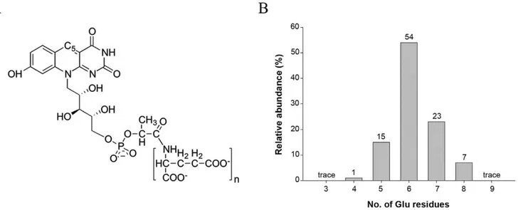
The coenzyme F420is a 5-deazaflavin derivative that has been
recently proposed to play a substantial role in the redox reactions ofMtb[3]. F420contains an isoalloxazine chromophore with a side
chain composed of ribitol and phospholactate moieties and a poly– glutamate tail of variable length (Figure 1A). The isoalloxazine chromophore of F420 is very similar to that of the flavins (FMN
and FAD), with the major difference being the atoms involved in the oxidoreductive reactions. Oxidoreduction of F420is achieved
by hydride transfer between a substrate molecule and C5of the
5-deazaflavin moiety, whereas transfer occurs to N5in FMN and
FAD. Despite its structural similarity to the flavins, F420 is
functionally similar to NAD(P)+
, being involved in hydride transfer reactions [for a review see [4] and references therein].
The number of known F420–dependent proteins in
mycobac-terial species is growing, and a few of such activities have been experimentally shown, including F420–dependent
glucose-6-phos-phate dehydrogenase (FGD) [5,6], deazaflavin–dependent nitror-eductase (Ddn) [7,8] and F420H2–dependent reductase (FDR) [9]
activities. Bioinformatic analyses have indicated the presence of three different F420–dependent families in Mtb with at least 28
members; the luciferase–like monooxygenase (LLM), pyridox-amine 59-phosphate oxidase (PNPOx), and the deazaflavin– dependent nitroreductase (DDN) families [3]. We have previously characterized the structure and function of FGD1 fromMtband showed that it has the annotated activity, providing reduced F420
for cell metabolism [5]. The reaction catalysed by FGD1 is equivalent to the first step in the pentose phosphate pathway, which normally provides NADPH for reductive biosynthetic reactions and maintenance of the cellular redox state. The enzyme Ddn uses the reduced F420produced by FGD1 to activate
a promising anti–TB prodrug PA–824 [10], which is currently in the second phase of clinical trials. The physiological role of Ddn, however, is yet to be identified. It has also been shown that reduced F420can convert NO2to NOin vitro, implying a possible
[11]. All these observations point to the fact that cofactor F420has
an important role in the physiology, and likely in the pathogenesis, ofMtb.
The Mtb genome [12] encodes a second protein that is homologous with FGD1 (36% protein sequence identity), anno-tated as FGD2. Expression of FGD2 (Rv0132c) is under control of the sigma factor SigF [13], which is expressed during stationary growth phase and under stress conditionsin vitro[14]. A complete genomic microarray analysis revealed that Rv0132c was down– regulated in aDsigFmutant ofMtbin late stationary phase [13]. We set out to determine whether Rv0132c does indeed bind the coenzyme F420and whether it has the annotated F420–dependent
glucose-6-phosphate dehydrogenase activity. We also noted that Rv0132c has a predicted N–terminal signal sequence that contains motifs suggestive of export via the twin-arginine translocation (Tat) pathway [15], together with post–translational lipid modification. Here we investigate the possible significance of the Rv0132c protein in F420 metabolism in Mtb. We report experimental
evidence regarding the cellular location of Rv0132c, consistent with post–translational lipidation of the protein. In addition, we show that the Rv0132c protein does bind the cofactor F420, has a
functional twin–arginine translocation (Tat) signal sequence, and is exported to the cell envelope by the Tat-dependent pathway. These results demonstrate that Rv0132c and theMtbTat pathway have a direct role in transferring the cofactor F420 across the
cytoplasmic membrane, and suggest that the known roles of F420
should be expanded to include activities in the cell envelope.
Materials and Methods
Mycobacterial strains and plasmids used in this study are in Table S1, and primers used in the amplification of the various constructs are detailed in Table S2. Full methodological details of bacterial growth, PCR amplification, cloning, homology model-ing, western blotting and immunoelectron microscopy are given in the Supporting Information (Text S1).
Sequence data
Sequence data for Rv0132c and its orthologues in various mycobacterial species were retrieved from the published (www.
ncbi.nlm.nih.gov/gene) and unpublished (http://www.sanger.ac. uk/cgi-bin/blast/submitblast/mycobacterium) sequences and the alignments were carried out using CLUSTALW [15].
Protein expression and purification
The ORF encoding Rv0132c was PCR–amplified from Mtb H37Rv genomic DNA (Text S1). When its amino acid sequence is compared with the homologous FGD1, Rv0132c has a 38-residue N-terminal extension, which was predicted by the program PRED–TAT [16] to be a signal sequence with a twin-arginine translocation (Tat) motif. For functional analysis, a construct Rv0132c–D38 was therefore prepared that encodes the Rv0132c protein without its predicted signal sequence and with an N– terminal His–tag, cleavable by TEV protease (Text S1). The Rv0132c protein was expressed in M. smegmatis mc24517 cells [17,18], grown for four days, after which the cells were lysed in 20 mM HEPES pH 7.5, 150 mM NaCl, 20 mM imidazole and 1 mMb–ME. After centrifugation, Rv0132c was purified from the supernatant by Ni2+
–affinity chromatography, cleavage of the His–tag and size exclusion chromatography (SEC) using the same buffer as for lysis (with no imidazole). Analytical SEC was used to determine the oligomeric state of the purified Rv0132c protein, using low molecular weight markers (conalbumin, 75 kDa; ovalbumin, 43 kDa; chymotrypsinogen, 25 kDa; and ribonuclease A, 13.7 kDa; GE Healthcare) to prepare a standard curve (Figure S1). The elution volume of Rv0132c protein was then used to estimate its molecular weight and oligomeric state.
Rv0132c activity experiments
Assays for the annotated FGD activity of Rv0132c were performed using previously published protocols [5,6], using appropriate concentrations of the purified Rv0132c, cofactor F420(25mM) and glucose-6-phosphate (0.01–1 mM). The change
in cofactor F420absorbance at 420 nm was monitored over 5 min
using UV–visible spectroscopy on a SpectraMAX microplate spectrophotometer (Molecular Devices). The reactions contained 100mL of 20 mM HEPES pH 7.0, 150 mM NaCl, 1 mMb–ME and were performed in 96–well format (Greiner bio–one, Germany).
Figure 1. Molecular structure of cofactor F420. (A) Schematic representation of cofactor F420, where n varies from 2–9 in different microorganisms. (B) Mass spectrometry analysis of cofactor F420bound to the purified Rv0132c–D38 protein showing the population of species differing in the number of glutamate residues in the poly-Glu tail.
Cofactor characterization and F420preparation
The protein off the gel filtration column was boiled at 100uC for 15 min and centrifuged at 160006g. The resulting supernatant was adjusted to pH,7 with formic acid and applied to a 10 mg C-18 reversed phase solid phase extraction (SPE) cartridge. The SPE cartridge was then washed with 300mL water and then 150mL of 50 mM ammonium bicarbonate. The alkaline elution was collected and concentrated to 10mL and was then diluted to 20mL with 20% acetonitrile. The resulting solution was back– filled into a static nanoelectrospray needle (tip diameter 4mm)
which was then mounted into a nanoelectrospray interface of a Finnigan LTQ FT mass spectrometer. Ion trap andioncyclotron resonance (ICR) cell data were obtained using a source voltage of 1.3 kV, capillary temperature of 225uC and capillary voltage of 26 V. MS/MS spectra were obtained by isolating key molecular ions and fragmenting using helium as the collision gas and 35% collision energy.
The F420coenzyme was purified from large scale preparations
of M. smegmatismc24517 cells expressingMtb–FGD1 or FbiABC (three ORFs involved in the biosynthesis of F420) constructs [4] in a
19.5–liter fermentor (New Brunswick Scientific). The purification was carried out using solvent extraction, ion exchange, adsorption and desalting chromatography steps as described before [4,19]. The absorption of F420 at 400 nm (e400= 25 mM21cm21) was
used to determine its concentration [20].
Tat–dependent translocation of Rv0132c
The first 42 residues of the Rv0132c protein were used to generate a BlaC fusion construct (Rv0132cSS–’BlaC), which was then transformed into M. smegmatis DlysDblaS (PM759) [21], M. smegmatis DlysDblaSDtatA (JM578) [22] and M. tuberculosis DblaC (PM638) [21] (Text S1). Transformants were tested for carben-icillin resistance by plating on media containing 50mg/mL carbenicillin, as an indication of Tat-dependent export of ’BlaC fusion constructs. For M. smegmatis and Mtb strains 500–1000 bacteria were plated on +/2 carbenicillin containing agar. Carbenicillin resistance (+) was scored when .90% of colonies plated grew on carbenicillin, whereas carbenicillin sensitivity (2) was defined in strains showing 0% (no single colony growth) on carbenicillin containing media (Text S1).
As an independent method of proving Rv0132c is exported out of the cytoplasm by the Tat pathway, epitope-tagged full-length Rv0132c (Rv0132c-HA) was introduced intoM. smegmatismc2155 (wild type) [23] and JM576 (DtatC mutant) [22]. Cells were fractionated after lysis and were then used for western blotting using an anti-HA primary antibody.
Subcellular localization of native Rv0132c inMtb
The subcellular localization of native Rv0132c in Mtb was determined by western blotting and immunoelectron microscopy (Text S1), using polyclonal antibodies raised against purified Rv0132c. The preimmune serum was used as a control. Whole cell lysate (WCL) derived fromMtbH37Rv cells was fractionated using differential ultracentrifugation to yield cell wall (CW), cytoplasmic membrane (CM), and soluble (SOL) fractions [24]. In addition, Mtblipoproteins were prepared using Triton X–114 partitioning [24,25]. For immunogold microscopy,MtbH37Ra cells were fixed and dehydrated in ethanol, pelleted and transferred to fresh resin in gelatin capsules for polymerization overnight at 60uC. Ultrathin sections (,80 nm) were cut with a 45 degree diamond knife
(Diatome) on an EM UC6 and collected onto 400–mesh nickel grids. For immunogold labelling, grids were incubated with either antiserum or preimmune serum, washed, blotted and transferred to drops of secondary antibody (goat anti–rabbit labelled with
10 nm gold, Sigma). Grids were washed, stained, air–dried and viewed in either a Philips CM12 or an FEI Tecnai 12 TEM, both operating at 120 kV.
Results
Rv0132c is an F420–binding protein
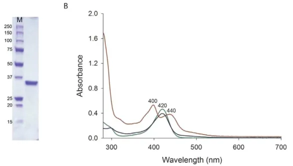
The Rv0132c protein, expressed in M. smegmatis from an Rv0132c–D38 construct lacking the predicted N–terminal signal sequence, was purified by Ni–NTA and SEC. The protein eluted in the Ni–NTA step was resolved as a single band on an SDS– PAGE gel (Figure 2A). Analysis of the purified protein by analytical SEC showed that the elution volume corresponded to a molecular weight of 75 kDa (Figure S1), consistent with a dimer in solution when compared with the monomer molecular weight of 35.2 kDa. The purified Rv0132c protein had a light yellow color that was retained after gel filtration, indicating that the source of the color remained bound to the protein.
To characterize the protein–bound yellow color, the purified protein was heat–denatured and the resulting yellow supernatant was analysed by mass spectrometry. This showed that bound F420
is indeed responsible for the yellow color of the protein. The mass spectrometry identified F420 molecules with varying lengths of
poly–glutamate tail, ranging from 4–8 glutamate residues (Figure 1B). This corresponds well with the range of F420species
extracted from wild typeM. smegmatiscells [5,19] and implies that the protein does not discriminate among F420species with different
numbers of glutamate residues. A similar promiscuity of binding has recently been shown for the enzyme Ddn, which binds F420-2
and F420-5 with similar affinity [26]. These findings are in line with
our hypothesis, from studies on FGD1, that the length of the F420
poly–glutamate tail may not affect reaction catalysis in F420–
dependent oxidoreductive enzymes [4].
The absorption spectrum of purified Rv0132c (Figure 2B) shows two absorption peaks, at 400 and 440 nm. In contrast, the F420
extracted from the purified Rv0132c shows a single peak at 420 nm, identical to that of the F420extracted fromM. smegmatis
(Figure 2B). This suggests that in the Rv0132c protein environ-ment the F420absorption spectrum is perturbed compared with its
free form. Considering that this recombinant Rv0132c–D38 lacks its native signal sequence for export and hence is restricted to the cytosol, our findings indicate that Rv0132c binds the F420cofactor
in the cytosol, independent of its signal sequence and final destination.
Rv0132c is incorrectly annotated
Of the two Mtb gene products annotated with FGD activity, FGD1 has been fully characterized with respect to structure and function [5]. Activity assays over a range of concentrations of enzyme and glucose-6-phosphate failed to detect any FGD activity for Rv0132c, however, under conditions where FGD1 was fully active (Figure 3). This indicates that Rv0132c is mis-annotated as an F420-dependent glucose-6-phosphate dehydrogenase. Sequence
alignments and homology modeling of Rv0132c (Text S1; Figure 4) show that the F420–binding residues identified in FGD1 are also
present in Rv0132c but that there are differences in the substrate binding site. In particular, three phosphate binding residues in FGD1, Lys198, Lys259, and Arg283 [5], are not present in Rv0132c. Superposition of the modeled Rv0132c on to the FGD1 experimental structure (Figure 4B) further shows that helixa9in
that whereas Rv0132c, like FGD1, has the ability to bind F420, the
two enzymes probably act on different substrates and catalyze different reactions. Our results provide experimental confirmation for the previous bioinformatic prediction that the Rv0132c protein would not have the annotated activity, based on sequence homology [27]. We conclude that Mtb possesses just a single enzyme with demonstrated FGD activity (FGD1) and suggest that the suffix 1 should be removed from FGD1 to prevent further confusion with the incorrectly–annotated FGD2.
Rv0132c and its signal sequence are conserved in pathogenic mycobacteria
Analysis of the Rv0132c sequence (Figure 4A) using the program PRED–TAT [16] predicts a 40-residue N-terminal signal sequence, with a twin-arginine translocation (Tat) motif. The signal sequence also contains a cysteine residue, within a lipobox motif, that implicates Rv0132c as a lipoprotein [27] destined for anchoring to the cell envelope after the post– translational addition of a lipid moiety. Although lipoproteins are mainly translocated across the cytoplasmic membrane using the Sec pathway, it has been reported that some lipoproteins are exported by the Tat system [28,29,30,31,32].
Sequence searches show that all members of theMtbcomplex (M. tuberculosis,M. bovis,M. africanum,M. canettiandM. microti) have homologues of Rv0132c with full conservation (99–100% identity) of the mature protein sequence and the signal sequence, including the Tat motif. Other pathogenic microbacteria such asM. kansasii, M. aviumandM. aviumsubsp.paratuberculosishave homologues with lower sequence identity (75–80%) that in most cases retain the Tat export sequence. In contrast, non-pathogenic mycobacteria such asM. smegmatis have homologues of Rv0132c that are of much lower sequence identity (30–40%) and all appear to lack a Tat signal sequence.
Rv0132c possesses a functional Tat signal sequence The Tat pathway is responsible for transporting folded proteins across the cytoplasmic membrane [33], being different from the Sec pathway, which transports proteins in an unfolded state [34]. Proteins are targeted to the Tat protein translocase, which includes three integral membrane proteins, TatA, TatB and TatC, using an N–terminal signal sequence containing a twin–arginine motif [35]. While the putative Rv0132c signal sequence resembles a Tat export sequence, past studies show that bioinformatic predictions of Tat substrates are problematic and experimental validation of Tat export is critical [36]. Of four tested programs to predict Tat signal sequences, PRED–TAT [16] was the only one to predict Rv0132c protein to be a Tat substrate, emphasizing the need for
Figure 2. SDS–PAGE and UV–visible spectra of the purified Rv0132c–D38 protein.(A) SDS–PAGE gel of the purified Rv0132c–D38 protein. (B) UV-visible spectra for purified Rv0132c (0.5 mg/mL in PBS, red), F420extracted fromM. smegmatiscells (50mM in PBS, green) and F420extracted from the purified Rv0132c (blue). M: molecular weight markers (kDa).
doi:10.1371/journal.pone.0045003.g002
Figure 3. Functional assay of Rv0132c–D38 protein. The FGD activity was assessed for Rv0132c–D38 protein and Mtb–FGD1 as a positive control.Mtb–FGD1 shows a decrease at 420 nm absorbance (green and red lines), whereas Rv0132c–D38 protein indicated no change in the absorbance (yellow and blue lines). The same results were observed using various concentrations of Rv0132c–D38 protein in the presence of different concentrations of glucose-6-phpsphate. The graph shows assays containing 1mM of each enzyme, 25mM F420with 0.1 mM (green and yellow lines) and 1 mM (red and blue lines) glucose-6-phosphate.
an experimental approach for verification. To this end we used a BlaC reporter system [22,36,37] to determine whether Rv0132c has a functional Tat signal sequence [22]. BlaC ofMtb(and BlaS forM. smegmatis) is a secretedb–lactamase that confers resistance tob–lactam antibiotics and can be used as a reporter for Tat– dependent export. This is because BlaC is normally exportedvia the Tat pathway [22] and, when expressed without its signal sequence, the truncated BlaC (’BlaC) is not exported, and does not conferb–lactam resistance [22]. In–frame fusion of a functional Tat signal sequence to ’BlaC, however, rescues export and b– lactam resistance [22]. In this way, signal sequences can be tested for their ability to promote Tat–dependent export [22]. Impor-tantly, the ’BlaC reporter only works with Tat (and not Sec) signal sequences.
To determine whether Rv0132c is a Tat substrate, we fused its signal sequence in frame with ’BlaC, forming an Rv0132cSS– ’BlaC construct (Text S1). WhereasM. smegmatisDblaSmutant is sensitive to carbenicillin, a b–lactam antibiotic, expression of Rv0132cSS–’BlaC conferred resistance to carbenicillin, indicating export (Table 1). Similarly, when expressed inM. tuberculosisDblaC, Rv0132cSS–’BlaC conferred resistance to carbenicillin (Table 1). When expressed in anM. smegmatisstrain lacking the Tat export channel (DblaSDtatA), Rv0132css–’BlaC failed to confer resistance to carbenicillin, confirming Tat dependent export (Table 1). The results obtained for Rv0132cSS–’BlaC were compared with theb– lactam resistance or sensitivity resulting from published controls: full length BlaC, truncated ’BlaC, and a Tat–dependent PlcBss– ’BlaC fusion [22]. PlcB is a demonstrated Tat substrate inMtband
Figure 4. Structural comparison of Rv0132c with FGD1.(A) Amino acid sequence alignment. The secondary structure elements for FGD1 [5] are shown above the sequence. FGD1 residues that hydrogen bond with F420or the phosphate group of glucose-6-phosphate are indicated below the sequence by F and asterisk, respectively. The twin arginines in the Tat motif and the critical cysteine residue in the lipobox motif are shown in red in the Rv0132c signal sequence. (B) Superposition of the FGD1 (orange) crystal structure on the modeled Rv0132c (cyan). The F420cofactor (green) bound to FGD1 is shown in stick representation. Replacement of helixa9with a smaller loop extends the active site cavity in Rv0132c. For details of FGD1 structure see [5].
doi:10.1371/journal.pone.0045003.g004
Table 1.Export of an Rv0132cSS–’BlaC1fusion protein is dependent on the Tat pathway.
Strain Genotype Carbenicillin Resistance2
Vector only BlaC ’BlaC1 PlcBss–’Blac Rv0132cSS–’Blac
M. smegmatisPM759 DblaS3 2
+ 2 + +
M. smegmatisJM578 DblaSDtatA3 2 2 2 2 2
M. tuberculosisPM638 DblaC4 2
+ 2 + +
1’BlaC = truncated BlaC lacking its native signal sequence.
2All strains were resistant to 20mg/mL kanamycin due to the vector resistance marker. The presence (
+) or absence (2) of carbenicillin resistance was determined by colony growth on LB–agar plates plus 20mg/mL kanamycin and 50mg/mL carbenicillin forM. smegmatisand 7AGT plates plus 20mg/mL kanamycin and 50mg/mL carbenicillin forM. tuberculosis. See materials and methods for additional experimental details.
3Carbenicillin resistance was determined after 4–7 days. 4Carbenicillin resistance was determined after 21 days.
doi:10.1371/journal.pone.0045003.t001
PlcBss–’BlaC is Tat exported and confers b–lactam resistance [22,36]. Rv0132cSS–’BlaC confers a similar level of b–lactam resistance as PlcBss–’BlaC. Taken together, these results demon-strate that Rv0132c has a functional Tat signal sequence.
Rv0132c is exported to the cell envelope by the Tat pathway
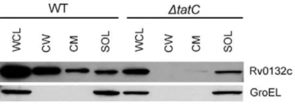
While the ’BlaC reporter experiments demonstrated the existence of a Tat signal sequence in Rv0132c, additional features in the mature domain of a protein are required for it to be Tat exported. This is because the mature domain of Tat-exported proteins must fold prior to export [38,39]. For this reason, we additionally tested whether export of the full length Rv0132c protein occursviathe Tat pathway. Rv0132c with a C-terminal HA epitope tag was expressed in wild type andDtatC M. smegmatis (Figure 5). Cells were harvested and lysed, and whole cell lysates (WCL) were fractionated using differential ultracentrifugation to generate cell wall (CW), cytoplasmic membrane (CM), and soluble (SOL) fractions (containing the cytosol). To determine cellular localization, equal cell equivalents of the fractions were separated on an SDS-PAGE gel for western blot analysis with anti-HA antibodies (Text S1). Rv0132c was detected as being exported to the cell wall and cell membrane fractions in wild typeM. smegmatis, but not in the DtatC mutant where the protein remained in the soluble cytosolic fraction. This analysis demonstrated that Rv0132c export is dependent on a functional Tat pathway. The
integrity of the cellular fractions was verified by Western blotting for GroEL (a cytoplasmic protein) as a control. The cell wall and cytoplasmic membrane fractions were free of cytoplasmic contamination, as shown by the lack of GroEL (Figure 5).
Native Rv0132c is targeted to the cell envelope inMtb
To determine the localization of the native Rv0132c protein in Mtb, anti–Rv0132c antiserum was used for western blotting and immunoelectron microscopy experiments (Text S1). Whole cell lysate (WCL) prepared fromMtbH37Rv cells was fractionated as above to yield cell wall (CW), cytoplasmic membrane (CM), and soluble (SOL) fractions. Equal cell equivalents of the fractions were subjected to western blot analyses, which showed that the native Rv0132c protein is present in the cell wall and membrane fractions ofMtb(Figure 6A). Triton X–114 fractionation ofMtb H37Rv whole cell lysate further showed that the native Rv0132c protein is present in the detergent–enriched fraction (Figure 6B). Triton X–114 is a non–ionic detergent which has been used extensively to partition hydrophilic proteins (i.e. soluble) from amphiphilic proteins (e.g. lipoproteins and integral membrane proteins) [40]. Given our prior results demonstrating the presence of a functional Tat signal sequence on Rv0132c, the Triton X-114 fractionation results are consistent with Rv0132c being a Tat exported lipoprotein that is exported to the cell envelope. The lipidation of the Rv0132c protein might also explain the reason for smeary bands of native Rv0132c in western blots (Figure 6). In contrast, the recombinant Rv0132c–D38 protein used as a control cannot be lipidated as it lacks the signal sequence, and shows a single sharp band. The integrity of the cellular fractions was verified by western blotting using GroEL antibody as a control for cytoplasmic contamination; no GroEL signal was detected in the CW and CM fractions (data not shown).
More evidence of the Rv0132c protein localization was obtained using immunoelectron microscopy ofMtbH37Ra cells using anti–Rv0132c antiserum. The results, which are consistent over a wide range of antiserum concentration, indicate that Rv0132c is indeed present mainly on the periphery of the cells (Figure 7).
Discussion
The presence of F420 in mycobacteria has captured much
attention since it was identified as being involved in the activation of a new anti–TB prodrug PA–824 [41]. In a search for enzymes
Figure 5. Rv0132c export is Tat dependent.Equalized whole cell lysates (WCL) from wild type (WT) andDtatC M. smegmatisexpressing Rv0132c-HA were fractionated to generate cell wall (CW), cytoplasmic membrane (CM), and soluble (SOL) fractions. Fractions were separated by SDS-PAGE and proteins were detected with an anti-HA antibody. Native GroEL was detected as a cytoplasmic control. Rv0132c-HA was exported to the CW and CM fractions in wild typeM. smegmatis, but it was not exported in the absence of a functional Tat pathway. doi:10.1371/journal.pone.0045003.g005
Figure 6. Subcellular localization of Rv0132c protein.(A) Western blots ofM. tuberculosisH37Rv subcellular fractions using 1/25000 dilution of anti–Rv0132c antiserum. Clear signals are found for the WCL, CW and CM fractions, but not for the SOL fraction. (B) Western blots of Triton X–114 treated fractions. The signal is present only in the DET fraction. WCL: whole cell lysate; CW: cell wall; CM: cytoplasmic membrane; SOL: soluble; AQU: aqueous fraction from Triton X–114 treatment; DET: detergent–enriched fraction from Triton X–114 treatment. In both panels recombinant Rv0132c– D38 protein (REC) is used as a positive control (0.7mg).
involved in PA–824 activation inMtb, two ORFs were identified, encoding the enzymes FGD1 [41] and Ddn [10]. It was subsequently shown that Ddn directly activates the prodrug using the reduced F420 provided by FGD1 [8]. Although the Mtb
genome has two ORFs encoding proteins annotated as having FGD activity, FGD1 (Rv0407) and FGD2 (Rv0132c) [12], no evidence has been reported for any involvement of Rv0132c in the activation of or resistance towards PA–824. This led us to question the function of Rv0132c and its F420–dependent nature; either
Rv0132c does not bind F420, or it does not have the annotated
activity, or is physically separated from Ddn where the activation process occurs.
Our results show unequivocally that Rv0132c is indeed an F420
-binding protein, but that it does not have the annotated FGD activity. Differences in the substrate-binding region of the active site are consistent with preference for a different, as yet unknown, substrate. What sets Rv0132c apart from FGD1, apart from the differences in the substrate binding site, is its possession (Figure 4A) of a signal sequence containing a twin–arginine translocation (Tat) motif, together with a lipobox motif that mediates lipid modifi-cation to produce a cell envelope–anchored lipoprotein. The full– length Rv0132c protein sequence, including its Tat motif, is extremely well conserved (99–100% identity) in theMtbcomplex (M. tuberculosis,M. bovis,M. africanum,M. microtiandM. canetti). In contrast, although homologues of Rv0132c can be found in non-pathogenic bacterial species they have much lower sequence identity with Rv0132c (,40%) and lack a Tat motif. This suggests a role for Rv0132c and its Tat motif in mycobacterial pathogenesis.
The Tat pathway transports folded proteins across energy– transducing membranes (e.g. the cytoplasmic membrane) [33], being different from the Sec pathway, which transports proteins in an unfolded state [34]. The majority of substrate proteins for the Tat pathway are enzymes that fold in the cytoplasm and require cofactor insertion therein prior to export; examples of the identified cofactors include molydopterin, haem, FAD and NADP+
[42]. We have shown here that the signal sequence possessed by Rv0132c, and by implication its homologues in other pathogenic mycobacteria, is functionally active in Tat-mediated export. In addition, using M. smegmatis we show Rv0132c is exported in a Tat-dependent manner and we further show the
native Rv0132c is exported to the cell envelope of pathogenicM. tuberculosis. This further implies translocation of F420 to the cell
envelope. This is the first report of F420translocation across the
cytoplasmic membrane in any F420–producing microorganism and
expands the number of Tat–dependent cofactors.
Putting our experimental results together, it is reasonable to conclude that the Rv0132c protein binds F420in the cytosol, after
which the Rv0132c–F420 complex is translocated across the
cytoplasmic membraneviathe Tat pathway, where the protein is anchored to the cell envelope. Although the specific biochemical function of Rv0132c is not yet known, its Tat–dependent translocation may have evolved in pathogenic mycobacteria to enable F420utilization for metabolic or biosynthetic processes in
the dense, lipid–rich cell wall. The conservation of Rv0132c and its specific Tat signal sequence in the Mtbcomplex, but not in non–pathogenic mycobacteria implies that the presence of F420
outside the cytosol is important to the pathogenesis of tuberculosis. There is growing evidence that F420 plays an important part in
defense against the host immune system and in non-replicating persistence ofMtb. Transition to the persistent state involves major changes in energy metabolism [43], and it has been suggested that F420, with its low redox potential, may have a role in reactions
associated with anaerobic survival [44].
Supporting Information
Figure S1 Calibration curve for estimation of Rv0132c–
D38 molecular weight using analytical SEC.The molecular weight calibration curve was obtained by plotting Kav values against LogMW of protein standards. The Kav value determined from the elution volume of Rv0132c–D38, indicated with *, corresponded to a molecular weight of 75.4 kDa, which is indicative of a dimer in solution.
(TIF)
Table S1 Mycobacterial strains and plasmids used in this study.
(DOCX)
Table S2 Primers used in the amplification of different Rv0132c constructs.
(DOCX)
Figure 7. Immunoelectron microscopy of theM. tuberculosisH37Ra cells using anti–Rv0132c antiserum.Electron micrographs are shown in which thin cryo–sectionedMtbcells are (A) treated with preimmune serum, and (B) treated with anti-Rv0132c antisera at a dilution of 1/200. The gold particles (indicated by arrowheads) are present mainly on the periphery of the cells in panel B, but are absent from the control panel (A). doi:10.1371/journal.pone.0045003.g007
Text S1 Supplementary methods. Details of bacterial growth; PCR amplification, cloning and preparation of constructs; western blotting; immunoelectron micros-copy and homology modeling.
(DOC)
Acknowledgments
M. smegmatis mc24517 was kindly provided by Professor W. R. Jacobs, Albert Einstein College of Medicine andM. tuberculosisH37Ra was a kind
gift from Professor G. Cook, University of Otago. We also thank Drs David Greenwood for mass spectrometry, Stephanie Dawes for valuable discussions, and Shaun Lott for help withMtbculture.
Author Contributions
Conceived and designed the experiments: GB MB EB. Performed the experiments: GB EP AT MF. Analyzed the data: GB EP MF MB. Contributed reagents/materials/analysis tools: GB EP AT MF MB. Wrote the paper: GB MB EB.
References
1. Global Tuberculosis Control (2010) World Health Organization
2. Duncan K, Barry CE, III (2004) Prospects for new antitubercular drugs. Curr Opin Microbiol 7: 460–465.
3. Selengut JD, Haft DH (2010) Unexpected Abundance of Coenzyme F420 -dependent enzymes in the Genomes of Mycobacterium tuberculosis and other Actinobacteria. J Bacteriol 192: 5788–5798.
4. Bashiri G, Rehan AM, Greenwood DR, Dickson JMJ, Baker EN (2010) Metabolic engineering of cofactor F420production in Mycobacterium smegmatis. PLoS ONE 5: e15803.
5. Bashiri G, Squire CJ, Moreland NJ, Baker EN (2008) Crystal structures of F420 -dependent glucose-6-phosphate dehydrogenase FGD1 involved in the activation of the anti-tuberculosis drug candidate PA-824 reveal the basis of coenzyme and substrate binding. J Biol Chem 283: 17531–17541.
6. Purwantini E, Daniels L (1996) Purification of a novel coenzyme F420-dependent glucose-6-phosphate dehydrogenase from Mycobacterium smegmatis. J Bacteriol 178: 2861–2866.
7. Dogra M, Palmer BD, Bashiri G, Tingle MD, Shinde S, et al. (2010) Comparative bioactivation of the novel anti-tuberculosis agent PA-824 in Mycobacteria and a subcellular fraction of human liver. Br J Pharmacol. 8. Singh R, Manjunatha U, Boshoff HI, Ha YH, Niyomrattanakit P, et al. (2008)
PA-824 kills nonreplicatingMycobacterium tuberculosisby intracellular NO release. Science 322: 1392–1395.
9. Taylor MC, Jackson CJ, Tattersall DB, French N, Peat TS, et al. (2010) Identification and characterization of two families of F420H2-dependent reductases from Mycobacteria that catalyse aflatoxin degradation. Mol Microbiol 78: 561–575.
10. Manjunatha UH, Boshoff H, Dowd CS, Zhang L, Albert TJ, et al. (2006) Identification of a nitroimidazo-oxazine-specific protein involved in PA-824 resistance inMycobacterium tuberculosis. Proc Natl Acad Sci U S A 103: 431–436. 11. Purwantini E, Mukhopadhyay B (2009) Conversion of NO2to NO by reduced coenzyme F420protects mycobacteria from nitrosative damage. Proc Natl Acad Sci U S A 106: 6333–6338.
12. Cole ST, Brosch R, Parkhill J, Garnier T, Churcher C, et al. (1998) Deciphering the biology ofMycobacterium tuberculosisfrom the complete genome sequence. Nature 393: 537–544.
13. Geiman DE, Kaushal D, Ko C, Tyagi S, Manabe YC, et al. (2004) Attenuation of late-stage disease in mice infected by theMycobacterium tuberculosismutant lacking the SigF alternate sigma factor and identification of SigF-dependent genes by microarray analysis. Infect Immun 72: 1733–1745.
14. DeMaio J, Zhang Y, Ko C, Young DB, Bishai WR (1996) A stationary-phase stress-response sigma factor from Mycobacterium tuberculosis. Proc Natl Acad Sci U S A 93: 2790–2794.
15. Larkin MA, Blackshields G, Brown NP, Chenna R, McGettigan PA, et al. (2007) ClustalW2 and ClustalX version 2. Bioinformatics 23: 2947–2948.
16. Bagos PG, Nikolaou EP, Liakopoulos TD, Tsirigos KD (2010) Combined prediction of Tat and Sec signal peptides with hidden Markov models. Bioinformatics 26: 2811–2817.
17. Bashiri G, Squire CJ, Baker EN, Moreland NJ (2007) Expression, purification and crystallization of native and selenomethionine labeled Mycobacterium tuberculosisFGD1 (Rv0407) using a Mycobacterium smegmatisexpression system. Protein Expr Purif 54: 38–44.
18. Braunstein M, Bardarov SS, Jacobs WRJ (2002) Genetic methods for deciphering virulence determinants of Mycobacterium tuberculosis. Methods Enzymol 358: 67–99.
19. Isabelle D, Simpson DR, Daniels L (2002) Large-scale production of coenzyme F420-5,6 by usingMycobacterium smegmatis. Appl Environ Microbiol 68: 5750– 5755.
20. Jacobson FS, Daniels L, Fox JA, Walsh CT, Orme-Johnson WH (1982) Purification and properties of an 8-hydroxy-5-deazaflavin-reducing hydrogenase fromMethanobacterium thermoautotrophicum. J Biol Chem 257: 3385–3388. 21. Flores AR, Parsons LM, Pavelka MS Jr (2005) Genetic analysis of the
beta-lactamases ofMycobacterium tuberculosisandMycobacterium smegmatisand suscepti-bility to beta-lactam antibiotics. Microbiology 151: 521–532.
22. McDonough JA, Hacker KE, Flores AR, Pavelka MS Jr, Braunstein M (2005) The twin-arginine translocation pathway ofMycobacterium smegmatisis functional and required for the export of mycobacterial beta-lactamases. J Bacteriol 187: 7667–7679.
23. Snapper SB, Melton RE, Mustafa S, Kieser T, Jacobs JWR (1990) Isolation and characterization of efficient plasmid transformation mutants of Mycobacterium smegmatis. Mol Microbiol 4: 1911–1919.
24. Gibbons HS, Wolschendorf F, Abshire M, Niederweis M, Braunstein M (2007) Identification of twoMycobacterium smegmatislipoproteins exported by a SecA2-dependent pathway. J Bacteriol 189: 5090–5100.
25. D’Orazio M, Folcarelli S, Mariani F, Colizzi V, Rotilio G, et al. (2001) Lipid modification of the Cu,Zn superoxide dismutase fromMycobacterium tuberculosis. Biochem J 359: 17–22.
26. Gurumurthy M, Mukherjee T, Dowd CS, Singh R, Niyomrattanakit P, et al. (2012) Substrate specificity of the deazaflavin-dependent nitroreductase from Mycobacterium tuberculosisresponsible for the bioreductive activation of bicyclic nitroimidazoles. FEBS J 279: 113–125.
27. Sutcliffe IC, Harrington DJ (2004) Lipoproteins ofMycobacterium tuberculosis: an abundant and functionally diverse class of cell envelope components. FEMS Microbiol Rev 28: 645–659.
28. Berks BC, Sargent F, De Leeuw E, Hinsley AP, Stanley NR, et al. (2000) A novel protein transport system involved in the biogenesis of bacterial electron transfer chains. Biochim Biophys Acta 1459: 325–330.
29. Gralnick JA, Vali H, Lies DP, Newman DK (2006) Extracellular respiration of dimethyl sulfoxide byShewanella oneidensisstrain MR-1. Proc Natl Acad Sci U S A 103: 4669–4674.
30. Thompson BJ, Widdick DA, Hicks MG, Chandra G, Sutcliffe IC, et al. (2010) Investigating lipoprotein biogenesis and function in the model Gram-positive bacteriumStreptomyces coelicolor. Mol Microbiol.
31. Valente FM, Pereira PM, Venceslau SS, Regalla M, Coelho AV, et al. (2007) The [NiFeSe] hydrogenase fromDesulfovibrio vulgarisHildenborough is a bacterial lipoprotein lacking a typical lipoprotein signal peptide. FEBS Lett 581: 3341– 3344.
32. Widdick DA, Hicks MG, Thompson BJ, Tschumi A, Chandra G, et al. (2011) Dissecting the complete lipoprotein biogenesis pathway inStreptomyces scabies. Mol Microbiol 80: 1395–1412.
33. Wickner W, Schekman R (2005) Protein translocation across biological membranes. Science 310: 1452–1456.
34. Stephenson K (2005) Sec-dependent protein translocation across biological membranes: evolutionary conservation of an essential protein transport pathway (review). Mol Membr Biol 22: 17–28.
35. Palmer T, Sargent F, Berks BC (2005) Export of complex cofactor-containing proteins by the bacterial Tat pathway. Trends Microbiol 13: 175–180. 36. McDonough JA, McCann JR, Tekippe EM, Silverman JS, Rigel NW, et al.
(2008) Identification of functional Tat signal sequences in Mycobacterium tuberculosisproteins. J Bacteriol 190: 6428–6438.
37. Marrichi M, Camacho L, Russell DG, DeLisa MP (2008) Genetic toggling of alkaline phosphatase folding reveals signal peptides for all major modes of transport across the inner membrane of bacteria. J Biol Chem 283: 35223– 35235.
38. DeLisa MP, Tullman D, Georgiou G (2003) Folding quality control in the export of proteins by the bacterial twin-arginine translocation pathway. Proc Natl Acad Sci U S A 100: 6115–6120.
39. Tullman-Ercek D, DeLisa MP, Kawarasaki Y, Iranpour P, Ribnicky B, et al. (2007) Export pathway selectivity ofEscherichia colitwin arginine translocation signal peptides. J Biol Chem 282: 8309–8316.
40. Malen H, Pathak S, Softeland T, de Souza GA, Wiker HG (2010) Definition of novel cell envelope associated proteins in Triton X-114 extracts ofMycobacterium tuberculosisH37Rv. BMC Microbiol 10: 132.
41. Stover CK, Warrener P, VanDevanter DR, Sherman DR, Arain TM, et al. (2000) A small-molecule nitroimidazopyran drug candidate for the treatment of tuberculosis. Nature 405: 962–966.
42. Berks BC, Palmer T, Sargent F (2003) The Tat protein translocation pathway and its role in microbial physiology. Advances in microbial physiology 47: 187– 254.
43. Shi L, Sohaskey CD, Kana BD, Dawes S, North RJ, et al. (2005) Changes in energy metabolism ofMycobacterium tuberculosisin mouse lung and under in vitro conditions affecting aerobic respiration. Proc Natl Acad Sci USA 102: 15629– 15634.