Multivariate Analyses of Rotator Cuff
Pathologies in Shoulder Disability
Jan F. Henseler1*, Yotam Raz1,3, Jochem Nagels1, Erik W. van Zwet2, Vered Raz3, Rob G. H. H. Nelissen1
1Department of Orthopaedics, Leiden University Medical Center, Postzone J-11-R, PO box 9600, 2300 RC Leiden, the Netherlands,2Department of Medical Statistics and Bioinformatics, Leiden University Medical Center, Leiden, the Netherlands,3Department of Human Genetics, Leiden University Medical Center, Leiden, the Netherlands
Abstract
Background
Disability of the shoulder joint is often caused by a tear in the rotator cuff (RC) muscles. Four RC muscles coordinate shoulder movement and stability, among them the supraspina-tus and infraspinasupraspina-tus muscle which are predominantly torn. The contribution of each RC muscle to tear pathology is not fully understood. We hypothesized that muscle atrophy and fatty infiltration, features of RC muscle degeneration, are predictive of superior humeral head translation and shoulder functional disability.
Methods
Shoulder features, including RC muscle surface area and fatty infiltration, superior humeral translation and RC tear size were obtained from a consecutive series of Magnetic Reso-nance Imaging with arthrography (MRA). We investigated patients with superior (supraspi-natus, n = 39) and posterosuperior (supraspinatus and infraspi(supraspi-natus, n = 30) RC tears, and patients with an intact RC (n = 52) as controls. The individual or combinatorial contribution of RC measures to superior humeral translation, as a sign of RC dysfunction, was investi-gated with univariate or multivariate models, respectively.
Results
Using the univariate model the infraspinatus surface area and fatty infiltration in both the supraspinatus and infraspinatus had a significant contribution to RC dysfunction. With the multivariate model, however, the infraspinatus surface area only affected superior humeral
translation (p<0.001) and discriminated between superior and posterosuperior tears. In
contrast neither tear size nor fatty infiltration of the supraspinatus or infraspinatus contribut-ed to superior humeral translation.
OPEN ACCESS
Citation:Henseler JF, Raz Y, Nagels J, van Zwet EW, Raz V, Nelissen RGHH (2015) Multivariate Analyses of Rotator Cuff Pathologies in Shoulder Disability. PLoS ONE 10(2): e0118158. doi:10.1371/ journal.pone.0118158
Academic Editor:Stephen E Alway, West Virginia University School of Medicine, UNITED STATES
Received:June 5, 2014
Accepted:January 5, 2015
Published:February 24, 2015
Copyright:© 2015 Henseler et al. This is an open access article distributed under the terms of the
Creative Commons Attribution License, which permits unrestricted use, distribution, and reproduction in any medium, provided the original author and source are credited.
Data Availability Statement:All relevant data are within the paper and its Supporting Information files.
Funding:This study is funded by the Dutch Arthritis Association (DAA), Dutch Arthritis Association and grant number 2013-1-060. The funder had no role in study design, data collection and analysis, decision to publish, or preparation of the manuscript.
Conclusion
Our study reveals that infraspinatus atrophy has the strongest contribution to RC tear pa-thologies. This suggests a pivotal role for the infraspinatus in preventing shoulder disability.
Introduction
Shoulder complaints are the second largest cause for musculoskeletal disability in the middle aged and older populations. Shoulder complaints restrict daily functioning due to pain and
limited arm mobility [1–3]. The majority of these complaints are caused by degenerative
rota-tor cuff (RC) pathologies, ultimately leading to RC tears [3–6]. Degenerative RC pathology is
the main cause of upper-limb related complaints in general practice [7,8], rheumatology [9]
and orthopaedic clinics [10]. The prevalence of RC tears in the general population is high
(20%) and increases with age (over 50% in the population above 65 years) [11–13].
The shoulder joint requires active stabilization from the RC muscles due to its
non-con-strained nature and dislocating forces of the prime shoulder movers (e.g. deltoids) [14–18]. In
contrast to other joints like the hip, bony structures in the shoulder joint do not provide the same level of passive stability. The lack of passive stability allows positioning of the arm in space with an unsurpassed range of motion. The trade-off for mobility in the shoulder joint re-quires active compensatory stabilization by the RC muscles: the supraspinatus (SSp), infraspi-natus (ISp), subscapularis and teres minor. In general, the SSp and ISp are more affected in RC
tearing compared to the other two muscles [19]. The most frequent torn RC muscle is the SSp
(i.e. superior tear), but a tear can progress posteriorly towards the ISp (i.e. posterosuperior tear). In RC tears the dynamic stabilization of the shoulder is lost. In the long term RC
dysfunc-tion can result in superior transladysfunc-tion of the humeral head towards the acromion [14,16,20–
24]. In large RC tears a decrease in acromiohumeral (AH) distance was reported [23,24]. In
ad-dition, fatty infiltration is also frequently found in RC muscle tear [21,25]. Both the AH dis-tance and fatty infiltration are associated with poor surgical outcome and increased
post-operative re-tear rates [26,27]. However, the intricate associations between superior translation
of the humeral head (i.e. decrease in AH distance), RC muscle atrophy, fatty infiltration and RC tear size are not yet fully understood.
We aim to identify predictors of superior translation of the humeral head in the presence of superior and posterosuperior RC tears based on Magnetic Resonance imaging with arthrogra-phy (MRA). We hypothesized that features of the RC muscle (muscle atroarthrogra-phy and fatty infiltra-tion) together with RC tear size will predict the degree of superior humeral head translation.
Methods
Patients
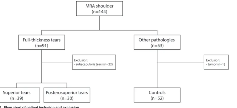
Patient cases were selected through the hospital financial administration with the diagnosis
of‘rotator cuff tear’(national Diagnosis Related Group code 1460) and‘rotator cuff tendinitis’
(DRG code 1450). Exclusion criteria consisted of poor quality MRA scans (motion artifacts, poor view delineation), tearing of the subscapularis, deltoid and/or teres minor, history of frac-tures of the shoulder, tumors, severe shoulder joint deformity (glenohumeral osteoarthritis or
rheumatoid arthritis grade III–IV), neurological denervation, stroke, muscular dystrophies,
and myositis. A flow chart summarizing patient inclusion and exclusion is presented inFig. 1.
144 patients with an MRA of the shoulder were identified and evaluated for inclusion. In total: 91 superior and posterosuperior RC tear patients were identified. 22 subscapularis tears and 1 tumor were excluded. 52 patients without a full-thickness RC tear were used as a control group. Of these, 8 patients had a partial thickness RC tear of the SSp, 8 patients were diagnosed with acromioclavicular (AC) osteoarthritis and 36 patients had unremarkable MRA scans.
MRA Imaging procedure
Fifteen minutes before MRA, injection of contrast fluid into the glenohumeral joint was per-formed via a posterior approach under fluoroscopic guidance. Patients had local intra-articular anesthesia with 10mL of 1% lidocaine and subsequently a diluted Gd-DTPA mixture (i.e. 10cc NaCl, 5ml Marcaine 0.25% and 0.2ml Gadolinium 1:200) was injected. All MRA scans were performed on a 1.5 Tesla Avanto Siemens MRI unit (Siemens AG, Erlangen, Germany) or Phil-ips Intera MRI unit (PhilPhil-ips Medical Systems, Best, the Netherlands) using a dedicated shoul-der coil and turbo spin-echo sequences. Patients were scanned in a supine position with the arm in neutral rotation.
Analyses of the images were performed on a PACS Workstation with Sectra IDS5 (Sectra Medical Systems AB, Linköping, Sweden) as monitor readings. Image analysis was carried out by two independent observers who were blinded to the MRA report and diagnoses of the pa-tients. As multiple planes and sequences were obtained following the institutional standard
shoulder MRA protocol, the T1-weighted coronal and sagittal plane (TR/TE 500–600/11–15,
Fig 1. Flow chart of patient inclusion and exclusion.
matrix 256; slice thickness 4mm, inter-slice gap 1mm, field of view of 15cm) were systematically evaluated.
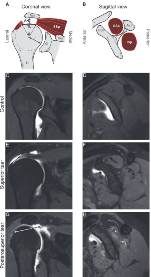
The methodology of the image evaluation is presented in Fig.2Aand1B. The osteometry
measurements performed on the coronal plane, at the widest point of the humeral head, are
vi-sualized inFig. 2A[28]. The radius (r) of the humeral head was measured at its widest point
using a circle-fit in the coronal plane. Superior translation of the humeral head was measured
as the AH distance [24,29,30] and was defined as the smallest distance between the most
crani-al articular cortex of the humercrani-al head, and the caudcrani-al corticcrani-al surface of the acromion
deter-mined in the coronal plane in the slice with the largest humeral head diameter (Fig. 2A). The
radius of the humeral head and the AH distance are reported in mm.
Representative scans of the RC from a control are presented inFig. 2C and 2D(coronal and
sagittal view, respectively), for the superior tears inFig. 2E and 2F(coronal and sagittal view,
respectively) and for the posterosuperior tearsFig. 2G and 2H(coronal and sagittal view,
re-spectively). In order to assess the anatomical features in a three-dimensional (3-D) perspective
we measured the RC tear dimensions in the coronal and sagittal plane [31]. The tear geometry
was measured on MRA as straight lines at the maximum anterior-to-posterior width in the
cor-onal plane and maximum lateral-to-medial length in the sagittal plane (TL,Fig. 2A) as
de-scribed previously [32]. The RC tear width in the coronal plane and length in the sagittal plane
are reported in mm. To further approximate the RC tear size we measured of the RC tear size relative to the geometrical centre of the humeral head in the coronal and sagittal plane. The
angle of the RC tear (ff, reported in degrees (°)) relative to the geometrical center of the humeral
head was measured with the same anterior-to-posterior points on the coronal plane and the
lateral-to-medial points of the tear in the sagittal plane (Fig. 2A). Subsequently, as the articular
surface of the humeral head approximates a sphere, the RC tear surface area in cm2could be
calculated with the following formula:
Tear surface area¼ ffcoronal
360
2phumerus
radius
ffsagittal
360
2phumerus
radius
To determine muscular mass as a measure for muscle atrophy, the cross-sectional surface areas (CSA) of the SSp and the ISp were measured on the scan with the anatomical glenoid neck and
base of the coracoid present, as illustrated inFig. 2B. The CSA of the SSp and the ISp are
re-ported in cm2. The presence of fatty infiltration was evaluated subjectively by examining either
the presence or absence of intramuscular fatty infiltration in the SSp and ISp muscles on the
coronal and sagittal T1-weighted images, as described previously with good inter-observer reli-ability [33,34].
Statistical analyses
For the comparison of continuous variables between the patient groups one-way analysis of variance (ANOVA) with post-hoc testing were performed. For the comparison of categorical
variablesχ2tests were performed.
The paired t-test was used to assess inter-observer differences. The Interclass correlation co-efficient (ICC, two-way random model with absolute agreement) was used to assess the
inter-observer reliability of the main continuous radiological variables. The Kappa (κ) was used to
assess the inter-observer consistency of the nominal dichotomous parameters. For
interpreta-tion, the criteria formulated by Cicchetti and Sparrow were used [35]:0.39, poor; 0.40 to
0.59, fair; 0.60 to 0.74, good; or0.75, excellent.
with the AH distance as numeric outcome, and SSp surface area (per cm2), ISp surface area (per cm2), SSp fatty infiltration (yes/no), ISp fatty infiltration (yes/no), age (years), gender and diagnosis (control/RC tear) as explanatory variables. Next, we performed a multivariate mixed model analysis with the AH distance as numeric outcome. The SSp and ISp surface area, SSp and ISp fatty infiltration, age, gender and diagnosis functioned all as explanatory variables into one multivariate model. The goal of such a multivariate model is to avoid confounding by as-sessing the effect of any single variable on the outcome, when all other variables are kept fixed. The categorical variables have a single effect and the continuous variables have an incremental effect. We performed similar analyses in the sub-population of RC tear patients. We therefore modified the explanatory variable diagnosis to indicate superior compared to posterosuperior tears. Also, we added tear size as an additional explanatory variable.
Statistical analyses were performed using SPSS Statistics (IBM Inc., Armonk, New York,
USA). Statistical significance was considered with p-values of0.05 (two-sided).
Results
Patient characteristics
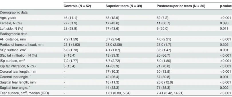
Patient characteristics were stratified for diagnosis and are summarized inTable 1. Compared
to controls, patients with a RC tear had a decreased AH distance (p = 0.02), and reduced
cross sectional surface area of the supraspinatus (SSp) and cross sectional surface area of the infraspinatus (ISp). For the control group, representative MRA images can be found inFig. 2panel C and D (coronal and sagittal view, respectively). For the superior tear group, representative MRA images can be found inFig. 2 panel E and F (coronal and sagittal view, respectively). For the posterosuperior tear group, representative MRA images can be found inFig. 2panel G and H (coronal and sagittal view, respectively). In panels E and G the length of the rotator cuff tear is measured in the coronal plane (arrow). In panel H fat infiltration of the SSp and the ISp is indicated (*).
doi:10.1371/journal.pone.0118158.g002
Table 1. Patient Characteristics.
Rotator cuff tears
Controls (N = 52) Superior tears (N = 39) Posterosuperior tears (N = 30) p-value
Demographic data
Age, years 46 (11.1) 58 (12.5) 62 (7.2) <0.001
Female, N (%) 27 (51.9) 17 (43.6) 11 (36.7) 0.393
Left side, N (%) 28 (53.8) 17 (43.6) 6 (20.0) 0.011
Radiographic data
AH distance, mm 7.2 (1.59) 6.7 (2.54) 4.0 (2.21) <0.001
Radius of humeral head, mm 23.1 (1.93) 23.0 (2.06) 23.0 (1.7) 0.302
SSp surface, cm2 5.0 (1.73) 4.1 (1.87) 3.6 (1.47) 0.001
SSp fat infiltration, N (%) 8 (15.4) 13 (33.3) 20 (66.7) <0.001
ISp surface, cm2 7.2 (1.77) 6.7 (2.72) 5.0 (1.80) <0.001
ISp fat infiltration, N (%) 8 (15.4) 14 (35.9) 21 (70.0) <0.001
Coronal tear length, mm - 17 (10.3) 30 (13.5) <0.001
Coronal tear angle,˚ - 42 (26.4) 67 (30.8) 0.001
Sagittal tear length, mm - 18 (11.3) 26.6 (12.9) <0.001
Sagittal tear angle,˚ - 44 (33.3) 71 (35.3) 0.002
Tear surface, cm2, median (IQR) - 1.61 (0.80, 5.34) 7.41 (3.42, 14.21) <0.001
The presented p-values are obtained through a one-way ANOVA. For the comparison of nominal variablesχ2tests were performed.
surface area of the SSp and ISp muscles (p<0.001 and p = 0.002, respectively) (Fig. 3A).
Pa-tients with a RC tear were significantly older compared to controls (p<0.001). Patients with a
RC tear had more fatty infiltration in SSp and ISp muscles (p<0.001 for both), compared to
controls. Lastly, within the control group, the radius of the humeral head was strongly
correlat-ed with the surface of the SSp and ISp (Pearson correlation 0.566 and 0.350; p<0.001 and p =
0.04, respectively), whereas within the RC tear group only weak correlation was found between the radius of the humeral head and the surface of the SSp and ISp (Pearson correlation 0.203 and 0.120; p = 0.09 and p = 0.33, respectively) (Table 1).
Next, we assessed whether these shoulder features could discriminate between patients with superior and posterosuperior tears. The AH distance was significantly reduced in patients with
a posterosuperior tear (p = 0.002), as well as the ISp surface area (p = 0.007) (Fig. 3B). In
addi-tion, the length and angle of tear in the sagittal plane are larger (p = 0.01 and p = 0.046, respec-tively) and subsequently the RC tear size is also larger (p = 0.02) in patients with a
posterosuperior tear. Furthermore, ISp fatty infiltration is more abundant in patients with pos-terosuperior tears (p<0.001).
Inter-observer reliability and consistency
The inter-observer reliability are provided inTable 2. Every image was analyzed by two
re-searchers independently. The AH distance measurements did not significantly differ between the two observers. Although small, there were statistical significant differences found for the
ISp surface area and RC tear size. The ICC for the AH distance was 0.9 (p<0.001) and for the
SSp and ISp surface areas 0.7 (p<0.001). This is considered good to excellent inter-observer
re-liability. For the dichotomous variables, theκfor the presence or absence (yes/no) of muscle
fatty infiltration in either the SSp or ISp was 0.7 (p<0.001). This indicates good consistency be-tween the two observers.
The AH distance in RC tears and controls
The relation between the AH distance and muscle atrophy as the surface area (per cm2) and fatty infiltration (yes/no) features of the RC in controls and RC tear patients is summarized in
Table 3.
Fig 3. Means and standard errors of the means of the acromiohumeral distance and the cross sectional surface area of the supraspinatus and infraspinatus between the patient groups.Compared to controls:*p<0.05;**p<0.001
First the association of each RC feature as explanatory variable for the AH distance was as-sessed individually using univariate analyses. With the univariate model, the muscle surface area and fatty infiltration of both the SSp and the ISp muscle, age and diagnosis were associated
with the AH distance (Table 3). The surface area of the SSp and ISp had an effect size of
0.37mm (p = 0.002) and 0.56mm (p<0.001) on the AH distance, respectively. This indicates an
increase in AH distance of 0.37mm per 1cm2 SSp surface area or 0.56mm increase per 1cm2 ISp surface area. In the absence (no) of the SSp or ISp fatty infiltration there was an increase in
AH distance of 2.38mm (p<0.001) or 1.88mm (p<0.001), respectively. Overall, the RC tear
group had a 1.68mm smaller AH distance compared to the control group.
Next, we applied a multivariate model adjusted for age and gender to assess the contribu-tions of the SSp and ISp surface areas and fatty infiltration on the AH distance between
con-trols and RC tear patients (Table 3). In this model the ISp surface area remained influential
indicating a significant contribution to the AH distance (0.5mm, p<0.001). Additionally, the
absence (no) of fatty infiltration in the SSp was associated with an increase in AH distance (1.2mm, p = 0.039). In contrast, the SSp surface area, ISp fatty infiltration and the diagnosis (i.e. presence of an RC tear) did not significantly contribute to the AH distance.
Table 2. Inter-observer difference and reliability.
Interobserver difference Reliabilty testing
Mean (SE) 95%-CI p-value ICC 95%-CI p-value
AH distance, mm 0.2 (0.09) -0.03–0.34 0.099 0.9 0.89–0.95 <0.001
Radius of humeral head, mm 0.4 (0.11) 0.17–0.63 0.001 0.8 0.70–0.86 <0.001
SSp surface, cm2 0.2 (0.13) -0.54–0.48 0.117 0.7 0.63–0.81 <0.001
ISp surface, cm2 0.5 (0.17) 0.16–0.83 0.005 0.7 0.61–0.81 <0.001
Tear surface, cm2 1.2 (0.58) 0.12–2.38 0.031 0.8 0.63–0.84 <0.001
The inter-observer differences for the main continuous radiological features are obtained through paired t-tests. The ICC is obtained for reliability testing. doi:10.1371/journal.pone.0118158.t002
Table 3. Contributors to acromiohumeral distance in RC tears and controls.
Variable Univariate models Multivariate model
Effect size 95%-CI p-value Effect size 95%-CI p-value
Diagnosis (control)† 1.68 0.831–2.529 <0.001 0.76 -0.116–1.637 0.088
Surface area, cm2
SSp 0.37 0.136–0.616 0.002 -0.07 -0.340–0.197 0.597
ISp 0.56 0.384–0.719 <0.001 0.52 0.304–0.725 <0.001
Fat infiltration (no)†
SSp 2.38 1.540–3.211 <0.001 1.12 0.055–2.179 0.039
ISp 1.88 1.009–2.740 <0.001 0.04 -0.993–1.071 0.940
Age, yr -0.05 -0.087–-0.020 0.002 0.01 -0.030–0.044 0.707
Gender (male)† -0.39 -1.281–0.505 0.391 -0.77 -1.670–0.129 0.092
Univariate modelling evaluated the relation of each variable with the AH distance individually. Multivariate modeling evaluated the combined parameter estimates of the effect sizes of the variables on the AH distance in RC tears and controls.
†Categorical parameters have a single effect in the models, with fat infiltration (absence compared to presence), gender (male compared to female) and diagnosis (control compared to RC tear).
The AH distance in superior and posterosuperior tears
The associations of muscle atrophy and fatty infiltration features of the RC and the AH
dis-tance for superior and posterosuperior tears are summarized inTable 4. Using the univariate
model, the contribution of ISp surface area, SSp and ISp fatty infiltration significantly to AH
distance significantly differ between superior and posterosuperior tears (Table 4). In the
uni-variate models, the ISp surface area had an effect size of 0.61mm per cm2 on the AH distance. The absence of fatty infiltration had a single effect of 2.29mm and 1.34mm for the SSp and ISp,
respectively. The tear surface had a negative effect (-0.2mm, p<0.001), indicating a smaller AH
in larger tears. Superior tears showed a larger AH distance compared to posterosuperior tears (2.68mm, p = 0.009).
With the multivariate model, however, only the ISp surface area remained associated with
the AH distance (increase of 0.53 mm AH per 1cm2ISp surface increase,Table 4). The model
included the surface areas and fatty infiltration of the SSp and ISp and RC tear size with adjust-ments for age and gender. The superior tears had a larger AH distance compared to
posterosu-perior tears (1.62mm, p = 0.009,Table 4). We could not find that the tear surface contributed
to the AH distance using the multivariate model, while a significant effect was found with the
univariate model (Table 4).
Discussion
Superior translation of the humeral head is a hallmark of advanced stage RC disease and leads to narrowing of the sub-acromial space with subsequent deterioration of shoulder function [14,16,20–24]. In the current study we found a decline in AH distance and increase in muscle atrophy and fatty infiltration in RC tears compared with controls. We found a similar decline in posterosuperior RC tears compared with superior RC tears. The ISp surface area most ro-bustly differs between controls and RC tears and between posterosuperior and superior RC tears using a multivariate model.
Table 4. Contributors to acromiohumeral distance between superior and posterosuperior RC tears.
Variable Univariate models Multivariate model
Effect size 95%-CI p-value Effect size 95%-CI p-value
Diagnosis (superior tear)† 2.68 1.512–3.846 <0.001 1.62 0.412–2.820 0.009
Surface area, cm2
SSp 0.09 -0.075–0.261 0.272 -0.06 -0.485–0.354 0.755
ISp 0.61 0.383–0.828 <0.001 0.53 0.197–0.857 0.002
Fat infiltration (no)†
SSp 2.29 1.087–3.495 <0.001 0.72 -0.972–2.408 0.399
ISp 1.34 0.049–2.620 0.042 -1.13 -2.527–0.273 0.113
Tear surface, cm2 -0.20 -0.288–-0.105 <0.001 -0.05 -0.162–0.51 0.304
Age, yr -0.05 -0.109–0.014 0.131 -0.01 -0.060–0.057 0.690
Gender (male)† 0.05 -1.299–1.401 0.940 -0.33 -1.616–0.954 0.608
Univariate modelling evaluated the relation of each variable with the AH distance individually. Multivariate modeling evaluated the combined parameter estimates of the effect sizes of the variables on the AH distance in superior and posterosuperior RC tears.
†Categorical parameters have a single effect in the models, with fat infiltration (absence compared to presence), gender (male compared to female) and diagnosis (superior tear compared to posterosuperior tear).
In the current study we investigated the contributions of the SSp and ISp muscle surface area and fatty infiltration and tear size to AH distance. We found that the larger posterosuper-ior RC tears resulted in a smaller AH distance with more pronounced muscle atrophy and fatty
infiltration compared to superior RC tears in agreement with previous studies [23,24].
Howev-er, in previous studies only individual RC features were assessed in relation to the AH distance,
either RC tear size or fatty infiltration [21,23–25]. An association between the AH distance and
RC tear size on ultrasound was found, but only for larger RC tear sizes [23]. Although we
found a twofold increase in RC tear size for the posterosuperior tears compared to the superior tears, the contribution was less prominent compared to muscle atrophy and fatty infiltration. We are the first to apply an integrated model for multiple predictors to assess the AH distance in RC tears. With this model we identified the ISp surface area as the most important contribu-tor to the AH distance. Therefore, we suggest the ISp surface area as a diagnosis prediccontribu-tor in RC disease.
The literature concerning the indication and timing of both surgical and conservative treat-ment for RC tears remains sparse. Still, pain scores and functional outcome results remain vari-able. Pain mediated shoulder adductor co-activation of the teres major and latissimus dorsi can
compensate for lost RC function [18,36,37]. Although the glenohumeral stability can partially
be restored with RC repair or tendon transfer surgery, similar gain is observed when
strength-ening the remaining RC and surrounding shoulder muscles [38–41]. This warrants further
studies on the effect of pain medication such as corticosteroids treatments and physical therapy in order to exercise the remaining shoulder muscles and prevent them from degeneration. This ultimately will center the humeral head effectively onto the glenoid and retain shoulder func-tionality [14–18].
In the current study muscle surface area was measured on MRA of the shoulder. MRA pro-vides a more accurate assessment of the RC tears in comparison to conventional MRI and ul-trasound due to the contrast distending the joint capsule, outlining the intra-articular
structures and dissemination into abnormalities [32,42,43]. Furthermore, several clinical score
have been described to qualitatively assess RC features clinically. However, these classifications for RC muscle atrophy, fatty infiltration and tendon retraction did not reach satisfactory
inter-observer reliability in previous studies [33,44]. We demonstrated that semi-quantitative
mea-surements obtained with MRA of RC features are reliable, and subsequently could be used for statistical modeling. The relation between and the measurements of RC features used in the current study and these common shoulder classification systems used in the clinic will need further study.
Muscle atrophy and fatty infiltration are well known contributors to muscle degeneration in
muscles dystrophies, myopathies, muscle aging and denervation [34,45]. Fatty infiltration is a
non-specific response to local or systemic damage and considered as a common outcome in muscle degeneration, including muscular atrophy. However the causality of fatty infiltration in muscle degeneration pathogenesis is not fully resolved. Goutallier et al. reported that fatty
infil-tration in the RC muscles only occurs in presence of a RC tear [46]. However, Ashry et al. and
others found progression of fatty infiltration in shoulder muscles with aging in patients without
RC tears [34,47]. We confirmed an association between RC tear and fatty infiltration, although
fatty infiltration was not found in all RC tears. We observed fatty infiltration also in the control group without RC tears. This suggests that fatty infiltration may only be indirectly associated with RC tears. Furthermore, it supports the idea that muscle atrophy and fat infiltration are
in-dependently associated processes [48].
patients. Therefore the differences between RC tears and control groups could be under-esti-mated. Fatty infiltration was measured qualitatively, and accurate measurements of fatty infil-tration should be quantitative. However excellent correlations were reported between previous
quantitative measurements and a qualitative visual rating, as used in the current study [21].
Moreover, ideally longitudinal studies should unravel the rate and mechanism of progression from superior to posterosuperior tears and the progress of fatty infiltration of muscles towards the development of RC tears.
Despite the limitation of the current study, we demonstrated an association between the AH
distance and RC muscle’s fatty infiltration and muscle atrophy. The AH distance, which
re-flects glenohumeral stability as a sign of RC dysfunction, is mostly affected by the remaining infraspinatus size, whereas the RC tear size has a lesser effect. This indicates a pivotal role for the infraspinatus within the RC muscles in preventing excessive superior translation of the humeral head.
Author Contributions
Performed the experiments: JFH YR. Analyzed the data: JFH YR. Contributed reagents/materi-als/analysis tools: EWvZ. Wrote the paper: VR JHF. Study design: JFH JN RGHHN.
References
1. Nakajima D, Yamamoto A, Kobayashi T, Osawa T, Shitara H, et al. (2012) The effects of rotator cuff tears, including shoulders without pain, on activities of daily living in the general population. J Orthop Sci 17: 136–140. doi:10.1007/s00776-011-0186-4PMID:22249436
2. Roquelaure Y, Ha C, Leclerc A, Touranchet A, Sauteron M, et al. (2006) Epidemiologic surveillance of upper-extremity musculoskeletal disorders in the working population. Arthritis Rheum 55: 765–778. doi:10.1002/art.22222PMID:17013824
3. Greving K, Dorrestijn O, Winters JC, Groenhof F, van der Meer K, et al. (2012) Incidence, prevalence, and consultation rates of shoulder complaints in general practice. Scand J Rheumatol 41: 150–155. doi:10.3109/03009742.2011.605390PMID:21936616
4. Oh LS, Wolf BR, Hall MP, Levy BA, Marx RG. (2007) Indications for rotator cuff repair: a systematic re-view. Clin Orthop Relat Res 455: 52–63. doi:10.1097/BLO.0b013e31802fc175PMID:17179786
5. Dorrestijn O, Greving K, van der Veen WJ, van der Meer K, Diercks RL, et al. (2011) Patients with shoulder complaints in general practice: consumption of medical care. Rheumatology (Oxford) 50: 389–395. doi:10.1093/rheumatology/keq333PMID:21047806
6. Matsen FA III. (2008) Clinical practice. Rotator-cuff failure. N Engl J Med 358: 2138–2147. doi:10. 1056/NEJMcp0800814PMID:18480206
7. Bot SD, van der Waal JM, Terwee CB, van der Windt DA, Schellevis FG, et al. (2005) Incidence and prevalence of complaints of the neck and upper extremity in general practice. Ann Rheum Dis 64: 118– 123. doi:10.1136/ard.2003.019349PMID:15608309
8. van der Windt DA, Koes BW, de Jong BA, Bouter LM. (1995) Shoulder disorders in general practice: in-cidence, patient characteristics, and management. Ann Rheum Dis 54: 959–964 PMID:8546527
9. Vecchio P, Kavanagh R, Hazleman BL, King RH. (1995) Shoulder pain in a community-based rheuma-tology clinic. Br J Rheumatol 34: 440–442 PMID:7788173
10. Dunn WR, Schackman BR, Walsh C, Lyman S, Jones EC, et al. (2005) Variation in orthopaedic sur-geons' perceptions about the indications for rotator cuff surgery. J Bone Joint Surg Am 87: 1978–1984. doi:10.2106/JBJS.D.02944PMID:16140812
11. Sher JS, Uribe JW, Posada A, Murphy BJ, Zlatkin MB. (1995) Abnormal findings on magnetic reso-nance images of asymptomatic shoulders. J Bone Joint Surg Am 77: 10–15 PMID:7822341
12. Moosmayer S, Tariq R, Stiris MG, Smith HJ. (2010) MRI of symptomatic and asymptomatic full-thick-ness rotator cuff tears. A comparison of findings in 100 subjects. Acta Orthop 81: 361–366. doi:10. 3109/17453674.2010.483993PMID:20450423
14. Steenbrink F, de Groot JH, Veeger HE, van der Helm FC, Rozing PM. (2009) Glenohumeral stability in simulated rotator cuff tears. J Biomech 42: 1740–1745. doi:10.1016/j.jbiomech.2009.04.011PMID: 19450803
15. Hansen ML, Otis JC, Johnson JS, Cordasco FA, Craig EV, et al. (2008) Biomechanics of massive rota-tor cuff tears: implications for treatment. J Bone Joint Surg Am 90: 316–325. doi:10.2106/JBJS.F. 00880PMID:18245591
16. McCully SP, Suprak DN, Kosek P, Karduna AR. (2006) Suprascapular nerve block disrupts the normal pattern of scapular kinematics. Clin Biomech (Bristol, Avon) 21: 545–553. doi:10.1016/j.clinbiomech. 2006.02.001PMID:16603286
17. Lugo R, Kung P, Ma CB. (2008) Shoulder biomechanics. Eur J Radiol 68: 16–24. doi:10.1016/j.ejrad. 2008.02.051PMID:18511227
18. Steenbrink F, Meskers CG, Nelissen RG, de Groot JH. (2010) The relation between increased deltoid activation and adductor muscle activation due to glenohumeral cuff tears. J Biomech 43: 2049–2054. doi:10.1016/j.jbiomech.2010.04.012PMID:20452596
19. Melis B, Nemoz C, Walch G. (2009) Muscle fatty infiltration in rotator cuff tears: descriptive analysis of 1688 cases. Orthop Traumatol Surg Res 95: 319–324. doi:10.1016/j.otsr.2009.05.001PMID: 19586809
20. Lehtinen JT, Belt EA, Kauppi MJ, Kaarela K, Kuusela PP, et al. (2001) Bone destruction, upward migra-tion, and medialisation of rheumatoid shoulder: a 15 year follow up study. Ann Rheum Dis 60: 322–326 PMID:11247859
21. van de Sande MA, Stoel BC, Obermann WR, Lieng JG, Rozing PM. (2005) Quantitative assessment of fatty degeneration in rotator cuff muscles determined with computed tomography. Invest Radiol 40: 313–319 PMID:15829828
22. Hirooka A, Wakitani S, Yoneda M, Ochi T. (1996) Shoulder destruction in rheumatoid arthritis. Classifi-cation and prognostic signs in 83 patients followed 5–23 years. Acta Orthop Scand 67: 258–263 PMID: 8686464
23. Keener JD, Wei AS, Kim HM, Steger-May K, Yamaguchi K. (2009) Proximal humeral migration in shoul-ders with symptomatic and asymptomatic rotator cuff tears. J Bone Joint Surg Am 91: 1405–1413. doi: 10.2106/JBJS.H.00854PMID:19487518
24. Henseler JF, de Witte PB, de Groot JH, van Zwet EW, Nelissen RG, et al. (2013) Cranial translation of the humeral head on radiographs in rotator cuff tear patients: the modified active abduction view. Med Biol Eng Comput. doi:10.1007/s11517-013-1057-2PMID:24370855
25. Fuchs B, Weishaupt D, Zanetti M, Hodler J, Gerber C. (1999) Fatty degeneration of the muscles of the rotator cuff: assessment by computed tomography versus magnetic resonance imaging. J Shoulder Elbow Surg 8: 599–605 PMID:10633896
26. Meyer DC, Wieser K, Farshad M, Gerber C. (2012) Retraction of supraspinatus muscle and tendon as predictors of success of rotator cuff repair. Am J Sports Med 40: 2242–2247. doi:10.1177/
0363546512457587PMID:22926748
27. Kim JR, Cho YS, Ryu KJ, Kim JH. (2012) Clinical and radiographic outcomes after arthroscopic repair of massive rotator cuff tears using a suture bridge technique: assessment of repair integrity on magnetic resonance imaging. Am J Sports Med 40: 786–793. doi:10.1177/0363546511434546PMID:
22307079
28. Rozing PM, Obermann WR. (1999) Osteometry of the glenohumeral joint. J Shoulder Elbow Surg 8: 438–442 PMID:10543596
29. Nagels J, Verweij J, Stokdijk M, Rozing PM. (2008) Reliability of proximal migration measurements in shoulder arthroplasty. J Shoulder Elbow Surg 17: 241–247. doi:10.1016/j.jse.2007.07.011PMID: 18234527
30. van de Sande MA, Rozing PM. (2006) Proximal migration can be measured accurately on standardized anteroposterior shoulder radiographs. Clin Orthop Relat Res 443: 260–265. doi:10.1097/01.blo. 0000196043.34789.73PMID:16462449
31. Davidson JF, Burkhart SS, Richards DP, Campbell SE. (2005) Use of preoperative magnetic reso-nance imaging to predict rotator cuff tear pattern and method of repair. Arthroscopy 21: 1428. doi:10. 1016/j.arthro.2005.09.015PMID:16376230
32. van der Zwaal P, Thomassen BJ, Urlings TA, de Rooy TP, Swen JW, et al. (2012) Preoperative agree-ment on the geometric classification and 2-dimensional measureagree-ment of rotator cuff tears based on magnetic resonance arthrography. Arthroscopy 28: 1329–1336. doi:10.1016/j.arthro.2012.04.054 PMID:22885159
classification system to increase reliability. Am J Sports Med 40: 1728–1734. doi:10.1177/ 0363546512452714PMID:22753846
34. Ashry R, Schweitzer ME, Cunningham P, Cohen J, Babb J, et al. (2007) Muscle atrophy as a conse-quence of rotator cuff tears: should we compare the muscles of the rotator cuff with those of the deltoid? Skeletal Radiol 36: 841–845. doi:10.1007/s00256-007-0307-5PMID:17508210
35. Cicchetti DV, Sparrow SA. (1981) Developing criteria for establishing interrater reliability of specific items: applications to assessment of adaptive behavior. Am J Ment Defic 86: 127–137 PMID:7315877
36. Henseler JF, Nagels J, Nelissen RG, de Groot JH. (2014) Does the latissimus dorsi tendon transfer for massive rotator cuff tears remain active postoperatively and restore active external rotation? J Shoulder Elbow Surg 23: 553–560. doi:10.1016/j.jse.2013.07.055PMID:24135419
37. Steenbrink F, de Groot JH, Veeger HE, Meskers CG, van de Sande MA, et al. (2006) Pathological mus-cle activation patterns in patients with massive rotator cuff tears, with and without subacromial anaes-thetics. Man Ther 11: 231–237. doi:10.1016/j.math.2006.07.004PMID:16890886
38. Gerber C, Maquieira G, Espinosa N. (2006) Latissimus dorsi transfer for the treatment of irreparable ro-tator cuff tears. J Bone Joint Surg Am 88: 113–120. doi:10.2106/JBJS.E.00282PMID:16391256
39. Henseler JF, Nagels J, van der Zwaal P, Nelissen RGHH. (2013) Teres major tendon transfer for pa-tients with massive irreparable posterosuperior rotator cuff tears: Short-term clinical results. Bone Joint J 95-B: 523–529. doi:10.1302/0301-620X.95B4.30390PMID:23539705
40. Ainsworth R, Lewis JS. (2007) Exercise therapy for the conservative management of full thickness tears of the rotator cuff: a systematic review. Br J Sports Med 41: 200–210. doi:10.1136/bjsm.2006. 032524PMID:17264144
41. Moosmayer S, Lund G, Seljom US, Haldorsen B, Svege IC, et al. (2014) Tendon repair compared with physiotherapy in the treatment of rotator cuff tears: a randomized controlled study in 103 cases with a five-year follow-up. J Bone Joint Surg Am 96: 1504–1514. doi:10.2106/JBJS.M.01393PMID: 25232074
42. de Jesus JO, Parker L, Frangos AJ, Nazarian LN. (2009) Accuracy of MRI, MR arthrography, and ultra-sound in the diagnosis of rotator cuff tears: a meta-analysis. AJR Am J Roentgenol 192: 1701–1707. doi:10.2214/AJR.08.1241PMID:19457838
43. Steinbach LS, Palmer WE, Schweitzer ME. (2002) Special focus session. MR arthrography. Radio-graphics 22: 1223–1246 PMID:12235350
44. Lippe J, Spang JT, Leger RR, Arciero RA, Mazzocca AD, et al. (2012) Inter-rater agreement of the Gou-tallier, Patte, and Warner classification scores using preoperative magnetic resonance imaging in pa-tients with rotator cuff tears. Arthroscopy 28: 154–159. doi:10.1016/j.arthro.2011.07.016PMID: 22019235
45. Klatte-Schulz F, Pauly S, Scheibel M, Greiner S, Gerhardt C, et al. (2012) Influence of age on the cell bi-ological characteristics and the stimulation potential of male human tenocyte-like cells. Eur Cell Mater 24: 74–89 PMID:22791374
46. Goutallier D, Postel JM, Bernageau J, Lavau L, Voisin MC. (1994) Fatty muscle degeneration in cuff ruptures. Pre- and postoperative evaluation by CT scan. Clin Orthop Relat Res: 78–83 PMID:8020238
47. Melis B, Wall B, Walch G. (2010) Natural history of infraspinatus fatty infiltration in rotator cuff tears. J Shoulder Elbow Surg 19: 757–763. doi:10.1016/j.jse.2009.12.002PMID:20363160