142
ARTICLE
Early respiratory evaluation should be
carried out systematically in patients
with multiple sclerosis
Avaliação respiratória precoce deveria ser feita sistematicamente em pacientes com
esclerose múltipla
Fernanda Machado Taveira1, Antônio Lúcio Teixeira2, Renan Barros Domingues3
Multiple sclerosis (MS) afects motor pathways and is as-sociated with reduced muscle strength, including the respira-tory muscles1-5. Respiratory complications are recognized as the leading causes of morbidity and mortality in patients with advanced MS3-4. Nearly half of the MS patients in advanced stages die as a consequence of respiratory complications, such as aspiration pneumonia, atelectasis, and respiratory failure2,4-6. he progression of respiratory dysfunction occurs as a result of the weakness and fatigue of respiratory mus-cles (inspiratory and expiratory) and failure to maintain the
airways free of secretions7. Abnormalities were shown in both expiratory (abdominal muscles and internal intercostal) and in-spiratory muscles (diaphragm and external intercostal) among MS patients2.
Some authors have suggested that the decrease in respi-ratory muscle strength in MS depends on the size and loca-tion of lesions in the central nervous system (CNS). Other fac-tors may also determine respiratory involvement in MS, such as corticosteroids myopathy, inlammatory cytokines like tumor necrosis factor (TNF), spasticity, incoordination, and
1Physical therapist, Master’s degree in Neurosciences, Federal University of Minas Gerais, Belo Horizonte MG, Brazil;
2MD, PhD, Professor of Neurology, Department of Internal Medicine, School of Medicine, and Neuroscience Postgraduation Program, Federal University of Minas Gerais, Belo Horizonte MG, Brazil;
3MD, PhD, Professor, Department of Pathology, Santa Casa School of Health Sciences, Vitória ES, Brazil and Neuroscience Postgraduation Program, Federal University of Minas Gerais, Belo Horizonte MG, Brazil.
Correspondence: Renan Barros Domingues; Rua Professor Almeida Cousin 125 / sala 1.310; 29050-565 Vitória ES - Brasil; E-mail: contato@renandomingues.med.br
Conflict of interest: There is no conflict of interest to declare.
Received 13 August 2012; Received in final form 24 September 2012; Accepted 01 October 2012.
ABSTRACT
The present study aimed at evaluating respiratory parameters in multiple sclerosis (MS). The sample comprised 30 patients with MS diagno-sis and 30 healthy subjects, matched by gender and age. Neurological assessment, expanded disability status scale (EDSS), manovacuom-etry, and peak flow (PEF) were performed. Patients with MS had lower values of maximum inspiratory (MIP) and expiratory (MEP) pressures and PEF compared to healthy controls. It was shown that respiratory impairment may be present in MS patients with low functional disability by EDSS. The data suggest that manovacuometry and PEF determination should be carried out systematically in patients with MS, and may be a reliable tool for the early detection of respiratory impairment allowing early respiratory rehabilitation.
Key words: multiple sclerosis, respiratory function, respiratory function tests.
RESUMO
O presente estudo teve como objetivo avaliar a função respiratória em pacientes com esclerose múltipla (EM). A amostra foi composta por 30 pacientes com diagnóstico de EM e 30 sujeitos saudáveis, pareados de acordo com o gênero e a idade. Foram realizados avaliação neurológi-ca, determinação da escala expandida do estado de incapacidade (EDSS), manovacuometria e “peak flow”. Pacientes com EM apresentaram pressões inspiratória (PI) e expiratória (PE) máximas inferiores e menor PFE quando comparados aos controles saudáveis. Identificou-se que o comprometimento respiratório pode ocorrer em pacientes com EM, mesmo com baixo grau de comprometimento funcional estimado pelo EDSS. Os dados do presente estudo sugerem que a avaliação respiratória com manovacuometria e PFE deve ser realizada sistematicamente em pacientes com EM, e pode ser uma ferramenta útil para detectar precocemente comprometimento respiratório, podendo sugerir a neces-sidade de reabilitação pulmonar precoce em pacientes com EM.
143
Fernanda Machado Taveira et al. MS: respiratory evaluation postural changes2,3. However, there is still little evidence
re-garding this.
he present study aimed at evaluating respiratory function in a population of MS patients with conventional respiratory parameters, such as maximum inspiratory pressure (MIP), maximum expiratory pressure (MEP), and peak low (PEF).
METHODS
he study was conducted in Multiple Sclerosis Clinic at
Hospital da Santa Casa de Misericórdia de Vitória. hirty MS patients diagnosed according to the international panel cri-teria by Polman et al.8 were included. he Control Group con-sisted of 30 healthy subjects matched by gender and age.
Patients with temporomandibular disorders (TMD), re-missions, infections, and other neurological diseases poten-tially compromising respiratory capacity were not included.
All participants were submitted to complete neurologic ex-amination and determination of Expanded Functional Disability Scale (EDSS). According to the EDSS score, patients were divid-ed into two groups: EDSS bellow 4 and EDSS equal or above 4. Measurement of MIP and MEP was performed with a mano-vacuometer (model M120; Commercial Medicine, São Paulo, Brazil), and measurement of peak expiratory low (PEF) was per-formed with “peak low” (Model Asses, full range 60–880 L/min; Respironics New Jersey, Inc., USA). Müller maneuver was used for MIP measurement9, and the Valsalva one was used for MEP9. PEF measurement was carried out as previously described10.
Statistical analysis
Data were analyzed with the GraphPad Prism version 5.0 and R statistical software version 2.14.1. he conidence in-terval was of 95% and the signiicance level was set as p<0.05. Veriication of data normal distribution was performed us-ing the Kolmogorov-Smirnov test. To compare medians between MS patients and controls, we used the nonparametric Mann-Whitney’s test. Median MIP, MEP, and PEF of patients with EDSS bellow 4 were compared with those of controls. he same proce-dure was conducted with patients with EDSS equal or above 4.
RESULTS
Of the 30 patients analyzed, 26 (86.67%) were female and 4 (13.33%) were male. he mean age was 36.66±11.78 years-old. he mean EDSS was 2.63±1.65. MS forms were: relapsing-remit-ting including 26 (86.66%); secondary progressive, 3 (10.0%); and primary progressive, 1 (3.33%).
Two patients (6.66%) were not using immunomodulators or immunosuppressants. Among the 28 patients receiving spe-ciic MS drugs, 46.42% were using beta-interferon 1A 44 µcg, 14.28% glatiramer acetate, 14.28% beta-interferon 1A 30 µcg,
10.71% beta-interferon 1B SC 9600000UI, 3.57% azathioprine, and 3.57% beta-interferon 1A 22 µcg.
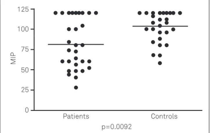
he MIP of patients was 81.4±30.91 cmH2O and of controls 103.93±18.63 cmH2O (p=0.0092), as in Fig 1. he MEP measured in patients was 85.4±21.23 cm H2O and 100.66±15 cmH2O in the Control Group (p=0.0034), as seen in Fig 2. he PEF of MS patients was 370.16±92.11 L/min. and of controls was 436.5±79.51 L/min. (p=0.0051), as in Fig 3.
he comparisons of median MIP, MEP, and PEF of both groups according to the EDSS and controls are shown in Table.
Fig 1. Maximum inspiratory pressure (MIP) in patients and controls.
p=0.0092
Patients Controls 125
MIP
100
75
50
25
0
Fig 3. Peak flow in patients and controls.
p=0.0034
125
100
75
50
25
0
Fig 2. Maximum expiratory pressure (MEP) in patients and controls.
p=0.0051
Patients Controls
750
P
eak fl
o
w 500
250
144 Aq o q t 71(3):142-145 DISCUSSION
he present study showed a reduction in respiratory func-tion parameters in MS patients compared to healthy con-trols. Compromising of respiratory function is well-known in patients with advanced MS. In our study, most patients had minimal disability and low EDSS score suggesting that respi-ratory dysfunction can be identiied even in patients with minimal to moderate disability. Few previous studies have al-ready showed that respiratory muscle strength and function-al capacity may be early afected in MS patients11,12.
MIP was signiicantly lower in MS patients. his inding is in line with previous studies showing reduced levels of inspiratory pressure in MS1,3,5,13-19. he same occurred with MEP, and lower expiratory pressure was already shown in MS1,3,4,5,13-19. It was not possible to assess which of these two pressures is afected earlier in the course of MS, since this was a cross-sectional study.
PEF rate was signiicantly lower in MS patients. Only few studies have addressed it in MS, but in most of them a decrease in PEF in patients with MS was shown, such as in Savci et al.1, Buyse et al.15, and Chiara et al.19. PEF is an easy-to-perform and in-expensive method, and the present study showed that it is poten-tially useful in evaluating respiratory function in patients with MS. When we subdivided patients according to the EDSS and compared with controls, diferences were found in MEP and PFE between patients and controls, regardless the EDSS being lower than four or above. his reinforces the notion that respiratory dysfunction may be found in fully deambu-latory MS patients with low functional disability. here were no diferences of the MIP between patients and controls when we analyzed the two subgroups of patients according to the EDSS. his may have happened because the number of
patients in the subgroups was low, not allowing a signiicant diference to appear.
The present study has clear limitations. No correla-tions were performed with the type of therapy used by the patient. However, patients during relapses or infections were excluded.
It is unknown if immunomodulatory and immunosuppres-sive therapies potentially afect respiratory function in MS.
Correlations between respiratory impairment and lesions of the cervical spinal cord and brainstem were not investigat-ed. It is possible that lesions in these CNS regions may be part of the respiratory impairment pathophysiology.
Patients with all forms of the disease were enrolled. hose with the progressive forms of MS were not excluded, but the number of subjects with secondary or primary progres-sive forms was very small and had probably little inluence in the inal results. In addition, respiratory dysfunction was found in patients with minimal to moderate disability.
Previous studies showed that3,5,18,19 breathing exercises may reduce the risk of respiratory complications and that early detection of decreased respiratory muscle strength is very important in this preventive process.
Further studies are needed in order to clarify the relation-ship between lesions of the cervical spinal cord and brain-stem respiratory symptoms in patients with MS and to ex-amine separately the respiratory impairment in patients with diferent forms of the disease and stages of evolution.
A systematic and simple evaluation of respiratory param-eters can be useful for the detection of early reduction in re-spiratory muscle strength, thus possibly allowing to establish an evaluation of early respiratory rehabilitation programs in patients with MS.
Table. !" # f$ % ! #& !' !#" ! ( )# %$&!( )$ "" '$ ",# %$ *+ w,$ (w$ $ #e#( - "# % (!$ # (",
ee % !#d((.$$& #%$ %%!" ,!- !()"(( '""e -$.
Controls (n=25) Median (min–max)
EDSS <4 (n=20)
Median (min–max) p-value
EDSS ≥4 (n=5)
Median (min–max) p-value
M /0 (cmH2O)
86 (28–120) 80 (28–120) 0.62 60 (48–120) 0.86 M 10 (cmH2O)
100 (68–120) 84 (32–120) 0.0087 80 (60–100) 0.045 0P1 (cmH2O)
420 (330–710) 370 (150–600) 0.0179 370 (200–400) 0.0208
23 4:56557 to8 294:56556 to8 4: 9; <=o>9? @ @;67 xxx BCt8 t tDC.
1. @vD@,3 7CE3 7D?,A <7aF tC.@6E57 t>C<x t7D
5 oGf7Dto7Cx D D Dt875CtCD C o .?BC RHBC IJ:1365-171.
2. K o C7 <R, KovDL,?D5 M. R to85D C7voC v57t
75CtCD C o .9L NO OO13:449-454.
3. F8 ?r,4 GCQ R A,SHo<H ALF t C.L7 xo5Qx Do7t oC tC
oGffDt oG E><7 to85D C t77ToT 5 o7 5 oG C 5o78G7Dto77 o7>tH5CtCD C o .N oC4 HyUH J;31:162-172.
4. A CCo2FL5C CoA,K7C C:, tC.S oTHGVDD 8C txto
tHx BCt8 t t7 t7t>tH5CtCD C o .L to7 2008;76:311-316.
5. Ko C7<LFrovD R,
rtC 4, tC.L to85D C> < 7 7x to85D Ct77T7v C8x B Cx5CtCD C o t7t.
Ac H4Hy
2xLHBC W:747-751.
6. 2 tC8:r,?5LFX Q8C 5 Q@, tC.Y tH7TE7H7Dx
6t5t8xD Go t7t>tH5CtCD C o .SC7LHBC 2007;21:595-602.
145
FZ [ n \n ]\^ \_h \]`T\vZ b[ \Zc\g i^ jk[Z lm b[ \c`[ pZs \gu \c b`n
7i y\l_h ` \ g z {, |b gg \g}\ WO, yZ[Zb[ \ ^~i Insuficiência respiratória crônica nas doenças neuromusculares: diagnóstico e tratamento. J Bras Pneumol 2007;33:81-92.
8. Polman CH, Reingold SC, Banwell B, et al. Diagnostic criteria for multiple sclerosis: 2010 Revisions to the McDonald Criteria. Ann Neurol 2011;69:292-302.
9. Souza RB. Pressões respiratórias estáticas máximas. J Pneumol 2002;28:155-164.
10. Nunn AJ, Gregg I. New regression equations for predicting peak expiratory flow in adults. Br Med J 1989;298:1068-1072.
11. Motl RW, Goldman MD, Benedict RH. Walking impairment in patients with multiple sclerosis: exercise training as a treatment option. Neuropsychiatr Dis Treat 2010;16:767-774.
12. Bosnak-Guclu M, Guclu-Gunduz A, Nazliel B, et al. Comparison of functional exercise capacity, pulmonary function and respiratory muscle strength in patients with multiple sclerosis with different disability levels and healthy controls. J Rehabil Med 2012;44:80-86.
13. Altintas A, Demir T, Ikitimur HD, et al. Pulmonary function in multiple sclerosis without any respiratory complaints. Clin Neurol Neurosurg 2007;109:242-246.
14. Smeltzer SC, Skurnick JH, Troiano R, et al. Respiratory function in multiple sclerosis. Utility of clinical assessment of respiratory muscle function. Chest 1992;101:479-484.
15. Buyse B, Demedts M, Meekers J, et al. Respiratory dysfunction in multiple sclerosis: a prospective analysis of 60 patients. Eur Respir J 1997;10:139-145.
16. Mutluay FK, Gürses HN, Saip S. Effects of multiple sclerosis on respiratory functions. Clin Rehabil 2005;19:426-432.
17. Foglio K, Clini E, Facchetti D, et al. Respiratory muscle function and exercise capacity in Multiple Sclerosis. Eur Respir J 1994;7:23-28.
18. Klefbeck B, Nedjad JH. Effect of inspiratory muscle training in patients with multiple sclerosis. Arch Phys Med Rehabil 2003;84:994-999.