Abstract
Tissue engineering of the small intestine offers an alternative to long-term intravenous nutrition and transplantation in patients with intestinal failure. Initial work, although encouraging, is limited by the volume of neonatal tissue required to produce a small neomucosal cyst. Our novel approach is to implant tubular poly-lactide-co-glycolide (PGLA) foam scaffolds subcutaneously. The aim of this study was to investigate whether these scaffolds would support growth of intestinal neomucosa. PGLA scaffolds were implanted subcutaneously into 8 Lewis rats; after 5 weeks, ‘organoid units’ were injected into the lumens. Tissue was assessed histologically after harvesting and quantitative immunohistochemistry was performed using antibodies against vascular endothelial growth factor (VEGF), vascular endothelial growth factor receptor 2 (VEGF-R2), fibroblast growth factor basic (bFGF) and fibroblast growth factor receptor 2 (FGF-R2). At 4 weeks post organoid unit implantation, clearly recognisable mucosa and submucosa was present on the luminal surface of the scaffold. Densities of VEGF and VEGF-R2 positive cells increased with time post organoid unit implantation. This pilot study demonstrates that it is possible to tissue engineer small intestinal neomucosa using subcutaneously implanted PLGA scaffolds. The yield of the process compares favourably to the published literature. Further work is required to optimise the technique.
Key words: Tissue engineering, Scaffold, Intestine, Angiogenesis
* Address for correspondence: Dr DAJ Lloyd
Department of Gastroenterology,
St Mark’s Hospital and Academic Institute, Watford Road,
Harrow, HA1 3UJ, UK
Telephone: +44 20 88695806 Fax: +44 20 82354001 E-mail: [email protected]
Introduction
Short bowel syndrome (SBS) is characterised by gastrointestinal fluid loss, malabsorption and progressive malnutrition; if fluids, electrolytes and nutrients are not replaced, patients become dehydrated and malnourished. This may ultimately be fatal. The commonest cause of SBS is loss of functional absorptive intestinal surface usually due to extensive small bowel resection. Patient survival is dependent on long-term parenteral nutrition, with its associated morbidity and mortality. With the provision of parenteral nutrition in the home setting, survival is quoted at 75% at 5 years and 60% at 10 years; patients with very short residual intestine (<50cm) have the worst prognosis (Messing et al., 1999). Surgical treatment to lengthen the bowel has been described in children but is of limited value in adults (Bianchi, 1980). Small intestinal transplantation can be considered but it requires aggressive immunosupression (Abu-Elmagd et al., 2002) and survival rates are not yet as high as other solid organ grafts. The one year graft survival for intestinal grafts has recently been quoted at 65% with patient survival at 77% (Grant et al., 2005). Scarcity of donor organs is another major limitation to transplantation, especially in the paediatric population.
A novel approach to the treatment of SBS has been the development of tissue engineered small bowel constructs. The technique behind this concept is to employ an artificial biodegradable scaffold to promote soft tissue growth and support an epithelium within its lumen (Gabe et al., 2004; Vacanti, 2003). Polylactic acid (PLA), polyglycolic acid (PGA) and their copolymers have been used in small intestinal tissue engineering in rodents as synthetic structures to support the adherence and growth of ‘organoid units’ (Choi et al., 1998; Mooney et al., 1994). These organoid units consist of intestinal epithelial stem cells, enterocytes and stromal cells. They are derived from neonatal intestinal tissue by a process of limited digestion using collagenase and dispase which yields a mixture of intestinal crypts and villi (Evans et al., 1992). Vacanti and colleagues have developed a technique in which organoid units are placed onto a tubular copolymer scaffold which is then implanted into the peritoneum of rodent models (Choi et al., 1998). They have demonstrated the development of a “cyst like” structure consisting of a neomucosal surface with the histological characteristics of small intestine; physiological studies reveal an electric potential across the neomucosa suggesting active transport (Choi et al., 1998). These cystic structures have been
A PILOT STUDY INVESTIGATING A NOVEL SUBCUTANEOUSLY IMPLANTED
PRE-CELLULARISED SCAFFOLD FOR TISSUE ENGINEERING OF INTESTINAL
MUCOSA
DAJ Lloyd1 *, TI Ansari2, P Gundabolu3, S Shurey2, V Maquet4, PD Sibbons2, AR Boccaccini5 and SM Gabe1,3
1 St Mark’s Hospital and Academic Institute, Harrow, UK. 2 Northwick Park Institute for Medical Research, Harrow,
UK. 3 Division of Surgery, Anaesthetics and Intensive Care, Imperial College, London, UK. 4 Centre for Education
and Research on Macromolecules, University of Liege, Liege, Belgium. 5 Department of Materials and Centre for
successfully incorporated into healthy rat small intestine (Kaihara et al., 1999a; Kaihara et al., 1999b; Kaihara et al., 2000; Kim et al., 1999) and indirect studies suggest that they may have absorptive potential (Grikscheit et al., 2004). Unfortunately, the yield of the published methodology remains low and a large quantity of donor tissue is required to produce each tissue engineered cyst (Choi et al., 1998; Grikscheit et al., 2004).
Our group has developed a poly-lactide-co-glycolide (PLGA) foam into a tubular structure with transverse and longitudinal porosity of controlled size. The scaffold produced has a wall thickness of 1mm and luminal diameter of 2mm. The pores within the scaffold are approximately 100µm in size and form transverse and longitudinal channels that allow cell migration throughout the scaffold (Day et al., 2004a). When implanted subcutaneously into a rat model there is progressive infiltration of cellular material into the scaffold with blood vessels present within this structure (Day et al., 2004a). Angiogenesis is vital to the survival and development of mucosal tissue within an artificial scaffold since, without vascularisation, gas and nutrient exchange relies almost entirely on passive diffusion. It is likely that angiogenesis is stimulated by growth factors such as vascular endothelial growth factor (VEGF) and fibroblast growth factor basic (bFGF) which are secreted by fibroblasts infiltrating the scaffold. VEGF is believed to be one of the major regulators of blood vessel formation (Ferrara, 2000) and bFGF is also highly angiogenic. Both VEGF and bFGF are thought to play an integral role in the neo-vascularisation of bioengineered tissues (Soker et al., 2000).
Our group has demonstrated that pre-implantation of scaffolds allows population by fibroblasts and initial angiogenesis, and have described the time course of cellular infiltration of tubular scaffolds and concomitant angiogenesis (Day et al., 2004b; Day et al., 2004a). It is our belief that a cellularised scaffold will facilitate the growth and development of implanted organoid tissue thus decreasing the number of organoid units required to develop a viable neomucosa. The purpose of this study was to evaluate a novel model of intestinal tissue engineering using pre-implanted subcutaneous scaffolds and to, using stereological methods, quantify those cells expressing growth factors involved in promoting angiogenesis.
Materials and Methods Animals
Experiments were performed using inbred male Lewis rats (Harlan UK Ltd). All animals were maintained and handled in accordance with the Animals (Scientific Procedures) Act 1986 and the study performed following guidelines stipulated by the UK Home Office. All animals were kept under standard laboratory conditions and fed a commercial pelleted diet.
Scaffold implantation
A tubular PLGA foam construct with a 2mm internal diameter and 1mm wall thickness was created using the
thermally induced phase separation (TIPS) technique described in our previous study (Day et al., 2004a). This construct was divided into 12 scaffolds each measuring 10mm in length. The scaffolds were sterilized by exposure to ultraviolet light. 2.2mm external diameter silicone tubing (Vygon UK Ltd) was placed inside each scaffold to maintain luminal patency. 8 adult male rats weighing 235-280g were anaesthetised using 0.25ml IM Hypnorm (0.315mg/ml fentanyl citrate and 10mg/ml fluanisone) and 1mg IP diazepam. 12 scaffolds (1-2 per animal) were implanted into subcutaneous pockets formed in the inguinal folds of the 8 rats. Skin was closed with 3/0 Mersilk® sutures (Ethicon, Johnson & Johnson Medical Ltd, UK) and the scaffold was left in situ for 5 weeks. 8mg/kg IM gentamicin was administered post-operatively.
Organoid unit manufacture
Organoid units were prepared using a modification of the method described by Evans et al. (Evans et al., 1992). A single 7 day old male Lewis rat was sacrificed by cervical dislocation. The complete length of small bowel was removed and cleaned free of mesentery. The intestine was opened along its length and divided into approximately 2mm lengths and washed 6 times in a solution of 2% glucose in Hanks’ balanced salt solution (HBSS). The tissue was then manually chopped into approximately 1mm3 pieces and digested for 25 minutes using a solution
of 300U/ml collagenase, Clostridium histiolyticum, type XI-S, (Sigma-Aldrich, Dorset, UK) and 0.1mg/ml dispase, Bacillus polymyxa (Gibco BRL, Life Technologies, UK) in HBSS at room temperature on an orbital shaker. The supernatant was discarded and the digestion was repeated twice. After the third digestion the supernatant was preserved and the reaction terminated by addition of Dulbecco’s modified Eagle’s medium (DMEM) supplemented with 2.5% foetal calf serum, 2% sorbitol and 60µg/ml penicillin and streptomycin solution (Gibco BRL, Life Technologies, UK). Organoid units were purified by three rounds of centrifugation at 250xg for 3 minutes followed by removal of supernatant and resuspension in supplemented DMEM. After the final centrifugation, the organoid units were resuspended in extracellular matrix (ECM) gel (Sigma-Aldrich, Dorset, UK) supplemented with 50ng/ml hepatocyte growth factor (HGF) (PeproTech EC Ltd, UK).
Organoid implantation and harvest
Tissue preparation
Following fixation, the length of each explanted tissue was measured. The scaffold was then divided transversely into equal quarters. Each quarter was placed cut face down into a cassette and processed to paraffin using routine laboratory techniques. One full face 5µm section was cut and stained with haematoxylin and eosin (H&E) for routine examination. Additional sections were cut at 3µm for immunohistochemsitry and mounted onto ATPS coated slides.
Immunohistochemistry
Sections were de-paraffinised in xylene, re-hydrated by passing through decreasing concentrations of industrial methylated spirit (IMS) (100%-70%) and rinsed in distilled water. For VEGF receptor-2 (VEGF-R2) staining, sections underwent antigen retrieval using trypsin digestion for 30 minutes at 37oC (Sigma-Aldrich, Dorset, UK). All sections
were rinsed in distilled water and treated with 0.3% hydrogen peroxide in methanol for 30 minutes to block endogenous peroxidase activity before being washed in distilled water for 3x 3 minute cycles; the wash was then repeated with phosphate buffered saline (PBS). Sections were treated with normal goat serum 1:200 (Vector Laboratories, Peterborough, UK) for 20 minutes to prevent non-specific staining before being incubated with the primary antibody. Primary antibodies used were as follows: rabbit polyclonal VEGF (Santa Cruz Biotechnology Inc through Autogen Bioclear UK Ltd) 1:100 dilation in bovine serum albumin (BSA) incubated at 4oC for 48hours, rabbit
polyclonal VEGF-R2 (Abcam, Cambridge, UK) 1:50 dilution in BSA incubated at 4oC for 24 hours, rabbit
polyclonal FGF basic (bFGF) (Abcam, Cambridge, UK) 1:100 dilution in BSA incubated at 4oC for 24 hours, rabbit
polyclonal FGF receptor 2 (FGF-R2) (Abcam, Cambridge, UK) 1:100 dilution in BSA incubated at 4oC for 24 hours.
Sections were subsequently washed in PBS 3x 3 minute cycles and then incubated further with normal goat serum and anti rabbit secondary antibody (Vector Laboratories, Peterborough, UK) 1:200 dilution in PBS at room temperature for 2 hours for VEGF and VEGF-R2 and 30 minutes for bFGF and FGF-R2. Following another 3x 3 minute wash in PBS, sections were incubated with an Avidin Biotin-peroxidase Complex using the Vectostain Elite ABC kit (Vector Laboratories, Peterborough, UK). Immunoreactivity was visualized with a diaminobenzidine (DAB) substrate (Vector Laboratories, Peterborough, UK). Finally sections were counterstained with Gill’s haematoxylin for 1½ minutes.
Stereological analysis
The numerical density of positive staining cells was estimated using 2D stereological techniques for each growth factor and receptor. An unbiased counting frame generated by a digital software package (Kinetic Imaging Stereology 5.0) was projected onto the histological image. Starting at a random position at the top left corner and following a tessellating pattern, positive cells within the unbiased counting frame and obeying the rules of the frame were included in the final estimate. Each section was viewed at final magnification of x630 and an average of
40 fields of view were analysed for each growth factor or receptor. Data for each field was used to obtain the total number of cells per unit area (NA) using the following equation:
Where NA = number per unit area, ∑Q= sum of positive
cells counted, ∑P= sum of unbiased counting frames and
f
A = area of the frame.
Statistical analysis
The mean and standard error of the mean (SEM) of the numerical density of positively stained cells were calculated for each growth factor and receptor. The results were analysed using linear regression analysis (Sigma Stats version 2). Calculated p values are reported and were considered statistically significant if p<0.05.
Results Macroscopic appearance
All animals survived the duration of the study with no adverse effects. At weeks 0, 1, 2 and 4 after organoid unit implantation firm spheroid cyst like structures measuring approximately 10mm in length and 5mm in diameter were identified at the site of implantation.
Histological appearance
Low magnification histological examination of H&E stained sections harvested at 5 weeks after scaffold implantation but with no organoid implantation revealed no identifiable mucosa or submucosa. However, there were numerous inflammatory cells and fibrosis on the internal aspect of the scaffold. At higher magnification fibroblasts, macrophages, monocytes and scattered polymorphs were identified within the scaffold. Groupings of these cells within the scaffold were seen in longitudinal lacunae from the internal to external surface of the scaffold. The scaffold was well vascularised and completely encapsulated by a fibrous band of tissue. At one position, corresponding to the join in the scaffold, a cellular fibrin lattice connected the internal and external surfaces of the scaffold.
Figure 1: H&E staining of scaffold at 1 week after organoid implantation. 1 week after implantation of the organoid units there is no evidence of a luminal neomucosa. At this time point, 6 weeks after scaffold implantation, there is extensive infiltration of the PLGA scaffold with evidence of vascularisation. At higher magnification, fibroblasts, macrophages and monocytes are identifiable.
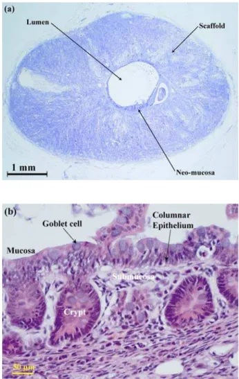
Figure 2: (a) H&E staining of scaffold at 4 weeks after organoid implantation. 4 weeks after organoid unit implantation there is evidence of mucosa and submucosa lining the entire circumference of the luminal surface of the scaffold. The appearance of the scaffold is similar to at 1 week after organoid implantation. (b) H&E staining of scaffold at 4 weeks after organoid implantation. At high magnification, the appearance of the mucosa and submucosa lining the luminal surface of the PLGA scaffold is similar to that of normal small intestinal tissue. There is columnar epithelium containing mucin producing goblet cells which has developed into crypts and villi in some areas. The submucosa contains macrophages, fibroblasts and other inflammatory cells.
Figure 3: Immunohistochemical staining with primary antibody against VEGF at 4 weeks after organoid implantation. Cells staining positively for VEGF are present at all time points although the density of positively staining cells increases with time. VEGF is present in vascular endothelial cells and is also secreted by fibroblasts. At 4 weeks after organoid unit implantation there is evidence of capillary formation with VEGF positive cells arranged in cylindrical structures.
lumen of the scaffold contained what appeared to be cellularised inorganic material, cellular debris and apoptotic cells together with necrotic debris and macrophages.
Growth factors and receptor quantification
Cells expressing VEGF, VEGF-R2, bFGF and FGF-R2 were present at all time points i.e. from pre-organoid unit implantation to 4 weeks post implantation. Stereological quantification was performed in two different regions, the morphological architecture was similar to that seen in well
scaffold, and the luminal region consisting of the mucosa and submucosa. There was a significant increase in the density of cells expressing VEGF and VEGF-R2 from week 0 (i.e. pre-organoid implantation) to week 4 within the scaffold (p<0.001 for both - see Figures 3 and 4(a)). The numerical density of cells expressing VEGF-R2 was consistently higher than the numerical density of cells expressing the accompanying growth factor in both the scaffold and the neo-mucosal region. Within the submucosal and mucosal region the density of cells expressing VEGF-R2 was considerably higher than those expressing VEGF. Neither bFGF nor FGF-R2 showed any statistically significant increase in the numerical density within the scaffold over the 4 weeks post implantation period. The numerical density of cells expressing FGF-R2 was higher than those expressing bFGF in the scaffold only. Within the submucosal and mucosal region, bFGF and FGF-R2 numerical densities were identical; however, it was higher than that observed within the scaffold (see Figure 4(b)).
Discussion
In this study we have successfully employed a PLGA scaffold, similar to that developed in our preliminary investigation (Day et al., 2004a), as a framework on which to grow tissue engineered neomucosa by implantation of organoid units derived from neonatal intestinal tissue. 5 weeks after implantation, the scaffold appearance was similar to that previously described (Day et al., 2004a). By week 4 after implantation of the organoid units, clearly recognisable mucosa and submucosa showing histological features similar to small intestine were present. The neomucosa consisted of a columnar epithelium containing goblet cells and there was organisation into crypts and villi. In conjunction with quantitative techniques, immunohistochemistry was used to evaluate a number of growth factors and their associated receptors. The presence of VEGF, VEGF-R2, bFGF and FGF-R2 clearly demonstrates that both the scaffold and the neomucosa are being vascularised, a precursor to neomucosal functionality.
2D stereological techniques were used for the first time to investigate cellularisation of the scaffold and tissue Figure 4: (a) Density of cells (NA) positively staining for VEGF and VEGF-R2. The numerical density of positively staining cells in the scaffold at weeks 0, 1, 2 and 4 and in the neomucosa at week 4 (indicated by *) is shown. Density of cells in the scaffold staining positively for VEGF and for VEGF-R2 increased with time. Error bars represent standard error of the mean. (b) Density of cells (NA) positively staining for bFGF and FGF-R2. The numerical density of positively staining cells in the scaffold at weeks 0, 1, 2 and 4 and in the neomucosa at week 4 (indicated by *) is shown. There is no significant change with time. Error bars represent standard error of the mean.
(a)
engineered neomucosa. Stereological techniques are unbiased and design based (Gundersen et al., 1987; Gundersen et al., 1988). Estimates derived from single planar counts (i.e. a single histological section) may have inherent bias associated with them unless stereological techniques are used. Depending upon where within the tissue the section is obtained and which field of view is examined results can be influenced or biased unless a systematic sampling regime is employed (Gundersen et al., 1987). Such inaccuracies are avoided by using stereological techniques and therefore comparisons between samples are more meaningful.
In addition to the use of a novel scaffold material and a more quantitative histological analysis, the methodology used in this study differs from that in the published literature (Choi et al., 1998; Grikscheit et al., 2004; Kaihara et al., 1999b; Kim et al., 1999) in two important aspects: the site of implantation and the timing of implantation relative to seeding with donor tissue. In this study the PLGA scaffolds were implanted subcutaneously rather than into the peritoneum. There are advantages and disadvantages to this approach. A peritoneal implantation site arguably provides a more anatomically and physiologically optimal environment for bowel growth and development than a subcutaneous implantation site. However the advantage of a subcutaneous scaffold is that it is more easily accessible allowing removal of the silicone inner tube and implantation of organoid units onto the cellularised scaffold. In addition, this technique also allows transcutaneous injection or infusion of modulatory peptides and growth factors directly into the construct. This may allow the yield of the process to be amplified and may also allow the construct to be used as an experimental model of bowel growth and regeneration. Furthermore, if these techniques were extrapolated to a clinical scenario, subcutaneous scaffold implantation would be significantly less hazardous than peritoneal implantation in patients who are likely to have undergone a number of abdominal operations with resultant scarring and formation of dense intraperitoneal adhesions.
The other major difference between the methodology described in this study and that of the Vacanti group is that in this study the scaffolds were implanted five weeks prior to seeding with organoid units rather than immediately after seeding. Previous work has demonstrated that within 4 weeks of subcutaneous implantation PLGA scaffolds become infiltrated with pro-angiogenic cellular material and there is evidence of early blood vessel formation (Day et al., 2004a). The initial concentrations of VEGF and VEGF-R2 positive cells in the non-mucosal tissue at week 0 (5 weeks after scaffold implantation but prior to organoid unit implantation) suggest that the onset of angiogenesis had begun prior to organoid unit implantation. VEGF-R2 is expressed almost exclusively by endothelial cells and it is therefore probable that endothelial cells had infiltrated the scaffold during the five weeks between scaffold implantation and seeding of organoid units. The increase in VEGF and VEGF-R2 seen in the outer non-mucosal region of the test tissue over the time course of the experiments strongly suggests progression of angiogenesis
after organoid unit seeding. It is difficult to compare absolute expression of either growth factors or their receptors in the highly organised neomucosa with that in the non-mucosal scaffold due to differences in tissue morphology and cell type.
The aim of scaffold pre-implantation was to improve the environment into which the organoid units were introduced with the aim of increasing the overall yield of the process. Although the work produced by Vacanti and colleagues is impressive, especially in respect to the range of intestinal mucosa that they have successfully engineered (Grikscheit et al., 2003a; Grikscheit et al., 2003b; Grikscheit et al., 2004; Kim et al., 1999), the methodology used remains inefficient. Approximately one entire neonatal rat intestine is required to produce the organoid units to populate successfully a single scaffold and produce a 1.5cm2 area of neomucosa (Choi et al., 1998). In this
study we were able to populate 9 scaffolds, each with an internal surface area of 0.6cm2, with organoid units
extracted from a single neonatal rat intestine. This represents an almost 4-fold improvement in yield compared with the published literature (Grikscheit et al., 2004).
This study is a pilot for further research using a subcutaneous model for intestinal tissue engineering. It is clearly important to extend the time course of this study in order to observe the development of the neomucosa. Future experiments will also investigate other methods to improve the yield of the tissue engineering process. One possible avenue is the use of exogenous growth factors to stimulate development of the neomucosa. These might be attached to the scaffold, injected transcutaneously into the scaffold, or might be administered systemically. A single published study has demonstrated increased neomucosal growth following administration of glucagon-like peptide 2 (GLP-2) (Ramsanahie et al., 2003). The further and optimal use of exogenous growth factors will depend on an increased knowledge of the spatial and temporal expression of the different growths factors and their receptors in order to allow more targeted use. Finally, it may be possible to use stem cell populations, rather than, or in combination with neonatal tissue to populate the scaffolds and thus reduce the requirement for neonatal tissue. Bone marrow cells have been used successfully in the tissue engineering of vascular structures (Hibino et al., 2005).
Conclusions
development and administration. However, at present the process of intestinal tissue engineering is inefficient with a large volume of neonatal tissue being required to produce a small volume of neomucosa. This study suggests that the yield of the process can be improved by pre-implantation of the bioresorbable PLGA scaffold prior to application of donor tissue. Our findings suggest that angiogenesis occurs during this pre-implantation period and continues after seeding of the scaffold with organoid units. Further work is needed to elucidate the optimal pre-implantation period, to investigate other ways to improve the yield of the tissue engineering process and to identify other potential sources of donor tissue.
Acknowledgements
This study was supported by the Sir Jules Thorne Charitable Trust and the TR Golden Charitable Trust. We are grateful to Dr Richard Day for input with the initial development of the PLGA scaffold.
References
Abu-Elmagd K, Bond G (2002) The current status and future outlook of intestinal transplantation. Minerva Chir 57: 543-560.
Bianchi A (1980) Intestinal loop lengthening—a technique for increasing small intestinal length. J Pediatr Surg 15: 145-151.
Choi RS, Riegler M, Pothoulakis C, Kim BS, Mooney D, Vacanti M, Vacanti JP (1998) Studies of brush border enzymes, basement membrane components, and electrophysiology of tissue-engineered neointestine. J Pediatr Surg 33: 991-996.
Day RM, Boccaccini AR, Maquet V, Shurey S, Forbes A, Gabe SM, Jerome R (2004a) In vivo characterisation of a novel bioresorbable poly(lactide-co-glycolide) tubular foam scaffold for tissue engineering applications. J Mater Sci Mater Med 15: 729-734.
Day RM, Boccaccini AR, Shurey S, Roether JA, Forbes A, Hench LL, Gabe SM (2004b) Assessment of polyglycolic acid mesh and bioactive glass for soft tissue engineering scaffolds. Biomaterials 25: 5857-5866.
Evans GS, Flint N, Somers AS, Eyden B, Potten CS (1992) The development of a method for the preparation of rat intestinal epithelial cell primary cultures. J Cell Sci 101: 219-231.
Ferrara N (2000) VEGF: an update on biological and therapeutic aspects. Curr Opin Biotechnol 11: 617-624.
Gabe SM, Day R, Boccaccini A (2004) Tissue engineering of the small intestine. In: Wnek GE and Bowlin GL, eds. Encyclopedia of Biomaterials and Biomedical Engineering. Marcel Dekker Inc, New York, 2004.
Grant D, Abu-Elmagd K, Reyes J, Tzakis A, Langnas A, Fishbein T, Goulet O, Farmer D (2005) 2003 report of the intestine transplant registry: a new era has dawned. Ann Surg 241: 607-613.
Grikscheit T, Ochoa ER, Srinivasan A, Gaissert H, Vacanti JP (2003a) Tissue-engineered esophagus:
experimental substitution by onlay patch or interposition. J Thorac Cardiovasc Surg 126: 537-544.
Grikscheit TC, Ochoa ER, Ramsanahie A, Alsberg E, Mooney D, Whang EE, Vacanti JP (2003b) Tissue-engineered large intestine resembles native colon with appropriate in vitro physiology and architecture. Ann Surg 238: 35-41.
Grikscheit TC, Siddique A, Ochoa ER, Srinivasan A, Alsberg E, Hodin RA, Vacanti JP (2004) Tissue-engineered small intestine improves recovery after massive small bowel resection. Ann Surg 240: 748-754.
Gundersen HJ, Bendtsen TF, Korbo L, Marcussen N, Moller A, Nielsen K, Nyengaard JR, Pakkenberg B, Sorensen FB, Vesterby A (1988) Some new, simple and efficient stereological methods and their use in pathological research and diagnosis. APMIS 96: 379-394.
Gundersen HJ, Jensen EB (1987) The efficiency of systematic sampling in stereology and its prediction. J Microsc 147: 229-263.
Hibino N, Shin’oka T, Matsumura G, Ikada Y, Kurosawa H (2005) The tissue-engineered vascular graft using bone marrow without culture. J Thorac Cardiovasc Surg 129: 1064-1070.
Kaihara S, Kim S, Benvenuto M, Kim BS, Mooney DJ, Tanaka K, Vacanti JP (1999a) End-to-end anastomosis between tissue-engineered intestine and native small bowel. Tissue Eng 5: 339-346.
Kaihara S, Kim SS, Benvenuto M, Choi R, Kim BS, Mooney D, Tanaka K, Vacanti JP (1999b) Successful anastomosis between tissue-engineered intestine and native small bowel. Transplantation 67: 241-245.
Kaihara S, Kim SS, Kim BS, Mooney D, Tanaka K, Vacanti JP (2000) Long-term follow-up of tissue-engineered intestine after anastomosis to native small bowel. Transplantation 69: 1927-1932.
Kim SS, Kaihara S, Benvenuto MS, Choi RS, Kim BS, Mooney DJ, Vacanti JP (1999) Effects of anastomosis of tissue-engineered neointestine to native small bowel. J Surg Res 87: 6-13.
Messing B, Crenn P, Beau P, Boutron-Ruault MC, Rambaud JC, Matuchansky C (1999) Long-term survival and parenteral nutrition dependence in adult patients with the short bowel syndrome. Gastroenterology 117: 1043-1050.
Mooney DJ, Organ G, Vacanti JP, Langer R (1994) Design and fabrication of biodegradable polymer devices to engineer tubular tissues. Cell Transplant 3: 203-210.
Ramsanahie A, Duxbury MS, Grikscheit TC, Perez A, Rhoads DB, Gardner-Thorpe J, Ogilvie J, Ashley SW, Vacanti JP, Whang EE (2003) Effect of GLP-2 on mucosal morphology and SGLT1 expression in tissue-engineered neointestine. Am J Physiol Gastrointest Liver Physiol 285: G1345-G1352.
Soker S, Machado M, Atala A (2000) Systems for therapeutic angiogenesis in tissue engineering. World J Urol 18: 10-18.
Discussion with Reviewers
A Curtis: Do you have evidence that proliferation and cell movement on these scaffolds match the normal in vivo situation in organogenesis?
Authors: Since we did not harvest tissues at hours and early days post application of the organoid units to the scaffolds, it is not possible to be certain that the organogenesis of the tissue grown in this study exactly follows the routes of proliferation and cell movement that occur in the normal in vivo situation. However, it is likely that to achieve the morphological status of these samples, the organogenic routes must be similar. It would be expected that cell signalling and feedback mechanisms associated with growth factors and specific sub-components of bowel organogenesis would have to be represented in a similar time and activity progression to induce the structural integrity seen in these samples. New studies will address this issue specifically.
DO Meredith: This is a very exciting method, however, a criticism that could be levelled is the use of neonatal tissue to produce the necessary mucosal tissue. If, ultimately, the goal is to move into human regeneration of the small intestine, how would this problem be overcome if there would be no source of neonatal tissue?
Authors: It may be possible to use bone marrow derived cells and/or stem cell populations, rather than, or in combination with neonatal tissue to populate the scaffolds and thus reduce the requirement for neonatal tissue. Studies have shown that bone marrow derived mesenchymal cells can differentiate into pericryptal myofibroblasts in both mice and humans (Brittan et al., 2002). A recent study has shown that in IL-10 knockout mice undergoing bone marrow transplantation, 30% of colonic subepithelial myofibroblasts are of bone marrow origin in normal mucosa, increasing to 45% in inflamed mucosa 3 months after transplantation (Bamba et al., 2005). It is unknown whether a similar process occurs in the neomucosa of intestinal constructs although there is a suggestion that a developing construct is of exclusive donor origin using Green Fluorescent Protein labelling (Grikscheit et al., 2003). However, more sensitive techniques may be required to investigate whether mesenchymal cells can become incorporated into a developing intestinal construct.
DO Meredith: Further down the line, do you think that the system could also be kept entirely in vitro until the time of final implantation?
Authors: It is possible that artificial scaffolds could be used to support the growth of intestinal neomucosa in vitro although this would necessitate a considerably better understanding of the various growth factors required for mucosal differentiation and development than is presently available. An in vitro model could be a useful tool for assessing the response of intestinal neomucosa to a range of pharmacological and other stimuli. However, the advantage of the in vivo model is that it allows vascularisation of the scaffold which would be difficult to achieve in vitro and maintain at final implantation. Vascularisation is essential to the support of the luminal neomucosa and it is unlikely that the neomucosa would survive if it were interrupted.
DO Meredith: The developed tissue seems functionally quite advanced. How would the proposed addition of growth factors enhance this already developed tissue? Authors: Targeted administration of exogenous growth factors has the potential to enhance proliferation of implanted tissue as well as encourage angiogenesis and neurogenesis. The aim of this would be to improve the yield of the tissue engineering process and thereby reduce the quantity of implanted tissue required to produce the neomucosa. In addition, exogenous growth factors can be used to augment mucosal absorptive properties. A single published study has investigated the effect of glucagon-like peptide 2 (GLP-2) on small intestinal constructs; exogenous GLP-2 administration resulted in increased villous height and increased expression of the Na+/glucose
co-transported (SGLT1) which was most marked when the intestinal construct was in continuity with native small intestine (Ramsanahie et al., 2003). The further and optimal use of exogenous growth factors will depend on an increased knowledge of the spatial and temporal expression of the different growths factors and their receptors in order to allow more targeted use.
Additional References
Bamba S, Otto WR, Lee CY, Brittan M, Preston SL, Wright NA (2005) The contribution of bone marrow to colonic subepithelial myofibroblasts in interleukin-10 knockout mice. Gut 54: A17.