Parasitological Confirmation and Analysis of
Leishmania
Diversity in Asymptomatic and
Subclinical Infection following Resolution of
Cutaneous Leishmaniasis
Mariana Rosales-Chilama1, Rafael E. Gongora1, Liliana Valderrama1, Jimena Jojoa1, Neal Alexander1, Luisa C. Rubiano1, Alexandra Cossio1, Emily R. Adams2, Nancy G. Saravia1, María Adelaida Gomez1
*
1Centro Internacional de Entrenamiento e Investigaciones Médicas-CIDEIM, Cali, Colombia,2Liverpool School of Tropical Medicine, Centre for Applied Health Research, Liverpool, United Kingdom
*mgomez@cideim.org.co
Abstract
Background
The contribution of individuals with subclinical infection to the transmission and endemicity of cutaneous leishmaniasis (CL) is unknown. Immunological evidence of exposure to Leish-maniain residents of endemic areas has been the basis for defining the human population with asymptomatic infection. However, parasitological confirmation of subclinical infection is lacking.
Methods
We investigated the presence and viability ofLeishmaniain blood and non-invasive muco-sal tissue samples from individuals with immunological evidence of subclinical infection in endemic areas for CL caused byLeishmania(Viannia) in Colombia. Detection ofLeishmania
kDNA was conducted by PCR-Southern Blot, and parasite viability was confirmed by ampli-fication of parasite 7SLRNA gene transcripts. A molecular tool for genetic diversity analysis of parasite populations causing persistent subclinical infection based on PCR amplification and sequence analysis of an 82bp region between kDNA conserved blocks 1 and 2 was developed.
Principal Findings
PersistentLeishmaniainfection was demonstrated in 40% (46 of 114) of leishmanin skin test (LST) positive individuals without active disease; parasite viability was established in 59% of these (27 of 46; 24% of total). Parasite burden quantified from circulating blood monocytes, nasal, conjunctival or tonsil mucosal swab samples was comparable, and ran-ged between 0.2 to 22 parasites per reaction. kDNA sequences were obtained from sam-ples from 2 individuals with asymptomatic infection and from 26 with history of CL, allowing
OPEN ACCESS
Citation:Rosales-Chilama M, Gongora RE, Valderrama L, Jojoa J, Alexander N, Rubiano LC, et al. (2015) Parasitological Confirmation and Analysis ofLeishmaniaDiversity in Asymptomatic and Subclinical Infection following Resolution of Cutaneous Leishmaniasis. PLoS Negl Trop Dis 9(12): e0004273. doi:10.1371/journal.pntd.0004273
Editor:Paul Andrew Bates, Lancaster University, UNITED KINGDOM
Received:March 5, 2015
Accepted:November 10, 2015
Published:December 11, 2015
Copyright:© 2015 Rosales-Chilama et al. This is an open access article distributed under the terms of the
Creative Commons Attribution License, which permits unrestricted use, distribution, and reproduction in any medium, provided the original author and source are credited.
Data Availability Statement:All relevant data are within the paper and its Supporting Information files.
Funding:MRC was supported by the COLCIENCIAS-CIDEIM Young Investigator agreement number 797-2009. This work was supported by COLCIENCIAS contract number 252-2010—code 222951928948, the Swiss National
genetic distance analysis that revealed diversity among sequences and clustering within theL. (Viannia) subgenus.
Conclusions
Our results provide parasitological confirmation of persistent infection among residents of endemic areas ofL. (Viannia) transmission who have experienced asymptomatic infection or recovered from CL, revealing a reservoir of infection that potentially contributes to the endemicity and transmission of disease. kDNA genotyping establishes proof-of-principle of the feasibility of genetic diversity analysis in previously inaccessible and unexplored para-site populations in subclinically infected individuals.
Author Summary
A variable and often high proportion of individuals residing in areas where cutaneous leishmaniasis is endemic are exposed toLeishmaniaparasites, yet do not develop symp-toms of disease. The role of this asymptomatic population in the transmission of disease is unknown and could interfere with the effectiveness of community or population-based control measures. Exposure toLeishmaniais indirectly assessed by immunological tests; however, immunological evidence does not discriminate between historical exposure to the parasite and actual presence of parasites without causing clinical manifestations. We sought to determine whether viableLeishmaniaare present in individuals with immunological evidence of asymptomatic infection. Our results showed that at least 24% of individuals having immunological evidence of subclinical or asymptomatic infection harboured liveLeishmania. These individuals may be at risk of activation of disease, or could represent an unperceived reservoir of parasites for vector-borne transmission. Char-acterization ofLeishmaniacausing asymptomatic infection has not been possible due to technical limits of detection of parasites in low grade infections. We developed a molecular method that allows genotypic analysis of parasites involved in subclinical infection and potentially provides a means to assess their involvement in transmission.
Introduction
Asymptomatic dermal or visceral leishmaniasis (VL) constitute a variable and sometimes high proportion of the naturally exposed population in endemic foci ofLeishmaniatransmission, ranging from 17 to 91% of incident infections [1–3]. Although xenodiagnosis has shown that sand flies can acquire infection from asymptomatic dogs in different settings ofL.infantum transmission [4–6], and even from vaccinated dogs [7], the epidemiological impact of asymp-tomatic infection in the transmission of leishmaniasis is unknown. Parasite persistence and via-bility have been demonstrated after treatment and clinical resolution of disease [8,9],
supporting the possibility that subclinically infected individuals can accumulate to constitute an important, unrecognized proportion of the population in endemic foci.
Prospective population-based studies of the natural history of cutaneous leishmaniasis (CL) in the Pacific coast and North-central regions in Colombia, and the Peruvian Andes showed that leishmanin skin test (LST) reactivity, and presence of scars compatible with history of CL, were risk factors for development of new active lesions [1,3,10]. Hence, re-activation of prior responsibility of the authors and does not necessarily
represent the official views of the National Institutes of Health. The funders had no role in study design, data collection and analysis, decision to publish, or preparation of the manuscript.
infection, whether clinically apparent or subclinical, is a potential source of incident disease. Attention has only recently focused on understanding host, parasite, entomological and epide-miological determinants of subclinical infection in order to enlighten the development of strat-egies for disease prevention and control [2].
Consensus criteria for subclinical or asymptomatic human infection are currently unavail-able primarily due to lack of a means to differentiate immunologically sensitizing exposure to Leishmania, from persistent infection. Screening for delayed type hypersensitivity response to Leishmaniaantigen orin vitroexpansion of memory T cells, and serological reactivity (in the case of asymptomatic VL) are used to define the infected population in endemic settings. How-ever, immunological reactivity may not be indicative of a persistent infection. Parasitological demonstration of asymptomatic infection has not been achieved in the context of endemic exposure to transmission of cutaneous leishmaniasis. Access to parasitological evidence of infection and quantitative data on infection may allow modeling to make projections of poten-tial impact [11] and assessment of the epidemiological contribution of persistent subclinical infection (following asymptomatic infection or successful treatment of symptomatic infection) to transmission.
Detection of parasite DNA and RNA by molecular methods have demonstrated respec-tively, the presence and viability ofLeishmaniaafter clinical cure [8,9], and in clinically normal mucosal tissues and peripheral blood monocytes during active disease [12]. To date, the devel-opment and use of molecular detection methods has been focused on determining the presence ofLeishmania, and quantifying parasite burden in clinical samples. Robust molecular tools for in-depth analysis ofLeishmaniagenome/transcriptome or genetic diversity such as next gener-ation sequencing and multilocus microsatellite typing (MLMT) require isolgener-ation of, or abun-dant parasites in tissue samples for adequate performance and in order to obtain informative results. These requirements have impeded the analysis of samples with low parasite burden such as those from subclinically infected individuals. PCR-based methods targeting polymor-phic high copy number coding or non-coding DNA sequences including kDNA,GP63, and cysteine peptidase b, among others [13], have partially overcome this impediment. However, standardized side-to-side comparisons between novel and validated methods for diversity anal-ysis including MLMT or isoenzyme typing have not been conducted.
This investigation sought to demonstrate the presence and viability ofLeishmaniain blood and non-invasive mucosal tissue samples from LST positive individuals without active disease from areas endemic for the transmission ofLeishmaniaspecies of theVianniasubgenus, and to design a molecular tool for genetic diversity analysis ofLeishmaniainvolved in subclinical infection. Parasitological demonstration of subclinical infection in individuals residing in foci of transmission, and the feasibility of molecular characterization ofLeishmaniapopulations found in persistent subclinical infection, provide bases to evaluate the contribution of the human population in the persistence and dissemination of CL.
Methods
Ethics statement
Study design
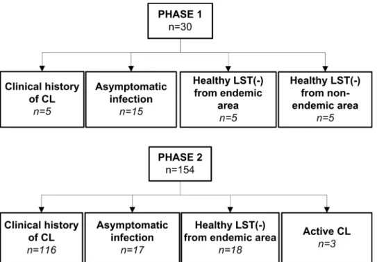
This descriptive study was conducted in two phases (Fig 1). An initial exploratory phase (Phase 1) was performed to determine the feasibility of molecular detection of persistent subclinical infection, which evaluated the presence ofLeishmaniakDNA in blood and mucosal swab sam-ples from members of five family households of rural areas of Tumaco (Nariño, Colombia), endemic for infections caused byL. (Viannia) species. Families/households were selected based on the presence of one household member (index case) with a parasitologically confirmed prior history of active disease as well as presence of compatible scars and positive leishmanin skin test reaction (LST), at least one co-habitant with asymptomatic infection based on LST reactivity in the absence of active or healed lesions, and a minimum of four co-habitants. In the second phase a larger scale assessment was designed to 1) establish the demonstrable presence ofLeishmaniain immunologically defined asymptomatic infection, and 2) to evaluate the per-sistence, viability and genotypic diversity ofLeishmaniaamong asymptomatically infected individuals and those with subclinical infections following prior symptomatic CL. This study was conducted in areas of endemic transmission of cutaneous leishmaniasis caused byL. (Viannia) species in the departments of Nariño and Risaralda in Colombia.
Following LST evaluation, the presence of parasites was assessed by PCR-Southern Blot for LeishmaniakDNA in blood monocytes, aspirates of lesion scars, and swab samples from nasal, tonsil, and conjunctival mucosa [14]. Parasite viability was determined by qRT-PCR of the Leishmania7SLRNA transcript [12], and genetic diversity ofLeishmaniawas assessed by geno-typing of the conserved region of the kDNA as described below.
Study subjects
184 subjects participated in this study. Phase 1 comprised a total of 30 individuals of which 25 were residents of endemic areas from rural communities of the municipality of Tumaco, department of Nariño and 5 were residents of non-endemic areas. Study groups were defined as follows: 1) Individuals with clinical history of CL (n = 5) confirmed by a typical scar of CL
[1] and positive LST result, 2) asymptomatic infection (n = 15), defined as residents of an endemic area of transmission of dermal leishmaniasis, having a positive LST and no evidence or history of dermal lesions or typical scars; and 3) healthy LST negative participants without history of leishmaniasis from endemic (n = 5) or non-endemic areas (n = 5) (Fig 1).
Phase 2 included 154 subjects: 103 participants were residents of a rural community in Pueblo Rico, Risaralda and 51 participants resided in rural communities of the municipality of Tumaco, department of Nariño. Study groups were as follows: 1) Individuals with clinical his-tory of CL (n = 116), 2) individuals with asymptomatic infection (n = 17), 3) healthy LST nega-tive participants from endemic areas (n = 18), and 4) individuals with acnega-tive parasitologically confirmed CL (n = 3) that served as positive controls (Fig 1).
Clinical samples
Blood monocytes, lesion/scar aspirates and duplicate swab samples from tonsil, conjunctiva and nasal mucosa were obtained from the study participants on one occasion. Duplicate sam-ples were obtained for the purpose of independent RNA and DNA extraction procedures. Blood monocytes were separated from a 10 mL sample of peripheral blood using 1-Step Mono-cytes following the manufacturer´s protocol (Accurate Chemical & Scientific Co.). MonoMono-cytes and lesion/scar aspirate samples were stored in TRIzol Reagent at -70°C until processing. RNA and DNA were extracted from blood monocytes using the AllPrep DNA/RNA Minikit (Qia-gen). Mucosal swab samples were stored at -20°C until processing, and DNA extracted from one of the two replicate swabs as previously reported [12,14]. RNA was extracted from the second swab sample using TRIzol followed by RNA cleanup using the RNeasy extraction Kit (Qiagen). Purified RNA was treated with DNAse I. All RNA samples were suspended in a final volume of 35μl of nuclease free water. Quantity and quality of nucleic acids were evaluated
using a NanoDrop2000 spectrophotometer.
Leishmania strains and kDNA sequences
Reference strains and clinical strains isolated from patients with CL were obtained from the CIDEIM BioBank (S1 Table). All strains were previously typed by monoclonal antibodies and/ or isoenzyme electrophoresis. Promastigotes were maintained at 26°C in RPMI medium sup-plemented with 10% heat-inactivated foetal bovine serum (Gibco), 1% glutamine, 100 U/ml penicillin and 100μg/ml streptomycin. Logarithmic phase promastigotes were harvested by
centrifugation, washed in phosphate-buffered saline (PBS), and solubilized in lysis buffer for DNA extraction. For the analyses of genetic diversity we also included kDNA sequences obtained from NCBI Genbank as summarized inS2 Table.
PCR, Southern blot and qRT-PCR
A 242 bp product of the humanGAPDHgene was targeted for amplification from all samples as a quality control procedure, using the primers Fw (5’- CTG GCC CTC TGC CCT CCT ACC A -3’) and Rv (5’- TTC CAT CCA GCC TGG GGC GAA -3’).L. (Viannia) minicircle kDNA was amplified from 100ng of DNA samples by PCR using the LV-B1 primers followed by Southern blot hybridization as previously described [15]. kDNA positive samples were evalu-ated by real time reverse transcriptase PCR to confirm the viability of parasites and estimate parasite burden using theLeishmania7SLRNA transcript as previously described [12] and using 10μl of the RNA sample. The single copy gene coding for the human TATA Box Binding
run by inclusion of purifiedL.(V).panamensisDNA as a positive control. Potential DNA carry-over or contamination was evaluated by inclusion of blank (water) and negative control (DNA of PBMCs from a healthy donor) samples. Standard curves for quantitation of parasite and human nucleated cells were constructed by ten-fold serial dilution of cDNA products obtained from 1x10⁷L. (V.)panamensispromastigotes and from the human U-937 promono-cytic cell line (1 x 10⁷cells), respectively. Specificity of the qRT-PCR products was assessed by analysis of the melt peak curve. Parasite burden was calculated by extrapolation to a standard curve and normalized to the number of human cells based on TBP expression. Real time detec-tion of amplificadetec-tion products was performed using SYBR Green Master Mix (Applied Biosys-tems) on a BioRad CFX-96 detection platform. For those kDNA positive samples that were below the limit of detection of the 7SLRNA qRT-PCR, a maximum likelihood estimate of 0.0001 parasites per reaction (0.00357 parasites per swab) was calculated based on the assump-tion of 10,000 minicircle kDNA copies and 250 copies of the 7SLRNA transcript per organism [16,17], and the samples volumes for each assay [18].
kDNA genotyping
Leishmaniastrains and kDNA positive samples from study participants were processed for genetic diversity analyses based on sequence comparison of the conserved region of Leish-maniaminicircle kDNA. A Nested PCR was designed to amplify the conserved block of Leish-maniakDNA. The external primers LVp1-Fw (5´- GAC ATG CCT CTG GGT AGG GGC GTT C -3´) and LVp1-Rv (5´- GGG TGG TAC GAT TTT GAC CCT AA -3´) were used for the first PCR reaction. Internal primers LVp1-Fw and LVp5-Rv (5´- CTG GGA TGC GCG GCC CAC TAT -3´) were used in the second PCR reaction (S1 Fig). Each 25μL of the first
PCR reaction mixture contained 0.8 mM of dNTP, 0.04 U/μL high fidelity Platinum Taq
poly-merase (Invitrogen), 2μL template DNA, 2.2 mM MgCl2, 1 X PCR buffer, and 0.4 nM of
LVp1-Fw and LVp1-Rv primers. The cycling reaction was as follows: 95°C for 5 min, followed by 35 cycles, each of 1 min at 95°C, 62°C for 30 sec and 72°C for 30 sec, and a final extension of 1 min at 72°C. The products of the first PCR were diluted 1:10 with ultrapure water and 2μL of
this dilution was used as template for the second PCR, which was performed under the condi-tions described above using LVp1-Fw and LVp5-Rv primer and set and an annealing tempera-ture of 59°C. Specificity of the tool was evaluated using total DNA extracted from human PBMCs obtained from healthy donors. Negative controls included within each reaction were PCR mix and DNA from human PBMCs. PCR products were separated in 1.3% agarose gels and products of approximately 180 bp were extracted and purified for the sequencing reaction using the QIAquick gel extraction kit (Qiagen). An 82bp fragment spanning conserved blocks 1 to 2 from the kDNA was selected for the analysis based on sequence variability in the inter-block regions. High resolution sequences were obtained by bi-directional Sanger sequencing (Macrogen-Korea) using LVp1-Fw and LVp5-Rv primers, and sequences edited and analyzed using BioEdit v7.2.5. Genetic distances were calculated using MEGA 6.0. Selection of the nucle-otide substitution model was based on MEGA 6.0 outputs for the model that best fitted the sequence data. Models analyzed included Jukes-Cantor, Hasegawa-Kishino-Yano and Tamura-Nei among others. Results for model use, based on the full sequence alignment indi-cated that the Jukes-Cantor model and G distribution best fitted the data. MEGA6 was also used to construct trees from resulting distance matrices.
Multilocus Microsatellite Typing (MLMT)
by PCR, as previously described [19]. The size of the microsatellites was determined by mobil-ity of the PCR products in 4.5% agarose gels. Genetic distances were estimated using MSA4.05 software and Populations-1.2.32, and neighbor joining and UPGMA trees were constructed using MEGA6.
Statistical analysis
D'Agostino and Pearson omnibus test [20] was used to test for departures of quantitative data from normal distributions. For parasite burdens, this was done on log-transformed data. The Mann Whitney U-Test was employed for statistical comparisons. Binomial proportion confi-dence intervals were calculated with the Wilson method using the 'binom' package in R [21]. Statistical significance was defined asp<0.05. Data were analyzed using Prism 5 software
(GraphPad Software, Inc., La Jolla, CA).
Results
Parasitological demonstration of persistent infection following
asymptomatic infection or clinical resolution of cutaneous leishmaniasis
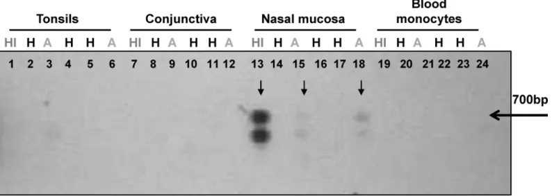
Amplification of humanGAPDHgene in 95% (114 of 120) of the DNA samples obtained from the 30 participants in the exploratory phase (Phase 1) corroborated the quality of the extracted DNA. The sixGAPDHnegative samples corresponded to swab samples from conjunctiva, reflecting difficulty in obtaining samples of a suitable quality from this potentially informative tissue.GAPDHnegative samples were excluded from further analysis. Four of the 5 individuals (80%) with prior history of CL (index cases), and 6 of 15 individuals (40%) with asymptomatic infection had one or more kDNA positive samples (Table 1). kDNA positive samples from the index case and from one or more asymptomatic cohabitants were found in three of the five households. A representative blot of samples from members of one household is shown inFig 2. kDNA positive samples were not found in any of the healthy individuals from either endemic (n = 5) or non-endemic (n = 5) areas.Based on the results of the exploratory analyses we proceeded to evaluate samples obtained from 154 individuals residing in CL endemic communities in Nariño and Risaralda for the presence ofLeishmaniakDNA. At least one sample was obtained from each study participant, however, 27 individuals did not consent to provide blood samples, 9 refused nasal swabs and 25 refused tonsil swabs. Amplification of the humanGAPDHgene was achieved in at least one
Table 1. Demonstration of feasibility of detecting asymptomatic infection among household members co-habitating with a prior symptomatic case using PCR-Southern blot ofLeishmaniakDNA (Phase1).
At least one kDNA positive sample
Family/ household
Index Case Clinical history of leishmaniasis and LST
(+)a(n/N)b Asymptomatic infection, LST(+)(n/N)a Healthy LST(-) co-habitant,(n/N)a
1 0/1 0/4 0/0
2 1/1 2/2 0/2
3 1/1 3/4 0/0
4 1/1 1/2 0/2
5 1/1 0/3 0/1
Total (%) 4/5 (80%) 6/15 (40%) 0/5 (0%)
aLST, Leishmanin skin test
bNumber of participants with at least one kDNA positive sample (n)/ Total number of participants (N)
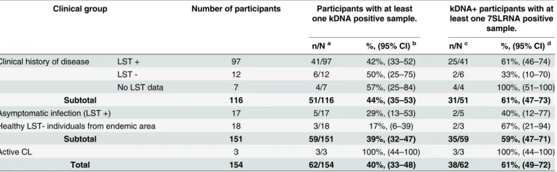
sample from each of the 154 participants, representing a total of 78% of samples (504 of 647 total samples). Sixty-two percent of GAPDH negative samples (88 of 143) corresponded to lesion scar aspirates, 33% were from conjunctival swabs and the remaining 5% were from blood monocytes, nasal and tonsil swabs. The one time sampling of study participants coupled to PCR ofLeishmaniakDNA and Southern blot hybridization, revealed parasite persistence in 40% (46/114) of LST+ individuals without active disease, composed of 41 of 97 LST+ partici-pants with a clinical history of CL and 5 of 17 asymptomatic infected individuals (Table 2). Nineteen (12%) individuals had more than one kDNA positive sample, for a total of 84 kDNA positive samples within the study population. Molecular demonstration of the presence of Leishmaniawas more frequent among individuals who had clinical evidence of prior symp-tomatic infection (44%, 51 of 116) than among LST positive asympsymp-tomatic infected individuals (29%, 5 of 17). kDNA positive samples (from blood monocytes, tonsil and nasal mucosa) were also detected in 3 of 18 skin test negative healthy individuals from endemic areas, and in the three positive control cases with active disease. Blood monocytes were the most frequently pos-itive sample for kDNA, being pospos-itive in 26% of individuals without active disease, followed by nasal and tonsil swabs that were positive in 14% of sampled individuals.
Quantification of parasite burden and demonstration of
Leishmania
viability
We quantified the parasite burden and assessed the viability ofLeishmaniaamong kDNA posi-tive samples by detection of RNA transcripts of theLeishmania7SLRNA gene. Transcripts were amplified from 59% of LST+ individuals in whomLeishmaniakDNA had been detected; from 25 of the 41 participants with clinical history of CL, and from 2 of the 5 asymptomatic infected individuals. Transcripts were also amplified from 6 of 10 individuals with a history of CL but with negative or unavailable LST results, and from 2 of the 3 LST- healthy individuals in the study communities from whomLeishmaniakDNA was amplified (Table 2). Amplifica-tion of the human TATA box binding protein (TBP) gene transcript was successful in 90% of
Fig 2. Detection ofLeishmaniakDNA in samples from individuals with asymptomatic infection who share a household with a historic symptomatic
case of CL.Leishmaniaminicircle kDNA was detected by PCR amplification and southern blot. Lanes 1–6: Tonsil swab samples; Lanes 7–12: conjunctival
swab samples; Lanes 13–18: nasal mucosa swab samples; Lanes 19–24: blood monocytes. HI: History of CL, H: Healthy, A: Asymptomatic. The arrow
denotes the 700bp band corresponding to the full length amplification product ofLeishmaniaminicircle kDNA. The lower band represents an unspecific kDNA PCR product as confirmed by sequencing.
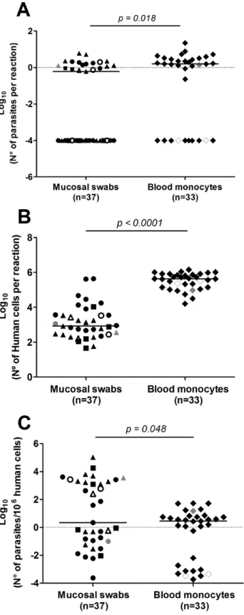
the 84 kDNA positive samples, supporting the quality and reliability of the amplified RNA. Twenty-eight kDNA positive samples were below the limit of detection of the 7SLRNA qRT-PCR (10 from blood monocytes and 18 from mucosal swab samples). Parasite loads ran-ged between 0.2 to 22 parasites per reaction. Absolute parasite loads and those normalized to the number of human nucleated cells per sample were slightly higher in blood monocytes than those from mucosal swab samples (Fig 3A–3C). No apparent differences in parasite loads of different mucosal tissues were observed. The presence of parasite RNA transcripts substantiates the viability ofLeishmaniawithin the sampled tissues and among a high proportion of individ-uals with subclinical infection.
Comparative analysis of
Leishmania
diversity by kDNA genotyping and
microsatellite typing
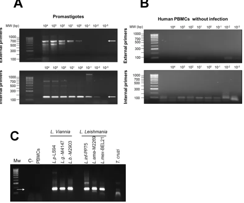
To explore relationships amongLeishmaniain individuals with subclinical infection and para-site strains isolated from patients with active disease within the same foci of transmission, we developed a nested PCR approach targeting a 180bp segment of the conserved region of Leish-maniaminicircle kDNA spanning blocks 1 to 3. The lower limit of detection of the nested PCR was 10−3promastigotes per reaction (Fig 4A). No cross-reactivity with human DNA was
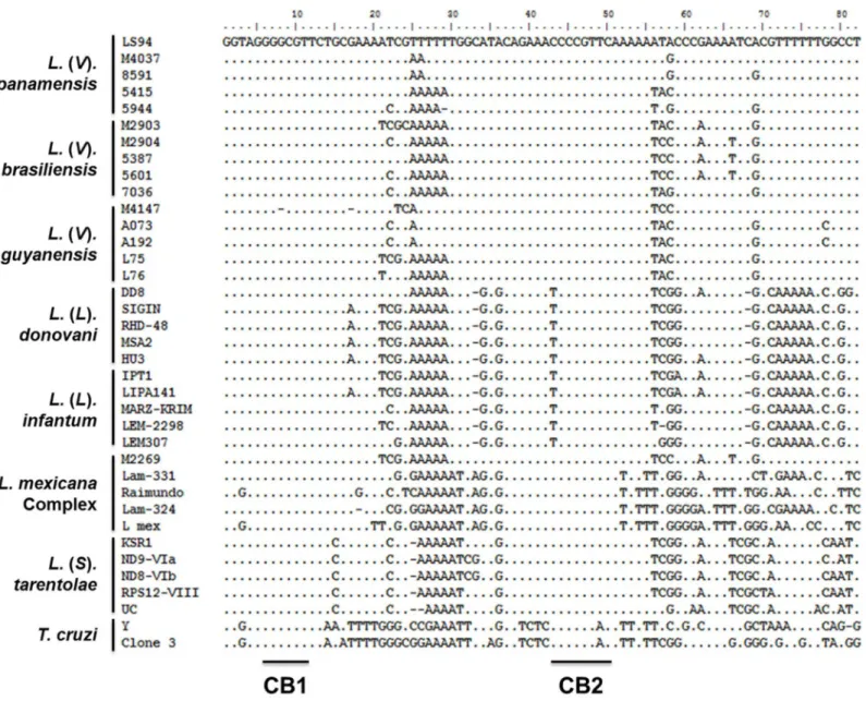
detected (Fig 4B). Amplification products were obtained fromLeishmaniaspecies pertaining to bothL. (Viannia) andL. (Leishmania) subgenera and a faint amplification product of ~180bp was detected inT.cruziDNA (Fig 4C). Although multiple sequencing attempts were performed onT.cruziamplification products, quality chromatograms were never obtained, reflecting the low-sensitivity cross reaction due to low homology of the LVp5 primer annealing site (S1 Fig). Alignment ofL.donovani,L.infantum,L.mexicana,L.tarentolae,L. (V.) brazi-liensis,L. (V.)guyanensis,L. (V.)panamensisandT.cruzikDNA sequences (S2 Table) showed that regions between conserved blocks 1 and 2 were polymorphic among species and strains of the same species (Fig 5).
Table 2. Detection ofLeishmaniakDNA and 7SLRNA gene transcript in clinical samples from subclinically infected individuals in endemic foci of
CL.
Clinical group Number of participants Participants with at least one kDNA positive sample.
kDNA+ participants with at least one 7SLRNA positive
sample.
n/Na %, (95% CI)b n/Nc %, (95% CI)d
Clinical history of disease LST + 97 41/97 42%, (33–52) 25/41 61%, (46–74)
LST - 12 6/12 50%, (25–75) 2/6 33%, (10–70)
No LST data 7 4/7 57%, (25–84) 4/4 100%, (51–100)
Subtotal 116 51/116 44%, (35–53) 31/51 61%, (47–73)
Asymptomatic infection (LST +) 17 5/17 29%, (13–53) 2/5 40%, (12–77)
Healthy LST- individuals from endemic area 18 3/18 17%, (6–39) 2/3 67%, (21–94)
Subtotal 151 59/151 39%, (32–47) 35/59 59%, (47–71)
Active CL 3 3/3 100%, (44–100) 3/3 100%, (44–100)
Total 154 62/154 40%, (33–48) 38/62 61%, (49–72)
aNumber of participants with kDNA positive sample (n)/ Total number of participants (N) b% participants with at least one kDNA positive sample and 95% Con
fidence Interval for proportions of kDNA positivity.
cNumber of participants with 7SLRNA positive sample (n) / Total number of participants with kDNA positive samples d% participants with at least one 7SLRNA positive sample and 95% Con
fidence Interval for proportions of 7SLRNA positivity.
Fig 3.Leishmaniaviability and parasite burden in kDNA positive samples.Absolute quantification of
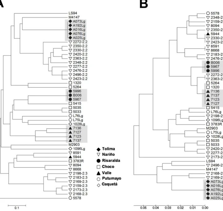
To explore the resolution and assess the reliability of the strain grouping achieved by kDNA genotyping, we performed a comparative analysis against multilocus microsatellite typing (MLMT). Genetic distances were calculated from MLMT data (S3 Table) and from kDNA sequences of a panel of 34L. (V.)panamensisand 9L. (V.)guyanensisstrains isolated from diverse locations within the Colombian territory (S1 Table). Results showed that both methods concurred in clustering of strains obtained from individuals with CL in foci of active figures, healthy LST- individuals from endemic area for CL.pvalues obtained from two tailed Mann-Whitney tests are shown.
doi:10.1371/journal.pntd.0004273.g003
Fig 4. Sensitivity and specificity of the nested PCR forLeishmaniaminicircle kDNA.Nested PCR amplification products from serial dilution curves of DNA fromL.V.panamensispromastigotes(A)and human PBMCs(B).The upper panel shows the first amplification product of 700 bp obtained with LVp1 primer sets (external primers) and the bottom panel shows the 180 bp product of the nested PCR obtained with LVp1-LVp5 primer sets (internal primers). Nested PCR amplification products from reference strains ofL.V.panamensis(L.p),L.V.guyanensis(L.g),L.V.braziliensis(L.b),L.infantum(L. inf),L.
amazonensis(L.ama),L.mexicana(L. mex) andT.cruzi(C).C-, negative control—water; PBMCs, DNA from uninfected human PBMCs.
transmission during outbreaks of CL, thus delimiting strains by time and location (Fig 6). Nev-ertheless, subgroups defined by MLMT such as zymodemes 2.2 and 2.3 ofL. (V.)panamensis were not clustered by kDNA genotyping, in line with the higher variability and rate of evolu-tion of kDNA compared to microsatellite sequences [13,22].
To estimate the best method for data analysis and clustering, we run in parallel Neighbor-Joining, UPGMA and Maximum Likelihood methods of kDNA sequences. As shown inS2 Fig, all clustering algorithms provided the same group distribution with the exception of clustering of database sequences obtained from theL.mexicanacomplex. Although it is recognized that bootstrap values are indicators of robustness for definition of the most appropriate clustering method, bootstrap values were low when using any of the above methods, potentially due the small sequence size (82bp) and the low overall variability among sequences (where the most
Fig 5. Analysis ofLeishmaniagenetic diversity.An 82bp sequence spanning conserved blocks 1 and 2 from theLeishmaniaminicircle kDNA was used for multiple sequence alignment. Sequences from five different strains were analyzed for eachLeishmaniaspecies, and from twoT.cruzistrains. Strain details are summarized inS1andS2Tables. CB: Conserved Block.
divergent strains—L.(V) panamensis 2363andL.infantum LLM735—showed 83% sequence homology). That all of the methods generated similar clustering and that clusters are biologi-cally concordant, supports their usefulness in these analyses. UPGMA clustering was selected for data interpretation given that clustering of the subgenera represented more accurately the Leishmaniataxonomy.
Considering the reported variability of kDNA sequences among clinical strains [16,23] and possible variations introduced during the PCR or sequencing reactions, we assessed the repro-ducibility of the method by re-amplifying and re-sequencing DNA samples from 13 strains
Fig 6. Genetic diversity analysis using minicircle kDNA genotyping allows clustering of strains isolated within foci of transmission during outbreaks of CL.UPGMA trees of distances calculated from MLMT data(A)and conserved block minicircle kDNA genotyping(B)from a panel of 34L.V.
panamensisand 9L.V.guyanensisclinical strains. Figure labels depict the geographical origin of strains within the Colombian territory. Shadowed codes denote groups of strains isolated from foci of transmission during outbreaks of infection.
belonging to different species of theVianniasubgenus obtained from the CIDEIM BioBank. As shown inS3 Fig, 100% sequence identity was obtained in two independent technical replicas in sequences from 10 of the 13 strains, and 99% sequence identity (corresponding to a 1bp change) in sequences from the remaining 3 strains. These results support the reproducibility of the method for analysis ofLeishmaniagenetic diversity using clinical samples and strains.
kDNA genotyping of samples from individuals with subclinical infection
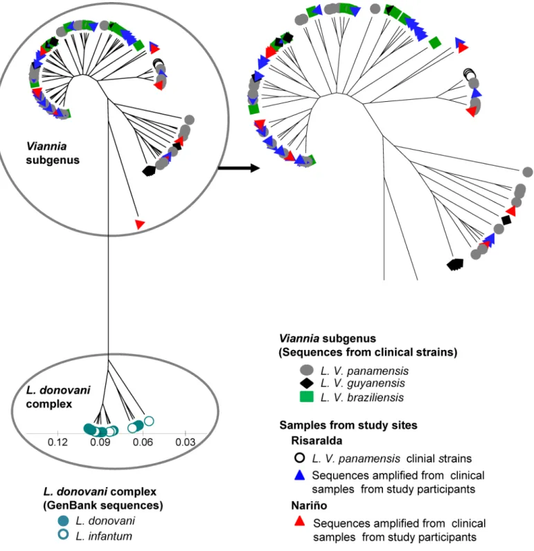
The intra-species variability of the target sequence and the sensitivity and specificity of the method forLeishmaniakDNA, support the potential of kDNA genotyping for the analysis of clinical samples with low parasite burden. We attempted to sequence nested PCR amplification products from 84 kDNA positive samples from 59 individuals. Fifty-four samples from 45 sub-clinically infected participants were successfully amplified by the nested PCR. However, PCR products from 33 of these samples, corresponding to 28 individuals with immunological or molecular evidence of subclinical infection (26 with prior history of CL, 2 with asymptomatic infection and one from a“healthy”LST- individual in the endemic site) were of sufficient qual-ity and quantqual-ity to provide accurate sequences. kDNA sequences were also obtained fromL. (V.)panamensisstrains isolated from the three patients with active CL.Sequences were analyzed alongside a panel of representativeL. (V.)panamensis,L. (V.) guyanensisandL. (V.)braziliensisstrains from diverse geographical origins within the Colom-bian territoryS1 Table), and database sequences fromL.donovani and L.infantumas out-groups (S2 Table). kDNA sequences from individuals with subclinical infection grouped within theL. (Viannia) cluster (Fig 7). However, the relationship betweenL. (V.)panamensis strains currently circulating in the focus of transmission and parasites involved in subclinical infections that have persisted over many years could not be discerned.
Discussion
Asymptomatic infections are common to all transmissible agents, constituting a variable segment of the spectrum of outcomes. The frequency of these inapparent infections varies according to the pathogenicity of the agent and the susceptibility of the exposed population. Investigations of the incidence of infection based on LST conversion revealed that 90% of inci-dent infections in a focus ofL. (V.)panamensistransmission in the municipality of Tumaco, Colombia, were asymptomatic [1], whereas only 17% of infections were asymptomatic in a focus ofL. (V.)peruvianatransmission in Peru [3]. In the current study, LST reactivity pro-vided an immunological marker for asymptomatic and subclinical infections, which were defined respectively as infections that result in skin test conversion but not disease, and persis-tent infection following clinical resolution of disease. Leishmanin reactivity induced by infec-tion, whether symptomatic or asymptomatic is typically long-lived and presumably sustained by antigen exposure. The amplification of kDNA and 7SLRNA transcripts from 40% and 24% of LST+ individuals, respectively, supports this assumption and demonstrates the technical fea-sibility of molecular detection ofLeishmaniain mucosal and blood monocyte samples from individuals with subclinical infections.
Although the relevance of asymptomatic and subclinical infections to public health is poorly understood, the detection ofLeishmaniain the absence of active disease in 40% of LST+ and a small proportion of LST- residents of endemic foci ofL. (Viannia) transmission provides an indication of the substantial magnitude of the population harboring parasites. Amplification of LeishmaniaDNA and RNA from 2 of 18“healthy”LST negative participants could reflect wan-ing delayed-type hypersensitivity responses [24] or a lower sensitivity threshold of this
least 12 of 116 participants with prior history of CL is also consistent with either of these sce-narios. Considering that sampling was conducted only one time and that tissue distribution
Fig 7. Analysis ofLeishmaniagenetic diversity in individuals with infection in the absence of disease.Sequences from the 82bp sequence spanning conserved blocks 1 and 2 fromLeishmaniaminicircle kDNA were analyzed. Shown is a UPGMA tree of the genetic distances of sequences from clinical samples obtained from study participants with subclinical infection (blue and red triangles) and strains isolated from study participants with active disease (yellow circles), alongside a panel of 34L. (V)panamensis, 10L. (V.)braziliensis, and 9L. (V)guyanensisclinical strains. Minicircle kDNA sequences retrieved from NCBI Genbank from9 L.donovaniand 8L.infantumstrains were used as outgroups.
and burden of parasites are likely to vary over time as shown with sequential samples of asymp-tomaticL.infantuminfection [25], and with the health status of each individual, the actual infected population is probably underestimated.
Subclinical infections represent a risk for reactivation and the triggering of pathogenesis. The recurrent behavior of dermal leishmaniasis in Latin America was clinically recognized in mucosal disease, long considered a secondary manifestation of prior cutaneous disease [26] or even prior asymptomatic infection [27]. Direct evidence of re-activation as a cause of recurrent leishmaniasis was provided by biochemical and genotypic analyses ofLeishmaniastrains iso-lated from primary lesions and from recurrent or new lesions following complete resolution of primary lesions [23]. Additionally, activation of CL following local trauma [28] and immuno-suppression [29–31] in individuals with asymptomatic infection or healed lesions provides evi-dence for the participation of immunological and inflammatory triggers in the activation/re-activation of CL. Population-based studies in areas of endemic transmission have identified both LST reactivity and the presence of scars compatible with history of CL as important risk factors for development of new incident lesions [1,3,10], underscoring the role of asymptom-atic and subclinical infection in the epidemiology and natural history of CL. Our results docu-menting the long-term presence of viableLeishmaniain blood monocytes and unaffected mucosal tissues in individuals with asymptomatic and subclinical infection support the indefi-nite persistence ofLeishmaniain the human host.
Through mechanistic mathematical modeling, Miller et.al. have recently shown that a small proportion of asymptomatically infected individuals (3.2%) with the highest parasitemias in VL endemic areas in Ethiopia, were responsible for infection of an average 62% of infected sand fly vectors [11]. Although parasite loads in unaffected mucosal tissues and blood mono-cytes (0.2 to 22 parasites per reaction, equivalent to 7 to 770 parasites per sample) of individu-als with subclinical and asymptomatic infection in our study population are relatively low compared to those with asymptomatic infection in VL endemic areas [11], these are within the range of parasite loads found in unaffected mucosal tissues in individuals with active CL [12]. Together with lessons learned from investigations of the infectivity of asymptomatic and even vaccinated dogs forLu.longipalpis, the principal vector of VL in the Americas, and from malaria elimination initiatives showing that asymptomatic carriers, in addition to being at risk of developing disease via reactivation of infection [32] can transmit infection to mosquitos [33,34], support the possibility that subclinically infected individuals may act as reservoirs for anthroponotic transmission ofL. (Viannia) species, which have traditionally been considered zoonoses. The importance of understanding the role of subclinical infection in the incidence and propagation of leishmaniasis has been recently recognized as a priority research area by the World Health Organization Expert Committee on the Control of the Leishmaniases [35].
A limiting factor in the study of subclinical infection is the technical feasibility for pheno-typic and genopheno-typic characterization ofLeishmaniafrom samples having low parasite burdens. To address this limitation we developed a strategy for analysis of genetic diversity based on nested PCR amplification and sequence genotyping of the conserved region ofLeishmania minicircle kDNA. The sensitivity of this strategy allowedLeishmaniakDNA sequences to be obtained from mucosal swab samples and blood monocytes from 28 of individuals with immunological (LST) or molecular (kDNA detection) evidence of subclinical infection, dem-onstrating the plausibility of approaching genotypic characterization ofLeishmaniacausing subclinical infection. Although the sensitivity of nested PCR was comparable to that of kDNA amplification and detection by southern blot (10−3promastigotes per reaction), partial
Regions within the conserved blocks ofLeishmaniakDNA minicircles were selected as targets for genetic diversity analysis based on limited yet potentially informative polymorphic character-istics. The methodology developed revealed diversity amongLeishmaniacausing subclinical infection in endemic foci of transmission ofL. (V.)panamensis, and clustering within theL. (Viannia) subgenus. Nevertheless, the limited number of active cases (n = 3) and the level of het-erogeneity in the amplified sequences did not allow relationships to be discerned between strains isolated from individuals with active disease at the time of the study andLeishmaniapersisting in the absence of disease. This could be due to variation in parasite populations during decades of transmission within the sites, or limitations of the genotyping methodology.
Comparative analysis of kDNA and MLMT genotyping in a panel of strains ofL. (Viannia) species showed clustering of strains isolated from disease outbreaks to be achieved by both meth-odologies, suggesting that kDNA genotyping could be exploited for future studies of the cycle and dynamics of transmission in active foci and during disease outbreaks. However, because phe-notypically distinguishable strains (eg.L. (V.)panamensisstrains pertaining to zymodemes 2.2 and 2.3) clustered by MLMT but not kDNA typing, micro-heterogeneity of kDNA sequences could impede the discernment of relationships among closely related parasites [36,37]. This out-come illustrates some of the limitations of kDNA genotyping, which include analysis of a single target sequence and the sequence length. Multi-target sequence analysis of polymorphic and high copy number sequences such as miniexon, Cytochrome B, GP63 or Cysteine proteinase B genes [38], and improved sensitivity for minicircle kDNA amplification could optimize the robustness of this approach to accessing subclinical infection and the parasites involved.
Our results provide parasitological confirmation of persistent infection in the absence of disease among residents of endemic areas of CL and a methodological approach to investigate the epidemiology and public health impact of subclinical infections. The novel exploitation of kDNA genotyping establishes proof-of-principle of the feasibility of genetic diversity analysis in parasite populations previously inaccessible and unexplored, and provides bases for more robust analyses of the relationships among these parasite populations.
Supporting Information
S1 Fig. Primer annealing sites. (DOCX)S2 Fig. Comparison of clustering algorithms for analysis of minicircle kDNA sequences. (DOCX)
S3 Fig. Verification of sequencing fidelity. (DOCX)
S1 Table. Clinical and reference strains ofL.Vianniaspecies. (DOCX)
S2 Table. GenBank kDNA sequences used in the analyses of genetic diversity. (DOCX)
S3 Table. MLMT profiles ofL.Vianniastrains. (DOCX)
Acknowledgments
Isabel Guasaquillo of the CIDEIM BioBank and research support team for isoenzyme and monoclonal antibody typing of the clinical strains included in this study. We also acknowledge the generous collaboration of the participants and their families. We thank the Secretary of Health of the Department of Risaralda (Secretaría de Salud Departamental de Risaralda) and staff of the Pueblo Rico Hospital for their technical and logistical support.
Author Contributions
Conceived and designed the experiments: MAG NGS. Performed the experiments: MRC REG. Analyzed the data: MRC REG LV JJ NA LCR AC ERA MAG NGS. Wrote the paper: MRC REG LV JJ NA LCR AC ERA MAG NGS.
References
1. Weigle KA, Santrich C, Martinez F, Valderrama L, Saravia NG Epidemiology of cutaneous leishmania-sis in Colombia: a longitudinal study of the natural history, prevalence, and incidence of infection and clinical manifestations. J Infect Dis. 1993, 168: 699–708. PMID:8354912
2. Singh OP, Hasker E, Sacks D, Boelaert M, Sundar S Asymptomatic Leishmania infection: a new chal-lenge for Leishmania control. Clin Infect Dis. 2014, 58: 1424–1429. doi:10.1093/cid/ciu102PMID:
24585564
3. Davies CR, Llanos-Cuentas EA, Pyke SD, Dye C Cutaneous leishmaniasis in the Peruvian Andes: an epidemiological study of infection and immunity. Epidemiol Infect. 1995, 114: 297–318. PMID:
7705493
4. Molina R, Amela C, Nieto J, San-Andres M, Gonzalez F, et al. Infectivity of dogs naturally infected with Leishmania infantum to colonized Phlebotomus perniciosus. Trans R Soc Trop Med Hyg. 1994, 88: 491–493. PMID:7570854
5. Michalsky EM, Rocha MF, da Rocha Lima AC, Franca-Silva JC, Pires MQ, et al. Infectivity of seroposi-tive dogs, showing different clinical forms of leishmaniasis, to Lutzomyia longipalpis phlebotomine sand flies. Vet Parasitol. 2007, 147: 67–76. PMID:17449184
6. Laurenti MD, Rossi CN, da Matta VL, Tomokane TY, Corbett CE, et al. Asymptomatic dogs are highly competent to transmit Leishmania (Leishmania) infantum chagasi to the natural vector. Vet Parasitol. 2013, 196: 296–300. doi:10.1016/j.vetpar.2013.03.017PMID:23562649
7. Fernandes CB, Junior JT, de Jesus C, Souza BM, Larangeira DF, et al. Comparison of two commercial vaccines against visceral leishmaniasis in dogs from endemic areas: IgG, and subclasses, parasitism, and parasite transmission by xenodiagnosis. Vaccine. 2014, 32: 1287–1295. doi:10.1016/j.vaccine.
2013.12.046PMID:24406392
8. Vergel C, Palacios R, Cadena H, Posso CJ, Valderrama L, et al. Evidence for leishmania (viannia) par-asites in the skin and blood of patients before and after treatment. J Infect Dis. 2006, 194: 503–511.
PMID:16845635
9. de Oliveira Camera P, Junger J, do Espirito Santo Silva Pires F, Mattos M, Oliveira-Neto MP, et al. Hae-matogenous dissemination of Leishmania (Viannia) braziliensis in human American tegumentary leish-maniasis. Trans R Soc Trop Med Hyg. 2006, 100: 1112–1117. PMID:16765391
10. Munoz G, Davies CR Leishmania panamensis transmission in the domestic environment: the results of a prospective epidemiological survey in Santander, Colombia. Biomedica. 2006, 26 Suppl 1: 131–144.
PMID:17361849
11. Miller E, Warburg A, Novikov I, Hailu A, Volf P, et al. Quantifying the contribution of hosts with different parasite concentrations to the transmission of visceral leishmaniasis in Ethiopia. PLoS Negl Trop Dis. 2014, 8: e3288. doi:10.1371/journal.pntd.0003288PMID:25356795
12. Romero I, Tellez J, Suarez Y, Cardona M, Figueroa R, et al. Viability and burden of Leishmania in extra-lesional sites during human dermal leishmaniasis. PLoS Negl Trop Dis. 2010, 4.
13. Schonian G, Kuhls K, Mauricio IL Molecular approaches for a better understanding of the epidemiology and population genetics of Leishmania. Parasitology. 2011, 138: 405–425. doi:10.1017/
S0031182010001538PMID:21078222
15. Vergel C, Walker J, Saravia NG Amplification of human DNA by primers targeted to Leishmania kineto-plast DNA and post-genome considerations in the detection of parasites by a polymerase chain reac-tion. Am J Trop Med Hyg. 2005, 72: 423–429. PMID:15827280
16. Noyes HA, Reyburn H, Bailey JW, Smith D A nested-PCR-based schizodeme method for identifying Leishmania kinetoplast minicircle classes directly from clinical samples and its application to the study of the epidemiology of Leishmania tropica in Pakistan. J Clin Microbiol. 1998, 36: 2877–2881. PMID:
9738037
17. Michaeli S, Podell D, Agabian N, Ullu E The 7SL RNA homologue of Trypanosoma brucei is closely related to mammalian 7SL RNA. Mol Biochem Parasitol. 1992, 51: 55–64. PMID:1565138
18. Solomon AW, Holland MJ, Burton MJ, West SK, Alexander ND, et al. Strategies for control of trachoma: observational study with quantitative PCR. Lancet. 2003, 362: 198–204. PMID:12885481
19. Oddone R, Schweynoch C, Schonian G, de Sousa Cdos S, Cupolillo E, et al. Development of a multilo-cus microsatellite typing approach for discriminating strains of Leishmania (Viannia) species. J Clin Microbiol. 2009, 47: 2818–2825. doi:10.1128/JCM.00645-09PMID:19587302
20. D'Agostino R, Pearson ES Tests for Departure from Normality. Empirical Results for the Distributions of b2 andp
b1. Biometrika. 1973, 60: 613–622.
21. Agresti A, Coull BA Approximate is better than ''Exact'' for interval estimation of binomial proportions. The American Statistician. 1998, 52: 119–126.
22. Lymbery AJ, Thompson RC The molecular epidemiology of parasite infections: tools and applications. Mol Biochem Parasitol. 2012, 181: 102–116. doi:10.1016/j.molbiopara.2011.10.006PMID:22027028
23. Saravia NG, Weigle K, Segura I, Giannini SH, Pacheco R, et al. Recurrent lesions in human Leish-mania braziliensis infection—reactivation or reinfection? Lancet. 1990, 336: 398–402. PMID:1974943
24. Weigle KA, Valderrama L, Arias AL, Santrich C, Saravia NG Leishmanin skin test standardization and evaluation of safety, dose, storage, longevity of reaction and sensitization. Am J Trop Med Hyg. 1991, 44: 260–271. PMID:2035747
25. dos Santos Marques LH, Gomes LI, da Rocha IC, da Silva TA, Oliveira E, et al. Low parasite load esti-mated by qPCR in a cohort of children living in urban area endemic for visceral leishmaniasis in Brazil. PLoS Negl Trop Dis. 2012, 6: e1955. doi:10.1371/journal.pntd.0001955PMID:23272263
26. Marsden PD Mucosal leishmaniasis ("espundia" Escomel, 1911). Trans R Soc Trop Med Hyg. 1986, 80: 859–876. PMID:3037735
27. Walton BC, Chinel LV, Eguia y Eguia O Onset of espundia after many years of occult infection with Leishmania braziliensis. Am J Trop Med Hyg. 1973, 22: 696–698. PMID:4200811
28. Wortmann GW, Aronson NE, Miller RS, Blazes D, Oster CN Cutaneous leishmaniasis following local trauma: a clinical pearl. Clin Infect Dis. 2000, 31: 199–201. PMID:10913426
29. Coura JR, Galvao-Castro B, Grimaldi G Junior Disseminated American cutaneous leishmaniasis in a patient with AIDS. Mem Inst Oswaldo Cruz. 1987, 82: 581–582. PMID:3507921
30. Mortazavi H, Salehi M, Kamyab K Reactivation of cutaneous leishmaniasis after renal transplantation: a case report. Case Rep Dermatol Med. 2014, 2014: 251423. doi:10.1155/2014/251423PMID:
24826350
31. Mirzabeigi M, Farooq U, Baraniak S, Dowdy L, Ciancio G, et al. Reactivation of dormant cutaneous Leishmania infection in a kidney transplant patient. J Cutan Pathol. 2006, 33: 701–704. PMID:
17026523
32. Lindblade KA, Steinhardt L, Samuels A, Kachur SP, Slutsker L The silent threat: asymptomatic parasi-temia and malaria transmission. Expert Rev Anti Infect Ther. 2013, 11: 623–639. doi:10.1586/eri.13.
45PMID:23750733
33. Bousema T, Okell L, Felger I, Drakeley C Asymptomatic malaria infections: detectability, transmissibil-ity and public health relevance. Nat Rev Microbiol. 2014, 12: 833–840. doi:10.1038/nrmicro3364
PMID:25329408
34. Kirchgatter K, Tubaki RM, Malafronte Rdos S, Alves IC, Lima GF, et al. Anopheles (Kerteszia) cruzii (Diptera: Culicidae) in peridomiciliary area during asymptomatic malaria transmission in the Atlantic Forest: molecular identification of blood-meal sources indicates humans as primary intermediate hosts. Rev Inst Med Trop Sao Paulo. 2014, 56: 403–409. PMID:25229220
35. Control of the leishmaniasis: report of a meeting of the WHO Expert Committee on the Control of Leish-maniases, Geneva, 22–26 March 2010.
37. Spithill TW, Grumont RJ, Mitchell GF Characterization of isolates and clones of Leishmania by analysis of kinetoplast DNA. J Cell Biochem. 1984, 24: 103–112. PMID:6725424