Fabrication and Characterization of Spongy Denuded
Amniotic Membrane Based Scafold for Tissue
Engineering
Ehsan Taghiabadi, M.Sc.1, 2, Sima Nasri, Ph.D.1, Saeed Shaieyan, M.D.2, Sasan Jalili Firoozinezhad, M.Sc.2, Nasser Aghdami, M.D., Ph.D.2*
1. Department of Biology, Faculty of Science, Payame NOOR University, Tehran, Iran
2. Department of Regenerative Biomedicine at Cell Science Research Center, Royan Institute for Stem Cell Biology and Technology, ACECR, Tehran, Iran
*Corresponding Address: P.O.Box: 16635-148, Department of Regenerative Biomedicine at Cell Science Research Center, Royan Institute for Stem Cell Biology and Technology, ACECR, Tehran, Iran
Email: [email protected]
Received: 28/Aug/2013, Accepted: 29/Jan/2014
Abstract
Objective: As a biological tissue material, amniotic membrane (AM) has low immuno-genicity and to date has been widely adopted in clinical practice. However, some features such as low biomechanical consistency and rapid biodegradation is limited the application of AM. Therefore, in this study, we fabricated a novel three-dimensional (3D) spongy scaf-fold made of the extracellular matrix (ECM) of denuded AM. Due to their unique charac-teristics which are similar to the skin, these scaffolds can be considered as an alternative option in skin tissue engineering.
Materials and Methods: In this experimental study, cellular components of human amniotic membrane (HAM) were removed with 0.03% (w/v) sodium dodecyl sulphate (SDS). Quantita-tive analysis was performed to determine levels of Glycosaminoglycans (GAGs), collagen, and deoxyribonucleic acid (DNA). To increase the low eficiency and purity of the ECM component, especially collagen and GAG, we applied an acid solubilization procedure hydrochloridric acid (HCl 0.1 M) with pepsin (1 mg/ml). In the present experiment 1-ethyl-3-(3-dimethyl aminopro-pyl) carbodiimide hydrochloride (EDC)/N-hydroxysuccinimide (NHS) cross linker agent was used to improve the mechanical properties of 3D lyophilized AM scaffold. The spongy 3D AM scaffolds were speciied, by scanning electron microscopy, hematoxylin and eosin (H&E) stain-ing, a swelling test, and mechanical strength and in vitro biodegradation tests. Human fetal ibroblast culture systems were used to establish that the scaffolds were cytocompatible.
Results: Histological analysis of treated human AM showed impressive removal of cel-lular components. DNA content was diminished after treatment (39 ± 4.06 μg/ml vs. 341 ± 29.60 μg/ml). Differences were observed between cellular and denude AM in matrix colla-gen (478 ± 18.06 μg/mg vs. 361 ± 27.47 μg/mg).With the optimum concentration of 1 mM NHS/EDC ratio1:4, chemical cross-linker agent could signiicantly increase the mechani-cal property, and resistance to collagenase digestion. The results of 2, 4, 6-Trinitrobenze-nesulfonic acid (TNBS) test showed that cross-linking eficiency of AM derived ECM scaf-folds was about 65% ± 10.53. Scafscaf-folds treated with NHS/EDC cross-linker agent by 100 μg/ml collagenase, lost 75% of their dry weight after 14 days. The average pore size of 3D spongy scaffold was 160 µm measured from scanning electron microscope (SEM) im-ages that it is suitable for cell penetration, nutrients and gas change. In addition, the NHS/ EDC cross-linked AM scaffolds were able to support human fetal ibroblast cell prolifera-tion in vitro. Extracts and contact prepared from the 3D spongy scaffold of AM showed a signiicant increase in the attachment and proliferation of the human fetal ibroblasts cells.
Conclusion: The extra-cellular matrix of denuded AM-based scaffold displays the main properties required for substitute skin including natural in vitro biodegradation, similar physical and mechanical characterization, nontoxic biomaterial and no toxic effect on cell attachment and cell proliferation.
Keywords: Extracellular Matrix, Skin Substitute, Biodegradable
Cell Journal(Yakht eh), Vol 16, No 4, Wint er 2015, Pages: 476- 487
Citation: Taghiabadi E, Nasri S, Shafieyan S, Jalili Firoozinezhad S, Aghdami N. Fabrication and characteriza
Introduction
The skin, which is the largest tissue in human body, is constructed of three layers epidermis, dermis and hypodermis. It performs a main function in protecting the human body from much chemical and mechanical damage from the surrounding environment. The loss of skin can occur for various reasons, such as thermal trauma, genetic disorders, chronic wounds, burns or even surgical interventions (1). Because of the low immunogenicity of donor skin and the limited avail-ability of donor skin sources, skin grafts are unable to provide complete recovery of the skin rendering them unsuitable for widespread use. Several years ago, great attempts were made to fabricate substitute human skin. Tissue engineering is an impressive way to develop skin substitutes and improve the wound healing. One fundamental purpose of cell biologists in tissue engineering is to induce cell proliferation using appropriate cell culture conditions (2, 3). The aim of study to construct a novel three-dimensional (3D) scaffold that improves cell proliferation and imitates the structure of the skin. Collagen is a main component of human connective tissues, particularly in soft skin tissues (4). Collagen is one of the best biomaterials for different applications (5, 6). Advan-tages include excellent biocompatibility, low toxicity and excellent biodegradability (7, 8). Despite these advantages, low mechanical consistency and the fast degradation rate of uncross-linked natural scaffolds such as collagen are the main problems that restrict its application. Thus, cross-linking of the collagen scaffolds is an impressive procedure to optimize their mechanical strength and to regulate the biodegrada-tion rate. Glycosaminoglycans (GAGs) are another principal component of the skin tissue that is asso-ciated with skin repair. Being the most hydrophilic molecule exist in the natural tissue such as skin, this characteristic GAGs is of vital role in water
mainte-nance (9). GAGs can be better modiied by function -al groups such as hydroxyl and carboxyl chains (10). GAGs also perform general functions in intermedi-ary skin-cell biological procedures, cell migration,
growth, granulation tissue matrix formation, inlam -matory response temperance, re-epithelialization and scarring (11). In addition, cellular attachment, growth and differentiation depends on denatured collagen structure.
Human amniotic membrane (HAM) has alots of
speciications that cause it applicable biomaterial.
The extra cellular matrix of amnion composed of
collagen I, collagen III, collagen IV and ibronec -tin. It is cheap and quickly takes, and its availabil-ity is virtually unrestricted. HAM has been pre-sented to consult applicable wound protection and have a symbolic outcome on pain reduction (12). 3D natural and synthetic scaffolds play a main function in maintenance of cell proliferation and tissue regeneration (13). Interactions between cells and the extracellular matrix (ECM) are responsi-ble for the control of cell action. So, cells grown in a 2D monolayer cannot cope with the relative complexity of the in vivo micro-environment. For
example, it has been suggested that cells cultivated on 2D layer such as culture plates, lose numerous critical signals, important regulators, and tissue phenotypes. Cells growing in 3D have different propagation capacity, extracellular matrix synthe-sis, cell congestion, and metabolic functions (13).
In general, easily degradation components of AM causesa loss of mechanical property (14). So, our hypothesize in this study, are cross-linking ECM components may change the degradation rate and
biomechanical speciications, thus improving their
and it is a zero-length cross-linking agent (15, 16). Therefore, we fabricated 3D spongy scaf-fold derived amniotic membrane (AM) specially collagen component with chemical cross-linker NHS/EDC. The porosity of sponge-like scaffold was assessed by in vitro cultured of human fetal
ibroblasts (FBs).
Materials and Methods
Harvest and preparation of HAMs
In this experimental study, after written informed consent was obtained, human placentas were taken from HAMs bank, part of the public cord blood bank in the Royan Institute, with Ethical Committee Ap-proval. All placenta donors were serologically
nega-tive for human immunodeiciency virus, hepatitis vi -rus type B, hepatitis vi-rus type C, and syphilis.
The placentas were washed 3 times by phos-phate-buffered saline (PBS, pH=7.4, Gibco, USA)
in a class 2 laminar low. After separation of AM
from the underlying chorionand cut into pieces of approximately 5×5 cm2. The pieces were stored in PBS containing 1.5% dimethyl sulfoxide (DMSO)
at -70˚C for up to ive months.
Decellularization of HAM
The HAM was thawed then rinsed 3 times with PBS (Gibco, USA) and then incubated in hypotonic tris buffer (10 mM tris) (Merck, Germany), pH=8.0 including ethylenediaminetetraacetic acid (EDTA, 0.1% w/v) (Sigma, USA) at 4˚C for 16 hours. The AM was then put in 0.03% (w/v) solution sodium do-decyl sulphate (SDS) (Merck, Germany) in tris-buff-ered saline (TBS) (Sigma, USA) containing EDTA (0.1% w/v, pH=7.6) and shaken at room temperature for 24 hours. In the next step, the AM was washed in TBS (pH=7.6). The AM was incubated in a buffer contain [50 mM tris hydrochloric acid (HCl), 10 mM magnesium chloride], pH=7.5, (Sigma, USA) for 3 hours at 37˚C, on the shaker, then rinsed 3 times with PBS (Gibco, USA) (17).
DNA quantitative assay
A DNA quantitative assay was undertaken in ive
denuded AM samples selected randomly, with to-tal DNA extracted using a DNA assay kit (Roche, Germany) according to the manufacturer’s instruc-tions. Optical density (OD) was measured at 260
nm with a micro-plate luorescence reader (Ther
-mo, USA). A standard curve was mapped to calcu-late the DNA concentration. Intact AM was used as the control.
Manufacturing AM spongy scaffold
A solution of HCl 0.1 M, pepsin and freeze dried
powder of acellular HAM were mixed to a inal
concentration of, 1 mg/ml, and, respectively. The mixed solution was added into a 24 wells and
frozen at −70˚C for 24 hours. The scaffold size could be adjusted by (regulating) the appropriate volume of the (constructing) solution. The sponge AM scaffold was fabricated by lyophilizing for 24 hours (18). The procedure of cross-link was done for 24 hours at 25˚C in ethanol 95% (Merck, Gera-many) containing 1 mM NHS/EDC (Sigma, USA) with a ratio of 1:4. Afterwards, the cross-linking reaction was stopped by elimination of NHS/EDC solution and adding with 0.1 M Na2HPO4 solution then washing with distilled H2O more three times remove un-reacted chemicals. The scaffold was lyophilized for another 24 hours and sterilized by ethanol 70% (Merck, Germany).
Histology and microscopy
Cellular AM, acellular AM and 3D spongy
scaf-fold for light microscopy were ixed using 10%
(w/v) neutral-buffered formalin (Sigma, USA)
de-hydrated and embedded in parafin wax. Sections
were cut using a microtome at 6 µm and stained with hematoxylin and eosin (H&E), collagen, GAGs and Russell-Movat stain. All histological sections were viewed using an olympus BX71 light microscope (Olympus, Germany).
Collagen analysis
were normalized with 0.5 mg of dry AM.
GAG analysis
The GAG content of acid-hydrolyzed experi-mental groups was determined using sulfated GAG kit (Biocolor, UK) according to the manufacturer’s instruction (19, 20). GAG levels were obtained by measuring absorbance at 656 nm and extrapolating values from a standard curve of chondroitin sul-phate B (Blyscan, UK). Data is expressed as µg/ mg of AM groups.
Determination of extent of cross-linking
The 2, 4, 6-trinitrobenzenesulfonic acid (TNBS) as-say was used to determine the amount of free amino groups in each of the experimental AM groups. The test samples were weighed and reacted with 0.5 ml of a 4% (w/v) NaHCO3 solution and 0.5 ml of a freshly made solution of 0.05% (w/v) TNBS. After reaction for 2 hours at 40˚C, 1.5 ml of 6 M HC1 was added and the samples were hydrolyzed at 60˚C for 90 minp -utes. The reaction mixture was diluted with distilled water (2.5 ml), cooled to room temperature and the absorbance at 420 nm was measured using a
micro-plate luorescence reader (Thermo, USA). Controls
(blank samples) were prepared using the same pro-cedure, except that HCl was added prior to the TNBS solution. The absorbance of the blank samples was subtracted from each sample absorbance. The ab-sorbance was correlated to the concentration of free amino groups using a calibration curve obtained with glycine in an aqueous NaHCO3 solution (0.1 mg/ml), where the relationship between absorbance and con-centration of primary amino groups was expressed as percent. The extent of cross-linking of 3D spongy scaffold was calculated using the following equation
(21). Results were the average of ive independent
measurements.
Absorbance of crosslinked scaffold
Absorbance of uncrosslinked scaffold Cross-linking degree (%)=
Scanning electron microscopy
After ixation of scaffolds in 2.5% glutaralde -hyde and 0.1 M sodium phosphate buffer, pH=7.2 for 12 hours , scaffolds were immersed in 1% os-mium tetroxide for 1 hour, dehydrated in ethanol and dried. Then, the scaffolds were subjected to scanning electron microscopy (18). For scanning
electron microscope (SEM), the 3D spongy AM scaffold was further dried with carbon dioxide in a critical point dryer (Balzers, Liechtenstein) and coated with gold in a sputter coater (Hitachi, To-kyo, Japan) before examination under a KYKY-EM3200, Germany, SEM with an accelerating voltage of 24 kV. For assessment average pore size of scaffolds, the poresize of 30 pores on each of the 5 SEM photos were measured.
In vitro collagenase degradation
Collagenase cleaves triple helical collagen at a
speciic site of chains (Gly 775-Leu/Ile 776). Dry spongy scaffolds (40 mg/ml) were loated at 6 well
cell culture containing 1 ml of PBS buffer (pH=7.4,
Gibco, USA) with collagenase (100 μg/ml) for
3, 7, 14, and 21 days. A reaction was incubated at 37˚C. At the end of the test periods, the weight of residual dry spongy scaffolds was assessed fol-lowing three times washing in distilled water and
inally lyophilization. The rate of degradation was
calculated from the weight of the remaining scaf-fold. The control group was untreated scafscaf-fold.
PBS swelling property
The water absorption capacity of the AM
scaf-folds were speciied by swelling scafscaf-folds in PBS
at 37˚C, pH=7.4. The scaffolds with (40 mg) were separately immersed into PBS solution for 5 min-utes, 1, 2, 3, 4, 5, 6, 24, 48 and 72 hours. The water of scaffolds were re moved and then weighed. For calculation of water absorption in the swollen scaf-fold was used of this equation:
Ww-Wd
Wd
Water absorption=
Ww=weight of the swollen scaffold, and Wd =weight
of the dry scaffold (18).
Cell viability assay-MTS test
MTS assessment applied in this study for evalua-tion of cell viabilities and proliferaevalua-tion rate (22).
Fe-tal ibroblasts (104 cells/well) were seeded in 96-well plates for 72 hours at 37˚C. After 72 hours complete
proportion of 5:1 were exposed for 3 hours at 37˚C in
a humidiied atmosphere containing 5% CO2. After this time, absorbance was measured at 490 nm with a plate reader (Thermo, USA).
Biocompatibility of 3D spongy HAM scaffold
Human fetal ibroblast proliferation and cell
metabolic activities in scaffolds was assessed by measuring the mitochondrial dehydrogenase ac-tivity using 3-(4, 5-dimethylthiazol-2-yl)-5-(3- carboxymethoxyphenyl)-2-(4-sulfophenyl)-2H-tetrazolium (MTS) assay (Sigma, USA), according to the manufacturer’s protocol. Scaffolds were lo-cated in 24-wells plates. Cells were seeded on the scaffolds (105 cells in 400 ml per well) and
cul-tured for 3, 7, 14 and 21 days at 37˚C/ 5% CO2
in ibroblast medium (10% (v/v) fetal bovine se -rum, 4 mM glutamine, penicillin (100 U/ml) and
streptomycin (100 mg/ml) in Dulbeccoʼs Modiied
Eagle Medium: Nutrient Mixture F-12 (DMEM/
F12). After the addition of 20 μl MTS per well and subsequent incubation for 3 hours at 37˚C, 5%
CO2, the absorption was measured at 490 nm (23).
Cell morphology
Cell morphology was distinguished using SEM. 15 mm scaffolds were put in 24-well culture dishes
and cells were seeded on top. Human fetal ibro -blasts were cultivated on the airside of the scaf-folds with a density of about 105 cells per cm2 in
ibroblast medium (10% (v/v) fetal bovine serum,
4 mM glutamine, penicillin (100 U/ml), and strep-tomycin (100 mg/ml) in DMEM/F12. Cells were
cultivated at 37˚C and 5% CO2 for 7 days. After
7 days, scaffolds were ixed by immersion in 2%
(v/v) glutaraldehyde in 0.1% osmium tetroxide for 1 hour, dehydrated in ethanol and dried. Then, the scaffolds were subjected to scanning electron mi-croscopy. At every indicated time interval (3, 7, 14 and 21 days), the scaffolds were collected for ex-perimental analysis.
Cell metabolic activities in scaffolds
Cells in scaffolds were quantitatively evaluated
with MTS assay at 3, 7, 14 and 21 days. 100 μl of
culture medium was aspirated at 3, 7, 14 and 21
days, then supplemented with 20 μl of MTS solu -tion in 96 plates and incubated at 37˚C for 3 hours.
200 μl of supernatant was used to measure optical
density spectrophotometrically at 490 nm (20, 22),
using a microplate reader (Thermo, USA).
Statistical analysis
Statistical signiicance was assessed using
one-way analysis of variance (ANOVA), and the
mini-mum signiicant difference between individual
group means was calculated using the t test meth-od. For a comparison of 2 groups, a 2-tailed un-paired student t test was used. Values of p less than
0.05 were considered signiicant. All data were
reported as mean ± standard deviation (SD) (n=5).
Results
Histological comparison of intact and denuded HAMs
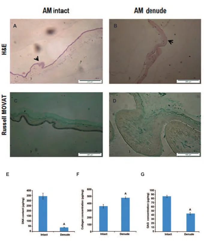
Intact and denuded HAMs were stained using H&E and dyes to determine whether the treatment successfully eliminated cellular components. For routine histology, all samples were embedded
us-ing parafin wax and sectioned and 5 sections at 6 μm were obtained and stained. H&E staining conirmed that the procedure was successful and
no cells were visible (Fig 1A, B). Russell MO-VAT staining demonstrated no obvious disruption to the sum of matrix histoarchitecture following treatment; the main structural component of HAM (collagen) appeared to have been preserved after decellularization (Fig 1C, D).
Quantiication of residual DNA following decel -lularization
The DNA content of HAM before treatment was
determined as (341 ± 29.60 μg/ml). After the de
-cellularization procedure, a signiicant decline to (39.38 ± 4.04 μg/ml) was observed (n=6, p<0.05,
ANOVA, Fig 1E).
Collagen and GAG analysis
Biochemical assays were undertaken to evaluate the ECM components after decellularization. The hydroxyproline content of intact AM was found to
be (361 ± 27.39 μg/mg); after treatment, a signii
-cant increase to 478 ± 14.42 μg/mg (n=5, p<0.05,
ANOVA) was observed (Fig 1F). GAGs form the major structural components of the ECM of tis-sues; their abundance in intact AM was found to be
85 ± 3.29 μg/mg. After treatment, a signiicant de
-crease to 43 ± 3.08 μg/mg (n=5, p<0.05, ANOVA)
Fig 1: Decellularization of human amniotic membrane (HAM): hematoxylin- and eosin (H&E)-stained native HAM (original magniication: ×20) Intact HAM (A), 0.03% (w/v) sodium dodecyl sulphate (SDS)-treated HAM (original magniication: ×20) (B), in each image, the arrows are indicating the apical surface of the HAM. Extracellular matrix (ECM) compositions were showed in intact AM, dendued AM and 3D AM scaffold (C, D) by using Russell-Movat staining (collagen, yellow) and (GAG, Green), Deoxyribonucleic acid (DNA) content of intact and denuded HAM was quantiied using a micro plate luorescence reader (E). Statistical differences between intact and denuded HAM groups; analysis of ECM components, including acid/ pepsin-soluble collagen, sulfated GAG (F, G). Statistical differences between collagen and GAG contents of intact HAM and 3D AM scaffold. (Data are shown as mean ± standard deviation), n=5 , A; P<0.001 and GAG; Glycosaminoglycan.
A B
C
E
D
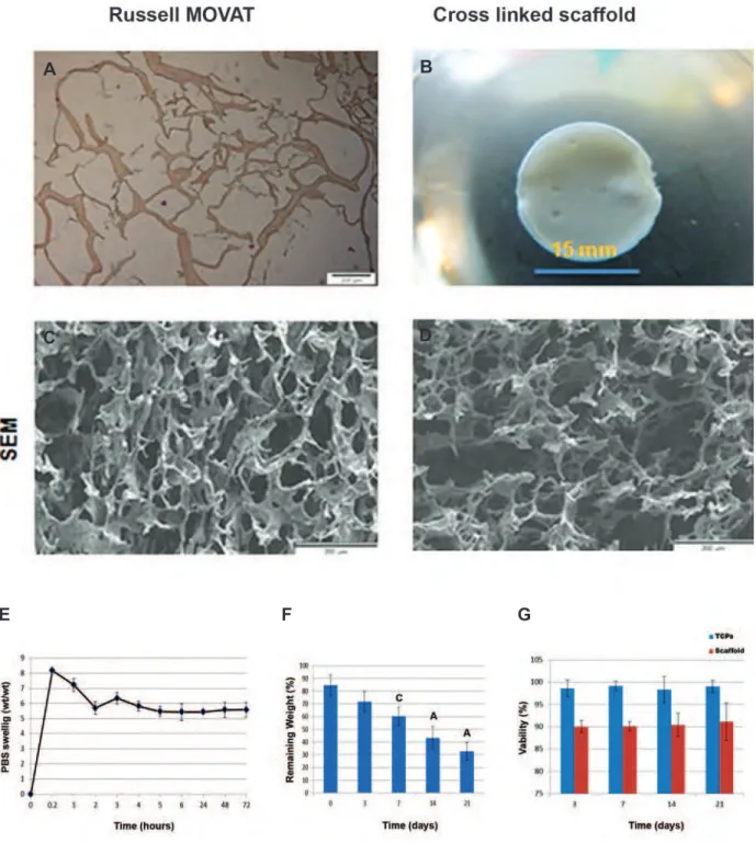
Scaffold characteristics
The main structural component of HAM (colla-gen) was showed by Russell MOVAT staining (Fig 2A). The thickness of 3D spongy scaffold in this study was about 4 mm to mimic the real thickness of human skin. The SEM observa-tion results (Fig 2B) showed the morphological characteristics of the 3D spongy AM scaffolds. The scaffold disclosed extremely interconnect-ed porous structures, and the pore wall surface appeared rough and homogeneous (Fig 2C, D). SEM images of cross-linked 3D spongy AM scaffolds indicated that it had an open porous
structure with pores ranging from 44 to 160 μm. The mean pore size was 90 μm and the average
porosity was 90%, that is suitable for cell pen-etration, nutrients and gas change.
Cross-linking degree
Cross-linking of biological tissue materials using water-soluble carbodiimide has received much attention in the field of biomaterials sci-ence (24). Therefore, the 3D spongy AM scaf-folds were cross-linked with EDC/NHS accord-ing to the general reaction mechanism. The results of the TNBS test showed that the cross-linking efficiency of AM derived ECM scaf-folds was about (65% ± 10.53).
PBS solution adsorption
We applied the swelling ratio test to assess wa-ter absorption capability and showed (Fig 2E) that without NHS/ EDC cross-linking, scaffolds
dissolved in water within 2 minutes and couldnʼt
maintain solid constructions. Our ECM compo-nents of 3D spongy AM scaffold cross-linked with NHS/ EDC presented a swelling ratio of approxi-mately 5 fold compared with dry weight scaffold. The results showed highly increased swelling
ra-tios at 5 minutes. Signiicant differences in swell -ing ratios were not observed at other selected time intervals (Fig 2E).
In vitro collagenase degradation
The biological degradation of the 3D AM sponge-like scaffold was characterized by measuring the decrease in weight. The rates were tested by in vitro enzyme assays using
col-lagenase I. Figure 2F shows that 100 μg/ml of
collagenase I solution decomposed the scaffold gradually over three weeks. The scaffold was 29.344 ± 4.87% of the original weight after 21 days of treatment. In vitro enzyme
biodegrada-tions were evaluated to show the time depend-ences of this scaffold.
Proliferation of cells directly in contact with scaf-folds
The extract cytotoxicity assay distinguished the effect of soluble components of 3D spongy AM scaffold on the viability of primary hu-man fetal dermal fibroblasts cells. Incubation of primary human fetal dermal fibroblasts with soluble extracts from intact AM, 3D spongy AM scaffold and tissue culture plate (TCP) dis-played different levels of cell viability accord-ing to MTS assay. Extracts prepared from the 3D spongy AM scaffold, showed no significant difference in the viability of the fetal fibroblasts cells compared to the TCP group (cells-only negative control) and 3D spongy AM scaffold after 14 and 21 days (n=6, p>0.05, ANOVA). The extracts from the 3D spongy AM scaffold did not display significant adverse effects on the viability of the fetal fibroblasts cells (Fig 2G).
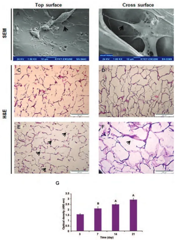
Cell morphology
The cell morphology of ibroblasts was studied
on the scaffolds after 7 days of culturing. SEM
images indicated ibroblast cells formed normal
spindle-shaped cells on all scaffolds (Fig 3A, B). As shown H&E images of scaffold without cell
(Fig 3C, D) and ibroblast cells were able to pen -etrate, attach and grow into the 3D structures of 3D spongy AM scaffold (Fig 3E, F) because of the presence of large pores.
Cell metabolic activities in scaffolds
Fig 2: 3D AM scaffold using Russell- Movat staining (collagen, yellow) and (GAG, Green) (A). Cross linked ECM derived AM scaffold produced by freeze dryer (B). SEM image of the surface (C). The cross section of the porous (D). PBS swelling ratio of ECM derived human AM scaffolds at different times (E). In vitro collagenase biodegradation; time course of weight remaining of ECM derived HAM scaffold, cross-linked with ratio (1:4) of NHS/EDC, after incubation in PBS containing 100 µg collagenase I, at 37˚C (F). Comparison results of effect of extract cytotoxicity of TCPs and scaffold groups on viability fetal fibroblast cells by MTS assay extract showed, (p>0.05) (G). (Data are shown as mean ± standard deviation). ECM; Extracellular matrix, AM; Amniotic membrane, GAG; Glycosaminoglycan, SEM; Scanning electronic microscopy, EDC; 1-ethyl-3-(3-dimethyl aminopropyl) carbodiimide hydrochloride, NHS; N-hydroxysuccin-imide, PBS; Phosphate-buffered saline, TCP; Tissue culture plates, n=5, A; P<0.001 and C; P<0.05.
A B
C
E
D
Fig 3: SEM images of fetal ibroblast cells attached (arrows are indicating ibroblast cells) to ECM derived HAM scaffolds, after 7 days at surface (A) and internal surfaces of 3D spongy scaffold (B) obtained by cross sectioning. H&E images before and after seeding cells, The light microscopy images of H&E images showed the external surface of scaffold without cell (C) and attachment of human fetal ibroblast cells at external surfaces of scaffold, the arrows are indicating attachment of fetal ibroblast cells, the cells are dark grey and the AM scaffolds are light red (D). H&E images show the internal surface of the scaffold without cell (E) attachment and growth of fetal ibroblast cells at internal surface of scaffold after 7 days (F). MTS results showed the metabolic activities of fetal ibroblast cells in ECM derived HAM scaffold. Statistical differences in metabolic activity at days 7, 14 and 21 with 3D HAM scaffold in days 3 (G). SEM; Scanning electronic microscopy, ECM; Extracellular matrix, HAM; Human amniotic membrane, H&E; Hematoxylin and eosin. (Data are shown as mean ± standard deviation (SD). (n=5, A; P<0.001 and B; P<0. 01).
A B
C
E
D
F
Discussion
AM is applied in surgery particularly for the re-construction of traumatic wounds and skin trans-plantation (12). HAM is an appropriate substitute for general skin for surgical use due to its availa-bility, low cost, and low risk of viral disease trans-mission and immunologic rejection. Basement membrane in human placenta-derived ECM could perform a functional component in the well re-generation of damaged basement membrane skin
tissue, adjust ibroblast and keratinocyte develop -ment and differentiation, and construct epithelial tissue (12). For a logical design of scaffolds for skin engineering, it is fundamental to study the features and effect of individual components of biomaterial. The overall aim of this study was to develop an acellular matrix scaffold suitable for tissue engineering applications in the form of a 3D scaffold and as a cell delivery system (24). The de-cellularization procedure must eliminate the main sources of immunogenic response including cel-lular components, membrane antigens, and solu-ble proteins, so blocking initiation of immune re-sponse and later latest degradation of the acellular matrix transplanted in to the patient (17). A number of methods for the removal of cells from HAM have been investigated with varying degrees of success (25, 26). In most cases, when assessing cell removal and maintenance of matrix struc-ture, the methods used failed to remove all of the cells and cellular components from the tis-sue matrix. In this experiment, the decellulariza-tion procedure of was accomplished according
to a modiied protocol that has been previously
used on HAM (17).
The AM was decellularized by EDTA, SDS in two steps without the use of nuclease (DNAse and RNAse) unlike in other studies (17), and were impressive in terms of elimination of the cellular component. During the decellularization proce-dure in this study the hypotonic buffer lyses the cells by swelling the water in the cells and SDS, which is an ionic detergent, attaches to cell mem-branes and causes the destruction of the lipid bi-layer. EDTA and the pH of the buffers blocked the activation of proteases during cell lysis (17). Re-sults of the procedure to eliminate cells from HAM showed the loss of cells but retention of DNA in the matrix.
Results of the hydroxyproline assays (Fig 1F)
indicated that the decellularization process did not lead to loss of collagen, elastin, or GAG content
of the tissue. There was a statistically signiicant
increase in all the structural components; this in-crease was probably as a result of extraction (by dry weight) of other soluble constituents (soluble proteins, lipids, nucleic acids).
Assessment of the hydroxyproline content using a collagen kit (Fig 1F) and Russel MOVAT stain-ing, (Fig 1A, B), (Fig 2A) showed that the decellu-larization method did not lead to a decrease of the collagen contentin the AM. Collagen is an impor-tant component for cell proliferations and tissue body formation. It provides some of the mechani-cal properties such as adhesive and tensile strength.
There was a statistically signiicant increase in this
structural component of ECM compared to intact AM; the main reason for this increase maybe an elicitation of other soluble protein and lipids con-stituents.
Cultivation of cells in 2D monolayer cannot provide an adequate in vivo micro-environment
for proliferation (26, 27). To fabricate an appro-priate 3D scaffold in skin tissue engineering,
vari-ous deinitive factors to consider include pore size
range, mechanical strength, biodegradability. AM dissolves because of endogenous enzymatic deg-radation of AM matrix during 1 week (28). For better use of AM in tissue engineering, it should be reinforced against enzymatic degradation.
Col-lagen ibers constitute the main structure of AM
which can easily undergo cross-linking, by bridges are made between the collagen chains (29, 30). Re-cently, EDC/NHS one of the cross-linker agents, has been utilized to improve mechanical proper-ties in collagen (10), collagen-chitosan (11), and collagen-phosphorylcholine to obtain suitable tis-sue engineered corneal substitutes. NHS/EDC are presumed to be water-soluble and non-toxic cross-linking agents because they can be made from urea
derivatives (15). Cross-linking has been conirmed
in-creased the water containing capacity. Because of the high water absorption feature, the sponge-like matrices were optimal for cells to culture in (27).
The degradation data presented gradual weight loss of the scaffold at selected time intervals (Fig 2F). Our scaffold was composed by NHS/EDC, was degraded by collagenase I and after it had de-composed; the scaffold lost its structural properties.
When constructing the skin graft, the estab-lishment of the dermis over the model was ap-parently accelerated by the application of skin cells to the graft (28). Fibroblast cells perform active roles in a diversity of biological proce-dures such as the production of collagen, GAG
and ECM proteins. In particular, ibroblast cells
produce intra/extracellular cytoskeleton tension forces which allow for interaction with the ECM
(29). SEM observations showed the fetal ibro -blast cells seeded in the scaffold that they
prolif-erated normally, conirming the beneit of these
materials to cell growth (Fig 3A, B).
The interconnected pores within the scaffold provided the space status for interactions of bio-logical cytokines and growth factors released from
keratinocyte and ibroblast cells (30, 31).The re
-sulting data from seeding fetal ibroblast cells on the scaffold was signiicant proliferation on the
day 21compared to 3 day, which displayed that not only the cell proliferation was promoted, but the individual collagen constructing abilities were also enhanced (Fig 3G). As our scaffold has dem-onstrated the ability to increase collagen secretion, it is potentially a good biomaterial for wound heal-ing in skin tissue engineerheal-ing. Our 3D spongy AM scaffold hasexcellent potential because of its suita-ble pore size, the great swelling ratio and good cy-tocompatibility. The skin medicine and therapeutic
wound dressing market is signiicant. Bio-func -tions of traditional dressings in the past are only for keeping the wound dry and preventing infec-tion. In clinical applications, we know that moist and warm surroundings aid repair of wounds to the skin. Effective scaffolds must investigate several main factors including skin tissue evaluation s,
tis-sue deiciency managements, humidity containing equilibrium, infection preventions, inlammation
controls and dermatological wound edge progres-sion enhancing in animal model. Other issues that need to be considered are; the patient healthy con-ditions (e.g. diabetes, burns), the injury type being
created by physical or chemical damage, and the environmental properties. We will continue focus-ing on these important options about skin tissue en-gineering skin wound dressings in future studies.
Conclusion
A diversity of biological scaffolds has been made with distinctive biochemical, biomechanical, and morphological properties. Different procedures
may be used to fabricate organ-speciic scaffolds
for tissue engineering. In this study, HAM-derived ECM scaffolds composed of various ECM compo-nents were created as a biological scaffold for skin tissue engineering. Human ECM scaffolds were constructed from HAM via pulverization, decel-lularization, and lyophilization. We found that the sponge-like AM-derived ECM scaffold provided an optimal pore size and water absorption for hu-man skin cell growth. This scaffold could be de-graded by collagenase I, which demonstrates its biodegradability. Our results show that HAM-de-rived ECM scaffold could be useful in skin tissue engineering due to its physico-mechanical prop-erties, which may improve the quality of wound healing.
Acknowledgments
The authors would like to thank the Royan
Insti-tute for inancially supporting this project. This re -search was the thesis of master student from Basic Science Faculty of Payame nor University, Tehran.
There is no conlict of interest in this article.
References
1. Shokrgozar MA, Fattahi M, Bonakdar S, Ragerdi Kashani I, Majidi M, Haghighipour N, et al. Healing potential of mesenchymal stem cells cultured on a collagen-based scaffold for skin regeneration. Iran Biomed J. 2012; 16(2): 68-74.
2. van der Veen VC, van der Wal MB, van Leeuwen MC, Ul
-rich MM, Middelkoop E. Biological background of dermal substitutes. Burns. 2010; 36 (3): 305-321.
3. Uijtdewilligen PJ, Versteeg EM, Gilissen C, van Reijmers
-dal SV, Schoppmeyer R, Wismans RG, et al. Towards em
-bryonic-like scaffolds for skin tissue engineering: identii
-cation of effector molecules and construction of scaffolds.
J Tissue Eng Regen Med. 2013. (In Press).
4. Duan X, Sheardown H. Crosslinking of collagen with den-drimers. J Biomed Mater Res A. 2005; 75(3): 510-518.
5. Brohem CA, Cardeal LB, Tiago M, Soengas MS, Barros
SB, Maria-Engler SS. Artiicial skin in perspective: con
-cepts and applications. Pigment Cell Melanoma Res.
2011; 24(1): 35-50.
6. Stoker AW, Streuli CH, Martins-Green M, Bissell MJ. De
tis-sue function. Curr Opin Cell Biol. 1990; 2(5): 864-874.
7. Nam K, Kimura T, Funamoto S, Kishida A. Preparation of
a collagen/polymer hybrid gel designed for tissue
mem-branes. Part I: controlling the polymer-collagen
cross-linking process using an ethanol/water co-solvent. Acta Biomater. 2010; 6(2): 403-408.
8. Ruszczak Z. Effect of collagen matrices on dermal wound healing. Adv Drug Deliv Rev. 2003; 55(12): 1595-1611.
9. Liu Y, Grifith M, Watsky MA, Forrester JV, Kuffova L, Grant
D, et al. Properties of porcine and recombinant human
collagen matrices for optically clear tissue engineering applications. Biomacromolecules. 2006; 7(6): 1819-1828.
10. Rafat M, Li F, Fagerholm P, Lagali NS, Watsky MA,
Munger R, et al. PEG-stabilized carbodiimidecrosslinked
collagen-chitosan hydrogels for corneal tissue engineer-ing. Biomaterials. 2008; 29(29): 3960-3972.
11. Liu W, Deng C, McLaughlin CR, Fagerholm P, Lagali NS,
Heyne B, et al. Collagen–phosphorylcholine interpenetrat-ing network hydrogels as corneal substitutes. Biomateri-als. 2009; 30(8): 1551-1559.
12. Niknejad H, Peirovi H, Jorjani M, Ahmadiani A, Ghanavi
J, Seifalian AM. Properties of the amniotic membrane for
potential use in tissue engineering. Eur Cell Mater. 2008; 15: 88-99.
13. Groeber F, Holeiter M, Hampel M, Hinderer S,
Schenke-Layland K. Skin tissue engineering--in vivo and in vitro ap
-plications. Clin in Plast Surg. 2012; 39(1): 33-58.
14. Ma DH, Lai JY, Cheng HY, Tsai CC, Yeh LK. Carbodiimide
cross-linked amniotic membranes for cultivation of limbal epithelial cells. Biomaterials. 2010; 31(25): 6647-6658.
15. Lai JY, Li YT. Functional assessment of cross-linked po
-rous gelatin hydrogels for bioengineered cell sheet carri-ers. Biomacromolecules. 2010; 11(5): 1387-1397.
16. Fujisato T, Tomihata K, Tabata Y, Iwamoto Y, Burczak K,
Ikada Y. Cross-linking of amniotic membranes. J Biomater Sci Polym Ed. 1999; 10(11): 1171-1181.
17. Wilshaw SP, Kearney JN, Fisher J, Ingham E. Production
of an acellular amniotic membrane matrix for use in tissue engineering. Tissue Eng. 2006; 12(8): 2117-2129.
18. Wang HM, Chou YT, Wen ZH, Wang CZ, Chen CH, Ho
ML. Novel biodegradable porous scaffold applied to skin regeneration. PloS One. 2013; 8(6): e56330.
19. Snyder SL, Sobocinski PZ. An improved 2, 4, 6-trini
-trobenzenesulfonic acid method for the determination of amines. Anal Biochem. 1975; 64(1): 284-288.
20. Park SN, Park JC, Kim HO, Song MJ, Suh H. Characteri
-zation of porous collagen/hyaluronic acid scaffold
modi-ied by 1-ethyl-3-(3-dimethylaminopropyl) carbodiimide
cross-linking. Biomaterials. 2002; 23(4): 1205-1212.
21. Ma DH, Lai JY, Cheng HY, Tsai CC, Yeh LK. Carbodiimide
cross-linked amniotic membranes for cultivation of limbal epithelial cells. Biomaterials. 2010; 31(25): 6647-6658. 22. Han J, Ma I, Hendzel MJ, Allalunis-Turner J. The
cyto-toxicity of gamma-secretase inhibitor I to breast cancer cells is mediated by proteasome inhibition, not by gamma-secretase inhibition. Breast Cancer Res. 2009; 11(4): R57. 23. Kuijpers AJ, Engbers GH, Krijgsveld J, Zaat SA, Dankert J, Feijen J. Cross-linking and characterisation of gelatin
matrices for biomedical applications. J Biomater Sci Pol
-ym Ed. 2000; 11(3): 225-233.
24. VonVersen-Hoynck F, Syring C, Bachmann S, Moller DE.
The inluence of different preservation and sterilisation steps
on the histological properties of amnion allografts--light and scanning electron microscopic studies. Cell Tissue Bank. 2004; 5(1): 45-56.
25. Luo JC, Li XQ, Yang ZM. Preparation of human acellu
-lar amniotic membrane and its cytocompatibility and
bio-compatibility. ZhongguoXiu Fu Chong Jian Wai Ke Za Zhi.
2004; 18(2): 108-111.
26. Eves PC, Beck AJ, Shard AG, Mac Neil S. A chemically
deined surface for the co-culture of melanocytes and ke
-ratinocytes. Biomaterials. 2005; 26(34): 7068-7081.
27. Boyd M, Flasza M, Johnson PA, Roberts JS, Kemp P. In
-tegration and persistence of an investigational human liv-ing skin equivalent (ICX-SKN) in human surgical wounds. Regen Med. 2007; 2(4): 363-370.
28. Fioretti F, Lebreton-DeCoster C, Gueniche F, Yousi M,
Humbert P, Godeau G, et al. Human bone marrow-derived
cells: an attractive source to populate dermal substitutes.
Wound Repair Regen. 2008; 16(1): 87-94.
29. Hunt NC, Shelton RM, Grover L. An alginate hydrogel ma
-trix for the localised delivery of a ibroblast/keratinocyte
co-culture. Biotechnol J. 2009; 4(5): 730-737.
30. Joshi PG, Nair N, Begum G, Joshi NB, Sinkar VP, Vora
S. Melanocyte-keratinocyte interaction induces calcium
signalling and melanin transfer to keratinocytes. Pigment
Cell Res. 2007; 20(5): 380-384.
31. SedaTigli R, Ghosh S, Laha MM, Shevde NK, Daheron L,