to Study Ischemia
Ex Vivo
Syamantak Majumder, M. Ilayaraja, Himabindu Reddy Seerapu, Swaraj Sinha, Jamila H. Siamwala, Suvro Chatterjee*
Vascular Biology Lab, AU-KBC Research Centre, Anna University, Chennai, India
Abstract
Background:Ischemia is a pathophysiological condition due to blockade in blood supply to a specific tissue thus damaging the physiological activity of the tissue. Differentin vivomodels are presently available to study ischemia in heart and other tissues. However, no ex vivo ischemia model has been available to date for routine ischemia research and for faster screening of anti-ischemia drugs. In the present study, we took the opportunity to develop anex vivo model of partial ischemia using the vascular bed of 4thday incubated chick embryo.
Methodology/Principal Findings:Ischemia was created in chick embryo by ligating the right vitelline artery using sterile surgical suture. Hypoxia inducible factor- 1 alpha (HIF-1a), creatine phospho kinase-MB and reactive oxygen species in animal tissues and cells were measured to confirm ischemia in chick embryo. Additionally, ranolazine, N-acetyl cysteine and trimetazidine were administered as an anti-ischemic drug to validate the present model. Results from the present study depicted that blocking blood flow elevates HIF-1a, lipid peroxidation, peroxynitrite level in ischemic vessels while ranolazine administration partially attenuates ischemia driven HIF-1aexpression. Endothelial cell incubated on ischemic blood vessels elucidated a higher level of HIF-1a expression with time while ranolazine treatment reduced HIF-1a in ischemic cells. Incubation of caprine heart strip on chick embryo ischemia model depicted an elevated creatine phospho kinase-MB activity under ischemic condition while histology of the treated heart sections evoked edema and disruption of myofibril structures.
Conclusions/Significance:The present study concluded that chick embryo partial ischemia model can be used as a novelex vivo model of ischemia. Therefore, the present model can be used parallel with the knownin vivo ischemia models in understanding the mechanistic insight of ischemia development and in evaluating the activity of anti-ischemic drug.
Citation:Majumder S, Ilayaraja M, Seerapu HR, Sinha S, Siamwala JH, et al. (2010) Chick Embryo Partial Ischemia Model: A New Approach to Study IschemiaEx Vivo. PLoS ONE 5(5): e10524. doi:10.1371/journal.pone.0010524
Editor:David S. Milstone, Brigham and Women’s Hospital, United States of America
ReceivedNovember 10, 2009;AcceptedApril 16, 2010;PublishedMay 7, 2010
Copyright:ß2010 Majumder et al. This is an open-access article distributed under the terms of the Creative Commons Attribution License, which permits unrestricted use, distribution, and reproduction in any medium, provided the original author and source are credited.
Funding:This work was supported by a grant from KB Chandrasekhar- Research Foundation. The funders had no role in study design, data collection and analysis, decision to publish, or preparation of the manuscript.
Competing Interests:The authors have declared that no competing interests exist.
* E-mail: [email protected]
Introduction
Ischemia is a condition in which blockade in blood flow leads to restricted oxygen and nutrient supply to a part of the body. Cardiac ischemia is the compromised blood flow and oxygen supply to heart muscle. The aim of developing ischemic animal model is to study the basic processes or potential therapeutic interventions in ischemia associated diseases, and the extension of patho-physiological knowledge, which will lead to improve medical treatment of human ischemic stroke. Ischemic stroke has a complex pathophysiology involving the interplay of many different cells and tissues such as neurons, glia, endothelium, cardiomyocytes and the immune system. These events cannot be mimicked satisfactorily inin vitro. Thus a large portion of stroke research is conducted on animals. Presently two ischemic animal models are well accepted by the scientific community. Artery ligation model is created in animal heart by ligating the left coronary artery [1]. Occlusion model is another well accepted model of ischemia, which is created by permanently occluding the middle cerebral artery [2]. However, these different animal models
are less feasible for routine laboratory testing of ischemia effects. Additionally, preliminary screening of any anti-ischemic drug needs to be carried out by usingin vitromodels or by using other models similar toin vivo conditions. In the present scenario, only fewin vitroischemia models such as cerebral ischemia model based on organotypic hippocampal slice cultures and tissue culture ischemia model of oxygen/glucose deprivation are documented [3,4]. However, the models are not well conceived and mostly control the oxygen level to create hypoxia in the tissue but not specifically ischemia.
recovery of ischemia by anti-ischemic drugs validated our novel approach of developing anex vivoischemia model.
Results
Partial ischemia elevated the reactive oxygen species generation in tissues
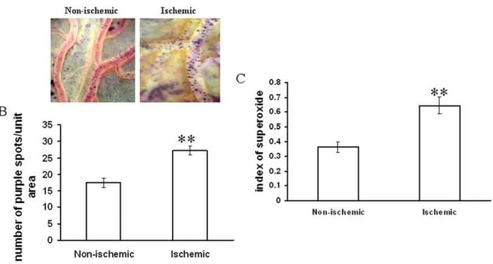
Different ROS parameters like superoxide, hydrogen peroxide, peroxynitrite and lipid peroxidation. were evaluated in the ischemic vessels of chick embryo (Fig. 1) using different redox probes. Mapping of lipid peroxidation was carried out using the thiobarbituric acid (TBA) protocol. A higher level of lipid peroxidation was observed in the proximal areas in relation to the ischemic zone while lipid peroxidation level reduced in distal areas (Fig. 2A,2B). Image analysis and colorimetric measurement of superoxide in ischemic tissues demonstrated 40% and 42% increase in superoxide level respectively (Fig. 3). Measurement of hydrogen peroxide using amplex red depicted that ischemia attenuated hydrogen peroxide production by 25% (image analysis) and 27% (fluorimetric measurement) respectively (Fig. 4). Nitric oxide (NO) level in the ischemic vessels was measured using diamino fluorescein diacetate (DAF-2DA) fluorescence probe. There was no significant difference in NO production between ischemic and non-ischemic vessels was observed (Fig. 5A,5B). Peroxynitrite generation in the ischemic vessels was measured using dihydrorhodamine (DHR123) fluorescence probe. Partial ischemia elevated the peroxynitrite level in tissue by 33% compared to non-ischemic controls (Fig. 5C). Image analysis and
fluorimetric measurement of free thiol in ischemic tissues using 5-chloromethylfluorescein diacetate (CMFDA) elaborated 48% and 42% reductions respectively in free thiol level under partial ischemia treatments (Fig. 6).
Partial ischemia dependent higher HIF-1a release induced the neo-vascularization in the adjacent vessels
HIF-1a, a hypoxia driven factor, is a well known pro-angiogenic factor as reported earlier [5]. We evaluated the concept and studied the angiogenesis pattern of the vascular bed adjacent to the ischemic area. Analysis of the vascular bed images using Angioquant software revealed that ischemia elevated the neo-vascularization in chick embryo vascular bed in a time dependent manner. Analysis of number of vessel complexes, vessel length, vessel size and junctions revealed 4, 5.5, 4.5 and 11 fold increase respectively after the ischemia treatment (Fig. 7). Increase in vascularization can also be due to backfilling of initially occluded vessels. However, critical analysis of the images from ischemic vessels showed that the vessels were formed newly from the existing vessels and thus can be concluded that increase in the vessel density is due to the neo-angiogenesis in that area.
Partial ischemia in chick embryo vessels elevated the expression level of HIF-1ain a time-dependent manner
HIF-1a expression was used as a biochemical marker to quantify the level of ischemic stress in tissues. Relative expression of HIF-1a in ischemic vessels was measured using quantitative
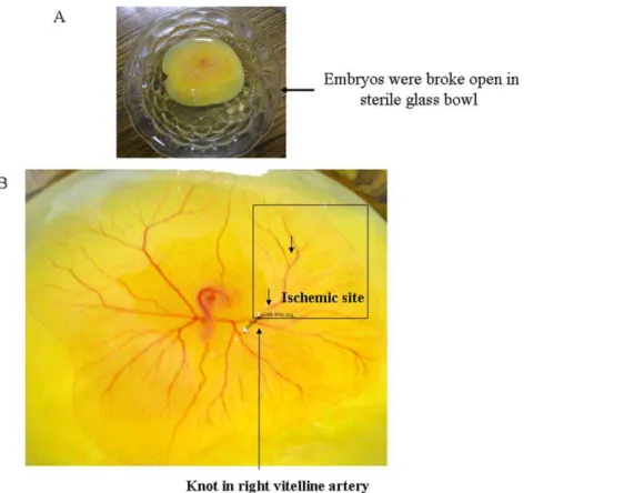
Figure 1. Creating partial ischemia in chick embryo vascular bed using ligation model.(A) Image showed the embryo with the whole yolk material has been placed in the sterile glass bowls. The bowls were covered with sterile glass leads and placed in a chamber maintaining 37uC and relative humidity. (B) Image represents the exact approach taken to block the blood flow to the specific area of the chick embryo vascular bed. Black arrow showed the suture that has ligated the right vitelline artery thus blocking the blood flow in the surrounding vascular bed. The box showed the ischemic area of the vascular bed.
doi:10.1371/journal.pone.0010524.g001
RT-PCR technique. The vessels were ligated and incubated for different durations ranging from 0 to 120 min. Quantitative analysis of the band intensities revealed a dose dependent increase in HIF-1aexpression in the ischemic vessels. HIF-1aexpression level was elevated by 1.5, 2.6 and 3.3 fold after 30, 120 and 240 min of ischemia treatments respectively. However, no significant increase in HIF-1a expression was observed in the ischemia-treated tissues ligated for 10 min (Fig. 8A). This data suggested that partial ischemia was successfully improvised in the chick embryo vascular bed using the strategy of right vitelline artery ligation model.
Ranolazine (RL) treatment attenuated HIF-1a expression and protected the free glutathione (GSH) levels in ischemic vessels
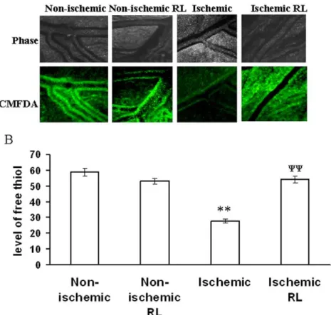
RL, a well known anti-ischemic drug was used in the present study to recover ischemia-dependent tissue injury. Free glutathi-one (GSH) being the quencher of cellular ROS protects the cells and tissue from ROS mediated damage. In the present study, we measured the level of GSH using CMFDA fluorescence probe. The result depicted that ischemia attenuated the cellular GSH level by 53% while RL administration recovered the GSH level by 52% in the ischemic vessels of chick embryo (Fig. 9A,9B). The expression level of HIF-1a was measured in the ischemic vessels under RL treatment using quantitative RT-PCR technique. Ischemia elevated the level of HIF-1a expression by 38% while
RL administration attenuated HIF-1a expression in ischemic vessels by 48% (Fig. 8B). However, RL did not affect the expression level of HIF-1aand level of GSH under non-ischemic condition (Fig. 9A,9B). This data elaborated that RL administra-tion protected chick embryo vessels from ischemia induced damage.
Partial ischemia interferes with cellular morphology of EC grown on the ischemic bed
Morphological analysis of EC grown on the ischemic bed was carried out to evaluate the effect of ischemia on cellular health. EC grown on collagen-coated cover slips were treated under partial ischemia by incubating it on the top of ischemic vascular bed. Images showed a higher number of round cells under ischemic condition. This data demonstrated that ischemic vascular bed driven secondary ischemia in EC interfered with cellular morphology (Fig. 10A).
To verify the effect of withdrawing ischemia on cellular morphology, we studied the morphology of the cells by placing the ischemia treated cells on non-ischemic vascular bed. Ischemia reduced the number of normal cells by 32% while placing back the EC on a non-ischemic bed reversed back the number of round cells by 19% (Fig. 10B). This data suggested that ischemia dependent cellular damage can be partially reversed by with-drawing ischemic stress.
Ischemic vascular bed driven secondary ischemia compromised cellular viability and induced cellular apoptosis
Analysis of apoptosis in EAhy926 cells under partial ischemia treatment was carried out using annexin V-FITC apoptosis detection kit. Result depicted that partial ischemia treatment is apoptotic to cells thus facilitating the binding of annexin V-FITC to the outer part of the plasma membrane (Fig. 10C). Partial ischemia induced endothelial apoptosis by 8 fold compared to non-ischemic control (Fig. 10C). Cellular viability of EC under partial ischemia treatment was assayed using trypan blue dye. Images of the cells revealed that partial ischemia increased the number of trypan blue stained cells by 8 fold (Fig. 10D).
Ischemic vascular bed elevated ROS generation in EC
Two different ROS parameters were studied in EC under partial ischemia treatment. Superoxide and lipid peroxidation level in cells were measured after incubating EC on ischemic vascular bed using nitro blue tetrazolium (NBT) and TBA protocols respectively. Partial ischemia promoted superoxide generation in EC by 27% while the level of lipid peroxidation under partial ischemia treatment was elevated by 40% compared to non-ischemic control (Fig. 11A,11B). This data suggested that ischemic vascular bed improvised secondary ischemia in EC that in turn elevated ROS level in EC.
Partial ischemia reduced the GSH concentration in EC
The level of free thiol in EC was measured using CMFDA fluorescent probe. Partial ischemia reduced the level of GSH in EC by 32% (Fig. 11C, 11D).
Anti-ischemic drug treatments improved the cellular health condition and preserved the GSH level under partial ischemia
To evaluate the protecting role of antioxidant and anti-ischemic drug, we performed morphology counting and GSH measurement
Figure 2. Mapping lipid peroxidation in ischemic vascular bed.
(A) Representative image showed the way section has been incised out from the vascular bed of chick embryo to map the lipid peroxidation status in the ischemic vascular bed. (B) Lipid peroxidation was measured by following the TBA protocol. After ligation of right vitelline artery, embryos were incubated for 2 h. An equal six sections of the chick embryo vascular bed was taken and the lipid peroxidation was measured. Data were normalized by taking the weight of the tissues.*P,0.05 and **P,0.001 versus C,D,E and F. (n = 5).
Figure 3. Effect of ischemia on superoxide generation in vitelline vascular tissue.(A) Vessels were probed with NBT for 1 h. Next, the images of the vessels having the purple formazen crystal were acquired with Nikon Cool Pix camera adapted to a stereo microscope. Images are the representative of 5 individual experiments. (B) The number of purple formazen crystals in the vessels was counted manually. Data were normalized by taking the area of the vessels and plotting as number of purple spots/unit area. **P,0.001 versus non-ischemic. (n = 5) (C) Ischemic vessels were incubated with NBT for 1 h followed by homogenization of the tissue and centrifugation of the suspension at 6500 g for 10 min. The supernatant was then read at 560 nm. Data were normalized with the weight of the tissue. **P,0.001 versus non-ischemic. (n = 3).
doi:10.1371/journal.pone.0010524.g003
Figure 4. Effect of ischemia on hydrogen peroxide generation in vitelline vascular tissue.(A) Ischemia treated vessels were probed with amplex red for 30 min. Fluorescent and bright filed images were acquired with DP71 CCD camera adapted to Olympus IX71 microscope. Images are the representative of 5 individual experiments. (B) The fluorescent intensities of the images were calculated using Adobe Photoshop 7.0 and plotted. Fluorescent intensities were considered as directly proportional to the level of hydrogen peroxide produced. **P,0.001 versus non-ischemic. (n = 5) (C) Ischemic tissues were probed with amplex red for 30 min followed by homogenization and centrifugation at 6500 g for 10 min. The supernatant was then read at excitation/emission of 570/585 nm. Values obtained were normalized with the weight of the tissues. *P = 0.023 versus non-ischemic. (n = 3). doi:10.1371/journal.pone.0010524.g004
Figure 5. Effect of ischemia on NO generation and peroxynitrite level in vitelline vascular tissue.(A) NO production by the ischemic vessels was measured using DAF-2DA fluorescent probe. Ischemic vessels were incubated for 10 min with 1mM DAF-2DA dissolved in HBS containing 1 mM
L-arginine. Images were taken under 4X objective of the microscope. Images are the representative of 3 individual experiments. (B) The green luminosity of the fluorescent images was analyzed using Adobe Photoshop 7.0. Luminosity of the images was directly proportional to the level of NO produced thus plotted as index of NO production. **P,0.001 versus non-ischemic. (n = 3) (C) Peroxy nitrite generation was measured using DHR123 fluorescent probe. Tissues were incubated with DHR123 for 2 h followed by homogenization and centrifugation for 5 min at 6500 g. Supernatant was read at excitation/ emission of 500/536 nm and the values were normalized with the weight of the tissues and plotted. *P = 0.03 versus non-ischemic. (n = 5).
doi:10.1371/journal.pone.0010524.g005
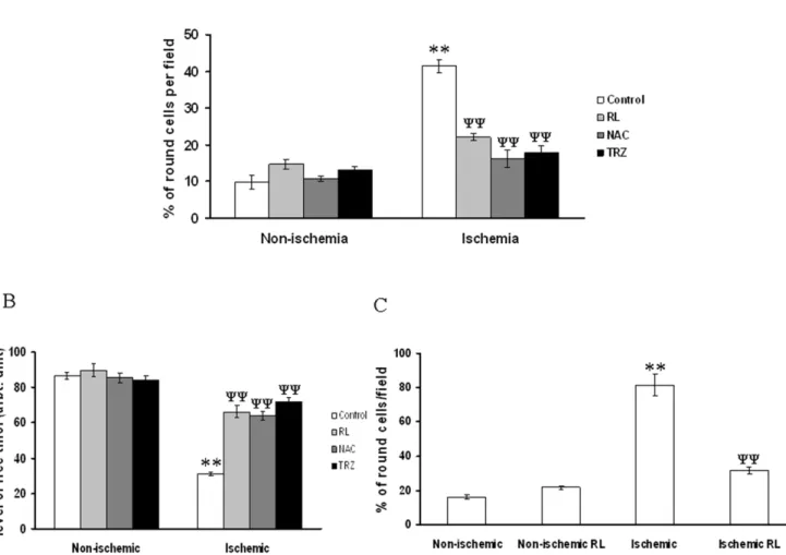
experiments in ischemia treated EC in the presence and absence of anti-ischemic drugs. Results depicted that ischemia induced rounding up of EC while RL, N-acetyl cysteine (NAC) and trimetazidine (TRZ) treatments protected cells from ischemia induced stress. However, non-ischemic cells were insensitive to RL, NAC and TRZ treatment (Fig. 12A). GSH measurement in ischemia treated EC using CMFDA elaborated that ischemia reduced the level of GSH while RL, NAC and TRZ potentiate the cells to maintain normal GSH concentration under ischemia (Fig. 12B). However, RL did not show any effect on the level of GSH in non-ischemia treated EC. Morphology of H9C2 cells under ischemia was studied with and without RL administrations. Ischemia promoted the rounding up of H9C2 cells while administration of RL protected the cells from ischemia induced rounding up (Fig. 12C). However, non-ischemic cells were insensitive to RL administration.
Partial ischemia induced HIF-1aexpression in EC while RL attenuated ischemia induced over expression of HIF-1a
To evaluate the protective effect of RL, HIF-1a mRNA expression was measured in RL treated EC under partial ischemia treatments. Quantitative RT-PCR data elaborated that ischemia induced HIF-1aexpression in EC by 6 and 11 fold in 30 min and 60 min ischemia treated cells respectively (Fig. 13A). Administra-tion of RL blocked ischemia induced HIF-1a expression by 1.8 fold (Fig. 13B).
Ischemic vascular bed mediated partial ischemia in caprine heart elevated the level of creatine phospho kinase-MB (CK-MB) activity in the heart tissues
To evaluate the efficacy of the present model on creating secondary ischemia in heart tissues, we incubated the caprine heart
Figure 7. Studying the angiogenesis pattern in vessels adjacent to ischemic area.(A) Angiogenesis pattern of the vessels adjacent to the ischemic area were followed for 3 h. The images of the vessels adjacent to the ischemic area were taken with Nikon Cool Pix camera adapted to a stereo microscope. Images were converted to gray scale and presented. Inset showed the magnified images from ischemia group which revealed formation of new vessels in the area. Yellow arrows showed the formation of new vessels in the area. Images are the representative of 5 individual experiments. (B) Analysis of the images were carried out using Angioquant software. Number of separate vessel complexes was analyzed by the software for different time point. Data presented as fold increase with time. **P,0.001 versus respective time point under non-ischemia. (C) Similarly, the software calculated the length of the vessels in different time of incubation. Data presented as fold increase with time. *P,0.05 and **P,0.001 versus respective time point under non-ischemia. (D) The size of the vessels was calculated by the Angioquant software. Data presented as fold increase with time. **P,0.001 versus respective time point under non-ischemia. (E) The number of junctions in the vessels were calculated by the software and plotted. *P,0.05 and **P,0.001 versus respective time point under non-ischemia.
doi:10.1371/journal.pone.0010524.g007
tissue on the ischemic vascular bed. To confirm ischemia in the heart tissues, we measured the activity of CK-MB, a known marker of myocardial ischemia. Biochemical analysis of the heart tissue sample depicted that ischemic vascular bed mediated ischemia in caprine heart tissues elevated the enzyme activity of CK-MB by 72% (Fig. 14A). Elevation of CK-MB activity in caprine heart elucidated that ischemic vascular bed of chick embryo successfully induced secondary ischemia in caprine heart tissues.
Partial ischemia caused edema and higher intra-muscular gap in caprine heart
Caprine heart tissues were incubated on the ischemic vascular bed and fixed with 10% formalin. Parafilm wax embedded tissue sections were stained using hematoxylin and eosin stains. Images of the sections depicted that ischemic vascular bed promoted edema and an elevated intra-mascular gaps in caprine heart tissue (Fig. 14B). This data suggested that chick embryo ischemic vascular bed induced secondary ischemia in caprine heart tissue.
Discussion
Different animal models are available for tissue specific ischemia such as embolic middle cerebral artery occlusion and endovascular
filament middle cerebral artery occlusion. Middle cerebral artery occlusion is achieved by injecting particles like blood clots or artificial spheres into the carotid artery of animals as an animal model of ischemic stroke. Thromboembolic MCAo is achieved either by injecting clots that were formed in vitro [6] or by endovascular instillation of thrombin [7]. The thromboembolic model is closest to the pathophysiology of human cardioembolic stroke. But, the fundamental problem in ischemia research is the lack of easy to develop routine research models, which should offer a regular testing platform for routine research and drug testing for ischemia. The present study focused to develop a partial ischemia model by using vascular bed of 96 h grown chick embryo. The present work aimed to develop a basic ischemia model instead tissue or organ specific one, which would cater the scientists in academic and industry to understand the fundamental aspects of ischemia implications and will also help in faster screening of anti-ischemic drugs. Although chick embryo never been used before for creating ischemia, efforts were made to develop chick embryo based flow-manipulation model for shear stress associated research [8–10]. Hogers et al., 1999 have taken two approaches to manipulate the blood flow in chick embryos [8]. First, one of the three major vitelline veins was ligated and secondly these vitelline veins were ligated permanently with a microclip until cardiac septation was completed. This venous clip model is one of the major approaches to control the blood flow in the vascular bed of chick embryo [8,9]. However, these studies are directed towards evaluating the consequences of low blood flow on embryo development and cardiovascular malformations while the present study aims to develop anex vivopartial ischemia model by using a similar approach to control the blood flow in chick embryo vascular bed. We ligated the right vitelline artery of chick embryo that blocks the blood-flow of corresponding areas of vascular bed in the chick embryo.
Work of Lee et al., 2000 and different other groups suggested that an early stage elevation of HIF-1a expression under myocardial ischemia is a protective measure by the cell to protect itself from the ischemia induced damage [11–16]. We measured the level of HIF-1aexpression in the ligated chick embryo model by considering it as one of the early markers of ischemic tissue damage, and we also found that HIF-1aexpression level elevated in affected vascular bed and also in cells cultured on the ischemic vascular bed. The work of Yamamotoet al (1983) demonstrated that lipid peroxidation plays a major role in ischemia driven cellular damages [17]. Works of different groups have also reported that a higher level of lipid peroxidation during ischemia and ischemic reperfusion causes cellular damage and cell death [17–19]. Yamamotoet al., 1983 have shown that administration of a-tocopherol, a ROS scavenger and a blocker of lipid peroxidation improves the cellular health under ischemia [17]. Similarly EC incubated on the ischemic vascular bed of chick embryo evoked a higher level of lipid peroxidation indicating that primary ischemia in the vascular bed spreads secondary ischemia in the cells and tissues grown on the affected vascular bed.
ROS generation during ischemia is a consequence of ischemia mediated tissue damage [20–22]. The burst of ROS during ischemia reperfusion is considered as one of the key factors in the development of myocardial stunning after regional ischemia [23,24]. Generation of ROS during ischemia [25–27] are considered as an important factor in the development of cardiac preconditioning [28,29]. Work of Beckeret al., 1999 concluded that significant superoxide generation occurs during ischemia before reperfusion from the ubisemiquinone site of the mitochondrial electron transport chain [25]. All together these studies signify that ischemia induced ROS generation by cellular systems plays a major role in ischemia mediated tissue
Figure 8. RL based recovery of GSH level in ischemic vitelline vascular tissue.(A) Level of free thiol in ischemic tissues treated with and with out RL were measured using CMFDA. Ischemic vessels were incubated with 100mM of RL during the period of treatment. Next, the
ischemic tissues were incubated with CMFDA for 30 min and imaged with DP71 CCD camera. Images are the representative of 5 individual experiments. (B) The fluorescent intensities of the images were calculated using Adobe Photoshop 7.0 and plotted. Fluorescent intensities were considered as directly proportional to the level of free thiol in tissues. **P,0.001 versus non-ischemic andyy
damage. Similarly, in the present study, we observed a higher level of superoxide generation in both ischemic tissues and in cells treated with the ischemic vascular bed. However, we observed that blocking of right vitelline artery suppressed hydrogen peroxide production. Hydrogen peroxide is known to confer protection to ischemic tissues. The work of Yadaet al. (2006) showed that endogenous hydrogen peroxide, in cooperation with NO, plays an important cardioprotec-tive role in coronary ischemia reperfusion injuryin vivo[30]. It has been reported that hypoxia activates catalase pool in the cells. A classic work by Oshinoet al. (1975) demonstrated that the rate of hydrogen peroxide production in the ‘‘haemoglobin-free’’, ‘‘non-circulatory’’ perfused liver of rats decreased to almost half in the livers of starved and phenobarbital-pretreated rats [31]. Enhanced superoxide generation and simultaneous fall in hydrogen peroxide in the present partial ischemia model indicates a reduced dismutation of superoxide by its classical SOD pathway, which is the prime source of hydrogen peroxide. In stead superoxide possibly interacts with NO to form peroxynitrite that is evident from the results of the present study. Work of Scadutoet al., 1988 depicted that the content of GSH in the rat declined to 40% of control values during 35 min of renal artery occlusion [32]. In the present partial ischemia model, the level of GSH, a potent quencher of ROS was suppressed. We deem that low GSH level under partial ischemia leads to higher ROS
generation that caused damage to EC and cardiomyofibroblast cells as observed in the present study. The result of present work further ascertains that deteriorating functional parameters of the EC is the combinational effect of ischemia and associated ROS generation from the ischemic vascular bed.
RL, a specific inhibitor of late sodium current reduces sodium overload and hence ameliorates disturbed ion homeostasis in cardiac cells [33–39]. In the present study we administered RL as an anti-ischemic drug and evaluated its activity on different functional and biochemical parameters of the cells and tissues under ischemia. As observed by different groups, present study also suggests that RL can protect the cells and tissues from secondary ischemia developed by incubating the cells on ischemic vascular bed of chick embryo. The results offered another line of support that the present model is capable of inducing ischemia in cells and tissues. Reports from earlier findings described the protective role of NAC after ischemia or ischemic reperfusion injury [40,41]. In addition to RL, we used these two well known anti-ischemic drugs NAC and TRZ [42,43] to elucidate the efficacy of these drugs in recovering ischemia based cellular injury in the present model. NAC and TRZ both protected EC from ischemic vascular bed driven secondary ischemia.
Figure 9. HIF-1aexpression level in ischemic vitelline vascular tissue.(A) A time dependent HIF-1aexpression in the ischemic tissues were
quantified using quantitative RT-PCR technique. After treatment mRNA was isolated from the tissues using RNA extraction kit followed by quantitative RT-PCR. Total extracted RNA was quantified by running a 2%agarose gel containing ethidium bromide. Total RNA loading for cDNA conversion was then normalized from the band luminosity of total RNA from different samples. Amplified products using specific primers for HIF-1a was then run on 2% agarose gel and visualized. The band luminosity was then quantified and plotted. Band intensities were considered as directly proportional to the HIF-1amRNA level in the tissues. Images are the representative of 3 individual experiments. *P = 0.012 and **P,0.001 versus 0 min time point. (n = 3) (B) HIF-1aexpression level was measured in ischemic vascular bed with RL treatment. RL was treated during the period of ischemia. RL attenuated ischemia induced HIF-1aexpression. Images are the representative of 3 individual experiments. **P,0.001 versus non-ischemia andyy
P,0.001 versus ischemia. (n = 3). doi:10.1371/journal.pone.0010524.g009
CK-MB activity has been shown to increase in certain patients with ischemic stroke, sub-arachnoid hemorrhage and head trauma in the absence of any clinically evident acute coronary syndrome [31,44–47]. The temporal pattern of elevation was typically gradual and sustained for several days, unlike myocardial infarction, in which CK-MB peaks and falls within the first 24 h of coronary artery occlusion [48]. Similarly, our study depicted that CK-MB activity is high when the caprine heart tissue sample was incubated on the ischemic vascular bed of chick embryo. As reported earlier by Zhaiet al., 2000, we also observed a similar histology pattern of the ischemic heart tissue sample where marked interstitial edema and evidence of intramascular gaps were observed when incubated under ischemic vascular bed driven ischemia [49]. These observations demonstrate that ligation mediated partial ischemia in chick vascular bed could create secondary ischemia in the caprine heart tissues as well.
The ligation of right vitelline artery in chick embryo causes partial ischemia in the surrounding vascular bed, which can further be used to create secondary ischemia in cells and other tissues under in vitro condition. The present model offers some unique features for routine ischemia related drug testing such as 1) less ethical issues 2) easy to develop model 3) real time observation of physiological parameters such as blood flow and angiogenesis and 4) culturing of cells and tissues of one’s choice on the ischemic bed makes the model very plastic. However, the present model
also has limitations in its present form. The limitations are 1) First of all, the model cannot be tissue or organ specific as it is possible in animal models. 2) Secondly, the model cannot produce intermittent ischemia or ischemic reperfusion injury, which is prevalent in human. 3) Unlike adult tissues, the avian embryo during early stages of development obtains oxygen through direct diffusion into the tissues (until day 5–6). In subsequent stages the oxygen diffuses into blood circulating through the extra-embryonic chorioallantoic membrane. However, due to blockade in artery, blood flow to that specific area is blocked which will partially hinder with the diffusion properties of free oxygen to the vessels. Therefore, a partial blockade in both oxygen and nutrient diffusion was obtained in the present model and thus termed as partial ischemia model.
Particularly, during dynamic exercise, normal coronary arteries dilate, whereas stenotic arteries constrict. This exercise-induced vasoconstriction has been associated with the occurrence of myocardial ischemia and has been believed to be the result of endothelial dysfunction, with a reduced release or production of NO, increased sympathetic stimulation, enhanced platelet aggregation with release of thromboxane A2 and serotonin or a passive collapse of the disease-free wall segment within the stenosis (the Bernoulli effect) or a combination of any of these [50]. It is not possible to exert exercise like effects on the present model of ischemia. However, work is in progress in our laboratory to modulate the blood flow in a
Figure 10. Effect of ischemic vascular bed driven secondary ischemia on EC.(A) The number of round and normal cells were counted from the bright field images. Data were normalized by counting the total number of cells per field and presenting it as percentage of cell. **P,0.001 versus non-ischemic. (n = 7) (B) Number of normal cells were counted from each field and plotted as percentage of normal cells. Ischemia induced reduction in normal cell number was calculated while reversibility was observed when the cells kept back on normal vessels. **P,0.001 versus preischemic andyy
P,0.001 versus postischemic. (n = 7). (C) Apoptosis was measured in ischemia treated EC using annexin-V FITC detection kit. The total number of cells and the number green positive cells per field were counted and plotted. Images are the representative of 3 individual experiments. **P,0.001 versus non-ischemic. (n = 3) (D) Cellular viability under ischemia treatment was measured using trypan blue dye. Trypan blue stained cells were taken as dead cells. The total number of cells and the number green positive cells per field were counted and plotted. Images are the representative of 7 individual experiments. **P,0.001 versus non-ischemic. (n = 7).
specified rhythm specifically in a defined vessel in the vascular bed by using macro-manipulator to achieve the intermittent and modulated ischemia along with reperfusion effects.
The results of the present work also suggested that anti-ischemic efficacy of any drug can be evaluated in the present model before studying the drug in any other animal models. Therefore, the present model can be used as an additional model along with other in vivo ischemia models to reduce animal experimentations.
Materials and Methods
Materials
Dulbecco’s modified Eagle’s medium (DMEM) was purchased from PAN-Biotech GmbH, Am Gewerbepark, Aidenbach. Fetal bovine serum was from Invitrogen Life technologies. Diamino fluorescein diacetate, 5-chloromethylfluorescein diacetate, amplex red, dihydrorhodamine 123 (DHR) was purchased from Moleculer Probe, Oregon, USA. RNA isolation spin prep kit was from Medox Inc., India. cDNA Mulv reverse transcriptase and taq polymerase was purchased from New-England Biolabs, MA and Finnzymes, Espoo, Finland respectively. RL [N-(2,6-dimethylphe-nyl)-2-[4-[2-hydroxy-3-(2-methoxyphenoxy) propyl]piperazin-1-yl]acetamide] and TRZ was purchased from Sigma Chemicals,
St. Louis, MO, USA. Nitro blue tetrazolium and NAC was purchased from Hi-Media, Bangalore, India. All other chemicals were of reagent grade and were obtained commercially.
Cell culture
Immortalized endothelial hybrid cell lines, EAhy926 were gifted from Dr C.J.S. Edgell. H9C2 myofibroblast cells were purchased from NCCS, Pune, India. Both the cells were maintained and cultured in DMEM supplemented with 10% FBS (v/v) and 1% penicillin (w/v) and streptomycin (w/v).
Animals
Fertilized white Leghorn chick (Gallus domesticus L.) eggs were obtained from Poultry Research Station, Nandanam, Chennai, TN, India and incubated at 37–38uC, with the blunt end up and at a relative humidity of 70–80%. Caprine heart samples were collected from the government authorized slaughter houses.
Creating partial ischemia on chick embryo vascular bed using artery ligation model
In the early stages of chick embryo development, two distinct circulatory systems are established in an embryonic system, one for the embryo itself maintaining the blood circulation inside the
Figure 11. Level of different reactive oxygen species and GSH in ischemic EC.(A) Level of lipid peroxidation in ischemic vascular bed treated EC was quantified using TBA protocol. EC were treated under ischemia for 2 h followed by processing as mentioned in the materials and methodssection. Data were normalized by counting the number of cells using hemocytometer. Elevated lipid peroxidation was noted under ischemia treatment. *P = 0.012 versus non-ischemic. (n = 5) (B) Superoxide production by ischemia treated EC was measured using NBT. Cells were treated under ischemia for 2 h followed by incubation with NBT for another 2 h. The formed formazen dye was extracted from the cells as mentioned in methodology. Data were normalized by counting the number of cells using hemocytometer. **P,0.001 versus non-ischemic. (n = 5) (C) Ischemia treated EC were probed with CMFDA to measure the level of GSH in the cells. Cells were treated with ischemic vascular bed driven secondary ischemia for 2 h followed by probing with CMFDA for 30 min. Images were acquired by DP71 camera adapted to an Olympus IX71 microscope. Images are the representative of 5 individual experiments. (D) The green luminosity from the CMFDA probed fluorescent images were measured using Adobe Photoshop version 7.0. Green luminosity of 40 different cells from 5 different sets of experiment were measured and plotted. Ischemia reduced the level of free thiol in EC. **P,0.001 versus non-ischemic. (n = 5).
doi:10.1371/journal.pone.0010524.g011
embryo body and another vitelline system extending into the egg sac, which helps the embryo to get nutrients from the yolk and diffused oxygen from the air. Blocking the circulation of any of the vitelline artery can interfere with nutrient and oxygen diffusion to the blood thus can create a partial ischemia. In the present study, ischemia was created in the vascular bed of chick embryo by using a modified protocol as mentioned by Voset al., 2003 where they studied the hemodynamics due to blockade in right vitelline vein [51]. 96 h grown chick embryos were broke open in a sterile glass bowls (Fig. 1A). Only embryos that showed no bleeding or deformities were selected. The material was subdivided into artery ligated embryos and control embryos. In the ligation group, a small incision in the yolk sac membrane was made. Subsequently, the right vitelline artery was blocked using a surgical suture (Fig. 1B). Cessation of blood flow proximal to the ligation was confirmed under stereo microscopic surveillance. The sutures were made up of 0.1 mm thick bio-friendly and biodegradable surgical thread. All experiments were performed ex vivo. During the experiments, all eggs were placed on a thermo element of 37uC/humidity and covered with a sterile glass lead.
Preparation of the tissue samples for measuring HIF-1a and different ROS parameters
After partial ischemia, vascular bed of the chick embryo was surgically incised out using a sterile surgical scissor. For mapping lipid peroxidation, the whole vascular bed along with the embryo was taken off from the yolk sac and placed in Petri plate containing PBS buffer. Next, the vascular bed was dissected out from the embryo into six equal sections. At the end of the dissection, the embryo left untouched while the whole vascular bed were sectioned to six equal portions. The left non-ligated portion was taken as our control tissue. Tissues were then washed 3–4 times with HBS in order to wash out all the yolk material and the blood from the vessels. Care was taken to avoid any damage to the vascular bed of chick embryo. Next, respective tissue samples were processed according to the experimental need.
Treatment of different cells under ischemia using the ischemic vascular bed of chick embryo
Work of different groups showed that mammalian cells can be grown easily using the hen’s yolk as growth media [52,53].
Figure 12. RL driven recovery of ischemia treated EC activity.(A) Number of round cells per field were counted from the bright field images. Data were normalized by counting the total number of cells and presenting as percentage of round cells per filed. RL, NAC and TRZ attenuated ischemia driven rounding up of the cells. No effect of RL, NAC and TRZ were observed under non-ischemic condition. **P,0.001 versus non-ischemic
andyy
P,0.001 versus ischemia. (n = 7) (B) Green luminosity from the images was measured using Adobe Photoshop version 7.0. Fluorescence intensities of 40 cells from 5 different sets of experiments were calculated and plotted. **P,0.001 versus non-ischemic andyy
P,0.001 versus ischemia. (n = 5). (C) Number of round cells was counted per field from the bright field images. Data were normalized by counting the total number of cells in each field and plotting as percentage of round cells per field.**P
,0.001 versus non-ischemic andyy
EAhy926 cells and H9C2 cells were grown in collagen coated cover glasses for overnight at 37uC/5%CO2. Equal numbers of cells were seeded on cover slips according to the experimental need. Next, the cover glasses containing the cells were placed on top of the ischemic vessels thus allowing the cells to be in direct contact with the ischemic area.
Measurement of free glutathione using CMFDA
Measurement of GSH was performed using the CMFDA fluorescence probe as mentioned else where [54]. Ischemia was created in the blood vessels by following the protocol as mentioned earlier followed by incubating the cells on the top of the ischemic vessels. For RL based recovery experiment, vessels were treated with RL during the period of ischemia while cells were pretreated with RL for 15 min followed by incubation on RL treated ischemic vessels. Both tissues and cells were washed with PBS and incubated with CMFDA for 30 min. Next, the unbound CMFDA was washed of by washing with PBS for 2 times. Fluorescence
images of the CMFDA bound tissues and cells were taken using Olympus IX71 microscope adapted with a DP71 camera. Next, fluorescence intensities of the images were calculated using Adobe Photoshop version 7.0.
Measurement of superoxide using NBT
Superoxide in ischemic vessels and cells were measured using NBT. Briefly, partial ischemia was created in the vessels by blocking the blood flow while the ischemic vascular bed was used to treat the cells under ischemia. Next, vessels and cells were washed with PBS and incubated with NBT for 2 h. For imaging, the vessels were washed with PBS and the images of the NBT stained vessels were taken with Nikon Cool Pix camera adapted to a stereo microscope. For colorimetric analysis of the sample, tissues and cells were homogenized and centrifuged at 6500 g to remove the debris. Absorption of the supernatant was taken in BioRad Elisa reader at 560 nm.
Measurement of peroxynitrite using dihydrorhodamine
DHR123 was used to quantify the level of peroxynitrite in ischemic vessels and cells. After ligation of the vessels, the chick embryos were kept for 2 h and the vascular bed was incised out using a sterile scissor. Tissues were washed with PBS and incubated with the DHR123 probe for 1 h. After 1 h incubation, the tissues and cells were washed with PBS and homogenized with a glass homogenizer. Homogenized samples were then centrifuged at 6500 g for 5 min and the supernatant was read at excitation/ emission of 500/536 nm.
Hydrogen peroxide measurement using amplex red
H2O2level in ischemic vessels of chick embryo was measured using H2O2 specific fluorescence probe, amplex red. Ischemic vessels were incised out and washed with PBS for 3–4 times. Next, the tissues were incubated with amplex red for 30 min. For imaging, tissues were washed with PBS for 2–3 times and fixed on glass cover slips and the images were taken under 4X objective of Olympus IX71 microscope. For fluorimetric analysis, tissues were homogenized and centrifuged. The reading of the supernatant was taken at excitation/emission of 570/585 nm.
Nitric oxide measurement using DAF-2DA fluorescence probe
NO levels in the ischemic chick embryo vessels were measured using the DAF-2DA fluorescence probe. Ischemic vessels were excised out and washed with PBS. Next, the vessels were incubated with DAF-2DA and 1 mM L-arginine mixture for 20 min. After washing, the DAF-2DA probed vessels were fixed on the glass cover slide and imaged under 4X objective of Olympus IX71 microscope. The fluorescence intensity from the vessels was measured using the Adobe Photoshop version 7.0.
Lipid peroxidation using TBA protocol
We evaluated lipid peroxides by measuring TBARS using a technique modified from Bromont et al., 1989 [18]. Ischemic vascular bed and cells were homogenized in 5vol ice-cold 0.05 M phosphate buffer (pH 7). 1 ml buffer solution and 1.5 ml trichloro-acetic (TCA)-thiobarbituric acid (TBA)-HCl reagent prepared in 0.85N HCL with 13.5% wt/vol TCA and 0.33% wt/vol TBA and deoxygenated by bubbling with nitrogen were added to 0.2 ml homogenate. Samples were then placed in sealed tubes containing nitrogen and were heated for 15 minutes in a boiling water bath. After cooling, 1 ml TCA 70% wt/vol was added, and the precipitate was removed by centrifugation at 2,500 g for 10
Figure 13. HIF-1alevel in ischemic EC.(A) HIF-1aproduction was
measured in EC treated for different time under ischemia. Cells were grown on collagen coated cover glasses and placed on the top of the ischemic vascular bed of chick embryo. RNA was isolated from the ischemia treated EC using RNA extraction kit. Loading of total RNA for cDNA conversion was normalized by running a gel for total RNA and calculating the band intensities for each set. After amplification, the band intensities from the representative images were calculated and plotted as a graph. Ischemia induced HIF-1aexpression in EC in a time dependent manner. **P,0.001 significantly different than 0 min. (n = 5) (B) RL was administered to evaluate its effect on ischemia induced HIF-1aexpression by EC. EC grown on collagen coated cover glasses were placed on ischemic chick embryo vascular bed followed by RNA extraction from the EC using RNA extraction kit. Loading of total RNA for cDNA conversion was normalized by running a gel for total RNA and calculating the band intensities for each set. RL attenuated ischemia induced HIF-1aexpression by EC. **P,0.001 versus ischemic. (n = 5). doi:10.1371/journal.pone.0010524.g013
minutes. The fluorescence of the supernatant was measured on Varian Carry Eclipse Fluorescence Spectrophotometer at 553 nm with an excitation wavelength of 515 nm. The level of fluores-cence was taken as an index of lipid peroxidation both in tissue and cellular systems.
Cell viability assay using trypan blue
Viability experiment was performed following the protocol as mentioned earlier [55]. In brief, EAhy926 cells were grown to about 60% confluence in collagen coated cover slips. The cells were then subjected to ischemia treatment. Media containing trypan blue (0.4 mg/ml) was added to the cells and incubated for another 15 min. After incubation, the media was removed and PBS was added to all the wells. Random images were acquired using Nikon Cool Pix camera adapted to a Motic inverted microscope and the numbers of cells having a blue colored nucleus were counted from the images.
Apoptosis measurement using annexin V-FITC apoptosis detection kit
EAhy926 cells were grown overnight on collagen coated cover slips and treated under partial ischemia using the ischemic vascular bed of chick embryo. Next, treated cells were processed using the protocol as supplied by the manufacturer (Merck, Calbiochem, EMD Chemicals Inc., Darmstadt, Germany). Fluorescence images of the cells were taken and the number of annexin V-FITC positive cells were counted per field.
Angiogenesis pattern of the vascular bed of chick embryo adjacent to the ischemic area
HIF-1a is a known inducer of angiogenesis [5]. In the present study, we wanted to evaluate the effect of secreted HIF-1a from the ischemic vessels on angiogenesis pattern of the adjacent vessels which are non ischemic. To study the same, we followed the
angiogenesis pattern of the vessels, which is just adjacent to the ischemic vessels. The images of the vessels at different time interval were acquired during the period of ischemia using a Nikon Cool Pix camera adapted with a stereo microscope. Images were then analyzed using Adobe Photoshop version 7.0 and Angioquant software [56].
Morphology counting of the cells under ischemia
EAhy926 and H9C2 cells were incubated on the top of the ischemic vascular bed. After 2 h incubation, cells were imaged with Nikon Cool Pix camera adapted to Motic inverted microscope. For reversible experiment, cells were first imaged and incubated under ischemia. After treatment, cells were again imaged and kept on the top of a normal vascular bed for another 2 h. After 2 h incubation, cells were imaged. For RL, NAC and TRZ based recovery experiments, cells were pretreated with RL, NAC and TRZ for 15 min placed on the top of ischemic vessels treated with RL, NAC and TRZ respectively.
RNA isolation and quantitative RT-PCR
Total RNA was isolated from ischemia treated EC and ischemic tissue itself using spin prep kit (Medox Inc) and quantified and normalized according to the band intensities. cDNA synthesis was performed on 200 ng of RNA using mulv reverse transcriptase (New-England Biolabs) and PCR was performed using 50 ng cDNA. The reactions were carried out for 30 cycles at 92uC denaturation for 1 min, 57uC annealing for 2 min and 72uC elongation for 3 min. The forward primer sequence is AGATCTCGGCGAAGTAAA-GAGTCTGAAGT-3 and the reverse primer sequence is 5-AGCATCCTGTACTGTCCTGTGGTGACTTGT-3 for human HIF-1awhile forward primer sequence is 5- ACTTCAGCAGACT-CAGAC-3 and the reverse primer sequence is 5-GTT-CCAATGTTCCTTTTC-3 for avian HIF-1a, which amplifies the human and avian HIF-1a sequence (3376 bp–3691 bp) with a
Figure 14. Effect of ischemic vascular driven secondary ischemia on caprine thoracic goat heart strip.(A) Caprine thoracic goat hearts were obtained from government authorized slaughter houses. Thin section of the hearts were made using surgical blade and placed on the top of ischemic vascular bed of chick embryo and incubated for 2 h. Next, the heart strips were homogenized in HBS containing protease inhibitor and centrifuged at 10,000 g. The CK-MB activity was then measured in the supernatant and the values obtained were plotted after normalizing with the weight of the tissues. Ischemia induced the CK-MB activity of the heart muscle. **P,0.001 versus non-ischemic. (n = 3) (B) Heart strips were treated under ischemia and fixed with 10% formalin. Next, the strips were embedded in paraffin wax and sectioned. The thin sections were stained with hematoxin-eosin stain and imaged with Nikon Cool Pix camera adapted to a stereo microscope. Ischemia induced edema and an elevated intra-mascular gaps as showed by the black arrows. Images are the representative of 5 individual experiments.
209 bp product. The density of each band in the 2% agarose gel was quantified using Bio-RAD Gel Quantification software.
Histology of the ischemia treated cardiac tissue samples
The biopsies were performed in the laboratory condition following the institutional and national ethical guideline. Mini-mum four endomyocardial samples were collected from different regions of the left side of the interventricular septum. Samples were then treated under ischemia by incubating it on ischemic vascular bed. For RL based recovery experiment, the heart tissues were pre-treated with RL for 15 min followed by incubation on ischemic vascular bed treated with RL during the period of incubation. After treatment, samples were fixed with 10% formaldehyde. Next, the samples of the non-ischemic and ischemic treated group were embedded in paraffin wax, sectioned into 5mm slices and stained with the hematoxin-eosin stain. Next, the
slides were observed under the 4X objective of a stereo microscope and imaged with Nikon Cool Pix camera adapted with the microscope. The technician responsible for examining the slides had no information about the sample types thus avoiding the biasness.
Studying CK-MB activity of the ischemia treated heart tissue samples
Samples isolated using the above protocol was treated under ischemia. Next, the cardiac tissues were homogenized with 1X HBS (0.4 gm NaCl, 0.0135 gm Na2HPO4.2H2O, 0.6 gm HEPES for 100 ml, pH-7.2) containing leupeptin (1mM), aprotinin
(1mM), phenyl methane sulphonyl fluoride (1 mM). Briefly,
samples were homogenized with HBS using the glass homogeniz-er. Next, the homogenized samples were centrifuged in 8000 g for 10 min. During the process of preparing samples the temperature was maintained at 4uC. Supernatants were taken out and stored in
285uC. CK-MB activities of the samples were measured with in 24 h of sample preparation. CK-MB activity was measured using the enzyme activity kit.
Statistical analysis
All the experiments were performed in triplicate (n = 3) unless otherwise specified. Data is presented as mean+ SE. Data was analyzed using the one-way ANOVA test, the Studentt-test and the Tukey post hoc test, as appropriate. P values smaller or equal to 0.05 were used as the criterion for a statistically significant difference.
Acknowledgments
The authors wish to thank Ms. M. Krishna Priya and Mr. Sree Rama Chaitanaya Sridhara for their insight reading and assistance in preparation of the manuscript.
Author Contributions
Conceived and designed the experiments: SM SC. Performed the experiments: SM MI HRS SS JHS. Analyzed the data: SM MI. Wrote the paper: SM SC.
References
1. Aoki N, Bitterman H, Brezinski ME, Lefer AM (1988) Cardioprotective actions of human superoxide dismutase in two reperfusion models of myocardial ischaemia in the rat. Br J Pharmacol 95: 735–740.
2. Marshall RC, Powers-Risius P, Reutter BW, Schustz AM, Kuo C, et al. (2003) Flow heterogeneity following global no-flow ischemia in isolated rabbit heart. Am J Physiol Heart Circ Physiol 284: 654–667.
3. Laake JH, Haug FM, Wieloch T, Ottersen OP (1999) A simplein vitromodel of ischemia based on hippocampal slice cultures and propidium iodide fluores-cence. Brain Res Brain Res Protoc 4: 173–184.
4. Wieloch T, Rytter A, Tobias C (2003)In vitroischemia model. WO/2003/ 091405.
5. Maxwell PH, Dachs GU, Gleadle JM, Nicholls LG, Harris AL, et al. (1997) Hypoxia-inducible factor-1 modulates gene expression in solid tumors and influences both angiogenesis and tumor growth. Proc Natl Acad Sci USA 94: 8104–8109.
6. Dittmar M, Spruss T, Schuierer G, Horn M (2003) External carotid artery territory ischemia impairs outcome in the endovascular filament model of middle cerebral artery occlusion in rats. Stroke 34: 2252–2257.
7. Dittmar MS, Vatankhah B, Fehm NP, Schuierer G, Bogdahn U, et al. (2006) Fischer-344 rats are unsuitable for the MCAO filament model due to their cerebrovascular anatomy. J Neurosci Methods 156: 50–54.
8. Hogers B, DeRuiter MC, Gittenberger-de Groot AC, Poelmann RE (1999) Extraembryonic venous obstructions lead to cardiovascular malformations and can be embryolethal. Cardiovasc Res 41: 87–99.
9. Groenendijk BC, Van der Heiden K, Hierck BP, Poelmann RE (2007) The role of shear stress on ET-1, KLF2, and NOS-3 expression in the developing cardiovascular system of chicken embryos in a venous ligation model. Physiology (Bethesda) 22: 380–389.
10. Wang Y, Dur O, Patrick MJ, Tinney JP, Tobita K, et al. (2009) Aortic arch morphogenesis and flow modeling in the chick embryo. Ann Biomed Eng 37: 1069–1081.
11. Lee SH (2000) Early expression of angiogenesis factors in acute myocardial ischemia and infarction. N Engl J Med 342: 626–633.
12. Baranova O, Miranda LF, Pichiule P, Dragatsis I, Johnson RS, et al. (2007) Neuron-specific inactivation of the hypoxia inducible factor 1alpha increases brain injury in a mouse model of transient focal cerebral ischemia. J Neurosci 27: 6320–6332.
13. Siddiq A, Ayoub IA, Chavez JC, Aminova L, Shah S, et al. (2005) Hypoxia-inducible factor prolyl 4-hydroxylase inhibition. A target for neuroprotection in the central nervous system. J Biol Chem 280: 41732–41743.
14. Siddiq A, Aminova LR, Ratan RR (2007) Hypoxia inducible factor prolyl 4-hydroxylase enzymes: center stage in the battle against hypoxia, metabolic compromise and oxidative stress. Neurochem Res 32: 931–946.
15. Hamrick SE, McQuillen PS, Jiang X, Mu D, Madan A, et al. (2005) A role for hypoxia-inducible factor-1alpha in desferoxamine neuroprotection. Neurosci Lett 379: 96–100.
16. Bergeron M, Yu AY, Solway KE, Semenza GL, Sharp FR (1999) Induction of hypoxia-inducible factor-1 (HIF-1) and its target genes following focal ischaemia in rat brain. Eur J Neurosci 11: 4159–4170.
17. Yamamoto M, Shima T, Uozumi T, Sogabe T, Yamada K, et al. (1983) A possible role of lipid peroxidation in cellular damages caused by cerebral ischemia and the protective effect of alpha-tocopherol administration. Stroke 14: 977–982.
18. Bromont C, Bralet MJ (1989) Increased lipid peroxidation in vulnerable brain regions after transient forebrain ischemia in rats. Stroke 20: 918–924. 19. Lucas DT, Szweda LI (1998) Cardiac reperfusion injury: Aging, lipid
peroxidation, and mitochondrial dysfunction. Proc Natl Acad Sci USA 95: 510–514.
20. Kwak KH, Han CG, Lee SH, Jeon Y, Park SS, et al. (2009) Reactive oxygen species in rats with chronic post-ischemia pain. Acta Anaesthesiol Scand 53: 648–656.
21. Hayashi T, Saitou Y, Nose K, Nishioka T, Ishii T, et al. (2008) Efficacy of carvedilol for ischemia/reperfusion-induced oxidative renal injury in rats. Transplant Proc 40: 2139–2141.
22. Miller MJ (2001) Preconditioning for cardioprotection against ischemia reperfusion injury: the roles of nitric oxide, reactive oxygen species, heat shock proteins, reactive hyperemia and antioxidants. Can J Cardiol 17: 1075–1082. 23. Bolli R, Marban E (1999) Molecular and cellular mechanisms of myocardial
stunning. Physiol Rev 79: 609–634.
24. Klawitter PF, Murray HN, Clanton TL, Angelos MG (2002) Reactive oxygen species generated during myocardial ischemia enable energetic recovery during reperfusion. Am J Physiol Heart Circ Physiol 283: 1656–1661.
25. Becker LB, Vanden Hoek TL, Shao ZH, Li CQ, Schumacker PT (1999) Generation of superoxide in cardiomyocytes during ischemia before reperfusion. Am J Physiol Heart Circ Physiol 277: 2240–2246.
26. Kehrer JP, Park Y (1991) Oxidative stress during hypoxia in isolated-perfused rat heart. Adv Exp Med Biol 283: 299–304.
27. Vanden Hoek TL, Li C, Shao Z, Schumacker PT, Becker LB (1997) Significant levels of oxidants are generated by isolated cardiomyocytes during ischemia prior to reperfusion. J Mol Cell Cardiol 29: 2571–2583.
28. Baines CP, Goto M, Downey JM (1997) Oxygen radicals released during ischemic preconditioning contribute to cardioprotection in the rabbit myocar-dium. J Mol Cell Cardiol 29: 207–216.
29. Vanden Hoek TL, Becker LB, Shao Z, Li C, Schumacker PT (1998) Reactive oxygen species released from mitochondria during brief hypoxia induce preconditioning in cardiomyocytes. J Biol Chem 273: 18092–18098.
30. Yada T, Shimokawa H, Hiramatsu O, Haruna Y, Morita Y, et al. (2006) Cardioprotective role of endogenous hydrogen peroxide during ischemia-reperfusion injury in canine coronary microcirculationin vivo. Am J Physiol Heart Circ Physiol 291: 1138–1146.
31. Oshino N, Jamieson D, Chance B (1975) The properties of hydrogen peroxide production under hyperoxic and hypoxic conditions of perfused rat liver. Biochem J 146: 53–65.
32. Scaduto RC, Gattone VH, Grotyohann LW, Wertz J, Martin LF (1988) Effect of an altered glutathione content on renal ischemic injury. Am J Physiol Renal Physiol 255: 911–921.
33. Spedding CB, Patmore ML, McCormack JG (1993) Protective effects of ranolazine in guinea-pig hearts during low-flow ischaemia and their association with increases in active pyruvate dehydrogenase. Br J Pharmacol 109: 748–750. 34. Gralinski MR, Black SC, Kilgore KS, Chou AY, McCormack JG, et al. (1994) Cardioprotective effects of ranolazine (RS-43285) in the isolated perfused rabbit heart. Cardiovascular Research 28: 1231–1237.
35. McCormack JG, Barr RL, Wolff AA, Lopaschuk GD (1996) Ranolazine stimulates glucose oxidation in normoxic, ischemic, and reperfused ischemic rat hearts. Circulation 93: 135–142.
36. Hasenfuss G, Maier LS (2008) Mechanism of action of the new anti-ischemia drug ranolazine. Clin Res Cardiol 97: 222–226.
37. Allely MC, Alps BJ (1988) The effects of the novel anti-anginal compound RS 43285 on myocardial conduction in the anaesthetized dog. Br J Pharmacol 93: 375–382.
38. Allely MC, Alps BJ (1990) Prevention of myocardial enzyme release by ranolazine in a primate model of ischaemia with reperfusion. Br J Pharmacol 99: 5–6.
39. Matsumura H, Hara A, Hashizume H, Maruyama K, Abiko Y (1998) Protective effects of ranolazine, a novel anti-ischemic drug, on the hydrogen peroxide-induced derangements in isolated, perfused rat heart: comparison with dichloroacetate. Jpn J Pharmacol 77: 31–39.
40. Knuckey NW, Palm D, Primiano M, Epstein MH, Johanson CE (1995) N-acetylcysteine enhances hippocampal neuronal survival after transient forebrain ischemia in rats. Stroke 26: 305–310.
41. Koksal C, Bozkurt AK, Cangel U, Ustundag N, Konukoglu D, et al. (2003) Attenuation of ischemia/reperfusion injury by N-acetylcysteine in a rat hind limb model. J Surg Res.111: 236–239.
42. Iqbal S, Baziany A, Hussain M, James S, Wright S, et al. (2002) Trimetazidine as a potential neuroprotectant in transient global ischemia in gerbils: a behavioral and histological study. Brain Res 22: 1–7.
43. Hauet T, Tallineau C, Goujon JM, Carretier M, Eugene M, et al. (1998) Efficiency of trimetazidine in renal dysfunction secondary to cold ischemia-reperfusion injury: a proposed addition to University of Wisconsin solution. Cryobiology 37: 231–44.
44. Fabinyi G, Hunt D, McKinley L (1977) Myocardial creatine kinase isoenzyme in serum after subarachnoid hemorrhage. J Neurol Neurosurg Psychiatry 40: 818–820.
45. Kaste M, Somer H, Konttinen A (1978) Heart type creatine kinase isoenzyme (CK MB) in acute cerebral disorders. Br Heart J 40: 802–805.
46. Hackenberry LE, Miner ME, Rea GL, Woo J, Graham SH (1982) Biochemical evidence of myocardial injury after severe head trauma. Crit Care Med 10: 641–644.
47. Ay H, Arsava EM, Saribas O (2002) Creatine Kinase-MB Elevation After Stroke Is Not Cardiac in Origin Comparison With Troponin T Levels. Stroke 33: 286–289.
48. Puleo PR, Guadagno PA, Roberts R, Scheel MV, Marian AJ, et al. (1990) Early diagnosis of acute myocardial infarction based on assay for subforms of creatine kinase-MB. Circulation 82: 759–764.
49. Zhai P, Eurell TE, Cotthaus R, Jeffery EH, Bahr JM, et al. (2000) Effect of estrogen on global myocardial ischemia-reperfusion injury in female rats. Am J Physiol Heart Circ Physiol 279: 2766–2775.
50. Kaufmann PA, Mandinov L, Seiler C, Hess OM (2000) Impact of exercise-induced coronary vasomotion on anti-ischemic therapy. Coron Artery Dis 11: 363–369.
51. Vos SS, Ursem NTC, Hop WM, Wladimiroff JW, Gittenberger-de Groot AC, et al. (2003) Acutely altered hemodynamics following venous obstruction in the early chick embryo. The Journal of Experimental Biology 206: 1051–1057. 52. Hartmanna C, Wilhelmsona M (2001) The hen’s egg yolk: a source of
biologically active substances. World’s Poultry Science Journal 57: 13–28. 53. Fujii DK, Gospodarowicz D (1983) Chicken Egg Yolk-Supplemented Medium
and the Serum-Free Growth of Normal Mammalian Cells.In Vitro19: 811–817. 54. Markovic J, Borra´s C, Ortega A, Sastre J, Vin˜a J, et al. (2007) Glutathione Is Recruited into the Nucleus in Early Phases of Cell Proliferation. J Biol Chem 282: 20416–20424.
55. Majumder S, Muley A, Kolluru GK, Saurabh S, Tamilarasan KP, et al. (2008) Cadmium reduces bio-availability of nitric oxide by impairing phosphorylation of endothelial nitric oxide synthase. Biochem Cell Biol 86: 1–10.