EMAST Is Associated with a Poor Prognosis in
Microsatellite Instable Metastatic Colorectal
Cancer
Sabine Venderbosch1,3, Shannon van Lent—van Vliet1, Anton F. J. de Haan2,
Marjolijn J. Ligtenberg1,4, Monique Goossens1, Cornelis J. A. Punt3, Miriam Koopman5, Iris D. Nagtegaal1*
1Department of Pathology, Radboud university medical center, PO Box 9101–6500 HB, Nijmegen, The Netherlands,2Department for Health Evidence, Radboud university medical center, PO Box 9101–6500 HB, Nijmegen, The Netherlands,3Department of Medical Oncology, Academic Medical Center, University of Amsterdam, PO Box 22660–1100 DD, Amsterdam, The Netherlands,4Department of Human Genetics, Radboud university medical center, PO Box 9101–6500 HB, Nijmegen, The Netherlands,5Department of Medical Oncology, University Medical Center Utrecht, PO Box 85500–3508 GA, Utrecht, The Netherlands
*Iris.Nagtegaal@radboudumc.nl
Abstract
Purpose
To determine the frequency and prognostic value of elevated microsatellite alterations at se-lected tetranucleotide repeats (EMAST) in metastatic colorectal cancer (mCRC) patients in relation to microsatellite instability (MSI) status and MSH3 protein expression.
Material and Methods
The frequency of EMAST was evaluated in mCRC patients with MSI tumors and microsatel-lite stable (MSS) tumors. A microsatel-literature overview was performed to compare the frequency of EMAST in our study with existing data. Immunohistochemistry for MSH3 was compared with EMAST status. Outcome was studied in terms of overall survival (OS) of mCRC pa-tients with MSI and MSS tumors.
Results
EMAST was evaluated in 89 patients with MSI tumors (including 39 patients with Lynch syn-drome) and 94 patients with MSS tumors. EMAST was observed in 45.9% (84 out of 183) of patients, with an increased frequency in MSI tumors (79.8% versus 13.8%, p<0.001). We found no correlation between EMAST and MSH3 protein expression. There was no effect of EMAST on prognosis in patients with MSS tumors, but patients with MSI / non-EMAST tu-mors had a significantly better prognosis than patients with MSI / EMAST tutu-mors (OS: HR 3.22, 95% CI 1.25-8.30).
Conclusion
Frequency of EMAST was increased in mCRC patients with MSI tumors, compared to MSS tumors. Our data suggest that the presence of EMAST correlates with worse OS in these
a11111
OPEN ACCESS
Citation:Venderbosch S, van Lent—van Vliet S, de
Haan AFJ, Ligtenberg MJ, Goossens M, Punt CJA, et al. (2015) EMAST Is Associated with a Poor Prognosis in Microsatellite Instable Metastatic Colorectal Cancer. PLoS ONE 10(4): e0124538. doi:10.1371/journal.pone.0124538
Academic Editor:Hiromu Suzuki, Sapporo Medical
University, JAPAN
Received:June 27, 2014
Accepted:March 15, 2015
Published:April 17, 2015
Copyright:© 2015 Venderbosch et al. This is an open access article distributed under the terms of the
Creative Commons Attribution License, which permits unrestricted use, distribution, and reproduction in any medium, provided the original author and source are credited.
Data Availability Statement:Due to ethical restrictions, relevant data are available upon request fromc.punt@amc.uva.nl.
Funding:This study was supported by a grant from the Dutch Colorectal Cancer Group (DCCG). The funders had no role in study design, data collection and analysis, decision to publish, or preparation of the manuscript.
Competing Interests:The authors have declared
patients. There was no effect of EMAST on the prognosis of patients with MSS tumors. A limitation of our study is the small number of patients in our subgroup analysis.
Introduction
Colorectal cancer (CRC) carcinogenesis is a multistep process in which different pathways are involved, among which microsatellite instability (MSI) is important [1–3]. MSI is characterized by a deficient mismatch repair system, which leads to cancer development through the accu-mulation of unrepaired frame shift mutations in simple repeat sequences or microsatellites [4]. To date several mismatch repair (MMR) proteins have been identified in humans: MSH2, MSH3, MSH6, MLH1 and PMS2. MSH2 forms a heterodimer with MSH6 or MSH3, giving rise to MutSαor MutSβ, respectively [5]. MutSαrecognizes single base-pair mismatches and small insertion-deletion loops (IDLs), whereas MutSβpreferentially recognizes larger mis-matches and IDLs. Furthermore, MLH1 and PMS2 form MutLα, which acts as a molecular matchmaker. In addition to the primary MMR defect, secondary loss of MMR proteins can occur as a consequence ofMSH3andMSH6frame shift mutations promoted byMLH1
inacti-vation [6,7] or because of MSH3 and MSH6 protein degradation in tumors not expressing their heterodimeric partner MSH2 [8,9]. As a result, single or combined defects of MMR sub-units (MutSα, MutSβand MutL) can variably underlie the genetic instability of MSI tumors. Germline alterations of MMR genes are the cause of MSI in Lynch syndrome patients [10]. MSI is also observed in 10–20% of patients with sporadic CRC, usually due to promoter hyper-methylation of theMLH1gene [11,12]. MSI tumors have distinctive features, such as location
in the proximal colon, a high incidence of lymphocytic infiltrate, a poorly differentiated, mu-cinous or signet ring histology [13]. MSI tumors are associated with a favorable prognosis in early stage colon cancer [14].
A distinct form of MSI is observed in several types of cancers and is called‘elevated micro-satellite alterations at selected tetranucleotide repeats’(EMAST) in contrast to mono-, and di-nucleotide based instability in common MSI [15–20]. Only a few studies describe this subtype in a small number of CRC patients [21–24]. EMAST has not been linked to major defects in DNA mismatch repair. Heterogeneous and reduced protein expression of MSH3 was observed in association with EMAST in CRC [21–24]. More recent reports suggest that MSH3 deficiency is the cause of EMAST in human CRC cells [25,26]. The link between MSH3 and EMAST sug-gests an acquired effect, as no germ line mutation inMSH3has ever been demonstrated [4].
There is a broad range in the prevalence of EMAST is CRC and the biological significance of EMAST in CRC is not clear. Only one article described an association with outcome for stage II/III CRC patients.[27]
Only limited data is available regarding EMAST or MSH3 expression in CRC patients. In the current study we evaluated the frequency of EMAST in MSI and microsatellite stable (MSS) CRC tumors. In addition, we assessed in an exploratory analysis the role of EMAST as a prognostic biomarker in metastatic CRC (mCRC) patients.
Material and Methods
Patient populations
Data were derived from mCRC patients included in two large phase III studies: CAIRO (Clini-calTrials.gov NCT00312000) (n = 820) and CAIRO2 (n = 755) (Clini(Clini-calTrials.gov
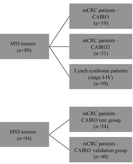
formalin-fixed paraffin-embedded (FFPE) material of the primary tumor was part of the initial protocol in both studies. To determine the frequency and prognostic value of EMAST in mCRC patients with MSI tumors we selected 50 mCRC patients with MSI tumors treated in both CAIRO studies. Since MSI is relatively rare in mCRC we combined the patients of the CAIRO (n = 19) and the CAIRO2 (n = 31) study. No validation cohort could be selected for MSI patients. To further evaluate the relation between EMAST and MSI, we retrieved 39 tu-mors from CRC patients (anonymous samples) with known Lynch syndrome (stage I-IV) from our own database (that has been set up conform the guidelines of the local medical ethical committee (Commissie Mensgebonden Onderzoek Radboudumc) with written informed con-sent of the patients, from which use of tissue is approved for this study). To determine the fre-quency and prognostic value of EMAST in mCRC patients with MSS tumors we selected 54 patients of the CAIRO study with comparable characteristics (test group). Patients within the test group were all treated with first-line capecitabine monotherapy for at least 3 cycles, locali-zation of the primary tumor in colon or recto- sigmoid which was resected, WHO performance score 0, normal baseline serum lactate dehydrogenase (LDH) concentration, and had not re-ceived prior adjuvant chemotherapy. In addition, we randomly selected 40 additional mCRC patients with MSS tumors treated in the same CAIRO study as a validation group. (Fig 1)
Fig 1. Flowchart of selected CRC patients to determine the frequency and prognostic value of EMAST.
EMAST analysis
Genomic DNA was extracted from four to eight manually microdissected 30μm section of
FFPE tissue of the primary tumors. Areas containing>50% tumor cells were selected by micro-scopic evaluation on a reference slide stained with H&E. Genomic DNA from microdissected tissues was isolated using the QIAamp DNA micro kit (Qiagen, Valencia, CA) following the manufacturer’s instructions. DNA concentration was determined at 260 nm using the Nano-drop ND-1000 spectrophotometer (NanoNano-drop Technologies, Inc., Wilmington, DE, USA). EMAST analysis was performed in duplicate on normal and tumor DNA of the selected pa-tients. EMAST status was determined by PCR and GeneScan analysis using five tetranucleotide markers: MYCL1, D8S321, D9S242, D20S82 and D20S85 (S1 Table) [23]. A tumor was defined EMAST if at least two of the five markers showed instability and non-EMAST if only one or none of the markers showed instability [22].
Patients were analyzed for the frequency and prognostic value in four different groups: pa-tients with combined MSI and EMAST tumors (MSI / EMAST), papa-tients with combined MSI and non-EMAST tumors (MSI / non-EMAST), patients with combined MSS and EMAST tu-mors (MSS / EMAST) and patients with combined MSS and EMAST tutu-mors (MSS / non-EMAST). The frequency of EMAST was compared for patients with MSI and MSS tumors. The outcome was analyzed within the group of patients with MSI tumors (excluding the Lynch syndrome patients) for EMAST compared to non-EMAST tumors and within the group of pa-tients with MSS tumors for EMAST compared to non-EMAST tumors.
Immunohistochemistry MSH3
Immunohistochemistry (IHC) was performed on tissue microarrays (TMA) of the primary tu-mors of 549 eligible randomized patients in the CAIRO study as previously described.[30] 4μm slides were cut of every TMA and mounted on glass. Xylene and ethanol were used for
deparaffinization and dehydration of the TMA slides. Water and phosphate-buffered saline (PBS) were used for washing of the slides. Endogenous peroxidase activity was blocked with 3% hydrogen peroxide in PBS for 30 min and slides were washed with water, after which heat-induced epitope retrieval was performed. The slides were stained with a monoclonal antibody against MSH3 (clone ERP4334; Epitomics—an Abcam company, Burlingame, CA, USA), dilu-tion 1:5000. Two independent investigators performed the scoring, and if the slide scoring was not unambiguous, the opinion of a third investigator (pathologist IDN) was final. Staining pat-tern of the MSH3 protein was evaluated by using the normal epithelial, stromal and inflamma-tory cells as internal control. Low MSH3 protein expression was defined as<85% brown staining of cell cores in tumor cells and high MSH3 protein expression was defined as85% brown staining of cell cores in tumor cells and not applicable if neither tumor nor stromal cells showed MSH3 protein expression [21].
category.[30] Hypermethylation status of theMLH1gene promoter and theBRAFV600E
mu-tation status, was assessed as described previously [30–32].
Statistical analysis
For the EMAST analysis, patients were divided into two categories: EMAST and non-EMAST tumors. The association between EMAST and MSH3 protein expression was investigated with a logistic regression model with independent factors group and MSH3 expression. OS was de-fined as the time from the date of randomization to the date of death from any cause. OS curves were estimated using the Kaplan—Meier method and compared using a Cox proportional haz-ard model. All tests were two-sided and p<0.05 was considered as statistically significant. All analyses were conducted using the SAS system version 9.2.
Literature search strategy, inclusion criteria, and data extraction
We reviewed the literature on the frequency of EMAST in CRC patients with MSI and MSS tu-mors. A search was conducted of Medline, PubMed, and the Cochrane Library from January 1990 to April 2014 with an English-language restriction, using the following search terms: EMAST, tetranucleotide repeat, in combination with colon cancer and colorectal cancer. Origi-nal publications were selected if the abstract contained data for patients with EMAST. In case of duplicate publications, the most recent and/or most complete study was included. Publica-tions were excluded if frequency of EMAST was limited to either patients with MSI or MSS tumors.
Results
Prevalence of EMAST
Overall, EMAST was observed among 45.9% of a total of 183 tumors (Table 1). Frequency of EMAST was significantly higher among patients with MSI tumors compared to MSS tumors: 79.8% compared to 13.8% (p<0.001).
In patients with MSS / EMAST tumors instability was generally shown at 2 EMAST loci (69.2%, 9 out of 13), whereas in patients with MSI / EMAST tumors instability was frequently shown at 4 (33.8%, 24 out of 71), or 5 (50.7%, 36 out of 71) EMAST loci (Fig 2A). The highest
Table 1. Prevalence of EMAST and non-EMAST tumors in the different patient groups.
EMAST non-EMAST Total number of patients pvalue
Patients with MSI tumors
mCRC 84.0% 16.0% 50
Lynch syndrome 74.4% 25.6% 39 0.113
Total 79.8% 20.2% 89
Patients with MSS tumors
Test group of patients 11.1% 88.9% 54
Validation group of patients 17.5% 82.5% 40 0.160
Total 13.8% 86.2% 94
All patients Total 45.9% 54.1% 183
pvalue represent heterogeneity between groups
Abbreviations: EMAST = elevated microsatellite alterations at selected tetranucleotide repeats, MSI = microsatellite instability, MSS = microsatellite stability
frequency of instability in EMAST tumors was demonstrated at the D20S82 locus (91.7%, 77 out of 84), followed by the MYCL1 locus (86.9%, 73 out of 84), the D9S242 locus (84.5%, 71 out of 84), the D8S321 locus (72.6%, 61 out of 84) and the D20S85 locus (65.5%, 55 out of 84) (Fig 2B).
EMAST and MSH3
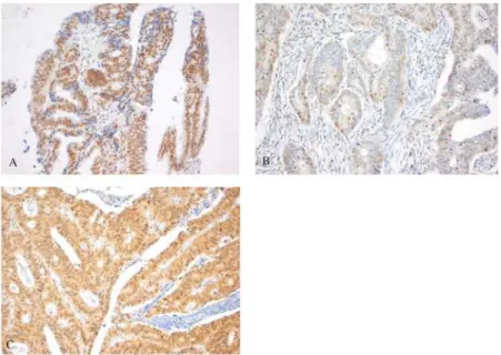
The majority of mCRC patients (n = 381, 69.4%) had a high expression of MSH3 in tumor cells (Fig 3). 21.1% of tumors demonstrated nuclear heterogeneity by expression of both positive and negative nuclei upon MSH3 IHC staining (Fig3A–3C). Both MSH3 expression and EMAST status was known in 139 patients. Heterogeneous or high MSH3 protein expression was not correlated to EMAST status (p = 0.088 and p = 0.856, respectively).
Outcome of patients with MSI tumors
Patients with MSI / EMAST tumors were mostly female (52% versus 22%, respectively, p = 0.038) (Table 2). Moreover, EMAST tumors were more frequently located above the recto-sigmoid area (93% versus 63%, p = 0.006).
Fig 2. Frequency of instable EMAST markers (A) and frequency of affected EMAST loci (B), subdivided by patients with MSI and MSS tumors.
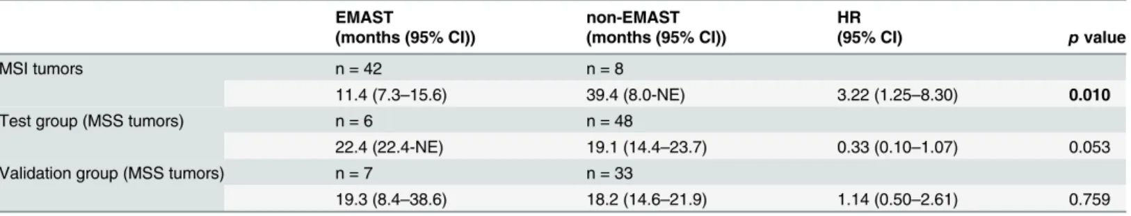
Median OS was significantly worse for patients with MSI / EMAST compared to MSI / non-EMAST tumors treated in both CAIRO studies (11.4 versus 39.4 months, respectively, HR 3.22, 95% CI 1.25–8.30) (Table 3).
MSI / EMAST and the relation to MMR proteins
The distribution of loss of MMR proteins in mCRC tumors is summarized inFig 4A. Most pa-tients with MSI tumors showed loss of MLH1 and/or PMS2 protein expression (72.0%, 36 out of 50 patients). Loss of MSH2 and/or MSH6 protein expression was found in 18.0% (9 out of 50) of patients. These patients are likely Lynch or Lynch-like syndrome patients. Only 7.1% (3 out of 42) of patients with a MSI / EMAST tumor showed loss of expression of the MSH6 pro-tein, compared to 62.5% (5 out of 8) of patients with MSI / non-EMAST tumors.
Hypermethylation of theMLH1gene promoter (32 out of 40 patients) andBRAFmutations
(24 out of 40 patients) were limited to the patients with a MSI / EMAST tumor (p<0.001 and p = 0.004 respectively).
EMAST in patients with Lynch syndrome
In order to further analyze the relation of MSI and EMAST we selected 39 patients with known Lynch syndrome (with germ line mutations inMSH2gene (n = 11),MLH1gene (n = 10), MSH6gene (n = 10) andPMS2gene (n = 8)). The majority of this population showed EMAST
(29 out of 39 patients). None of the patients showed hypermethylation of theMLH1gene
pro-moter, and all tumors wereBRAFwild-type. Nine out of 10 patients presenting with
non-EMAST tumors had a germline mutation inMSH6(Fig 4B).
Outcome of patients with MSS tumors
Baseline patient and tumor characteristics for patients with MSS tumors (test group and valida-tion group), subdivided by EMAST and non-EMAST tumors are presented inTable 2. There
Fig 3. Staining pattern of MSH3 protein expression.Heterogeneous MSH3 protein expression (A), demonstrated by expression of both brown (positive) and blue (negative) nuclei upon MSH3 IHC staining. Low MSH3 protein expression was defined as<85% brown staining of cell cores in tumor cells (B) and high
MSH3 protein expression was defined as85% brown staining of cell cores in tumor cells (C).
was no significant difference in outcome for patients with EMAST compared to patients with non-EMAST tumors (Table 3).
Review of the literature
The literature search identified 7 studies in which EMAST was described in stage I-IV CRC pa-tients with MSI and MSS tumors [21–24,27,33,34]. Two studies were excluded: one study de-scribed the same population [23] and one study assessed the prevalence of EMAST solely in
Table 2. Baseline patient and tumor characteristics of patients with MSI and MSS tumors, subdivide by EMAST and non-EMAST tumors.
Patients with MSI tumors Patients with MSS tumors
Test group Validation group
EMAST non-EMAST
EMAST non-EMAST
EMAST non-EMAST
n = 42 n = 8 pvalue n = 6 n = 48 pvalue n = 7 n = 33 pvalue
Median age (range) 68 (34– 84)
59 (37–73) 0.131 72 (47– 77)
66 (34–79) 0.749 71 (58– 76)
67 (39–81) 0.161
Sex
male 20 (48%) 7 (88%) 0.038 5 (83%) 31 (65%) 0.651 5 (71%) 22 (67%) 0.338
female 22 (52%) 1 (22%) 1 (17%) 17 (35%) 2 (29%) 11 (33%)
WHO performance status
PS0 26 (62%) 4 (50%) 0.156 6 (100%) 48 (100%) - 3 (42%) 22 (67%) 0.045
PS1 14 (33%) 4 (50%) - - 3 (42%) 11 (33%)
PS2 2 (5%) - - - 1 (16%)
-Serum LDH
normal 32 (76%) 4 (50%) 0.110 6 (100%) 48 (100%) - 5 (71%) 23 (70%) 0.348
abnormal 10 (24%) 4 (50%) - - 2 (29%) 10 (30%)
Previous adjuvant therapy
yes 5 (12%) 2 (25%) 0.239 - - - 3 (42%) 5 (15%) 0.108
no 37 (88%) 6 (75%) 6 (100%) 48 (100%) 4 (58%) 28 (85%)
Localization of the primary tumor
colon 93 (93%) 5 (63%) 0.006 5 (83%) 42 (88%) 0.416 3 (42%) 20 (61%) 0.869
recto sigmoid - 2 (25%) 1 (17%) 6 (12%) - 1 (3%)
rectum 2 (5%) 1 (12%) - - 4 (58%) 11 (33%)
multiple tumor - - - 1 (3%)
unknown 1 (2%)
-Histology of the primary tumor
adenocarcinoma 22 (52%) 5 (63%) 0.144 5 (83%) 35 (73%) 0.183 7 (100%) 24 (73%) 0.141 mucinous adenocarcinoma (>50%
WHO)
16 (38%) 2 (25%) 1 (17%) 4 (8%) - 4 (12%)
adenocarcinoma + mucinous component
4 (10%) - - 6 (13%) - 4 (12%)
other - 1 (12%) - 3 (6%) - 1 (3%)
BRAFmutation status
mutation 24 (57%) - 0.004 - 5 (11%) 0.590 - - 0.677
wild-type 16 (38%) 7 (88%) 6 (100%) 40 (83%) 7 (100%) 31 (94%)
unknown 2 (5%) 1 (12%) - 3 (6%) - 2 (6%)
NOTE: Statistically significant results are set in bold
Abbreviations: MSI = microsatellite instability, MSS = microsatellite stability, EMAST = elevated microsatellite instability at selected tetranucleotide repeats
Table 3. Overall survival of patients with MSI and MSS tumors, subdivided by EMAST and non-EMAST tumors.
EMAST non-EMAST HR
(months (95% CI)) (months (95% CI)) (95% CI) pvalue
MSI tumors n = 42 n = 8
11.4 (7.3–15.6) 39.4 (8.0-NE) 3.22 (1.25–8.30) 0.010
Test group (MSS tumors) n = 6 n = 48
22.4 (22.4-NE) 19.1 (14.4–23.7) 0.33 (0.10–1.07) 0.053
Validation group (MSS tumors) n = 7 n = 33
19.3 (8.4–38.6) 18.2 (14.6–21.9) 1.14 (0.50–2.61) 0.759
NOTE: Statistically significant results are set in bold
Abbreviations: EMAST = elevated microsatellite instability at selected tetranucleotide repeats, MSI = microsatellite instability, MSS = microsatellite stability, CI = confidence interval, HR = hazard ratio, NE = not estimable
doi:10.1371/journal.pone.0124538.t003
Fig 4. Percentage of mCRC patients with MSI tumors and loss of MLH1 and/or PMS2 (MLH1 / PMS2) and MSH2 and/or MSH6, (MSH2 and MSH6) subdivided in patients with MSI / EMAST tumors and patients with MSI / non-EMAST tumors.(A). Percentage of patients with known Lynch syndrome and germ line mutation of the different MSI genes, subdivided in patients with MSI / EMAST tumors and patients with MSI / non-EMAST tumors (B).
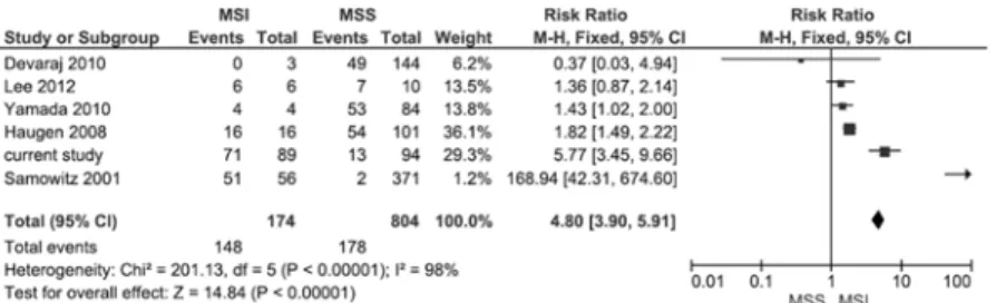
patients with MSS tumors [27]. Three studies had limited numbers of MSI tumors.Fig 5 sum-marizes a forest plot of the 5 published studies and the current study on the prevalence of EMAST in stage I-IV CRC patients with MSI and MSS tumors. EMAST is significantly more frequent in tumors with MSI (148/174) (RR 4.80, 95% confidence interval 3.90–5.91). Signifi-cant heterogeneity was observed.
Discussion
This study presents the analysis on the frequency and prognostic value of EMAST in mCRC patients. Although EMAST was observed in 45.9% of all mCRC patients, it was most pro-nounced in MSI tumors (79.8%). The frequency of EMAST among MSS tumors (13.8%) was much lower in our study compared to most studies in stage I-IV CRC [21–24,27,34]. The broad range of frequency (0.54–60.2%) of EMAST among MSS tumors described in literature [21–24,27,33,34] might be due the fact that there is no consensus on the definition of EMAST and the panel required for its diagnosis. Because of the polymorphic nature of tetranucleotide repeats in the current study we used stringent criteria for the definition of EMAST: at least two of the five tetranucleotide markers should show instability.
Despite the fact that several small studies (n = 3 to n = 56) [21–24,33,34] demonstrated that MSI invariably is associated with EMAST, the correlation between EMAST and MSI is not widely accepted. We found a high frequency of EMAST in MSI tumors, which was confirmed in a analysis of the existing literature in stage I-IV CRC (Fig 5). Only a small subset of patients with MSI tumors is EMAST. Interestingly, the majority of patients with MSI / non-EMAST tumors showed loss of MSH6. This is in line with the fact that MSH6 is only involved in mononucleotide mismatch repair [35,36]. Patients with MSI / non-EMAST tumors are more often male and tumors developed more frequently in the rectum, these characteristics are comparable to patients withMSH6germline mutations [37]. In our population of MSI tumors
the presence of EMAST is correlated withMLH1deficiency, which causes a total DNA
mis-match repair defect, both in Lynch syndrome as well as in the sporadic setting, confirming ear-lier observations [21,27].
Data about the EMAST phenotype in CRC are scarce and underlying mechanism(s) remain unclear. The earliest reports suggested that EMAST might be associated with mutations in the
TP53gene [16,17] or that environmental carcinogens may exacerbate this phenotype [38] in
cancers other than CRC. Later on, an association was made between loss of MSH3 and EMAST in CRC [21]. Due to the fact that MutSβhas a strong affinity for recognizing more than two un-paired nucleotides and genetic complementation of MSH3 deficiency in human cells increased stability at loci containing dinucleotide and tetranucleotide repeats [39], it was argued that that loss of MutSβdue to MSH3 inactivation may result in MSI not only at loci containing dinucle-otide repeats, but also at loci with tetranucledinucle-otide repeats, such as EMAST [21]. Recent studies
Fig 5. Forest plot for the association of prevalence of EMAST in patients with MSI compared to MSS tumors in stage I-IV CRC.
confirm the association between loss of MSH3 and EMAST in CRC, however the exact under-lying mechanism remains unknown [25,26]. We failed to demonstrate a correlation between MSH3 protein expression and EMAST. Actually, we did find the heterogeneous expression pat-tern of MSH3 in 21.1% of patients as described by others, although this was not correlated to EMAST. However our study had a small number of patients. Another possible explanation for the difference between our study and previous studies might be a potential undersampeling bias since we used one TMA specimen per tumor instead of full tumor slides.
We demonstrate that patients with MSI / non-EMAST tumors had a significantly better prognosis compared to patients with MSI / EMAST tumors. The addition of EMAST to MSI tumors seems to worsen overall prognosis and survival. The MSI / non-EMAST group would be expected to be enriched forMSH6deficiency since EMAST would be identified with a total
DNA mismatch repair defect, such as withMLH1orMSH2deficiency. Our results on the
cor-relation of EMAST with clinical outcome should be interpreted with caution due to the small number of patients in the different subgroups.
In summary, the frequency of EMAST among mCRC patients with MSS tumors is low compared to patients with MSI tumors. There was no correlation between EMAST and MSH3 protein expression. We did find a clear link between MSI and EMAST, and outcome was significantly better for mCRC patients with MSI / non-EMAST tumors. Further studies are warranted to elucidate the molecular basis for EMAST and to show whether the tetranu-cleotide alterations observed in EMAST tumors may have a functional impact by themselves or just represent bystander alterations in a subset of tumors with specific defects in DNA rep-lication and repair.
Supporting Information
S1 Table. Tetranucleotide microsatellite PCR primer sequences. (PDF)
Acknowledgments
This study was supported by a grant from the Dutch Colorectal Cancer Group (DCCG).
Author Contributions
Conceived and designed the experiments: SV SVV IDN. Performed the experiments: SV SVV. Analyzed the data: SV SVV AJFdH MJL IDN. Contributed reagents/materials/analysis tools: SV MG. Wrote the paper: SV SVV MJL CJAP MK IDN.
References
1. Jass JR (2007) Classification of colorectal cancer based on correlation of clinical, morphological and molecular features. Histopathology 50: 113–130. PMID:17204026
2. Samowitz WS (2008) Genetic and epigenetic changes in colon cancer. Exp Mol Pathol 85: 64–67. doi: 10.1016/j.yexmp.2008.03.008PMID:18482722
3. Snover DC (2011) Update on the serrated pathway to colorectal carcinoma. Hum Pathol 42: 1–10. doi: 10.1016/j.humpath.2010.06.002PMID:20869746
4. Grady WM, Carethers JM (2008) Genomic and epigenetic instability in colorectal cancer pathogenesis. Gastroenterology 135: 1079–1099. doi:10.1053/j.gastro.2008.07.076PMID:18773902
6. Baranovskaya S, Soto JL, Perucho M, Malkhosyan SR (2001) Functional significance of concomitant inactivation of hMLH1 and hMSH6 in tumor cells of the microsatellite mutator phenotype. Proc Natl Acad Sci U S A 98: 15107–15112. PMID:11742074
7. Ohmiya N, Matsumoto S, Yamamoto H, Baranovskaya S, Malkhosyan SR, Perucho M (2001) Germline and somatic mutations in hMSH6 and hMSH3 in gastrointestinal cancers of the microsatellite mutator phenotype. Gene 272: 301–313. PMID:11470537
8. Genschel J, Littman SJ, Drummond JT, Modrich P (1998) Isolation of MutSbeta from human cells and comparison of the mismatch repair specificities of MutSbeta and MutSalpha. J Biol Chem 273: 19895– 19901. PMID:9677427
9. Chang DK, Ricciardiello L, Goel A, Chang CL, Boland CR (2000) Steady-state regulation of the human DNA mismatch repair system. J Biol Chem 275: 29178. PMID:10979987
10. Jass JR (2006) Hereditary Non-Polyposis Colorectal Cancer: the rise and fall of a confusing term. World J Gastroenterol 12: 4943–4950. PMID:16937488
11. Kane MF, Loda M, Gaida GM, Lipman J, Mishra R, Goldman H et al. (1997) Methylation of the hMLH1 promoter correlates with lack of expression of hMLH1 in sporadic colon tumors and mismatch repair-de-fective human tumor cell lines. Cancer Res 57: 808–811. PMID:9041175
12. Cunningham JM, Christensen ER, Tester DJ, Kim CY, Roche PC, Burgart LJ et al. (1998) Hypermethy-lation of the hMLH1 promoter in colon cancer with microsatellite instability. Cancer Res 58: 3455– 3460. PMID:9699680
13. Jass JR, Do KA, Simms LA, Iino H, Wynter C, Pilla SP et al. (1998) Morphology of sporadic colorectal cancer with DNA replication errors. Gut 42: 673–679. PMID:9659163
14. Popat S, Hubner R, Houlston RS (2005) Systematic review of microsatellite instability and colorectal cancer prognosis. J Clin Oncol 23: 609–618. PMID:15659508
15. Ahrendt SA, Decker PA, Doffek K, Wang B, Xu L, Demeure MJ et al. (2000) Microsatellite instability at selected tetranucleotide repeats is associated with p53 mutations in non-small cell lung cancer. Cancer Res 60: 2488–2491. PMID:10811129
16. Xu L, Chow J, Bonacum J, Eisenberger C, Ahrendt SA, Spafford M et al. (2001) Microsatellite instability at AAAG repeat sequences in respiratory tract cancers. Int J Cancer 91: 200–204. PMID:11146445 17. Danaee H, Nelson HH, Karagas MR, Schned AR, Ashok TD, Hirao T et al. (2002) Microsatellite
instabil-ity at tetranucleotide repeats in skin and bladder cancer. Oncogene 21: 4894–4899. PMID:12118368 18. Singer G, Kallinowski T, Hartmann A, Dietmaier W, Wild PJ, Schraml P et al. (2004) Different types of
microsatellite instability in ovarian carcinoma. Int J Cancer 112: 643–646. PMID:15382045 19. Burger M, Denzinger S, Hammerschmied CG, Tannapfel A, Obermann EC, Wieland WF et al. (2006)
Elevated microsatellite alterations at selected tetranucleotides (EMAST) and mismatch repair gene ex-pression in prostate cancer. J Mol Med 84: 833–841. PMID:16924473
20. Choi YD, Choi J, Kim JH, Lee JS, Lee JH, Choi C et al. (2008) Microsatellite instability at a tetranucleo-tide repeat in type I endometrial carcinoma. J Exp Clin Cancer Res 27: 88. doi: 10.1186/1756-9966-27-88PMID:19116039
21. Haugen AC, Goel A, Yamada K, Marra G, Nguyen TP, Nagasaka T et al. (2008) Genetic instability caused by loss of MutS homologue 3 in human colorectal cancer. Cancer Res 68: 8465–8472. doi:10. 1158/0008-5472.CAN-08-0002PMID:18922920
22. Devaraj B, Lee A, Cabrera BL, Miyai K, Luo L, Ramamoorthy S et al. (2010) Relationship of EMAST and microsatellite instability among patients with rectal cancer. J Gastrointest Surg 14: 1521–1528. doi:10.1007/s11605-010-1340-6PMID:20844976
23. Lee SY, Chung H, Devaraj B, Iwaizumi M, Han HS, Hwang DY et al. (2010) Microsatellite alterations at selected tetranucleotide repeats are associated with morphologies of colorectal neoplasias. Gastroen-terology 139: 1519–1525. doi:10.1053/j.gastro.2010.08.001PMID:20708618
24. Yamada K, Kanazawa S, Koike J, Sugiyama H, Xu C, Funahashi K et al. (2010) Microsatellite instability at tetranucleotide repeats in sporadic colorectal cancer in Japan. Oncol Rep 23: 551–561. PMID: 20043121
25. Campregher C, Schmid G, Ferk F, Knasmuller S, Khare V, Kortum B et al. (2012) MSH3-deficiency initi-ates EMAST without oncogenic transformation of human colon epithelial cells. PLoS One 7: e50541. doi:10.1371/journal.pone.0050541PMID:23209772
26. Tseng-Rogenski SS, Chung H, Wilk MB, Zhang S, Iwaizumi M, Carethers JM (2012) Oxidative stress induces nuclear-to-cytosol shift of hMSH3, a potential mechanism for EMAST in colorectal cancer cells. PLoS One 7: e50616. doi:10.1371/journal.pone.0050616PMID:23226332
28. Koopman M, Antonini NF, Douma J, Wals J, Honkoop AH, Erdkamp FL et al. (2007) Sequential versus combination chemotherapy with capecitabine, irinotecan, and oxaliplatin in advanced colorectal cancer (CAIRO): a phase III randomised controlled trial. Lancet 370: 135–142. PMID:17630036
29. Tol J, Koopman M, Cats A, Rodenburg CJ, Creemers GJ, Schrama JG et al. (2009) Chemotherapy, bevacizumab, and cetuximab in metastatic colorectal cancer. N Engl J Med 360: 563–572. doi:10. 1056/NEJMoa0808268PMID:19196673
30. Koopman M, Kortman GA, Mekenkamp L, Ligtenberg MJ, Hoogerbrugge N, Antonini NF et al. (2009) Deficient mismatch repair system in patients with sporadic advanced colorectal cancer. Br J Cancer 100: 266–273. doi:10.1038/sj.bjc.6604867PMID:19165197
31. Tol J, Dijkstra JR, Klomp M, Teerenstra S, Dommerholt M, Vink-Borger ME et al. (2010) Markers for EGFR pathway activation as predictor of outcome in metastatic colorectal cancer patients treated with or without cetuximab. Eur J Cancer 46: 1997–2009. doi:10.1016/j.ejca.2010.03.036PMID:20413299 32. Heideman DA, Lurkin I, Doeleman M, Smit EF, Verheul HM, Meijer GA et al. (2012) KRAS and BRAF
mutation analysis in routine molecular diagnostics: comparison of three testing methods on formalin-fixed, paraffin-embedded tumor-derived DNA. J Mol Diagn 14: 247–255. doi:10.1016/j.jmoldx.2012. 01.011PMID:22425762
33. Samowitz WS, Holden JA, Curtin K, Edwards SL, Walker AR, Lin HA et al. (2001) Inverse relationship between microsatellite instability and K-ras and p53 gene alterations in colon cancer. Am J Pathol 158: 1517–1524. PMID:11290569
34. Lee SY, Miyai K, Han HS, Hwang DY, Seong MK, Chung H et al. (2012) Microsatellite instability, EMAST, and morphology associations with T cell infiltration in colorectal neoplasia. Dig Dis Sci 57: 72– 78. doi:10.1007/s10620-011-1825-5PMID:21773681
35. Papadopoulos N, Nicolaides NC, Liu B, Parsons R, Lengauer C, Palombo F et al. (1995) Mutations of GTBP in genetically unstable cells. Science 268: 1915–1917. PMID:7604266
36. Edelmann W, Yang K, Umar A, Heyer J, Lau K, Fan K et al. (1997) Mutation in the mismatch repair gene Msh6 causes cancer susceptibility. Cell 91: 467–477. PMID:9390556
37. Klarskov L, Holck S, Bernstein I, Okkels H, Rambech E, Baldetorp B et al. (2011) Challenges in the identification of MSH6-associated colorectal cancer: rectal location, less typical histology, and a subset with retained mismatch repair function. Am J Surg Pathol 35: 1391–1399. doi:10.1097/PAS.
0b013e318225c3f0PMID:21836479
38. Slebos RJ, Oh DS, Umbach DM, Taylor JA (2002) Mutations in tetranucleotide repeats following DNA damage depend on repeat sequence and carcinogenic agent. Cancer Res 62: 6052–6060. PMID: 12414628