1-Methyl-D-Tryptophan Potentiates TGF-
β
-Induced Epithelial-Mesenchymal Transition
in T24 Human Bladder Cancer Cells
Rodrigo Barbosa Oliveira Brito1, Camila Soares Malta1, Diego Mota Souza1, Luiz Henrique Gomes Matheus1, Yves Silva Teles Matos1, Chrisna Souza Silva1, Janaína
Mendes Ferreira1, Valeria Sutti Nunes2, Cristiane Miranda França3, Humberto Dellê1*
1Programa de Pós-graduação em Medicina, Universidade Nove de Julho (UNINOVE), São Paulo, Brazil, 2Lipids Laboratory (LIM-10), Endocrinology and Metabolism Division of Hospital das Clínicas, Faculty of Medical Sciences, University of São Paulo, São Paulo, Brazil,3Programa de Pós-graduação em Biofotônica em Ciências da Saúde, Universidade Nove de Julho (UNINOVE), São Paulo, Brazil
Abstract
Immune escape and metastasis are the hallmarks of several types of cancer including bladder cancer. One of the mechanisms involved in these processes has been linked to indoleamine 2,3-dioxygenase (IDO). Although IDO is classically recognized for its immu-nomodulatory property, it has presented nonimmunological effects in some tumors. TGF-β1 is believed to contribute to carcinoma development by modulating immunossupressive molecules, including IDO. In addition, TGF-β1 induces the epithelial-mesenchymal transi-tion (EMT), which is a critical step in the tumor invasiveness and metastasis. We investi-gated the role of MT and IDO modulation in the induction of EMT by TGF-β1 in T24 human bladder carcinoma cells. When T24 cells were incubated with the IDO inhibitor (MT, 1-methyl-D-tryptophan), with TGF-β1, and with MT+TGF-β1, a significant decrease of IDO expression and activity was observed. In addition, downregulation of e-cadherin and upre-gulation of n-cadherin and EMT transcription factors were induced by the treatments, con-firming the induction of EMT. siRNA-mediated knockdown of IDO decreased e-cadherin expression, but had no effect on EMT transcription factors. In the scratch-wound assay, the heightened migration process was intensified when the cells were incubated with MT+TGF-β1. These effects were associated with a robust inhibition of Akt activation. After inoculation of T24 cells under the kidney capsule of Balb/c nude, the cells were positive for IDO in the center of the cell infiltrate, being negative in the periphery, where EMT is high. In conclusion, inhibition of IDO by TGF-β1 and MT is associated with EMT in T24 human bladder carcinoma cells. MT has potentiating effect in TGF-β1-induced EMT, inde-pendently of IDO. This nonimmunological effect of MT should be considered if IDO is the target to avoid immune escape in bladder cancer.
OPEN ACCESS
Citation:Brito RBO, Malta CS, Souza DM, Matheus LHG, Matos YST, Silva CS, et al. (2015) 1-Methyl-D-Tryptophan Potentiates TGF-β-Induced Epithelial-Mesenchymal Transition in T24 Human Bladder Cancer Cells. PLoS ONE 10(8): e0134858. doi:10.1371/journal.pone.0134858
Editor:Michael Platten, University Hospital of Heidelberg, GERMANY
Received:January 28, 2015
Accepted:July 14, 2015
Published:August 12, 2015
Copyright:© 2015 Brito et al. This is an open access article distributed under the terms of the Creative Commons Attribution License, which permits unrestricted use, distribution, and reproduction in any medium, provided the original author and source are credited.
Data Availability Statement:All relevant data are within the paper and its Supporting Information files.
Funding:This work was supported by the São Paulo Research Foundation (FAPESP), grant 2012/04423-0,http://www.fapesp.br/.
Introduction
Urinary bladder cancer is the most common malignancy of the urinary system [1]. Although the most common form of human bladder cancer is non-muscle invasive (70% to 80%), 30% to 50% of these cases progress to a muscle-invasive form after repeated resections, which ulti-mately leads to cancer-specific death and metastasis [2].
Multiple mechanisms have been implicated in tumor invasiveness and metastasis, including the epithelial-mesenchymal transition (EMT). In this process, a polarized epithelial cell assumes a mesenchymal phenotype, which leads to a loss of cellular adhesion to the basement membrane, activation of motility and invasiveness, and production of extracellular-matrix-degrading enzymes [3]. Several molecules are engaged in the initiation of EMT, including the TGF-βsuperfamily proteins. TGF-β1 is an important inducer of EMT in several different types of tumors [4], including bladder cancer [5]. Certain genetic variations and increased plasma levels of TGF-βare potent predictors of bladder cancer risk [6,7].
Indoleamine 2,3-dioxygenase (IDO) is an enzyme that catalyzes the degradation of the amino acid tryptophan, which leads to accumulation of tryptophan metabolites, such as kynur-enine. As described by Munn et al., IDO has a role in the maternal-fetal interface and functions to protect embryos against the maternal immune system [8]. Subsequently, IDO has been explored for use as an effective immunomodulatory molecule. Because IDO is produced by many cell types of the immune system, including dendritic cells, macrophages, and regulatory T cells, IDO has been implicated in immune-mediated disorders, such as allograft rejection [9], autoimmune diseases [10] and escape from antitumor immunity in cancer [11]. Regarding cancer, IDO activity is present in many human tumor types as well as tumor-draining lymph nodes [11]; however, the role of IDO in tumor growth is still poorly understood. While IDO expression is correlated with a less favorable prognosis for many types of cancer including colo-rectal [12], uterine cervical [13], endometrial [14], breast [15], melanoma [16], ovarian [17], and lung cancer [18], IDO expression is also associated with recurrence-free survival of hepato-cellular patients [19] and the long-term survival of patients with renal carcinoma [20].
Although there is a controversy about the role of the IDO in cancer, molecules that can modu-late IDO-mediated pathways have been seen as promising for cancer treatment. In this context, 1-methyl-D-tryptophan, a competitive inhibitor of IDO, has been intensively studied as anti-cancer agent. Currently, the association of 1-methyl-D-tryptophan (MT) with docetaxel was used in a phase I clinical trial with patients with metastatic solid tumors [21]. However, this molecule may act independently of IDO activity [22], as well as like tryptophan to regulate mTOR pathway [23].
IDO expression has been found not only in infiltrating immune cells and tumor-draining lymph nodes but also in neoplastic cells. In bladder cancer, the T24 human transi-tional carcinoma cell line produces IDO constitutively in culture [24], even without involve-ment of the immune system, leading to the hypothesis that IDO may have a role in non-immune processes. Levina et al., demonstrated that upregulation of IDO in breast cancer cells increased cell proliferation and decreased apoptosis in a manner that was independent of the immunological effects of IDO [25].
Materials and Methods
Cell culture
Human bladder cancer T24 cells (HTB-4; American Type Culture Collection-ATCC, Manas-sas, VA, USA) were acquired from the cell bank of the Federal University of Rio de Janeiro. T24 cells were cultured in McCoy’s 5A Medium (Sigma-Aldrich, St. Louis, MO) supplemented with 10% fetal bovine serum and penicillin-streptomycin (Sigma-Aldrich, St. Louis, MO) and maintained at 37°C with 5% CO2.
To analyze the effect of TGF-β1 on IDO expression, T24 cells were seeded in 6-well plates (1X10⁵cells per well). The cells were incubated with 1 ng/ml, 5 ng/ml or 10 ng/ml of TGF-β1 (R&D Systems Inc., Minneapolis, MN) in serum-free McCoy’s 5A Medium for 48 h
(triplicate).
After determining the optimal concentration of TGF-β1 (5 ng/ml), T24 cells were seeded in 6-well plates and cultured until reaching 75% confluence. The cells were then maintained in serum-free McCoy’s 5A Medium for 48 h under the following four conditions in triplicate: only medium (control); medium containing 1 mM methyl-tryptophan (MT; 1-methyl-D-tryp-tophan, cat 452483, Sigma-Aldrich, St. Louis, MO); medium containing TGF-β1 (5 ng/ml); and medium containing MT plus TGF-β1. At the end of 48 h, supernatants were collected for kynurenine measurement, and the cellular monolayers were trypsinized for RNA extraction. This experiment was repeated for protein isolation.
Small interfering RNA (siRNA) transfection
In order to knockdown endogenous IDO, T24 cells were transfected with siRNA for IDO (Silencer Select inventoried 4392420, Ambion, Carlsbad, CA) or with a non-targeting control siRNA for transfection control (Silencer Select Negative Control inventoried 4390843,
Ambion, Carlsbad, CA), using Lipofectamine 3000 Reagent (Invitrogen, Carlsbad, CA). Briefly, the cells were cultured in 6 well-plates, and then were maintained in serum-free McCoy medium containing (per well) 1μg of siRNA, 5μl of P3000 Reagent, and 5μL of Lipofectamine 3000 Reagent, during 24 h. After transfection, the medium was removed and the cells were cul-tured for 24 h in McCoy’s 5A Medium supplemented with 10% fetal bovine serum at 37°C with 5% CO2. After this recovery time, the cells were incubated with TGF-β1 (5 ng/ml, for
48 h, in serum-free McCoy medium), exactly as previously described. The following four conditions in triplicate were performed: T24 cells previously transfected with control siRNA (si-Control), without TGF-β1; T24 cells previously transfected with IDO siRNA incubated without TGF-β1 (siIDO); T24 cells previously transfected with control siRNA incubated with TGF-β1 (TGF-β); and T24 cells previously transfected with IDO siRNA incubated with TGF-β1 (siIDO+TGF-β).
RT-qPCR
N-cad (forward 5’–TGCCCGGTTTCATTTAGGGG–3’and reverse 5’–TCCCTCAGGAACT GTCCCAT–3’; accession number X57548), TWIST (forward 5’–AGAGATGCAACT AAGCCCTCT–3’and reverse 5’–AAGCAGCTACTGACAGGCAC–3’; accession number Y11177), Snail (forward 5’–ACCACTATGCCGCGCTCTT–3’and reverse 5’–GGTCGT AGGGCTGCTGGAA–3’; accession number NM005985), and Slug (forward 5’–TGTT GCAGTGAGGGCAAGAA–3’and reverse 5’–GACCCTGGTTGCTTCAAGGA–3’; accession number NM003068) were used. Triplicate of each sample were heated at 95°C for 5 min and then subjected to 40 cycles of denaturation at 95°C for 15 sec, annealing at 60°C for 60 sec and extension at 60°C for 60 sec. These reactions were performed using an Applied Biosystems 7500 Real-Time PCR System (Applied Biosystems, Ca, USA). After real-time PCR, the cycle threshold (Ct) was determined for the housekeeping gene (TBP) as well as target genes using
the auto baseline and auto threshold conditions. Normalized gene expression data, usingΔΔCt
(ΔCtreference-ΔCttarget) and the formula 2-ΔΔCt, were utilized for further analysis.
Western blot
Western blot analysis was performed to analyze IDO expression and Akt/pAkt signaling path-way activation. Briefly, total protein was extracted from T24 cells by lysis buffer (50 mM Tris-Base, 1 mm EDTA, 0.5μM PMSF, 0.001% Triton X-100, 0.1 mM AEBSF, 0.08μM aprotinin, 4μM bestatin, 1.4μM E-64, 2.0μM leupeptin and 1.5μM pepstatin). One hundred micrograms of total protein were denatured and loaded on a 12% sodium dodecyl sulfate (SDS)-polyacryl-amide electrophoresis gel and then transferred to a nitrocellulose membrane by semi-dry elec-troblotting (Trans-Blot SD Semi-Dry Transfer Cell; Bio-Rad Laboratories, Hercules, CA). After blocking with non-fat milk, the membranes were incubated with 1:250 mouse anti-human IDO (MAB5412; Merck Millipore, Billerica, MA) or 1:1000 rabbit anti-human phospho-Akt (MAB 2965; Cell Signaling Technology Inc., Danvers, MA). After detection by enhanced chemiluminescence (ECL-detection system; Amersham Pharmacia Biotech Inc., Piscataway, NJ), the anti-IDO and anti-phospho-Akt membranes were stripped with stripping buffer (62.5 mM Tris-HCl, 2% SDS, 0,1 M 2-mercaptoethanol), and incubated with 1:5000 mouse anti-humanβ-actin (A1978; Sigma-Aldrich, St. Louis, MO) and 1:1000 rabbit anti-human Akt (MAB 4685; Cell Signaling Technology Inc., Danvers, MA), respectively. For detection, 1:5000 goat rabbit IgG HRP-conjugated (Sigma-Aldrich, St. Louis, MO) and 1:5000 goat anti-mouse IgG HRP-conjugated (Sigma-Aldrich, St. Louis, MO) secondary antibodies were used.
Kynurenine measurement
HPLC was performed to measure kynurenine in the cell culture supernatants. The samples were deproteinized by centrifugation at 5,000 g (15 min at 4°C) with 10% trichloroacetic acid (1:1, v/v). In parallel, a standard curve was constructed with the following concentrations: 0.5μM, 1.0μM, 2.0μM, 4.0μM, 8.0μM, and 16.0μM. After centrifugation, the supernatants and the standards were filtered through a 0.22μm syringe-loaded filter and resolved with a mobile phase of acetonitrile plus sodium acetate buffer (4:96, v/v), pH 4.7. A precolumn of 12.5X4.6 mm and a column of 250X4.6 mm (5μm; Agilent Hypesil ODS, Agilent Technolo-gies, Santa Clara, CA, USA) were used. The peak representing kynurenine was detected with the Chemstation Agilent software (G2170AA, Agilent Technologies, Santa Clara, USA).
Scratch-wound migration and transwell invasion assays
TGF-β1 (5 ng/ml), and TGF-β1 + MT. Cells maintained in serum-free medium represented the control. After a 24 h incubation with the treatment, supernatants were removed and fresh medium was added (10% FBS). One scratch per well was carried out using a 200μl pipette tip and four images per well were taken at 40X magnification under an inverted microscope (Ti-S; Nikon Corp., Tokyo, Japan). After three hours, additional images were acquired. Each scratch-wound area was calculated using the ImageProPlus 6.0 program (Media Cybernetics Inc., Bethesda, MD).
Transwell invasion assays were also performed. Transwells were fitted with membranes (6.5 mm diameter, 8.0μm pore size, Corning Inc., NY), coated with BD Matrigel Basement Membrane Matrix (BD Biosciences, San Jose, CA) at a concentration of 3 mg/ml, and then incubated at 37°C for 2 hours. Cells were treated as described for the scratch-wound assay, and then seeded on top of the Matrigel coating with serum-free medium (triplicate). The cells were allowed to migrate for 6 h and 24 h towards a chamber containing McCoy’s medium supple-mented with 20% fetal bovine serum. After incubation, the membranes were fixed in methanol and stained with hematoxylin. The number of migrated cells was determined under micro-scope magnification (400X).
Animal model for invasion analysis
All animal procedures were approved by the Nove de Julho University Institutional Animal Care and Use Committee.
Three male BALB/c nude mice (5–6 weeks old) were used. The mice were maintained in individually ventilated cages under filtered air (Alesco, Capivari, Sao Paulo, Brazil) with free access to water and food. Intraperitoneal injection of ketamine (100 mg/Kg; Ketamin-S, Sao Paulo, Brazil) and xylazine (10 mg/Kg; Rompun, Bayer, Leverkusen, Germany) was utilized to achieve general anesthesia. A 10 mm lumbar incision was performed to access the left kidney. T24 cells (1X10⁶) were resuspended in 10 microliters of PBS and inoculated under the kidney capsule using a 21G-needle attached to a 100μl-Hamilton syringe (Hamilton Company, Reno, Nevada). After careful inoculation, the capsular incision was subsequently cauterized. Mice were monitored daily for 28 days. On the 28thday, mice were anesthetized with ketamine/xyla-zine, followed by laparotomy and thoracotomy to verify metastasis. For each mouse, a midcor-onal section of the left kidney was fixed in Duboscq-Brazil solution for 60 minutes followed by post-fixation in buffered 10% formaldehyde solution until paraffin embedding. Sections (3μm thick) were stained with hematoxylin and eosin and then used for immunohistochemistry. His-tological analysis was used to verify the presence of T24 cells in the subcapsular space and renal parenchyma.
Immunohistochemistry
Paraffin sections were subjected to microwave irradiation in citrate buffer to enhance antigen retrieval. Biotin Blocking System (X0590, Dako Co, Denmark) and non-fat milk blocking were used before primary antibody incubation. Mouse anti-human IDO (MAB5412; Merck Milli-pore, Billerica, MA) (1:25) was used as the primary antibody for the analysis of IDO expression. To complete the sandwich, sections were incubated with LSAB+ System-HRP reagents (K0690; Dako Co, Denmark). Finally, DAB substrate-chromogen was used to complete the reaction (K346811; Dako Co, Denmark).
Statistical Analysis
non-parametric data, Kruskal-Wallis or Wilcoxon was applied. A p-value less than 0.05 was considered significant.
Results
TGF-
β
1 and MT inhibit IDO expression
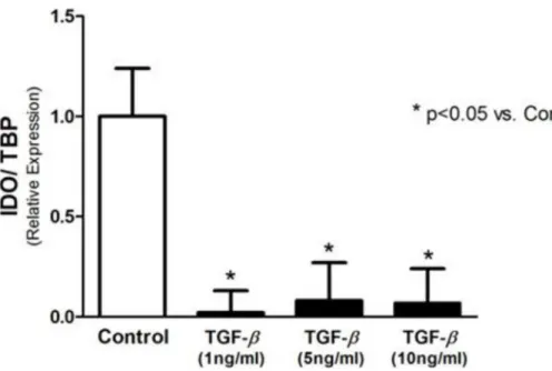
Incubation of T24 cells with TGF-β1 significantly decreased IDO expression (Fig 1), for all tested concentrations. As illustrated inFig 2A, treatment with MT and MT + TGF-β1 significantly reduced expression of IDO in T24 cells (relative expression of 0.16 ± 0.18 vs. 1.00 ± 0.01). This effect was also observed in the results of Western blotting for the IDO protein (Fig 2B). To assess IDO activity, kynurenine was measured in the supernatant of the T24 cells using HPLC. The concentration of kynurenine was markedly diminished after incubation with MT, TGF-β1, and MT + TGF-β1 (0.59 ± 0.12μM, 0.29 ± 0.02μM, and 0.38 ± 0.06μM, respectively, vs.
3.45 ± 0.16μM in the Control; p<0.0001) (Fig 2C).
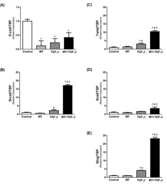
MT potentiates EMT gene expression
A significant decrease was observed in Ecad expression with MT and TGF-β1 treatments (Fig 3A). In contrast, Ncad expression significantly increased with TGF-β1 treatment (Fig 3B), an effect that was potentiated by the combination of TGF-β1 with MT (more than 8 fold vs. TGF-β1 alone). In addition, the expression of EMT-associated transcription factors (Twist, Snail and Slug) was increased by TGF-β1, and treatment with TGF-β1 in combination with MT intensified this effect (Fig 3C, 3D and 3E).
siRNA-mediated knockdown of IDO and expression of EMT markers
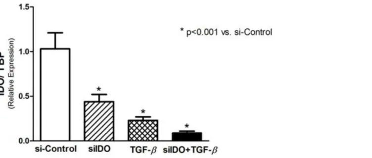
The use of siRNA was effectiveness in silencing IDO gene expression. As demonstrated in
Fig 4, siIDO significantly decreased the expression of IDO in T24 cells. A significant reduction of IDO expression was observed after stimulus with TGF-β1, effect that was intensified when
Fig 1. IDO expression analyzed by real-time PCR.IDO expression is downregulated by TGF-βin T24 cells.
associated with siIDO (Fig 4). Regarding the EMT markers, siIDO significantly reduced Ecad expression, and its association with TGF-β1 potentiates this effect, however, no statistical sig-nificance was found (Fig 5A). Although the siIDO showed effect on Ecad expression, siIDO had no effect in the expression of N-cadherin and EMT transcription factors (Twist, Snail, and Slug) (Fig 5).
Scratch-wound and transwell invasion assays
In the scratch-wound assay, the migration capacity of the T24 cells was evaluated by measuring the area occupied by migrating cells. As demonstrated inFig 6, 3 hours after introduction of Fig 2. MT and TGF-β1 treatment diminishes IDO expression and activity in T24 cells.(A) IDO expression by real-time PCR, (B) Western blotting for IDO, and (C) IDO activity assessed by kynurenine measurement in the supernatant.
the scratch-wound, MT and TGF-β1 induced a faster migration of T24 cells compared to untreated cells. This heightened migration process was intensified when the cells were incu-bated with TGF-β1 plus MT (Fig 6A and 6B).
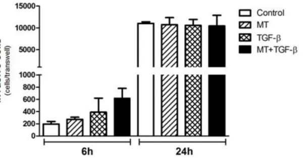
T24 cell invasion was studied using reconstituted Matrigel in transwell chambers. After 6 h of incubation, heightened invasion activity was observed when the cells were incubated with TGF-β1 plus MT, although the result was not statistically significant (Fig 7). After 24 h of incu-bation, no observable difference was detected between the different groups (Fig 7).
Akt activation
To analyze Akt pathway activation, Western blotting experiments were performed. As demon-strated inFig 8, incubation of T24 cells with MT, TGF-β1 or MT + TGF-β1 promoted robust inhibition of Akt activation.
Fig 3. Real-time PCR for EMT markers.Relative mRNA levels of (A) E-cadherin (epithelial marker), (B) N-cadherin (mesenchymal marker), and (C-E) EMT-associated transcriptional factors (Twist, Snail and Slug). Association of MT and TGF-β1 potentiated EMT in T24 cells, decreasing E-cadherin expression and upregulating inductor genes of EMT.*p<0.05 vs. Control;#p<0.05 vs. MT;†p<0.05 vs. TGF-β.
In vivo
analysis
To analyze IDO expressionin vivo, T24 cells were inoculated under the kidney capsule of nude mice. Four weeks after the inoculation, we identified T24 cells under the kidney capsule. Fur-thermore, T24 cells were found infiltrating the renal parenchyma toward the renal medulla (S1 Fig). Immunohistochemical analysis of IDO expression revealed that subcapsular and infiltrat-ing T24 cells were positive for IDO. However, the expression of IDO was specifically localized to the center of the cell infiltrate (Fig 9).
Discussion
In the present study, we investigated the non-immunological role of MT and IDO in cancer progression and dissemination, particularly in EMT. Bladder carcinoma T24 cells constitu-tively express IDO, even in the absence of an immune system. We were able to induce EMT in T24 cells with TGF-β1, and this effect correlates with downregulation of IDO. When we use MT or IDO siRNA, the EMT was potentiated, especially by MT.
EMT is a process in which epithelial cells acquire an invasive phenotype, leading to a sys-temic spread of the malignant cells. During EMT, specific genes are expressed that ultimately function to deregulate intracellular biochemical pathways, which contributes to the establish-ment of a malignant phenotype. In our study, we assessed the process of decreased expression of E-cadherin followed by increased N-cadherin expression, a process called“cadherin switch-ing,”to characterize EMT in T24 cells [29]. EMT plays an important role in bladder cancer, where motility, invasion, and anti-apoptotic mechanisms are required for metastasis [30].
TGF-β1 is a potent inducer of EMT in certain types of tissue, including bladder cancer. T24 cells express TGF-βreceptor 1, and siRNA-based inhibition of TGF-βreceptor 1 suppresses the motility and invasiveness of these cells [27]. In our study, T24 cells acquired mesenchymal characteristics, such as mesenchymal marker expression and motility, after TGF-β1 stimulus. Interestingly, TGF-β1 treatment induced EMT and inhibited IDO expression in T24 cells. In addition, MT, which also diminished IDO expression, intensified the TGF-β-driven EMT. Fig 4. Real-time PCR for IDO to prove the effectiveness of the IDO-siRNA to induce IDO knockdown.A negative control-siRNA was used for si-Control and TGF-βconditions. A significant reduction of IDO expression was observed in siIDO cells, and in TGF-β-stimulated cells. The association of siIDO and TGF-βinduced a more pronounced reduction of the IDO expression.
The siRNA-mediated knockdown of IDO significantly reduced the E-cadherin expression, but the mesenchymal markers were not altered. Because MT significantly increased the expres-sion of mesenchymal markers, these results indicate that IDO pathway possibly participates in the EMT of T24 cells, but MT has an effect potentiating TGF-β1-induced EMT independently of IDO. Metz et al demonstrated that MT has effect on dendritic cells independently of IDO inducing the mTOR pathway (Metz). It is important to note that mTOR is strongly associated with EMT in bladder cancer [31]. It is possible that MT mediates mTOR pathway in T24 cells, but this mechanism was not assessed in our study.
Our study is the first to examine the relationship between TGF-β1 and IDO in T24 cells. However, there are studies demonstrating that TGF-β1 may modulate IDO expression in Fig 5. Effect of siRNA-mediated knockdown of IDO on EMT markers.Relative mRNA levels of (A) E-cadherin (epithelial marker), (B) N-E-cadherin (mesenchymal marker), and (C-E) EMT-associated
transcriptional factors (Twist, Snail and Slug). siRNA-mediated knockdown of IDO was effective in reducing E-cad expression, and its association with TGF-βpotentiated this effect. However, knockdown of IDO alone had no effect in N-cad expression, as well as in the expression of EMT-transcription factors. Only TGF-β
acted inducing the expression of N-cad and EMT-transcription factors, and its association with siIDO did not change this effect.*p<0.05 vs. Control;#p<0.05 vs. MT.
certain types of cells. Yuan et al. demonstrated that while IDO expression can be induced in human fibroblasts after stimulation with INF-gamma, TGF-β1 abrogates this effect without affecting INF-gamma expression [32]. In contrast, TGF-β1 induced IDO expression in den-dritic cells via the PI3K/Akt and noncanonical NFK-βpathways, leading to a tolerogenic phe-notype [26]. The mechanisms linking TGF-βand IDO have not been completely elucidated, Fig 6. The scratch-wound assay was used to assess migration of T24 cells under MT and TGF-β1 stimulus.(A) Images were captured at 40X magnification under an inverted microscope, three hours after scratch formation. (B) Wound closure was significantly faster in MT + TGF-βcompared to Control.
but a more plausible mechanism has been described by Pallotta and Grohmann [33]. Their studies support the theory that TGF-βconfers regulatory effects on IDO synthesis. Further-more, IDO may exert its effects through a mechanism that is independent of its enzyme activity [33].
In our study, we assessed PI3K/Akt pathway activation using Western blotting for Akt/ phospho-Akt. Treatment with MT and TGF-β1 significantly decreased phospho-Akt levels, indicating that TGF-β-driven EMT in T24 cells is associated with the downregulation of this pathway. In many cell types, TGF-β-induced EMT is associated with PI3K/Akt pathway upre-gulation; however, in T24 cells it remains unclear. Al-Azayzih et al. analyzed phospho-Akt in T24 cells after incubation with TGF-β1 (as used in our experiments, 5 ng/ml), and they were unable to determine a difference in phospho-Akt levels [34]. The absence of effect on phospho-Akt levels may be due to the length of TGF-β1 treatment. While they used an incubation period of 24 hours, we used 48 hours. Furthermore, Iliopoulos et al. used Akt-/- cells to show that Akt1 knockdown promoted TGF-β-induced EMT in MCF10A cells. This effect was shown to be dependent on the expression of specific microRNAs [35]. In this context, the role of Akt in EMT may differ depending on the model system.
Fig 7. Analysis of the invasive potential of T24 cells by the Matrigel invasion assay.Although a greater level of invasive capacity was observed in T24 cells after 6 hours of TGF-βand MT + TGF-βtreatment, no statistical significance was observed. After a migration period of 24 hours, no difference was found.
doi:10.1371/journal.pone.0134858.g007
Fig 8. PI3K/Akt pathway analysis.PI3K/Akt pathway activation was assessed by Western blotting for Akt and phospho-Akt in T24 cell lysates. After 48 hours of incubation with MT, TGF-β, and MT + TGF-β, a decreased level of phospho-Akt was observed. Inhibition of the PI3K/Akt pathway is associated with EMT in T24 cells.
In addition to cadherin switching and significantly increased expression of EMT-related transcription factors (Twist, Snail, and Slug), T24 cells treated with TGF-βplus MT demon-strated increased migratory capacity and invasiveness. In addition toin vitroexperiments, T24 cells were inoculated in the kidney subcapsular space of Balb/c nude mice. After 4 weeks, T24 cells were observed in the renal parenchyma toward the medulla. Immunohistochemical analy-sis demonstrated that T24 cells in the center of the infiltrate were positive for IDO, while peripheral cells were IDO-negative. It is possible that the peripheral cells of the infiltrate lost IDO expression to acquire an invasive phenotype. Further studies are necessary to clarify this mechanism.
In conclusion, MT and downregulation of IDO are associated with EMT in T24 human bladder carcinoma cells, and this effect may be triggered by TGF-β1. Although IDO inhibition is attractive as anti-cancer therapy because IDO promotes tolerance to tumors, the nonimmu-nological effects mediated by MT and other IDO modulators deserve consideration in bladder cancer. As perspective, inhibitors of IDO must be combined with other classes of drugs in anti-cancer therapy to minimize possible adverse effects caused by these agents.
Fig 9. Immunohistochemistry for IDO expression in renal tissue 28 days after T24 cell inoculation.IDO-positive cells were found in the infiltrate center (arrow), while IDO-negative cells were predominantly found in the periphery of the infiltrate (arrowhead). IDO immunostaining was not observed in the renal parenchyma (asterisk).
Supporting Information
S1 Fig. Histology of the renal tissue after subcapsular inoculation of T24 cells.To analyze T24 cell invasionin vivo, we inoculated 1X10⁶cells under the kidney capsule. Histology (hema-toxylin and eosin staining) was assessed 28 days after inoculation. (A) T24 cells under the kid-ney capsule (arrowhead), and (B) T24 cells infiltrating the renal parenchyma toward the renal medulla (arrow). Renal parenchyma is indicated with asterisk.
(TIF)
Acknowledgments
We are sincerely grateful to Angela Batista Gomes dos Santos and Samara for their technical assistance.
Author Contributions
Conceived and designed the experiments: RBOB DMS JMF CMF HD. Performed the experi-ments: RBOB CSM DMS LHGM YTM CSS JMF VSN CMF HD. Analyzed the data: RBOB CSM DMS LHGM YTM CSS JMF VSN CMF HD. Contributed reagents/materials/analysis tools: VSN CMF HD. Wrote the paper: RBOB CSM DMS LHGM YTM CSS JMF VSN CMF HD.
References
1. Siegel R, Ma J, Zou Z, Jemal A. Cancer statistics. CA Cancer J Clin. 2014; 64(1): 9–29. doi:10.3322/ caac.21208PMID:24399786
2. Sylvester RJ, van der Meijden AP, Oosterlinck W, Witjes JA, Bouffioux C, Denis L, et al. Predicting recurrence and progression in individual patients with stage Ta T1 bladder cancer using EORTC risk tables: a combined analysis of 2596 patients from seven EORTC trials. Eur Urol. 2006; 49: 466–5. PMID:16442208
3. Kalluri R, Weinberg RA. The basics of epithelial-mesenchymal transition. J Clin Invest. 2009; 119(6): 1420–8. doi:10.1172/JCI39104PMID:19487818
4. Song J. EMT or apoptosis: a decision for TGF-beta. Cell Res. 2007; 17(4): 289–90. PMID:17426696
5. Fan Y, Shen B, Tan M, Mu X, Qin Y, Zhang F, et al. TGF-β-induced upregulation of malat1 promotes bladder cancer metastasis by associating with suz12. Clin Cancer Res. 2014;15; 20(6): 1531–41. doi:
10.1158/1078-0432.CCR-13-1455PMID:24449823
6. Wei H, Kamat AM, Aldousari S, Ye Y, Huang M, Dinney CP, et al. Genetic variations in the transforming growth factor beta pathway as predictors of ladder cancer risk. PLoS One. 2012; 7(12): e51758. doi:
10.1371/journal.pone.0051758PMID:23251617
7. Shariat SF, Kim JH, Andrews B, Kattan MW, Wheeler TM, Kim IY, et al. Preoperative plasma levels of transforming growth factor beta(1) strongly predict clinical outcome in patients with bladder carcinoma. Cancer. 2001; 92(12): 2985–2992. PMID:11753975
8. Munn DH, Zhou M, Attwood JT, Bondarev I, Conway SJ, Marshall B, et al. Prevention of allogeneic fetal rejection by tryptophan catabolism. Science. 1998;21; 281(5380): 1191–3.
9. DellêH, Noronha IL. Induction of indoleamine 2,3-dioxygenase by gene delivery in allogeneic islets pro-longs allograft survival. Am J Transplant. 2010; 10(8): 1918–24. doi:10.1111/j.1600-6143.2010.03190. xPMID:20636452
10. Katz JB, Muller AJ, Metz R, Prendergast GC. Indoleamine 2,3-dioxygenase in T-cell tolerance and tumoral immune escape. Immunol Rev. 2008; 222: 206–221. doi:10.1111/j.1600-065X.2008.00610.x
PMID:18364004
11. Uyttenhove C, Pilotte L, Théate I, Stroobant V, Colau D, Parmentier N, et al. Evidence for a tumoral immune resistance mechanism based on tryptophan degradation by indoleamine 2,3-dioxygenase. Nat Med. 2003; 9(10): 1269–74. PMID:14502282
13. Nakamura T, Shima T, Saeki A, Hidaka T, Nakashima A, Takikawa O, et al. Expression of indoleamine 2, 3-dioxygenase and the recruitment of Foxp3-expressing regulatory T cells in the development and pro- gression of uterine cervical cancer. Cancer Sci. 2007; 98: 874–81. PMID:17433037
14. Ino K, Yoshida N, Kajiyama H, Shibata K, Yamamoto E, Kidokoro K, et al. Indoleamine 2,3-dioxygen-ase is a novel prognostic indicator for endometrial cancer. Br J Cancer. 2006; 95: 1555–61. PMID:
17117179
15. Mansfield AS, Heikkila PS, Vaara AT, von Smitten KA, Vakkila JM, Leidenius MH. Simultaneous Foxp3 and IDO expression is associated with sentinel lymph node metastases in breast cancer. BMC Cancer. 2009; 9: 231–40. doi:10.1186/1471-2407-9-231PMID:19604349
16. Brody JR, Costantino CL, Berger AC, Sato T, Lisanti MP, Yeo CJ, et al. Expression of indoleamine 2,3-dioxygenase in metastatic malignant melanoma recruits regulatory T cells to avoid immune detection and affects survival. Cell Cycle. 2009; 8: 1930–4. PMID:19448397
17. Inaba T, Ino K, Kajiyama H, Yamamoto E, Shibata K, Nawa A, et al. Role of the immunosuppressive enzyme indoleamine 2,3-dioxygenase in the progression of ovarian carcinoma. Gynecol Oncol. 2009; 115: 185–92. doi:10.1016/j.ygyno.2009.07.015PMID:19665763
18. Astigiano S, Morandi B, Costa R, Mastracci L, D'Agostino A, Ratto GB, et al. Eosinophil granulocytes account for indoleamine 2,3-dioxygen- ase-mediated immune escape in human non-small cell lung cancer. Neoplasia. 2005; 7: 390–6. PMID:15967116
19. Ishio T, Goto S, Tahara K, Tone S, Kawano K, Kitano S. Immunoacti-vative role of indoleamine 2,3-dioxygenase in human hepatocellular carcinoma. J Gastroenterol Hepatol. 2004; 19: 319–26. PMID:
14748880
20. Riesenberg R, Weiler C, Spring O, Eder M, Buchner A, Popp T, et al. Expression of indoleamine 2,3-dioxygenase in tumor endothelial cells correlateswith long-term survival of patients with renal cell carci-noma. Clin Cancer Res. 2007; 13: 6993–7002. PMID:18056175
21. Soliman HH, Jackson E, Neuger T, Dees EC, Harvey RD, Han H, et al. A first in man phase I trial of the oral immunomodulator, indoximod, combined with docetaxel in patients with metastatic solid tumors. Oncotarget. 2014;30; 5(18): 8136–46. PMID:25327557
22. Agaugué S, Perrin-Cocon L, Coutant F, André P, Lotteau V. 1-Methyl-tryptophan can interfere with TLR signaling in dendritic cells independently of IDO activity. J Immunol. 2006;15; 177(4): 2061–71. PMID:
16887964
23. Metz R, Rust S, Duhadaway JB, Mautino MR, Munn DH, Vahanian NN, et al. IDO inhibits a tryptophan sufficiency signal that stimulates mTOR: A novel IDO effector pathway targeted by D-1-methyl-trypto-phan. Oncoimmunology. 2012;1; 1(9): 1460–1468. PMID:23264892
24. Byrne GI, Lehmann LK, Landry GJ. Induction of tryptophan catabolism is the mechanism for gamma-interferon-mediated inhibition of intracellular Chlamydia psittaci replication in T24 cells. Infect Immun. 1986; 53(2): 347–51. PMID:3089936
25. Levina V, Su Y, Gorelik E. Immunological and nonimmunological effects of indoleamine 2,3-dioxygen-ase on breast tumor growth and spontaneous metastasis formation. Clin Dev Immunol. 2012;173029. doi:10.1155/2012/173029PMID:22654951
26. Belladonna ML, Orabona C, Grohmann U, Puccetti P. TGF-βand kynurenines as the key to infectious tolerance. Trends Mol Med. 2009; 15(2): 41–49. doi:10.1016/j.molmed.2008.11.006PMID:19162548
27. Li Y, Yang K, Mao Q, Zheng X, Kong D, Xie L. Inhibition of TGF-beta receptor I by siRNA suppresses the motility and invasiveness of T24 bladder cancer cells via modulation of integrins and matrix metallo-proteinase. Int Urol Nephrol. 2010; 42(2): 315–23. doi:10.1007/s11255-009-9620-3PMID:19669587
28. Geng J, Fan J, Ouyang Q, Zhang X, Zhang X, Yu J, et al. Loss of PPM1A expression enhances inva-sion and the epithelial-to-mesenchymal transition in bladder cancer by activating the TGF-β/Smad sig-naling pathway. Oncotarget. 2014;30; 5(14): 5700–11.
29. Bryan RT, Tselepis C. Cadherin switching and bladder cancer. J Urol. 2010; 184: 423–31. doi:10. 1016/j.juro.2010.04.016PMID:20620393
30. van der Horst G, Bos L, van der Pluijm G. Epithelial plasticity, cancer stem cells, and the tumor-support-ive stroma in bladder carcinoma. Mol Cancer Res. 2012; 10(8): 995–1009. doi:10.1158/1541-7786. MCR-12-0274PMID:22714124
31. Shorning BY, Griffiths D, Clarke AR. Lkb1 and Pten synergise to suppress mTOR-mediated tumorigen-esis and epithelial-mesenchymal transition in the mouse bladder. PLoS One. 2011;19; 6(1): e16209. doi:10.1371/journal.pone.0016209PMID:21283818
32. Yuan W, Collado-Hidalgo A, Yufit T, Taylor M, Varga J. Modulation of cellular tryptophan metabolism in human fibroblasts by transforming growth factor-beta: selective inhibition of indoleamine 2,3-dioxygen-ase and tryptophanyl-tRNA synthet2,3-dioxygen-ase gene expression. J Cell Physiol. 1998; 177(1): 174–86. PMID:
33. Pallotta MT, Orabona C, Volpi C, Vacca C, Belladonna ML, Bianchi R, et al. Indoleamine 2,3-dioxygen-ase is a signaling protein in long-term tolerance by dendritic cells. Nat Immunol. 2011;31; 12(9): 870–8. doi:10.1038/ni.2077PMID:21804557
34. Al-Azayzih A, Gao F, Goc A, Somanath PR. TGFβ1 induces apoptosis in invasive prostate cancer and bladder cancer cells via Akt-independent, p38 MAPK and JNK/SAPK-mediated activation of caspases. Biochem Biophys Res Commun. 2012;12; 427(1): 165–70. doi:10.1016/j.bbrc.2012.09.035PMID:
22989755