Splicing-Related Features of Introns Serve to Propel
Evolution
Yuping Luo1,2*, Chun Li3, Xi Gong2, Yanlu Wang2, Kunshan Zhang1, Yaru Cui1, Yi Eve Sun1*, Siguang Li1,2*
1Stem Cell Translational Research Center, Tongji Hospital, Tongji University School of Medicine, Shanghai, China,2College of Life Sciences, Nanchang University, Nanchang, China,3Shanghai Stem Cell Institute, Institutes of Medical Sciences, Shanghai Jiao Tong University School of Medicine, Shanghai, China
Abstract
The role of spliceosomal intronic structures played in evolution has only begun to be elucidated. Comparative genomic analyses of fungal snoRNA sequences, which are often contained within introns and/or exons, revealed that about one-third of snoRNA-associated introns in three major snoRNA gene clusters manifested polymorphisms, likely resulting from intron loss and gain events during fungi evolution. Genomic deletions can clearly be observed as one mechanism underlying intron and exon loss, as well as generation of complex introns where several introns lie in juxtaposition without intercalating exons. Strikingly, by tracking conserved snoRNAs in introns, we found that some introns had moved from one position to another by excision from donor sites and insertion into target sties elsewhere in the genome without needing transposon structures. This study revealed the origin of many newly gained introns. Moreover, our analyses suggested that intron-containing sequences were more prone to sustainable structural changes than DNA sequences without introns due to intron’s ability to jump within the genome via unknown mechanisms. We propose that splicing-related structural features of introns serve as an additional motor to propel evolution.
Citation:Luo Y, Li C, Gong X, Wang Y, Zhang K, et al. (2013) Splicing-Related Features of Introns Serve to Propel Evolution. PLoS ONE 8(3): e58547. doi:10.1371/ journal.pone.0058547
Editor:Barbara Bardoni, CNRS UMR7275, France
ReceivedNovember 24, 2012;AcceptedFebruary 5, 2013;PublishedMarch 13, 2013
Copyright:ß2013 Luo et al. This is an open-access article distributed under the terms of the Creative Commons Attribution License, which permits unrestricted use, distribution, and reproduction in any medium, provided the original author and source are credited.
Funding:This work was supported by the National Key Basic Research Program of China Grants 2011CBA01106, 2011CB965102 and 2010CB945600; National Natural Foundation of China Grants 31171317, 30971473, 31271375 and 31271450. The funders had no role in study design, data collection and analysis, decision to publish, or preparation of the manuscript.
Competing Interests:The authors have declared that no competing interests exist. * E-mail: [email protected] (YL); [email protected] (YES); [email protected] (SL)
Introduction
Spliceosomal introns, one of the hallmarks of eukaryotic genomes, exist in eukaryotic protein-coding genes [1–4] and non-protein-coding genes [5–6]. After transcription, they are inevitably removed from corresponding RNA transcripts. Al-though more than thirty years have passed since the discovery of spliceosomal introns, fundamental questions about spliceosomal intron evolution, including evolutionary origins of introns, their ages, natural selection pressures imposed on them, as well as how introns are lost and gained during evolution, have only begun to be elucidated. Previous studies of intron loss and gain mainly focused on protein-coding genes [7–10], which are subjected to huge natural selection pressure because small changes in nucleotide sequences in exons (lost or gained) tend to drastically alter protein structures and functions. As a result, many alterations in protein encoding genes could not survive or leave traces during evolution, rendering previous studies on protein-encoding genes incapable of revealing the full spectrum of changes in intron dynamics during evolution.
Several lines of evidence suggest that half or more of mammalian transcriptomes consist of non-coding RNAs (ncRNAs), many of which are subjected to splicing [11–13]. Non-coding RNA genes are under less stringent selection pressure than protein-coding genes because their functional units are in general short and their other parts are easier to tolerate sequence alterations [14]. On the other hand, the presence of conserved
non-coding RNAs in introns of non-coding genes has facilitated tracking of intron loss and gain events. snoRNA, an abundant class of non-protein-coding RNAs, is widespread in eukaryotes from yeast to human. Because snoRNAs in gene clusters detected so far in fungi are conserved and encoded by independent genes or nested within introns of non-protein-coding host genes, it is appropriate to use snoRNA gene clusters as evolutionary conserved marks to track exon/intron modifications during evolution. Recently, by comparing conserved snoRNAs in introns, an alternative mechanism for intron loss through widespread degeneration of splicing signals (de-intronization) was uncovered inSaccharomyces[15], demonstrating the power of studying intron loss and gain in non-coding RNA genes.
intronic structures are more prone to translocation via unknown mechanisms. Based on these findings we propose a novel evolution mechanism, i.e., intronic structures serve as an additional motor to propel evolution.
Results
Intron distributions within three snoRNA gene clusters in fungi
In order to acquire insight into the evolutionary dynamics of introns in fungal non-coding RNA, we used multiple complete genome sequences and high-quality draft sequences in fungi as references for our analyses and systematically analyzed three snoRNA gene clusters, named snoRNA gene cluster I, II and III, which encode snR78-snR77-snR76-snR75-snR74-snR73-snR72, snR57-snR55-snR61 and snR41-snR70-snR51 snoRNAs, respec-tively. We found that nearly all analyzed fungi have these snoRNA cluster sequences. The structure features of these snoRNAs are such that they possess box C (5’-TGATGA-3’) and D (5’-CTGA-3’ or 5’-ATGA-3’) near their 5’ and 3’ ends together with one or two functional elements involved in guiding 2’-O-ribose methylation of rRNA [16]. We searched for splicing signals in sequences adjacent to snoRNAs in these clusters, and predicted 323 intronic snoRNAs from 460 analyzed snoRNAs (Figure 1), among which we have verified 83 of these introns by comparison of expressed sequence tags (ESTs) from these species to the corresponding genomic sequences and/or by reverse transcription polymerase chain reaction (RT-PCR) and sequencing (Table S1,S1, S2, S3). Although there is not enough EST data to verify the rest intronic snoRNAs, they have canonical 5’ splice sites, canonical 3’ splice sites and branch point sequences of intron and therefore are indeed intronic. The spliced RNAs from these fungal polycistronic snoRNA host genes, similar to spliced mammalian U22 snoRNA host gene RNA [17], are poorly conserved and lack long open reading frames, therefore having little potential for protein coding. In these three clusters, intron distribution among different fungal groups varies considerably. In Euascomycetes of Ascomycota (the species labeled in purple in Figure 1),Basidiomycota(the species labeled in green in Figure 1) and Zygomycota (Rhizopus oryzae), snoRNAs in these three clusters are all intronic; in some species of Hemiascomycetes except for Saccharomyces cerevisiae and its close relatives (the species labeled in dark blue in Figure 1) and Archiascomycetes(the species labeled in orange in Figure 1) most of them are within individual introns; whereas inS. cerevisiaeand its close relatives (the species labeled in baby blue in Figure 1), all of them arise from unspliced primary transcripts. This is presumably correlated with intron densities within the genome among different fungal species, since it is reported that at least for protein-encoding genes, the intron density is 2–3 introns/gene forEuascomycetesfungi Neurospora crassaandAspergillus nidulans[18–20], 0.9 intron/gene for Archiascomycetes fungi Schizosaccharomyces pombe [21], and 0.05 intron/gene [22] forHemiascomycetousfungiS. cerevisiae.
Intron presence/absence polymorphisms resulting from intron loss and gain events
Previous analyses of eukaryotic genomes revealed that the common ancestor of fungi and animals was intron-rich [23,24]. Thus, extensive ancestral intron loss was suggested to account for intron-poor eukaryotes. The current non-intronic snoRNA gene cluster I, II and III inS. cerevisiaeand its close relatives should result from intron loss events through splice-site degeneration mecha-nisms (de-intronization of intronic sequences) during fungi evolution (Figure 1), as proposed recently for non-coding RNA introns [15]. After de-intronization of snoRNA-associated intronic
sequences, the snoRNAs in these three intron-less clusters in S. cerevisiaeand its close relatives remain stable in sequence structure, including snoRNA number and order.
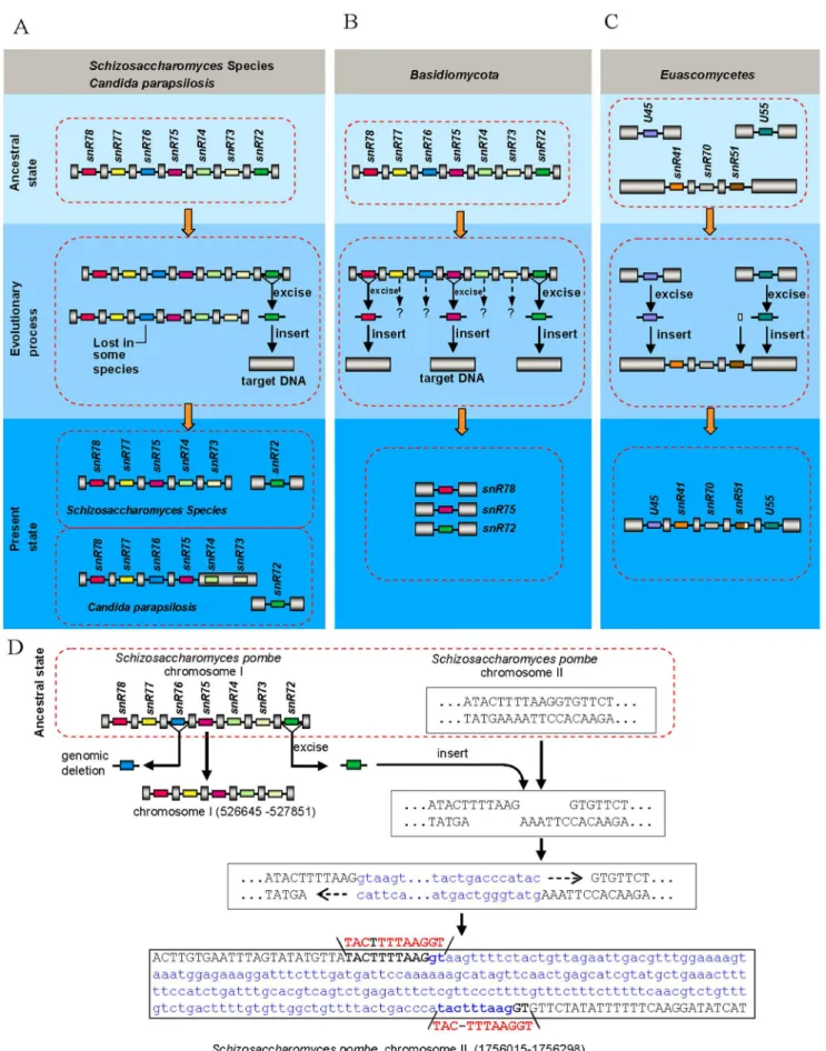
However, one third of snoRNA-associate introns in intronic snoRNA gene clusters show polymorphisms (Figure 1) among analyzed fungi. SnR76-associated introns in cluster I of all the Archiascomycetesspecies andR. oryzae(marked with open triangle in Figure 1), snR72-associated introns in cluster I of most fungal species (marked with filled triangle in Figure 1), as well as snR57-associated introns in cluster II of allEuascomycetesspecies andPichia guilliermondii(marked with filled star in Figure 1) were lost together with their intronic snoRNAs from snoRNA gene clusters, respectively. Sequence alignment showed that elimination of snR76-associated and snR57-associated introns possesses charac-teristics of the ‘‘genomic deletion model’’ postulated a while ago for intron loss in protein-coding genes [10]. Those characteristics include retention of residual intron sequences and a lack of biased loss of 3’ introns. These intron loss cases clearly result from genomic deletion events followed by subsequent divergence of remainder intronic sequences (Figures 1, 2). In addition, besides intron loss, we also observed intron gain events. U45-associated and U55-associated introns were inserted in snoRNA gene cluster III at both ends in nearly all the Euascomycetes species (species highlighted in purple in Figure 1). These intron presence/absence polymorphisms in intronic snoRNA gene clusters likely result from intron loss and gain events during fungi evolution, based on the presence or absence of introns in homologous positions of orthologous genes of widely divergent fungi that we observed (Figure 1).
Evidence for the origin of recently gained introns By tracking snoRNAs in cluster I, II and III in different fungal species, we found some intron gain events that are linked to intron loss events. InSchizosaccharomycesspecies and someCandidaspecies, the snR72-associated introns were lost in the cluster I sites, but reappeared in other places within the genome (Figure 3A). In addition, snR78, snR75 and snR72-associated introns in some species ofBasidiomycotaare no longer located in the snR72–78 poly-cistronic cluster, instead, they scatter over different sites in the genome (Figure 3B). Moreover, the U45 and U55-associated intron disappear from original sites and are inserted into snR41-snR70-snR51 poly-cistronic gene cluster in most species of Euascomycetes(Figure 3C), which provided direct evidence for the origin of some newly gained introns.
and gain of these snoRNA-associated introns. Given the loss of some snoRNA-associated introns from their donor sites and insertion into target sites with no homology between donor and recipient genes (Figure 3) and given that there exists short direct repeats (Figure 3D, Table S2) within some gained snoRNA-associated introns, we proposed a new model, named ‘‘excision-and-insertion’’ model, for intron loss and gain, i.e., they are excised from the donor sites (Figure 3) as complete or nearly complete intron units at the DNA level and got inserted into staggered double-strand breaks sites of the genome. Our results suggested that some introns might move from site to site within the genome without harboring transposon structures.
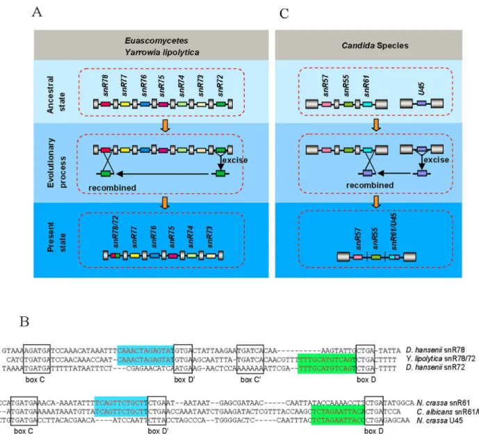
Obviously, not all excised snoRNA-associated introns could be inserted directly into the genome. What’s interesting is that some of the excised introns can recombine with other sequences to form reorganized introns. snR78/72-associated intron in all species of Euascomycetes fungi and one species of Hemiascomycetous fungi (Y. lipolytica) came from recombination between snR72-associated intron and snR78-associated intron (Figure 1, Figure 4A, 4B), which is a good indication for ‘‘reorganized intron evolution’’ event. The snR61/U45-associated intron structure in Candida species indicates an additional example of such evolution mechanism (Figure 1, Figure 4B, 4C). Finally, the snR73, snR74, snR76 and snR77-associated introns in some species of Basidiomycotafungi (Figure 3B) and U45 and U55-associaed introns in all fungi except forEuascomycetes(Figure 1) disappeared from the genome, suggesting another alternative fate of excised snoRNA-associated introns, which is permanent elimination from the genome. Together, these observations indicate that excised snoRNA-associated introns have three different fates: direct reinsertion into elsewhere within the genome (Figure 3), recom-bination with other sequences (Figure 4) or lost from genome (Figure 3B).
Besides short direct repeats mentioned above, we also observed short inverted repeats, ranging in size from 8 to 12 base pairs, with one repeat positioned within the end of an adjacent exon sequence and the other repeat near the opposite end of the inserted intron (Figure S4A). In order to know whether this kind of short inverted repeats exist in gained non snoRNA-associated intron, we analyzed a gained intron (intron B) which is inserted into intronic snR51 of cluster III at the position between conserved antisense functional sequence involved in guiding 2’-O-ribose methylation of rRNA and conserved box D (Figure 5A). In different species of Euascomycetesfungi, the inserted intron sequences are significantly divergent (Figure S5), and the surrounding exons demonstrate similar divergence (Figure S6), suggesting that the intron-gain event appears to have occurred in the ancestor of theEuascomycetes and may be a single intron-gain event followed by subsequent divergent changes of intronic sequences. After careful analysis of the inserted intron B and its flanking sequences, we found that intron B in the ancestor of theEuascomycetesand its flanking exons (conserved antisense functional sequence of snR51) also harbored short inverted repeats (Figure S4B). Moreover, we found this kind of inverted repeats also existed in recently gained introns resulting from repair of double-strand breaks (DSBs) accompanied by small segmental insertions inD. pulexprotein coding genes [29] (Figure
S4C), suggesting these short inverted repeats are likely related to the novel intron loss-gain event mentioned above.
Exon loss and splicing of complex introns
Previous work confirmed that fungal introns in protein encoding gene are typically short and exons are long relative to their mammalian counterparts [30], but recent work showed that some internal exons within polycistronic snoRNA gene clusters are small, even absent in Candida clade and more distantly related Hemiascomycete Y. lipolytica (15). The genomic deletion of internal exons in polycistronic snoRNA gene clusters lead to complex intron architectures, where several introns lie in juxtaposition without intercalating exons. Interestingly, we found that such complex introns are not specific toCandidaclades andY. lipolyticaof Hemiascomycetes, but also exist in Euascomycetes and Archiascomycetes (Figure 1), suggesting exon loss could be a common phenomenon in fungi evolution. Taken together, we found at least 61 exon loss events in these clusters in Hemiascomycetes, Euascomycetes and Archiascomycetes fungi (Table S3). Because much of the available genomic information is still incomplete, our results may only represent a subset of such complex introns in fungi.
The traditional splicing of spliceosomal introns is mediated by the spliceosome, which interacts with specific parts of the intron and the flanking exons to ensure accurate and efficient splicing [31]. Because these complex introns analyzed above are novel constitutions of eukaryotic gene, their splicing characteristics are still unknown. To track their splicing pattern, we systematically analyzed spliced products of polycistronic snoRNA precursors from cluster III in Euascomycetes fungal species. Polycistronic snoRNA gene cluster III in all Euascomycete species misses two interior exons, forming an ‘‘intron-intron-intron’’ structure (Figure 1, 5A). In addition, two snoRNA-associated introns, U45 and U55-associated introns (intron A and intron C) and one non-snoRNA-associated intronic sequence (intron B) are inserted in snoRNA gene cluster III (Figure 5A). We have compared expressed sequence tags (ESTs) from EST database ofEuascomycetes fungal species to their corresponding genome sequences and found 42 spliced products from cluster III transcripts. Among the 42 spliced products, 18 removed intron I, but retained intron II and III with their intronic snoRNA; 14 removed intron I and II, but retained intron III with their snoRNA; 10 removed all the three introns (Figure S7, S8, S9, S10, S11, S12, S13, S14).
We did not detect any spliced products that result from removal of intron II and/or intron III but retaining intron I or removal of intron III but retaining intron I and/or intron II. This indicates that intron I, II and III may be removed by stepwise splicing from the 5’ end to the 3’ end of the transcripts (Figure 5B), i.e., intron I was preferentially removed at first splicing step, and splicing of intron II can occur only after intron I has been removed, and followed by the removal of intron III. Alternatively, it is also possible that only intron I, which is next to an exon, contains a functional splicing donor, and this donor may pick any of the three functional splicing acceptors in the 3’end of each intron to conduct splicing, resulting in the products we detected.
To further verify the stepwise splicing pattern or alternative acceptor usage for such unusual introns, we performed RT-PCR Figure 1. The distribution of snoRNA-associated introns in fungal Polycistronic snoRNA gene cluster I, II and III.Cladogram showing Basidiomycota(green),Zygomycota(red) andAscomycota, subdivided intoHemiascomycetes(blue),Euascomycetes(purple), andArchiascomycetes (orange). snoRNA gene clusters I, II and III exist widespread in fungi genomes. The location where the snR76-, snR72- and snR57-associated intron were lost are marked with open triangle, filled triangle and filled star, respectively. snoRNAs are represented schematically by different colored boxes, introns as lines, and exons as gray pillars, with internal exons labeled by size. In detail: cluster I, snR78-snR77-snR76-snR75-snR74-snR73-snR72; cluster II, snR57-snR55-snR61; cluster III, snR41-snR70-snR51. All are drawn not to scale.
amplification with YLC2F1/YLC2R1 primer pair for Y.lipolytica cluster II, DhC2F1/DhC2R1 and DhC3F1/DhC3R1 primer pairs forDebaryomyces hanseniicluster II and III, respectively. We then cloned the RT-PCR products, sequenced them, and analyzed spliced products. Our result demonstrated that the unusual introns of cluster II and III inD. hanseniiandY. lipolyticacould potentially be stepwise spliced from the 5’ end to the 3’ end (Figure S1, S2, S3) or spliced via alternative splicing acceptor usage mentioned above. As expected, we couldn’t get any splice products containing intron I without intron II and/or intron III. This splicing pattern was further confirmed by additional RT-PCR analyses with specialized primer pairs DhC2F2/DhC2R1 and YLC2F2/YLC2R1, again, the first removal of intron II and/or intron III could never be detected. Such splicing patterns suggest that, consistent with intron splicing in protein encoding genes [31], 5’ exon sequences are required to ensure functionality of splicing donors to perform accurate and efficient splicing for non-coding RNA gene.
Discussion
We have performed systematic analysis of intron and exon architecture of three noncoding snoRNA gene clusters from available fungal genome databases and found that intron distributions in non-protein-coding genes among different fungal groups vary considerably (Figure 1). In three snoRNA gene clusters, all of the snoRNAs inS. cerevisiae and its close relatives reside in unspliced primary transcripts (Figure 1), potentially resulting from substantial intron loss via degeneration of their splicing signals [15]. After de-intronization of intronic sequences, the snoRNAs in the three intron-less clusters inS. cerevisiaeand its close relatives remain stable in sequence structure. However, one third of intronic snoRNAs in other fungi show presence/absence polymorphisms due to intron loss and gain events during evolution (Figure 1). In addition, the intronic snoRNA gene clusters had also experienced exon loss and we found 61 exon loss cases in Hemiascomycetes, Euascomycetesand Archiascomycetesfungi (Table S3). The presence of complex introns where multiple introns reside in juxtaposition is a clear indication for exon loss. Taken together, it appears that intron-containing sequences are more prone to structural changes than sequences without introns. Therefore splicing-related features of introns may serve as an additional motor to propel evolution, though how RNA splicing machinery influences excision of DNA elements remained to be determined. Analysis of the ultimate fate of the excised snoRNA-associated introns suggests that intron loss events could be independent in different lineages. For example, in Archiascomycetes fungi, the snR72-associated introns were inserted as a whole into target sites of genome, whereas in Euascomycetes fungi, the snR72-associated introns recombined with other sequences to form reorganized introns (Figure 1, Figures 3, 4), suggesting the excision of snR72-associated introns happened after the divergence ofArchiascomycetes fungi and Euascomycetes fungi and therefore were obviously independent events. This suggests that some intron sequences might be hot spots for excision.
How introns spread within the genome remains an unanswered question in evolution biology [5]. Identifying the origin of recently
gained introns is likely a key to understanding where new introns come from. For recently gained introns inCaenorhabditis elegansand Caenorhabditis briggsae, reverse splicing of preexisting introns [28] is the main mechanism for intron gain during recent nematode evolution [32]. However, studies of protein-coding genes in Daphnia population revealed that more than half of the recent gained introns were associated with short sequence repeats, which were formed via repair of staggered double-strand breaks. However, the sources of these gained introns, except for one, still remain unknown [29]. By tracking conserved snoRNAs in introns, we found that the gained introns by repairing double-strand breaks are derived from excised introns from other sites. The failure of previous studies to find the sources of recently gained introns in Daphniapopulation can be explained by the fact that there are no conserved sequences within these introns, making it difficult to track their origins. Our study demonstrated that intron loss and gain by a mode of excision from donor sites and reinsertion into the target sites (Figure 1, 3) may represent a novel mechanism underlying exon-intron structure evolution.
Besides short direct repeats mentioned above, we also found short inverted repeats associated with some recently gained snoRNA-associated introns (Figure S4A). These short inverted repeats differ from the inverted repeats of transposons. Transpo-sons consist of inverted repeats at both ends, which are recognized by transposase, followed by excision and re-insertion into a new location [32,33]. However, in some recently gained introns in this work, the short inverted repeats exist in target site and inserted exogenous DNA fragment, respectively. The function of the short inverted repeats remains to be revealed. We hypothesize that the short inverted repeats may act as sequence-specific guides for recognition between excised intron sequence and target sequence via base pairing. If the existence of short inverted repeats acts as guide for interactions between excised intron and target sequence, we would predict that intron removal from one location and insertion into another is site-specific, rather than random.
DNA sequences should have been subjected to enormous alterations during evolution. However, due to the presence of natural selection, many genomic alterations are erased without leaving a trace. snoRNA genes are conserved and their changes in the genome are more traceable, which are extremely suited for studying mechanisms underlying intron-exon loss and gain during evolution. Through this study, we revealed that introns could be movable elements in the genome to propel evolution, and hence intron-containing sequences are more prone to sustainable variations leading to evolution.
Materials
Fungal species for intron loss and gain analysis. As referenced by molecular systematic studies [24,34,35], we chose different fungal species in our intron loss and gain analysis: Saccharomyces cerevisiae, Saccharomyces paradoxus, Saccharomyces bayanus, Candida glabrata, Saccharomyces castellii, Saccharomyces kluyveri, Kluyver-omyces waltii, Ashbya gossypii, Pichia guilliermondii, DebaryKluyver-omyces hansenii, Clavispora lusitaniae, Lodderomyces elongisporus, Candida parapsilosis, Candida tropicalis, Candida dubliniensis, Candida albicans andYarrowia Figure 2. Examples of intron loss by genomic deletion.(A) Genomic deletion of most of an intron sequence which harbor snR76 from snoRNA cluster I inSchizosaccharomyces pombe(Sp) andRhizopus oryzae(Ro), respectively. (B) Genomic deletion of most of an intron sequence which harbor snR57 from snoRNA cluster II inNeosartorya fischeri(Nf),Aspergillus fumigatus(Afu),Aspergillus flavus(Afl),Pichia guilliermondii(Pg), respectively. (C) Genomic deletion of most of an intron sequence which harbor snR57 from snoRNA cluster II inPichia guilliermondii(Pg). snoRNAs are represented schematically by different colored boxes, introns as lines or in lowercase letters, and exons as gray pillar or in capital letters. Conserved sequences are shaded. All are drawn not to scale.
lipolytica belong to the basal species in Hemiascomycetes. Podospora anserine, Chaetomium globosum, Neurospora crassa, Magnaporthe grisea, Gibberella zeae, Gibberella moniliformis, Phaeosphaeria nodorum, Aspergillus flavus, Coccidioides immitis, Sclerotinia sclerotiorum, Aspergillus fumigatus, Neosartorya fischeri, Aspergillus terreus and Aspergillus nidulans are the basal species in Euascomycetes. Schizosaccharomyces japonicus, Schizo-saccharomyces pombe, SchizoSchizo-saccharomyces octosporus and Schizosacchar-omyces cryophilus belong to Archiascomycetes and Coprinopsis cinerea, Phanerochaete chrysosporium and Ustilago maydisare basal species of Basidiomycota.Rhizopus oryzaebelongs toZygomycota.
Survey of snoRNA gene cluster I, II and III sequences in fungi. To obtain sequences of snoRNA gene cluster I, II and III from above fungal species, we downloaded budding yeast box C/ D snoRNA sequences from the snoRNA database (http://people. biochem.umass.edu/fournierlab/snornadb) and used the sequenc-es ofS. cerevisiaesnoRNA cluster I, II and III sequences as query to search for their orthologs in other fungi from multiple complete genome sequences as well as high-quality draft sequences by the BLAST tool on the NCBI website (http://www.ncbi.nlm.nih.gov/ sutils/genom_tree.cgi).
gain by the mechanism of ‘‘excised and inserted’’. DNA insert site is show in capital letters, inserted sequence is show in lowercase letters and short direct repeats are marked in red.snoRNAs are represented schematically by different colored boxes, introns as lines, and exons as gray pillars. All are drawn not to scale.
doi:10.1371/journal.pone.0058547.g003
Figure 4. Recombination of snoRNA-associated intron.(A) Recombination between snR72-associated intron and snR78-associated intron in EuascomycetesandYarrowia lipolytica. snoRNAs are represented schematically by different colored boxes, introns as lines, and exons as gray pillars. All are drawn not to scale. (B) The sequences of snR78, snR72, snR78/72, snR61, U45 and snR61/U45. Conserved motifs C, D9, C9and D are indicated by boxed sequences. The functional sequence complementarity to the rRNA is in red. (C) Recombination between U45-associated intron and snR61-associated intron inCandidaSpecies. snoRNAs are represented schematically by different colored boxes, introns as lines, and exons as gray pillars. All are drawn not to scale.
Prediction of snoRNA-associated introns. The intron sequences inY. lipolytica, D. hansenii, S. cerevisiae, C. glabrataandK. lactis were downloaded from the Ge´nosplicing website (http:// genome.jouy.inra.fr/genosplicing/index), and the splicing pattern of these organisms were analyzed. In addition, introns and splicing elements of five diverse Fungi, two filamentous members of the Ascomycota,A. nidulansandN. crassa, a member of theBasidiomycota, Cryptococcus neoformans, and two well-studied members of the Ascomycota group of fungal organisms, S. cerevisiae and S. pombe were compared and analyzed [30]. From the analyses we accurately characterized conserved fungal intronic elements and predicted snoRNA-associated introns in snoRNA gene cluster I, II and III.
Splicing analysis of snoRNA-associated introns. The availability of genomic sequences and expressed sequence tag (EST) data of some fungi permitted the identification of intron for
these organisms by aligning ESTs to genomic sequences. snoRNA-associated introns in cluster I, II and III from some fungi were confirmed by the comparison of EST data from these species to the corresponding genomic sequences. In addition, the splicing of snoRNA-associated introns in cluster I and II ofC. albicans [15] and in cluster I, II and III ofS. pombewere confirmed previously. Fungal speciesD. hansenii,Y. lipolytica, N. crassa, C. glabrataandK. lactis were used for the experimental confirmation of intron structure and splicing pattern. These strains were grown in rich YPD medium (1% yeast extract, 1% peptone, 2% glucose) at 30uC. Escherichia coli strain TG1 [F’/supE, hsdg5, thig
(lac-proAB)] grown in 2YT(1.6% Bacto tryptone, 1% Bacto yeast extract, 0.5% NaCl) liquid or solid medium and were used for cloning procedures. Total RNA was extracted from cells grown on YPD medium with the use of guanidine thiocyanate/phenol-chloroform extraction. Reverse transcription was carried out in Figure 5. Intron gain and exon loss and splicing inEuascomycetessnR41-snR70-snR51 polycistronic cluster III.(A) Intron gain and exon loss inEuascomycetessnR41-snR70-snR51 polycistronic cluster III. snoRNAs are represented schematically by different colored boxes, introns as lines, and exons as gray pillars. (B) Stepwise splicing of snR41-snR70-snR51 polycistronic cluster inEuascomycetes. snoRNAs are represented schematically by different colored boxes, introns as lines, and exons as gray pillars. The 59splice site, branch site and 39splice site sequences are shown in blue letters for intron III and green letters for intron B.
doi:10.1371/journal.pone.0058547.g005
20mL reaction mixture containing 15mg of total cellular RNA treated with DNase, reverse primer and 500 mmol/L dNTPs. After being denatured at 65uC for 5 min and then cooled to 42uC, 200 units of M-MLV reverse transcriptase (Promega) were added and the extension was carried out at 42uC for 45 min. After reverse transcription, PCR was carried out. The primer pairs DhC2F1/DhC2R1 and DhC2F2/DhC2R1 were used for PCR amplification ofD. hanseniicluster II. The primer pairs YlC2F1/ YlC2R1 and YlC2F2/YlC2R1 were used for PCR amplification ofY. lipolyticacluster II. The primer pair DhC3F1/DhC3R1 was used for PCR amplification ofD. hanseniicluster III. The primer pair NcC2F1/NcC2R1 was used for PCR amplification of N. crassacluster II. The primer pair CgC2F1/CgC2R1 was used for PCR amplification of C. glabrata cluster II. The primer pair KlC2F1/KlC2R1 was used for PCR amplification of K. lactis cluster II.
DhC2F1: 5’-ACCTAAACTCTACTATAATG-3’ DhC2F2: 5’-GAAGTATTGGTATGTTTCAAC-3’ DhC2R1: 5’- GAGTTCTGAAGTATATTAAG-3’ YlC2F1: 5’- CTCACATACGACAAGACAATG-3’ YlC2F2: 5’-CACGACACTGAATGGTGAGTAC-3’ YlC2R1: 5’- TACGTTAGCTATAAATCAGGG-3’ DhC3F1: 5’- ATATATGGAATCACTGAAAG-3’ DhC3R1: 5’- CATGTATTCATAAGAATTGG-3’ NcC2F: 5’- TGGTTCGCACGGATAGA-3’ NcC2R: 5’- CCCACTAGACGCAAGAT-3’ CgC2F: 5’- AATTTTTTCAATGCTAATGGT-3’ CgC2R: 5’- AACGTATCTCCCCGTTTTCAA-3’ KlC2F: 5’- CTACCGATTCTAAATGATTAT-3’ KlC2R: 5’- GCCTTTCTATATTTCAAGTAT-3’
The RT-PCR amplified fragments were cloned into plasmid vectors to construct cDNA libraries. Then the clones from these libraries were sequenced to confirm the splicing snoRNA-associated introns.
Phylogenetic analysis
Sequences of the surrounding exons of snR51-associated nested introns inEuascomycetes snR41-snR70-snR51 polycistronic cluster III were got and aligned in Clustal X. Alignments without introns were used to build gene tree with neighbor joining and calculation of bootstrap values with MEGA 4.
Supporting Information
Figure S1 Splicing of cluster II introns in Yarrowia lipolytica.(A) snoRNA cluster II DNA sequence fromY. lipolytica. Coding regions for snoRNAs are underlined. The exons of the non-coding RNA are in capital letters. Introns are in lowercase letters. Conserved 5’splice canonical sequences are in blue. Branch-point sequences and the 3’splice canonical sequences are in red. Arrows mark the locations of the primers used for RT-PCR analysis. (B) Partial sequence of splice intermediate. Arrow indicates position where the first intron is removed. (C) Partial sequence of spliced end product. Arrow in red indicates position where the first and second introns are removed. Arrow in black indicates position where the third intron is removed. (D) Schematic diagram of the structure and expression of snoRNA gene cluster II fromY lipolytica.
(TIF)
Figure S2 Splicing of cluster II introns inDebaryomyces hansenii.(A) snoRNA cluster II DNA sequence fromD. hansenii. Coding regions for snoRNAs are underlined. The exons of the non-coding RNA are in capital letters. Introns are in lowercase letters. Conserved 5’splice canonical sequences are in blue.
Branch-point sequences and the 3’splice canonical sequences are in red. Shared nucleotide by two introns is in green and marked by red asterisk. Arrows mark the locations of the primers used for RT-PCR analysis. (B) Partial sequence of splice intermediate. Arrow indicates position where the intron I is removed. (C) Partial sequence of spliced end product. Arrow in red indicates position where intron I and II are removed. Arrow in black indicates position where intron III is removed. (D) Schematic diagram of the structure and expression of snoRNA gene cluster II from D. hansenii.
(TIF)
Figure S3 Splicing of cluster III introns from Debar-yomyces hansenii. (A) snoRNA cluster III DNA sequence from D. hansenii. Coding regions for snoRNAs are underlined. The exons of the non-coding RNA are in capital letters. Introns are in lowercase letters. Conserved 5’splice canonical sequences are in blue. Branch-point sequences and the 3’splice canonical sequences are in red. Arrows mark the locations of the primers used for RT-PCR analysis. (B) Partial sequence of splice intermediate. Arrow indicates position where the first intron is removed. (C) Partial sequence of spliced end product. Arrow indicates position where the first and second introns are removed. (D) Schematic diagram of the structure and expression of snoRNA gene cluster III fromD. hansenii.
(TIF)
Figure S4 Junction sequences of recently gained in-trons. (A) Junction sequences of recently gained snoRNA-associated intron in fungi. (B) Junction sequences of recently gained non snoRNA-associated intron in fungi. (C) Junction sequences of recently gained intron in protein coding gene of Daphnia. Intronic sequences are set in lowercase letters and flanking exon sequences in capital letters. The short inverted repeats sequences and short direct repeats sequences are indicated with black arrow and blue arrow, respectively.
(TIF)
Figure S5 Sequence alignment showing intron gains in Euascomycetes. Conserved intronic sequences are set in gray and flanking exon sequences in multicolor.
(TIF)
Figure S6 Neighbor-joining gene tree of the surrounding exon sequences of snR51-associated nested introns in Euascomycetes snR41-snR70-snR51 polycistronic cluster III.
(TIF)
Figure S7 Comparison of expressed sequence tag (EST)
from Euascomycetes species to their corresponding
genome sequences.Coding regions for snoRNAs are in gray, the exons of the non-coding RNA are in capital letters; introns are in lowercase letters; Conserved 5’splice canonical sequences are in blue, branch-point sequences and the 3’splice canonical sequences are in red. Arrows indicates position where the intron is removed. (TIF)
Figure S8 Comparison of expressed sequence tag (EST)
from Euascomycetes species to their corresponding
genome sequences.Coding regions for snoRNAs are in gray, the exons of the non-coding RNA are in capital letters; introns are in lowercase letters; Conserved 5’splice canonical sequences are in blue, branch-point sequences and the 3’splice canonical sequences are in red. Arrows indicates position where the intron is removed. (TIF)
Figure S9 Comparison of expressed sequence tag (EST)
genome sequences.Coding regions for snoRNAs are in gray, the exons of the non-coding RNA are in capital letters; introns are in lowercase letters; Conserved 5’splice canonical sequences are in blue, branch-point sequences and the 3’splice canonical sequences are in red. Arrows indicates position where the intron is removed. (TIF)
Figure S10 Comparison of expressed sequence tag (EST) fromEuascomycetesspecies to their correspond-ing genome sequences.Coding regions for snoRNAs are in gray, the exons of the non-coding RNA are in capital letters; introns are in lowercase letters; Conserved 5’splice canonical sequences are in blue, branch-point sequences and the 3’splice canonical sequences are in red. Arrows indicates position where the intron is removed.
(TIF)
Figure S11 Comparison of expressed sequence tag (EST) fromEuascomycetesspecies to their correspond-ing genome sequences.Coding regions for snoRNAs are in gray, the exons of the non-coding RNA are in capital letters; introns are in lowercase letters; Conserved 5’splice canonical sequences are in blue, branch-point sequences and the 3’splice canonical sequences are in red. Arrows indicates position where the intron is removed.
(TIF)
Figure S12 Comparison of expressed sequence tag (EST) fromEuascomycetesspecies to their correspond-ing genome sequences.Coding regions for snoRNAs are in gray, the exons of the non-coding RNA are in capital letters; introns are in lowercase letters; Conserved 5’splice canonical sequences are in blue, branch-point sequences and the 3’splice canonical sequences are in red. Arrows indicates position where the intron is removed.
(TIF)
Figure S13 Comparison of expressed sequence tag (EST) fromEuascomycetesspecies to their correspond-ing genome sequences.Coding regions for snoRNAs are in gray, the exons of the non-coding RNA are in capital letters;
introns are in lowercase letters; Conserved 5’splice canonical sequences are in blue, branch-point sequences and the 3’splice canonical sequences are in red. Arrows indicates position where the intron is removed.
(TIF)
Figure S14 Comparison of expressed sequence tag (EST) fromEuascomycetesspecies to their correspond-ing genome sequences.Coding regions for snoRNAs are in gray, the exons of the non-coding RNA are in capital letters; introns are in lowercase letters; Conserved 5’splice canonical sequences are in blue, branch-point sequences and the 3’splice canonical sequences are in red. Arrows indicates position where the intron is removed.
(TIF)
Table S1 Certificated snoRNA-associated introns. In-tronic snoRNA sequences are in gray, C and D boxes are indicated and functional sequences are in red.
(DOC)
Table S2 snoRNA-associated introns and adjacent ex-ons. Intronic sequences are set in lowercase letters and flanking exon sequences in capital letters. snoRNA sequences are in gray and short direct repeats sequences are in red. (DOC)
Table S3 Exon loss in fungi. Intronic sequences are set in lowercase letters and flanking exon sequences in capital letters. Conserved 5’splice canonical sequences are in blue, branch-point sequences and the 3’splice canonical sequences are in red.
(DOC)
Author Contributions
Conceived and designed the experiments: YL SL. Performed the experiments: YL XG YW. Analyzed the data: YL CL KZ YC SL. Contributed reagents/materials/analysis tools: YL SL. Wrote the paper: YL SL YES.
References
1. Jeffreys AJ, Flavell RA (1977) The rabbit beta-globin gene contains a large insert in the coding sequence. Cell 12:1097–1108.
2. Wang E, Sandberg R, Luo S, Khrebtukova I, Zhang L, et al. (2008) Alternative isoform regulation in human tissue transcriptomes. Nature 456: 470–476. 3. Tani T, Ohshima Y (1989) The gene for the U6 small nuclear RNA in fission
yeast has an intron. Nature 337:87–90.
4. Bhattacharya D, Lutzoni F, Reeb V, Simon D, Nason J, et al. (2000) Widespread occurrence of spliceosomal introns in the rDNA genes of ascomycetes. Mol Biol Evol 17: 1971–1984.
5. Gilbert W (1978) Why genes in pieces? Nature 271: 501.
6. Doolittle WF (1978) Genes in pieces: Were they ever together? Nature 272: 581– 582.
7. Cho S, Jin SW, Cohen A, Ellis RE (2004) A phylogeny of Caenorhabditis reveals frequent loss of introns during nematode evolution. Genome Res 14: 1207– 1220.
8. Lewin R (1983) How mammalian RNA returns to its genome. Science 219: 1052–1054.
9. Derr LK (1998) The involvement of cellular recombination and repair genes in RNA-mediated recombination inSaccharomyces cerevisiae. Genetics 148: 937–945. 10. Roy SW, Gilbert W (2006) The evolution of spliceosomal introns: patterns,
puzzles and progress. Nature Rev Gene 7: 211–221.
11. Jia H, Osakm M, Bogum GK, Stantonm LW, Johnsonm R, et al. (2010) Genome-wide computational identification and manual annotation of human long noncoding RNA genes. RNA 16:1478–1487.
12. Cabili MN, Trapnell C, Goff L, Koziol M, Tazon-Vega B, et al. (2011) Integrative annotation of human large intergenic noncoding RNAs reveals global properties and specific subclasses. Genes Dev 25:1915–1927. 13. Guttman M, Amit I, Garber M, French C, Lin MF, et al. (2009) Chromatin
signature reveals over a thousand highly conserved large non-coding RNAs in mammals. Nature 458: 223–227.
14. Brown JW, Marshall DF, Echeverria M (2008) Intronic noncoding RNAs and splicing. Trends Plant Sci 13:335–342.
15. Mitrovich QM, Tuch BB, De La Vega FM, Guthrie C, Johnson AD (2010) Evolution of yeast noncoding RNAs reveals an alternative mechanism for widespread intron loss. Science 330: 838–841.
16. Kiss T (2002) Small nucleolar RNAs: an abundant group of noncoding RNAs with diverse cellular functions. Cell 109:145–148.
17. Tycowski KT, Shu MD, Steitz JA (1996) A mammalian gene with introns instead of exons generating stable RNA products. Nature 379:464–466. 18. Dujon B, Sherman D, Fischer G, Durrens P, Casaregola S, et al. (2004) Genome
evolution in yeasts. Nature 430:35–44.
19. Galagan JE, Calvo SE, Borkovich KA, Selker EU, Read ND, et al. (2003) The genome sequence of the filamentous fungusNeurospora crassa. Nature 422: 859– 868.
20. Galagan JE, Calvo SE, Cuomo C, Ma L, Wortman JR, et al. (2005) Sequencing ofAspergillus nidulansand comparative analysis withA. fumigatusandA. oryzae. Nature 438:1105–1115.
21. Wood V, Gwilliam R, Rajandream MA, Lyne M, Lyne R, et al. (2002) The genome sequence ofSchizosaccharomyces pombe. Nature 415:871–880.
22. Hirschman JE, Balakrishnan R, Christie KR, Costanzo MC, Dwight SS, et al. (2006) Genome Snapshot: a new resource at theSaccharomycesGenome Database (SGD) presenting an overview of the Saccharomyces cerevisiaegenome. Nucleic Acids Res 34:D442–445.
23. Koonin EV (2009) Intron-dominated genomes of early ancestors of eukaryotes. J Hered 100:618–623.
24. Stajich JE, Dietrich FS, Roy SW (2007) Comparative genomic analysis of fungal genomes reveals intron-rich ancestors. Genome Biol 8:R223.
25. Torriani SF, Stukenbrock EH, Brunner PC, McDonald BA, Croll D (2011) Evidence for extensive recent intron transposition in closely related fungi. Curr Biol 21:2017–2022.
26. Yenerall P, Krupa B, Zhou L (2011) Mechanisms of intron gain and loss in Drosophila. BMC Evol Biol 19:364.
27. Bernstein LB, Mount SM, Weiner AM (1983) Pseudogenes for human small nuclear RNA U3 appear to arise by integration of self-primed reverse transcripts of the RNA into new chromosomal sites. Cell 32:461–472.
28. Coghlan A, Wolfe KH (2004) Origins of recently gained introns inCaenorhabditis. Proc Natl Acad Sci USA 101:11362–11367.
29. Li W, Tucker AE, Sung W, Thomas WK, Lynch M (2009) Extensive, Recent Intron Gains inDaphniaPopulations. Science 326:1260–1262.
30. Kupfer DM, Drabenstot SD, Buchanan KL, Lai H, Zhu H, et al. (2004) Introns and splicing elements of five diverse fungi. Eukaryot Cell 3:1088–1100. 31. Collins L, Penny D (2005) Complex spliceosomal organization ancestral to
extant eukaryotes. Mol Biol Evol 22:1053–1066.
32. Kuang H, Padmanabhan C, Li F, Kamei A, Bhaskar PB, et al. (2009) Identification of miniature inverted-repeat transposable elements (MITEs) and biogenesis of their siRNAs in the Solanaceae: new functional implications for MITEs. Genome Res 19:42–56.
33. Nicolas E, Lambin M, Hallet B (2010) Target immunity of the Tn3-family transposon Tn4430 requires specific interactions between the transposase and the terminal inverted repeats of the transposon. J Bacteriol 192:4233–4238. 34. Wolfe KH (2006) Comparative genomics and genome evolution in yeasts. Philos
Trans R Soc Lond B Biol Sci 361:403–412.