Silk Gland Gene Expression during
Larval-Pupal Transition in the Cotton Leaf Roller
Sylepta derogata
(Lepidoptera: Pyralidae)
Honghua Su1, Yuming Cheng1, Zhongyang Wang2, Zhong Li1, David Stanley3, Yizhong Yang1*
1School of Horticulture and Plant Protection of Yangzhou University, Yangzhou, Jiangsu, China, 225009, 2Yangzhou Termite Control Center, Yangzhou, Jiangsu, China, 225001,3USDA–Agricultural Research Service, Biological Control of Insects Research Laboratory, 1503 S. Providence Road, Columbia, MO, United States of America, 65203
Abstract
The cotton leaf roller,Sylepta derogata, is a silk-producing insect pest. While young larvae feed on the underside of leaves, the older ones roll cotton leaves and feed on the leaf edges, which defoliates cotton plants. The larvae produce silk to stabilize the rolled leaf and to balloon from used to new leaves. Despite the significance of silk in the biology of pest insect species, there is virtually no information on the genes involved in their silk pro-duction. This is a substantial knowledge gap because some of these genes may be valu-able targets for developing molecular pest management technologies. We addressed the gap by posing the hypothesis that silk gland gene expression changes during the transition from larvae to pupae. We tested our hypothesis using RNA-seq to investigate changes in silk gland gene expression at three developmental stages, 5thinstar larvae (silk producing; 15,445,926 clean reads), prepupae (reduced silk producing; 13,758,154) and pupae (beyond silk producing; 16,787,792). We recorded 60,298 unigenes and mapped 50,158 (larvae), 48,415 (prepupae) and 46,623 (pupae) of them to the NCBI database. Most differ-entially expressed genes in the 5thinstar larvae/prepupae libraries were relevant to nucleo-tide synthesis and maintenance of silk gland function. We identified down-regulated transcriptional factors and several genes involved in silk formation in the three libraries and verified the expression pattern of eight genes by qPCR. The developmental- and tissue-specific expression patterns of the fibroin light chain gene showed it was highly expressed during the larval silk-producing stage. We recorded highest expression of this gene in the larval silk gland, compared to other tissues, including midgut, hindgut, epidermis, Malpi-ghian tubes, hemolymph and fat body. These data are a genetic resource to guide selec-tion of key genes that may be targeted forin plantaand other gene-silencing technologies for sustainable cotton agriculture.
OPEN ACCESS
Citation:Su H, Cheng Y, Wang Z, Li Z, Stanley D, Yang Y (2015) Silk Gland Gene Expression during Larval-Pupal Transition in the Cotton Leaf Roller Sylepta derogata(Lepidoptera: Pyralidae). PLoS ONE 10(9): e0136868. doi:10.1371/journal. pone.0136868
Editor:Erjun Ling, Institute of Plant Physiology and Ecology, CHINA
Received:January 21, 2015
Accepted:August 9, 2015
Published:September 9, 2015
Copyright:This is an open access article, free of all copyright, and may be freely reproduced, distributed, transmitted, modified, built upon, or otherwise used by anyone for any lawful purpose. The work is made available under theCreative Commons CC0public domain dedication.
Data Availability Statement:All raw sequence reads files are available from the NIH Short Read Archive database (accession number(s) SRP058214).
Introduction
Insects representing at least 20 orders produce silk thread. Some produce silk only in the larval
or adult stages and others in both [1]. In a narrow sense, the term‘silk-producing insects’refers
to domesticated and wild species used in commercial silk industries, however, the silks of many insects, particularly pest species, are not commercially useful. Some silk-producing insects inflict tremendous damage on crops and forests. For example, neonates of the lackey moth,
Malacosoma neustria testaceaMotsch, a forest and fruit tree pest, produce silk, form a larval web, and then feed on new buds and leaves. All the leaves can be destroyed during population
outbreaks [2]. The rice leaf folder,Cnaphalocrocis medinalisGuenee (Lepidoptera: Pyralidae)
folds rice leaves vertically with silk, creating a sort of tent in which larvae feed on the rice
epicu-ticle and leaf tissue. The resulting damage leads to substantial rice production losses [3]. The
cotton leaf roller (CLR),Sylepta derogataFabricius, rolls cotton leaves into conical structures
and feeds on the edges of the rolled leaf, resulting in serious defoliation. The larvae use silk to stabilize their leaf cones and to balloon to other leaves. High pest populations significantly
decrease cotton production [4].
Although silk plays fundamental roles in the biology of these and other pests, information on their silk glands and mechanisms of silk production remains relatively scarce, particularly
compared to the abundant literature on silk production in the domesticated silk worm,Bombyx
mori[5]. This is a crucial lacuna because silk glands of insects in various orders develop from
different cell types and they are associated with different anatomical structures [6]. More to the
point, due to their actions in the biology of pest species, genes involved in silk production are potential targets for crop- and pest-specific molecular technologies designed to manage pests by silencing specific genes. We addressed the lacuna by posing the hypothesis that silk gland gene expression changes during the developmental transition from larvae to pupae. Here, we report on the outcomes of our transcriptomic analyses designed to test our hypothesis.
Materials and Methods
Insects
CLRs were collected from local velvetleaf,Abutilon theophrastiMedic (Malvaceae), a
cosmo-politan weed which grew naturally near the field in the Yangzhou University experimental
farm located at 32°420N, 119°40E. Neither the CLRs nor the velvetleaf are endangered and
pro-tected species. Thus, no specific permissions were required for this research). The insects were
maintained on fresh velvetleaf leavesab libat 27±1°C, 70%±7% relative humidity (RH), and
14L:10D in the laboratory. Fifth-instar larvae, prepupae and pupae (day 2) were anesthetized and the silk glands were isolated (20 glands/preparation, two biologically independent prepara-tions) for transcriptome sequencing. The resulting transcriptomes represent phases of the CLR life cycle characterized by high, reduced and no silk production.
cDNA library construction and Illumina sequencing
Total RNA was extracted with the SV Total RNA Isolation System (Promega, Madison, WI,
USA) following the manufacturer’s protocol. Quality and quantity of the RNA was determined
on a Nanodrop spectrophotomter (Thermo Fisher, Massachusetts, USA). cDNA library con-struction and Illumina sequencing were performed at the Beijing Berry Genomics Co. Ltd.
(Beijing, China). 20μg of pooled total RNA was fragmented in a Biorupter Cracker for 2 min
following the manufacturer’s protocol (Diagenode, Liège, Belgium). Fragments of 250–300 bp
were recovered and purified using the Oligotex mRNA MiniKit (Qiagen, Dusseldorf, Ger-many). Using these fragments as templates, random hexamer primers (6 bp) were employed to Control Center provided support in the form of salary
for author Zhongyang Wang, but did not have any additional role in the study design, data collection and analysis, decision to publish, or preparation of the manuscript. The specific role of this author is articulated in the‘author contributions’section.
synthesize first-strand cDNA using Superscript∏reverse transcriptase (Invitrogen, California,
USA). Second-strand cDNA was generated in reactions composed of buffer (10× blue buffer,
Enzymatics, Boston, USA), dNTPs (500μM), RNase H (2,000 UN, 1μl in 50μl reaction), and
DNA polymerase I (5μl in 50μl reaction). Following end repair and adaptor ligation, short
sequences were amplified by PCR and purified with a QIAquick PCR extraction kit (Qiagen, Venolo, the Netherlands), and sequenced on a HiSeq 2000 platform (San Diego, CA, USA). The reaction conditions were: 25°C, 10 min; 42°C, 50 min; hold at 4°C (for first-strand cDNA synthesis); 25°C, 2.5h (for second-strand cDNA synthesis); 20°C, 30 min (for end repair); 37°C, 30 min (for A attachment) and adaptor ligation was conducted at 20°C for 30 min.
The deep-sequencing dataset was deposited in NCBI GenBank Short Read Archive and the accession number SRP058214.
Assembly and functional annotation
Transcriptomede novoassembly was carried out with the short read assembly program, Trinity
(version 2012-06-08), which generated two classes of transcripts: clusters (prefix CL) and sin-gletons (prefix U). Transcripts larger than 200 bp were aligned by BLASTX to the Nr (release-2012-10-05) (ftp://ftp.uniprot.org/pub/databases/uniprot/previous_releases/), KEGG(release
63.0)) (http://www.genome.jp/), and COG (release-2009-03-31) (http://www.ncbi.nlm.nih.
gov/COG/))(E-value<10-5) databases. These databases retrieved proteins with the highest
sequence similarity to the given transcripts, along with their protein functional annotations. We then used the Blast2Go program for GO annotation and WEGO software to plot the GO annotation results.
Analysis of transcript expression differences between developmental
stages
The transcript expression abundances were calculated by Reads per Kilobase of exon model per Million mapped reads (RPKM), which eliminates the influence of different gene lengths and sequencing discrepancy on the calculation of expression abundance. The formula is:
RPKM ¼ total exon reads
mapped readsðmillionsÞ exon lengthðKBÞ
We set the q-value at<0.05 as the minimum false discovery rate for the statistically
signifi-cance differences in gene expression.
qPCR and data analysis
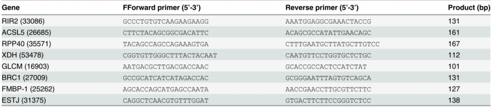
We isolated larvae, prepupae and pupae as described, collecting 30, 45, 60 silk glands from each group, with three independent biological replicates. RNA was isolated and cDNA pre-pared as described. qPCR was performed using the Mastercycler ep realplex (Eppendorf,
Germany). Gene specific primers (Table 1) were designed using Beacon Designer 7.6 and
syn-thesized by Sangon Biotech Co., Ltd (Shanghai, China). Glyceraldehyde 3-phosphate
dehydro-genase (GAPDH) was used as a stably expressed reference gene [7]. The reaction program was:
2 min at 95°C for 15 s, 55°C for 30 s, and 72°C for 30 s. Melting curves confirmed a single gene-specific peak and the absence of primer dimmer peaks. GoTaq qPCR Master Mix (Pro-mega, Madison, WI, USA) was used to measure the mRNA levels according to the
manufactur-er’s instructions. A five-fold dilution series was used to construct a relative standard curve to
genes was calculated by the comparative 2-ΔΔCTmethod [8] to identify the relative mRNA lev-els of the samples from different life stages.
Development- and tissue-specific expression of a gene encoding a
fibroin light chain (FLC)
Larvae, instars 1–5, prepupae, pupae, and adult females and males were collected (about 0.08 g
tissue/sample) with 3 independent biological replicates, frozen in liquid nitrogen and kept under -70°C until analysis.
The midgut, hindgut, silk gland, fat body, epidermis, and Malpighian tubes were isolated
from 5thinstar larvae (5 tissues per pool, 3 independent biological replicates). The larvae were
washed with distilled water twice and anesthetized on ice. The pleopod was cut with dissecting scissors and hemolymph was collected with pressure on the body using a pipettor. All of the
tis-sue samples were frozen in liquid nitrogen and kept under -70°C for analysis [9]. R NA was
iso-lated and cDNA prepared as described. Development- and tissue-specific qPCR analysis of
FLCexpression was performed as described.
Results
Transcriptome sequencing and sequence assembly
Before mapping the tags, the transcriptomes were preprocessed, yielding 42,040,007 clean reads and 60,298 unigenes. All the raw tags were filtered with reference sequences, leaving 15,445,926, 13,758,154, and 16,787,792 clean reads from the larval, prepupal and pupal
librar-ies, respectively (Table 2). Analysis of sequencing saturation showed that the number of
detected genes increased until the sequencing reads reached 3 million or more, indicating the Table 1. Primers used for qRT-PCR validation, all with Tm = 55°C. Gene IDs are in parenthesis.
Gene FForward primer (5’-3’) Reverse primer (5’-3’) Product (bp)
RIR2 (33086) GCCCTGTGTCAAGAAGAAGG AAATGGAGGCGAAACTACCG 131
ACSL5 (26685) CTTCTACAGCGGCGACATTC ACAGCGCCATATTGAACAGC 161
RPP40 (35571) TACAGCCAGCCAGAAAGTGA CTTTGAATGCTTATGCTTGTCC 167
XDH (53478) CGGTGTTGGGCTTTACTACAAT CAATGTTCCTGGTGCTCTGC 112
GLCM (16903) AATGACGCTTGACGACCAAC GCACCGCCACTCCATCTAT 101
BRC1 (27009) GCCGCATCATCATAGACCAC GCGGGAATTTAGTGTCAGCA 131
FMBP-1 (25262) AGCACCAGCATGAGCCAATA AACCGAACCTTGCGTTCTTC 127
ESTJ (31375) CAGGCTCAACGTGTTTGGAT GTGACTTCTTCCGGGTCTCC 138
doi:10.1371/journal.pone.0136868.t001
Table 2. Summary statistics of RNA-seq library sequencing and mapping.
Larvae Prepupae Pupae
Raw reads 15,556,751 13,851,700 17,189,289
Clean reads 15,445,926 13,758,154 16,787,792
Mapped reads 14,037,862 12,223,263 11,721,016
Mapped rate 90.88% 88.84% 69.81%
Mapped gene number 50,158 48,415 46,623
Total gene number 60,298 60,298 60,298
Mapped gene rate 83.18% 80.29% 77.32%
identified expressed reads were sufficient to represent the entire transcriptional information of the CLR genome.
Gene identification and annotation
All distinct sequences longer than 200 bp were searched against the NR, Swiss-prot, and KEGG
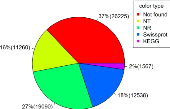
protein databases by BLASTX with a cut-off E-value of 1e-10. We recorded 19,090 (41.1% of all
distinct sequences; by NR), 12,538 (27.0%; Swiss-prot), 11,260 (24.3%; NT) and 1,567 (3.4%;
KEGG) transcripts (Fig 1). Identities of many genes (about 37%) were not found in available
databases, as expected due to the lack of transcriptomic information in tissues within pest species.
Gene expression profiles
We identified 50,158, 48,415, and 46,623 transcripts from the silk gland libraries. Among these, 37,846 transcripts were expressed in all three libraries, 44,897 were expressed in larval and pre-pupal libraries, and 39,873 were expressed in prepre-pupal and pre-pupal libraries. We recorded 40,771
transcripts expressed in larvae and pupae (Fig 2).
Expression of 38 genes changed (27", 11#) during the larval/prepupal transition. The
prepu-pal-pupal transition led to expression changes in 355 genes (137", 218#). The broader
excur-sion from larvae to pupae included up-regulation of 140 and down-regulation of 169 genes (Table 3; specific genes listed inS1 Table).
Fig 1. A pie chart showing proportions (with numbers in parentheses) of transcripts annotated by NR, Swiss-prot, NT and KEGG databases.
Genes related to silk formation
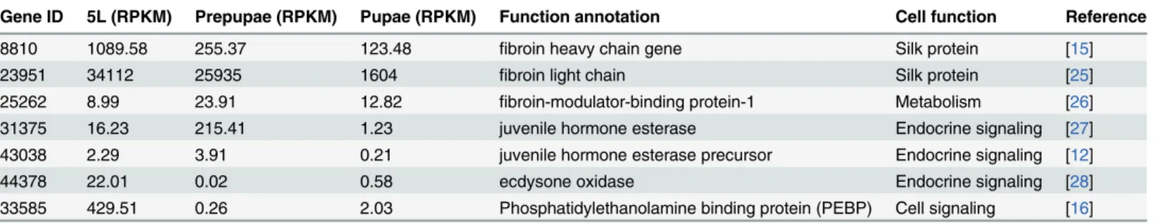
We selected seven silk gland genes that changed in expression during the developmental
excur-sion as directly or indirectly involved in silk formation (Table 4; partial sequences of specific
genes listed inS2 Table). Expression of genes encoding the heavy- and light-chain fibroin
pro-teins declined as the larvae moved beyond the silk-producing larval stage. A gene encoding a
fibroin-modulator-binding protein-1 is a transcriptional regulatory factor operating in theB.
morisilk gland. Expression of this gene increased by almost 3-fold as the larvae went into the
pre-pupal stage. Three genes act in endocrine signaling, which we take to indirectly influence silk production via their influence on silk gland developmental physiology as the insects moved from larvae to prepupae to pupae. We included a gene encoding a phosphatidylethanolamine binding protein (PEBP) because it acts in cell signaling and may indirectly influence silk pro-duction. Expression of this gene declined by a wide extent as the larvae advanced to prepupae. Fig 2. A Venn diagram showing overlap of silk gland genes expressed in larval, prepupal and pupal stages. 5L represents fifth instar larvae.
doi:10.1371/journal.pone.0136868.g002
Table 3. Differentially Expressed Genes across all libraries.
5thinstar larvae: Prepupae Prepupae:Pupae 5thinstar larvae:Pupae
Total 38 355 309
Up-regulated 27 137 140
Down-regulated 11 218 169
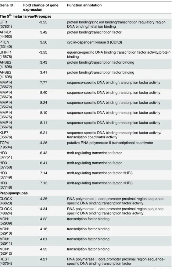
Genes encoding transcription factors
Development from larvae to pupae was attended by changes in expression of genes encoding a
range of transcription factors. Expression of 17 genes changed expression (14", 3#) going from
larvae to prepupae; 21 genes (14", 7#) changed expression in the transition from prepupae to
pupae. We listed these genes in a separate table (Table 5; specific gene sequences listed inS2
Table) to highlight the actions of their cognate proteins as transcription factors.
Some genes underwent very large changes in expression, 20 of which are listed inTable 6
(specific gene sequences listed inS2 Table). A gene encoding PEBP decreased in expression
by about 1400-fold and expression of a gene encoding a possible serine protease inhibitor increased by over 1200-fold. Apparent development regulation of several other genes, includ-ing genes encodinclud-ing a sugar transporter protein, a peroxidase, and an aldehyde oxidase I, went
through similar large-scale expression changes. We list these inTable 6to emphasize the scale
of expression changes.
Confirmation of gene expression
Expression analysis of 8 genes demonstrated parallel expression changes recorded by RNA-seq
and qPCR (Fig 3). Of particular interest, genes encoding a xanthine dehydrogenase (XDH), a
possible CRCT domain protein BRC1 (Brc1), and ribonuclease P protein subunit p40 (RPP0) were expressed in parallel.
FLC(ORF = 804 bp, encoding 267 amino acids) was obtained by RACE (GenBank accession
number: JX456150 [10]). During the larval stage,FLCexpression increased with larval
develop-ment, reaching a peak at the 5thinstar, then declined during the prepupal, pupal and adult
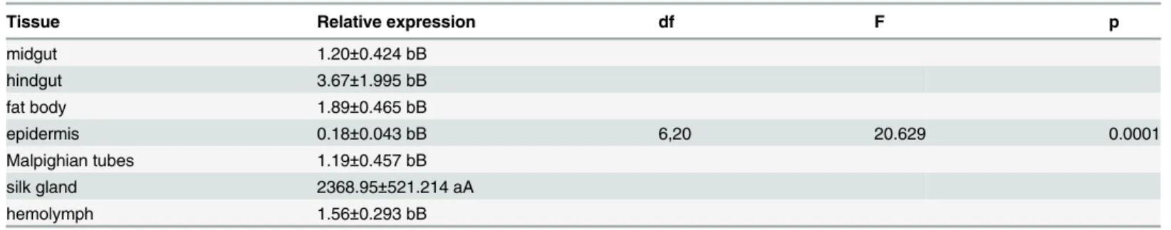
stages (Fig 4).FLCwas expressed, at very low levels, in the midgut, hindgut, fat body,
epider-mis, Malpighian tubes and hemolymph. Highest expression, by about 646 to 13,249-fold,
occurred in the silk gland (F= 20.629, df = 6, 20,P= 0.0001);FLCexpression was similar
among the other six tissues (F= 1.723, df = 5, 17,P= 0.2039) (Table 7).
Discussion
The data reported in this paper strongly support our hypothesis that silk gland gene expression changes during the developmental transition from larvae to pupae. Three points form a solid argument. One, we generated transcriptomes from silk glands prepared from larvae, prepupae and pupae, from which we identified 355 silk gland genes that changed expression during
development from 5thinstar larvae to pupae, understanding that not all 355 genes act directly
in silk production. Two, we confirmed expressed changes in 8 genes by qPCR, indicating our RNA-seq data reliably represents changes in gene expression. Three, 38 of the expression-altered genes encode transcription factors, which would influence expression of other genes. Table 4. Expression abundance of genes related to silk formation identified in the indicated citations. 5L = 5th instar larvae.
Gene ID 5L (RPKM) Prepupae (RPKM) Pupae (RPKM) Function annotation Cell function Reference
8810 1089.58 255.37 123.48 fibroin heavy chain gene Silk protein [15]
23951 34112 25935 1604 fibroin light chain Silk protein [25]
25262 8.99 23.91 12.82 fibroin-modulator-binding protein-1 Metabolism [26]
31375 16.23 215.41 1.23 juvenile hormone esterase Endocrine signaling [27]
43038 2.29 3.91 0.21 juvenile hormone esterase precursor Endocrine signaling [12]
44378 22.01 0.02 0.58 ecdysone oxidase Endocrine signaling [28]
33585 429.51 0.26 2.03 Phosphatidylethanolamine binding protein (PEBP) Cell signaling [16]
Table 5. Expression abundance of most differentially expressed transcription factors during larval to pupal development. Silk formation is highest in 5th instar larvae, lower in prepupae and does not occur in pupae.
Gene ID Fold change of gene expression
Function annotation
The 5thinstar larvae/Prepupae GFI1
(37831)
-3.55 protein binding/zinc ion binding/transcription regulatory region DNA binding/metal ion binding
ARRB1 (44963)
3.42 protein binding/transcription factor
PTEN (33140)
3.56 cyclin-dependent kinase 3 (CDK3)
UHRF1 (15676)
-3.55 equence-specific DNA binding transcription factor activity/protein binding
APBB2 (41696)
3.43 protein binding/transcription factor binding
APBB2 (41695)
3.41 protein binding/transcription factor binding
MMP14 (35672)
7.77 sequence-specific DNA binding transcription factor activity
MMP14 (35673)
8.40 sequence-specific DNA binding transcription factor activity
MMP14 (35674)
8.24 sequence-specific DNA binding transcription factor activity
MMP14 (35675)
8.10 sequence-specific DNA binding transcription factor activity
MMP14 (35676)
8.11 sequence-specific DNA binding transcription factor activity
KLF7 (35676)
6.21 sequence-specific DNA binding transcription factor activity/ transcription coactivator activity
TCP4 (19504)
-4.28 putative RNA polymerase II transcriptional coactivator
HR3 (37751)
6.43 molt-regulating transcription factor
HR3 (37750)
6.41 molt-regulating transcription factor
HR3 (37749)
7.14 molt-regulating transcription factor HHR3
HR3 (37748)
7.13 molt-regulating transcription factor HHR3
Prepupae/pupae CLOCK
(46823)
-4.25 RNA polymerase II core promoter proximal region sequence-specific DNA binding transcription factor activity
CLOCK (46824)
-4.34 RNA polymerase II core promoter proximal region sequence-specific DNA binding transcription factor activity
MDN1 (52909)
4.22 transcription factor binding
MDN1 (52910)
4.18 transcription factor binding
MDN1 (52911)
4.61 transcription factor binding
MDN1 (52912)
4.55 transcription factor binding
REST (43754)
4.21 RNA polymerase II core promoter proximal region sequence-specific DNA binding transcription factor
Taken together, these points support our inference that gene expression in CLR silk glands is under strong developmental control.
In silk gland biology, the transition represents a period of high silk production in 5thinstar
larvae, to reduced production in pre-pupae to atrophy of the silk gland in pupae. We surmise developmental regulation of gene expression makes up the proximal mechanism of these changes in silk gland biology. The biological significance of regulating silk gland gene expres-sion touches on several areas. As seen in many areas of biology, such as development of repro-ductive systems, silk production is restricted to the life stages in which silk contributes to CLR biology. Overall CLR fitness would be substantially reduced due to biological costs of allocat-ing resources toward maintainallocat-ing unnecessary silk glands and to protein-costly silk produc-tion. Perhaps more to the point, unnecessary silk could be directly deleterious to CLR and many other silk-producing insect species. We infer that silk gland gene expression is tightly regulated.
The major developmental hormones, juvenile hormone (JH) and 20-ecdysteroid (20E),
sig-nal the transition from larvae to pupae and also influence silk gland biology [11]. The JH
ana-log, methoprene, is applied to silkworms,B.mori, to extend the last larval instar and enhance
silk production [6]. JH esterase is responsible for the developmentally-regulated decline in JH
titers associated with pupation [12]. Our data show that JH esterase is expressed at low levels in
5thinstar larval silk glands, then increases by 13-fold in pre-pupal silk glands before declining
to near-zero in pupae. This is consistent with high silk production during cocoon formation and subsequent atrophy of the silk gland. Similarly, ecdysone oxidase (EO) reduces 20E titers Table 5. (Continued)
Gene ID Fold change of gene expression
Function annotation
DNMT1 (7480)
4.19 DNA cytosine-5 methyltransferase
KDM1A (23518)
4.12 transcription factor binding
35426 4.08 putative gonadotropin inducible transcription factor HR3
(37751)
-4.01 molt-regulating transcription factor
HR3 (37750)
-3.99 molt-regulating transcription factor
HR3 (37749)
-3.79 molt-regulating transcription factor HHR3
HR3 (37748)
-3.78 molt-regulating transcription factor HHR3
ZNF83 (44937)
-4.24 putative KRAB box and zincfinger C2H2 type domain containing protein
ZGPAT (26481)
3.93 sequence-specific DNA binding transcription factor activity
ZGPAT (26482)
3.90 sequence-specific DNA binding transcription factor activity
RTF1 (50796)
3.98 hypothetical protein KGM_10227
RTF1 (50798)
3.78 hypothetical protein KGM_10227
35424 3.64 putative gonadotropin inducible transcription factor MYNN
(27584)
4.92 sequence-specific DNA binding transcription factor activity
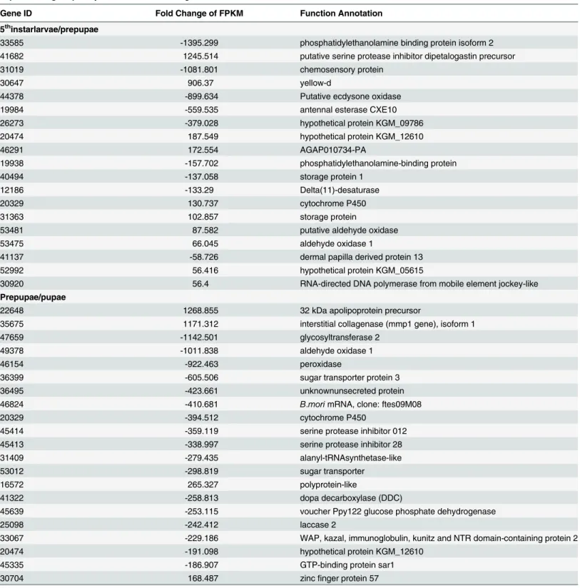
Table 6. Twenty of highly differentially expressed annotated genes in the 5th instar larvae/ prepupae and prepupae/pupae libraries based on expressed tag frequency.“-”indicates down regulation.
Gene ID Fold Change of FPKM Function Annotation
5thinstarlarvae/prepupae
33585 -1395.299 phosphatidylethanolamine binding protein isoform 2
41682 1245.514 putative serine protease inhibitor dipetalogastin precursor
31019 -1081.801 chemosensory protein
30647 906.37 yellow-d
44378 -899.634 Putative ecdysone oxidase
19984 -559.535 antennal esterase CXE10
26273 -379.028 hypothetical protein KGM_09786
20474 187.549 hypothetical protein KGM_12610
46291 172.554 AGAP010734-PA
19938 -157.702 phosphatidylethanolamine-binding protein
40494 -137.058 storage protein 1
12186 -133.29 Delta(11)-desaturase
20329 130.737 cytochrome P450
31363 102.857 storage protein
53481 87.582 putative aldehyde oxidase
53475 66.045 aldehyde oxidase 1
41137 -58.726 dermal papilla derived protein 13
52992 56.416 hypothetical protein KGM_05615
30920 56.4 RNA-directed DNA polymerase from mobile element jockey-like
Prepupae/pupae
22648 1268.855 32 kDa apolipoprotein precursor
35675 1171.312 interstitial collagenase (mmp1 gene), isoform 1
47659 -1142.501 glycosyltransferase 2
49378 -1011.838 aldehyde oxidase 1
46154 -922.463 peroxidase
36399 -605.506 sugar transporter protein 3
36495 -423.661 unknownunsecreted protein
46824 -410.681 B.morimRNA, clone: ftes09M08
20329 -394.512 cytochrome P450
45414 -359.119 serine protease inhibitor 012
45413 -338.997 serine protease inhibitor 28
31409 -279.435 alanyl-tRNAsynthetase-like
53012 -298.819 sugar transporter
16572 265.327 polyprotein-like
41322 -258.813 dopa decarboxylase (DDC)
45639 -253.115 voucher Ppy122 glucose phosphate dehydrogenase
25098 -242.412 laccase 2
33067 -229.186 WAP, kazal, immunoglobulin, kunitz and NTR domain-containing protein 2
20474 -191.098 hypothetical protein KGM_12610
45335 -186.907 GTP-binding protein sar1
30704 168.487 zincfinger protein 57
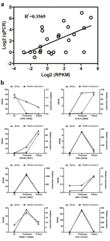
Fig 3. qPCR validation of 8 genes from three CLR developmental stages. (a) Comparison of
expression levels measured by RNAseq and qPCR for the selected 8 transcripts in three libraries (5L, Prepupae and Pupae; R2= 0.3569). (b) Comparison of expression levels of eight genes recorded by
RNAseq and qPCR. Each panel shows gene expression on the left y-axis, determined by RPKM, and relative gene expression determined by qPCR on the right y-axis.
by converting 20E into inactive 3-dehydroecdysone [13]. Expression of the gene encoding EO in silk glands declined by about 1100-fold to virtually zero in the larval-pre-pupal transition. We infer the decline in silk gland EO expression is consistent with increased 20E titers during silk production and preparation for the pupal molt. Altered expression of silk gland genes encoding enzymes that regulate JH and 20E titers aligns with the actions of these hormones in silk production during development.
Gene expression is influenced by a large number of transcription factors. We recorded sub-stantial changes in expression of genes encoding transcription factors in the larval-pupal
tran-sition. Among these, expression of 10 genes increased by>6-fold and expression of another
10 decreased by>3-fold. Genes encoding sequence-specific DNA-binding transcription factor
Fig 4. Expression pattern of a fibroin light chain gene at the indicated CLR developmental stages. Insect life stages are indicated on the vertical axis and relative gene expression (as fold-change) on the horizontal axis. Histogram bars annotated with the same letter are not significantly different.
doi:10.1371/journal.pone.0136868.g004
Table 7. Expression pattern of fibroin light chain gene in different tissues of CLR.
Tissue Relative expression df F p
midgut 1.20±0.424 bB
hindgut 3.67±1.995 bB
fat body 1.89±0.465 bB
epidermis 0.18±0.043 bB 6,20 20.629 0.0001
Malpighian tubes 1.19±0.457 bB
silk gland 2368.95±521.214 aA
hemolymph 1.56±0.293 bB
activities, MMP14 (35673–35676) increased by>8-fold. These large changes in expression of
an array of genes indicate the dynamic metabolic and developmental changes in the silk gland during silk production and silk gland atrophy during subsequent development into pupae.
In particular, expression of the molt-regulating transcription factors HR3 (37748–37751)
increased by 6- to 7-fold in the larval-pre-pupal transition, then decreased by circa 3-fold in the pre-pupal-pupal period. These proteins act in gene regulation during metamorphosis. Silk gland-specific changes in expression of these genes are associated with the changes in the struc-ture and function of the gland during the larval-pupal metamorphosis. HR3 genes are crucial to molting physiology at the organismal level and they trigger hypotheses about the actions of
these and other transcription factors in the silk gland [14].
Fibroin and sericin are the main silk proteins. Fibroin is made of heavy- and light-chain polypeptides incorporated with the 25 kDa protein, P25. Sericin is a glycoprotein responsible
for agglomerating the silk proteins [15]. Silk gland expression of gene 8810, encoding a fibroin
heavy chain, was highest in larvae, lower by about 4.3-fold in pre-pupae and still lower, by
about 9-fold, in pupae. We recorded similar data for aFLC, high expression in larvae, slightly
lower in pre-pupae and reduced by about 21-fold in pupae. This pattern is consistent with the silk-producing biology of the silk gland.
Our qPCR analysis showsFLCexpression gradually increased during larval development to
a peak at the 5thinstar. Because older larvae depend on silk, we infer theFLCexpression
pat-tern is consistent with the silk-producing and leave-rolling behavior of CLR larvae. Similarly,
silk glandFLCexpression was much higher compared to other tissues. Again, our
interpreta-tion isFLCacts in silk production.
Developmental regulation of silk gland gene expression led to very large changes in some genes. These rather striking changes are not unexpected because the silk glands undergo funda-mental changes from metabolically active, silk-producing tissues to regulated atrophy in a lim-ited time frame. The functions of some genes, such as yellow-d (30647), are not yet known well enough to generate meaningful commentary. Changes in other genes lead to useful hypotheses about their specific functions in the context of silk gland biology. The purpose of this first description of silk gland transcriptomes is to report on the large changes in gene expression. Detailed discussion of each gene would amount to speculations and we limit our discussion of individual genes to a phosphatidylethanolamine binding protein (PEBP), a peroxidase and a chemosensory protein.
PEBPs form a protein family, members of which occur in a very wide range of organisms,
including bacteria, plants and animals [16]. These proteins are mostly signal moieties with
sub-stantial importance in biomedicine [17]. Silk gland gene 33585 encodes a PEBP that declined
in expression (by almost 1400-fold) during the larval-pre-pupal excursion. The biological func-tions of PEBP in insects are not fully appreciated, but they probably act in intracellular
signal-ing roles, as reported in biomedical research. Reumer et al. [18] discovered theDrosophila
PEBP1 (CG18594) is expressed in larval fat bodies and in theDrosophilalarval hemocyte cell
line, l(2)mbn. The authors investigated the idea that PEBP conveys immune protection against bacterial infections. They showed that larvae overexpressing PBEP1 synthesized and released increased anti-microbial proteins into the hemolymph and that infection stimulated a 6.5-fold
increase in PBEP1 expression. They inferred thatDrosophilaPEBP1 acts in immunity. We do
not yet appreciate the specific role of this gene in silk glands, but surmise that PEBP acts in var-ious signaling functions.
Expression of a peroxidase (POX; 46154) was down-regulated by nearly 1,000-fold in the transition from prepupae to pupae. POXs act in many areas of insect biology, including cuticle tanning and anti-oxidant metabolism. A new role of POXs has recently come to light. POXs
expression of eggshell genes inDrosophila[20]. Most recently, Park et al. [21] reported that
POXs act in PGE2-mediated immune reactions to infection. We speculate that PGs act in silk
gland physiology and down-regulation of POX is congruent with the loss of function and break down of the silk gland.
Insects sense chemicals via soluble proteins, which act by binding air-borne chemicals, such as sex pheromones and plant volatiles. The soluble proteins, bound with their signal ligands, interact with specific chemoreceptors located on sensory nerve membranes, which leads to the transduction from chemical to electrical signals and subsequent integration within the central
nervous system [22]. Insects express two families of soluble olfactory proteins, odorant binding
proteins (OBPs) and chemosensory proteins (CSPs). CSPs are smaller than OBPs (100–120,
compared to 150–160 amino acids), although they act in similar functions [22]. CSPs are
expressed in antennae, but also in other, non-sensory, tissues and body parts, such as
anten-nae-removed heads, thoraces, abdomens, gonads, wings and pheromone glands [23]. The
broad distribution of these proteins led to suggestions that CSPs act in additional roles, possibly female reproduction, limb regeneration and embryonic development. We report that silk glands express one gene encoding a CSP, 31019, which declined in expression by about 1,000-fold during the larval-pre-pupal transition. We infer this protein acts in the silk gland during the silk-producing phase of pupation, possibly directly in silk production.
Arthropods, including pest insects, use silk in a very wide range of biological functions, including food capture, nests, guidelines, dispersal, reproduction, egg coatings and protective cocoons. Some silk-producing insects influence the structure of their environments. For an
unusual example, the aquatic insectHydropsyche siltalai(Tricoptera, Hydropsychidae,
net-spinning caddisflies) produces silk cup-like nets to capture small prey organisms. Statzner et al.
[24] reported that their silk is fixed to gravel pieces that results in gravel consolidation in
stream beds, which influences sediment erosion. The concept that insect silk production can influence local environments prompts our suggestion of substantial research into pest insect silk production.
Supporting Information
S1 Table. Differentially Expressed Genes across all libraries.
(DOC)
S2 Table. Gene IDs and the partial sequences.
(DOC)
Acknowledgments
Mention of trade names or commercial products in this article is solely for the purpose of pro-viding specific information and does not imply recommendation or endorsement by the U.S. Department of Agriculture. All programs and services of the U.S. Department of Agriculture are offered on a nondiscriminatory basis without regard to race, color, national origin, religion, sex, age, marital status, or handicap.
Author Contributions
References
1. Wang MQ, Cai WZ (2004) Silk and silk glands of insects. Entomol. Knowl 40(1): 90–95.
2. Klapwijk MJ, Csóka G, Hirka A, Björkman C (2013) Forest insects and climate change: long-term trends in herbivore damage. Ecol Evol 3(12): 4183–4196. doi:10.1002/ece3.717PMID:24324869
3. Xu J, Wang QX, Wu JC (2010) Resistance of cultivated rice varieties toCnaphalocrocis medinalis (Lep-idoptera: Pyralidae). J Econ Entomol 103(4):1166–1171. PMID:20857724
4. Di JX, Chen XS, Wu QJ, Xu NY, Xiao SH, Liu JG, et al. (2007) Preliminary study of damage caused by
Sylepta derogateFabricius (Lepidoptera: Pyralidae). Jiangsu Agricultural Sciences 5: 82–84. 5. Hou Y, Xia QY, Zhao P, Zou Y, Liu HL, Guan J, et al. (2007) Studies on middle and posterior silk glands
of silk worm (Bombyx mori) using two dimensional electrophoresis and mass spectrometry. Insect Bio-chem Molec 37: 486–496.
6. Sehnal F, Akai H (1990) Insect silk glands: their types, development and function, and effects of envi-ronmental factors and morphogenetic hormones on them. Int J Insect Morphol Embryol 19 (2): 79–132. 7. Zheng YT, Li HB, Lu MX, Du YZ (2014) Evaluation and validation of reference genes for qRT-PCR
nor-malization inFrankliniella occidentalis(Thysanoptera: Thripidae). PLoS One 9(10): e111369. doi:10. 1371/journal.pone.0111369PMID:25356721
8. Livak KJ, Schmittgen TD (2001) Analysis of relative gene expression data using real-time quantitative PCR and the 2(-ΔΔC(T))
method. Methods 25(4): 402–408. PMID:11846609
9. Wang X (2011) Cloning and expression of Csaqp1genes in rice stem borer,Chilo SuppressalisWalker (Lepidoptera: Pyralidae). M.Sc. Thesis, Yangzhou University. Available:http://www.shangxueba.com/ lunwen/v1473369.html. Accessed 19 December 2014.
10. Chen CX, Wang ZY, Gu GX, Yang YZ, Liang GH (2015). Cloning of a silk-producing gene in the cotton leaf rollerSylepta derogata(Lepidoptera: Pyralidae) and its function. J Environ Entomol: In press. 11. Zhao XM, Liu C, Jiang LJ, Li QY, Zhou MT, Cheng TC, et al. (2014) A juvenile hormone-transcription
factor Bmdimm-fibroin H chain pathway is involved in the synthesis of silk protein in silkworm,Bombyx mori. J Biol Chem Pii:jbc.M114 606921. [Epub ahead of print]
12. Kamimura M, Takahashi M, Kikuchi K, Reza AM, Kiuchi M (2007) Tissue-specific regulation of juvenile hormone esterase gene expression by 20-hydroxyecdysone and juvenile hormone inBombyx mori. Arch Insect Biochem Physiol 65(3):143–151. PMID:17570489
13. Yang HJ, Wang MX, Zhang P, Sabhar A, Malik FA, Bhaskar R, et al. (2011) Cloning and characteriza-tion of theBombyx moriecdysone oxidase. Arch Insect Biochem Physiol 78: 17–29. doi:10.1002/arch. 20436PMID:21678487
14. Xiong Y, Zeng H, Zhang Y, Xu D, Qiu D (2013) Silencing the HaHR3 gene by transgenic plant-mediated RNAi to disruptHelicoverpa armigeradevelopment. Int J Biol Sci 9(4): 370–381. doi:10.7150/ijbs.5929 PMID:23630449
15. Shimizu K, Ogawa S, Hino R, Adachi T, Tomita M, Yoshizato K (2007) Structure and function of 5 '-flanking regions ofBombyx morifibroin heavy chain gene: Identification of a novel transcription enhancing element with a homeodomain protein-binding motif. Insect Biochem Mol Biol 37(7): 713–
725. PMID:17550827
16. Li H, Huang F, Fan L, Jiang Y, Wang X, Li J, et al. (2014) Phosphatidylethanolamine-binding protein 4 is associated with breast cancer metastasis through Src-mediated Akt tyrosine phosphorylation. Onco-gene 33(37): 4589–4598. doi:10.1038/onc.2013.408PMID:24276246
17. Zhao J, O’Donnell VB, Balzar S, Croix CM St., Trudeau JB, Wenzel SE (2011) 15-lipoxgnease 1 inter-acts with phosphatidylethanolamine-binding protein to regulate MAPK signaling in human airway epi-thelial cells. Proc Natl Acad Sci USA 108(34): 14246–14251. doi:10.1073/pnas.1018075108PMID: 21831839
18. Reumer A, Bogaerts A, Van Loy T, Husson SJ, Temmerman L, Choi C, et al. (2009) Unraveling the pro-tective effect of aDrosophilaphosphatidylethanolamine-binding protein upon bacterial infection by means of proteomics. Dev Comp Immunol 33: 1186–1195. doi:10.1016/j.dci.2009.06.010PMID: 19545586
19. Tootle TL, Spradling AC (2008)DrosophilaPxt: a cyclooxygenase-like facilitator of follicle maturation. Develop 135: 839–847.
20. Tootle TL, Williams D, Hubb A, Frederick R, Spradling A (2011)Drosophilaeggshell production: identifi-cation of new genes and coordination by Pxt. PLoS One 6: e19943. doi:10.1371/journal.pone. 0019943PMID:21637834
21. Park J, Stanley D, Kim Y (2014) Role of peroxinectin in PGE2-mediated cellular immunity inSpodoptera
22. Pelosi P, Lovinella I, Felicioli A, Dani FR (2014) Soluble proteins of chemical communication: an over-view across arthropods. Front Physiol 5: 320. doi:10.3389/fphys.2014.00320PMID:25221516 23. Qiao HL, Deng PY, Li DD, Chen M, Jiao ZJ, Liu ZC, et al. (2013) Expression analysis and binding
experiments of chemosensory proteins indicate multiple roles inBombyx mori. J Insect Physiol 59(7): 667–675. doi:10.1016/j.jinsphys.2013.04.004PMID:23624070
24. Statzner B, Arens MF, Champagne JY, Morel R, Herouin E (1999) Silk-producing stream insects and gravel erosion: significant biological effects on critical shear stress. Water Resources Res 35: 3495–
3506.
25. Chaitanya RK, Dutta-Gupta A (2010) Light chain fibroin and P25 genes ofCorcyra cephalonica: Molec-ular cloning, characterization, tissue-specific expression, synchronous developmental and 20-hydro-xyecdysone regulation during the last instar larval development. Gen Comp Endocrinol 167(1):113–
121. doi:10.1016/j.ygcen.2010.02.007PMID:20171223
26. Takiya S, Saito S, Yokoyama T, Matsumoto D, Aizawa T, Kamiya M, et al. (2009) DNA-binding property of the novel DNA-binding domain STPR in FMBP-1 of the silkwormBombyx mori. J Biochem 146 (1):103–111. doi:10.1093/jb/mvp053PMID:19304790
27. Kontogiannatos D, Swevers L, Maenaka K, Park EY, Iatrou K, Kourti A (2013) Functional characteriza-tion of a juvenile hormone esterase related gene in the mothSesamia nonagrioidesthrough RNA inter-ference. PLoS One 20138 (9): e73834.