Stress Tolerance in
Francisella novicida
Jacob R. Chambers, Kelly S. Bender*
Department of Microbiology, Southern Illinois University, Carbondale, Illinois, United States of America
Abstract
The RNA-binding protein Hfq is recognized as an important regulatory factor in a variety of cellular processes, including stress resistance and pathogenesis. Hfq has been shown in several bacteria to interact with small regulatory RNAs and act as a post-transcriptional regulator of mRNA stability and translation. Here we examined the impact of Hfq on growth, stress tolerance, and gene expression in the intracellular pathogenFrancisella novicida. We present evidence of Hfq involvement in the ability ofF. novicidato tolerate several cellular stresses, including heat-shock and oxidative stresses, and alterations in
hfqgene expression under these conditions. Furthermore, expression of numerous genes, including several associated with virulence, is altered in ahfqmutant strain suggesting they are regulated directly or indirectly by Hfq. Strikingly, we observed a delayed entry into stationary phase and increased biofilm formation in thehfqmutant. Together, these data demonstrate a critical role for Hfq inF. novicidagrowth and survival.
Citation:Chambers JR, Bender KS (2011) The RNA Chaperone Hfq Is Important for Growth and Stress Tolerance inFrancisella novicida. PLoS ONE 6(5): e19797. doi:10.1371/journal.pone.0019797
Editor:Stefan Bereswill, Charite´-University Medicine Berlin, Germany
ReceivedNovember 2, 2010;AcceptedApril 16, 2011;PublishedMay 5, 2011
Copyright:ß2011 Chambers, Bender. This is an open-access article distributed under the terms of the Creative Commons Attribution License, which permits unrestricted use, distribution, and reproduction in any medium, provided the original author and source are credited.
Funding:This work was supported by the National Institute of Allergy and Infectious Diseases of the National Institutes of Health (NIH) through an Academic Research Enhancement Award#RAI082502A to K. S. Bender. The funders had no role in study design, data collection and analysis, decision to publish, or preparation of the manuscript.
Competing Interests:The authors have declared that no competing interests exist.
* E-mail: [email protected]
Introduction
Francisella tularensis is a gram-negative facultative intracellular pathogen and the etiologic agent of tularemia. Inhalation of as few as 10 organisms can result in development of pneumonic tularemia, a form of the disease associated with the highest morbidity and mortality. Due to its high infectivity and relative ease of dissemination,F. tularensishas been designated a category A select agent by the Centers for Disease Control and Prevention [1]. In humans, the most common form of tularemia is an ulceroglandular disease resulting from the bite of a bacteria-carrying arthropod vector or exposure to the infected blood of an animal through an abrasion in the skin [2]. Numerous arthropod vectors have been implicated in the transmission of tularemia and a wide range of animal hosts, including small mammals, birds, and humans, are susceptible to infection by F. tularensis [3]. Besides possessing a wide range of hosts, the pathogen is also quite resilient to environmental conditions and has been found to survive in mud or water for over a year [4].
Several studies have shown that F. tularensis pathogenicity is dependent on its ability to persist and replicate inside host cells, especially macrophages [1,5]. Several of the genes required for virulence are located within the Francisella pathogenicity island (FPI), though additional genes are likely to be required [6]. A number of genes in the FPI, such aspdpA and pdpB, have been shown to be required for intracellular bacterial growth and survival [7,8]. Genes outside the FPI have also been implicated in aspects of F. tularensis virulence and dissemination through the utilization of genome-wide screens [8,9]. While a limited number ofF. tularensisregulators are known, those shown to be involved in virulence include MglA, SspA, FevR, and RelA [10,11,12].
Recently, the F. tularensis subspecies holarctica strain LVS Hfq protein was reported to play a role in virulence, possibly by negatively regulating thepdpoperon of the FPI [13].
The RNA-binding protein Hfq is an important regulator of gene expression in a number of bacterial pathogens including Yersinia pseudotuberculosis,Neisseria meningitidis, and Salmonella enterica [14,15,16]. The Hfq protein was first identified as a host factor required for synthesis of bacteriophage QbRNA inEscherichia coli and belongs to the Sm and Sm-like family of proteins found in both prokaryotes and eukaryotes [17,18]. Hfq is present in many bacterial species and hfq mutants often exhibit pleiotropic phenotypes including defects in virulence, quorum sensing, growth rate, and stress tolerance [19,20,21,22,23].
Recent studies suggest that Hfq acts primarily as a post-transcriptional regulator that facilitates RNA-RNA interactions between small noncoding RNAs (sRNAs) and their mRNA targets [18,24]. Small RNAs are a novel group of gene regulators that typically act by base pairing with target mRNAs resulting in either the up- or down-regulation of protein synthesis. These sRNAs are often encoded intransand commonly rely on the Hfq protein for mediating the sRNA-mRNA interaction [25].
hfq deletion mutant, and the heterologous expression of the F. novicidaHfq in anE. coli hfqmutant. We demonstrate that the N-terminal half of the protein is most critical to heat stress tolerance and that the entire protein is unable to complement the phenotype of anE. coli hfqmutant. Not only does Hfq play a role in the response to salt and heat shock, as reported forF. tularensissubspeciesholarctica [13], it also plays a role in pH tolerance, peroxide stressors, entry into stationary phase, and biofilm formation inF. novicida. We also report the expression profile of a subset of genes previously identified by Meibom and coworkers to be regulated by Hfq inF. tularensis subspecies holarctica [13]. Thus Hfq and likely sRNAs appear to play a role in regulating global gene expression, stress resistance, and formation of biofilms inF. novicida.
Materials and Methods
Bacterial Strains and Growth Conditions
Bacterial strains and plasmids used in this study are listed in Table 1.F. novicidastrain U112 and all its derivatives were routinely grown at 37uC in Tryptic Soy (TS) broth (Difco) supplemented with 0.1% L-cysteine or on TS agar plates supplemented with 0.1% cysteine, unless otherwise noted.E. colistrains were grown in Luria-Bertani (LB) medium or on LB agar plates. When required, media was supplemented with chloramphenicol (50mg/ml), erythromycin
(250mg/ml), kanamycin (50mg/ml), or tetracycline (10mg/ml).
Strain construction
To construct a non-polarF. novicida hfqdeletion mutant, a 3,033 bp DNA cassette containing anermCORF flanked by 1,170 bp upstream and 1,128 bp downstream of the hfq (FTN_1051) ORF was constructed via fusion of three separate PCR products [28]. Primers for the cassette contained overlapping sequences and are listed in Table 2. The following PCR reaction mixture was used to amplify the upstream (primers FnHfqKO-upF/FnHfqKO-upR) and downstream (primers FnHfqKO-downF/FnHfqKO-downR) regions flanking the hfqORF fromF. novicidaas well as the 735 bpermCORF from pIDN4
[29] (primers FnHfqKO-ermF/FnHfqKO-ermR): 16PfuUltraTMII
Buffer (Stratagene), 0.25 mM each dNTP, 2.5 pmol each primer, 1ml
PfuUltraTM II Fusion HS DNA polymerase (Stratagene), and 1ml
template DNA in a total volume of 50ml. Reactions were cycled
according to the following program: 95uC denaturation for 2 min; followed by 6 cycles consisting of 95uC for 30 sec, 45uC for 30 sec, and 72uC for 70 sec; then 25 cycles consisting of 95uC for 30 sec, 54uC for 30 sec, and 72uC for 70 sec; and ending with a 6 min extension at 72uC. The resulting PCR products were gel purified using the QiaQuick Gel Extraction kit (Qiagen).
For the initial fusion reaction, 100 ng of each purified product was added to a PCR mixture as described above except no primers were added. The products themselves were used as primers. This initial fusion mixture was cycled according to the following program: 95uC denaturation for 15 sec; followed by 5 cycles consisting of 95uC for 15 sec, 55uC for 60 sec, and 72uC for 210 sec. The final fusion reaction contained 20ml of the initial fusion product, 2.5 pmol each of primers
FnHfqKO-upF and FnHfqKO-downR, 16PfuUltraTMII Polymerase
Buffer, 0.25 mM each dNTP, and 1mlPfuUltraTMII Fusion HS DNA
polymerase. This reaction was cycled using the following parameters: 25 cycles consisting of 95uC for 15 sec, 55uC for 60 sec, and 72uC for 210 sec; and ending with a 8 min extension at 72uC. Sequence analysis of the final fusion product was used to verify lack of sequence errors and that thehfqORF was replaced with theermCORF.
To construct the complementing plasmid pKK214hfq, the hfq coding region and 191 bp directly upstream were PCR amplified from F. novicida using primers FN_hfqProm-F-SmaI and FN-hfq.orf-EcoRI-R using a standard reaction mixture and cycling parameters. The product was digested with SmaI and EcoRI and ligated into SmaI-EcoRI-digested pKK214gfp [30], producing pKK214hfq. All constructs were verified by DNA sequence analysis and introduced into the appropriateF. novicidastrains by electroporation [31]. Transformants were screened and confirmed by PCR analysis and DNA sequencing, and the resulting mutant strains were designatedF. novicidaDhfqand complemented strain
Dhfq/pKK214hfq.
Table 1.Bacterial strains and plasmids used in this study.
Strains Genotype or description Source or reference
F. novicida
U112 Wild-typeF. tularensissubsp.novicidastrain U112 [56]
U112Dhfq U112Dhfq, EmR This study
U112 Tnhfq1 U112hfq:: EZ-Tn5,KAN-2.at ORF position 229 of 327 bp [37]
U112 Tnhfq2 U112hfq:: EZ-Tn5,KAN-2.at ORF position 133 of 327 bp [37]
E. coli
MC4100 araD139D(argF-lac)205 flb-5301 pstF25 rpsL150 deoC1 relA1 [24]
GS081 MC4100hfq-1 ::V(CmR) [24]
Plasmids
pKK214gfp Low-copy-number expression vector withgroELpromoter ofF. tularensisLVS,Dcat::gfp [30]
pKK214hfq hfqORF and 191 bp-upstream region inserted into pKK214gfp This study
pACYC177 Low-copy-number cloning vector, AmpR, KmR New England Biolabs
pACYCMchfq MC4100hfqORF and 163 bp upstream region inserted into pACYC177 This study pACYCFnhfq U112hfqORF and MC4100hfqpromoter region region inserted into pACYC177 This study
pIDN4 Source of erythromycin gene cassette [29]
pMAL-c4X Vector for protein purification containing maltose binding protein New England Biolabs
To complement anE. coli hfq mutant with theF. novicidaHfq protein under control of theE. coli hfqpromoter, the wild-typeF. novicida hfqORF was PCR amplified using primers EcFnHfq-FusF
and FN_hfq-R-PstI (Table 2) in a standard reaction mixture. The 163 bp promoter region of the native E. coli hfq ORF was amplified with primers Ec_hfq-up-F-BamHI and
EcHfqProm-Table 2.Primers used in this study.
Primer name Primer sequence (59- 39)
FN_hfqProm-F-SmaI TTTCCCGGGATGGAGGACGAAACTAAAGAGTTAGATAT
FN-hfq.orf-EcoRI-R CGGAATTCTTACTCGTGAATATTACCTTCATTC
FN_hfq-R-PstI GCAATCTGCAGTTACTCGTGAATATTA
FN_hfq-22F CAAGACCCGTTCTTAAATGC
FN_hfq-326R TCGTGAATATTACCTTCATTCTC
FN_hfq-151R AGCTGGAACTATAGTAGAAATAGCATGT
FN-hfq-ATG-F ATGTCAAGAATATCATCTTTACAAGACC
FN-hfq-330R-PstI GCAATCTGCAGTTACTCGTGAATATTA
FnHfqKO-ermF TAGATTCGAGGTACGGCTACAGTCTTTTGGCTAACACACACGCCATTC
FnHfqKO-ermR CGGTCTATGAACTTAGTGAGCGGATTAGTTTATGCATCCCTTAACTTACTTA
FnHfqKO-upF CTATGGTTTGGCTGGACCAACAGCTTC
FnHfqKO-upR AAGACTGTAGCCGTACCTCGAATCTATGTCTCACTTCCTTTTAA
FnHfqKO-downF AATCCGCTCACTAAGTTCATAGACCGAGTTCCATTGTGGAGTAATATTAG
FnHfqKO-downR TCCTCAAGAGGCACGAAACTTGGC
FN_hflX-373F AAACTTCAGGTTGAGTTGGCGCAG
FN_hflX-1013R TCAAGAGGCACGAAACTTGGCTTG
FN_hflX-501R TATCTCAAGCTGTGTCTCACCAGGTC
FN_miaA-25F GCTGGACCAACAGCTTCAGGTAAA
FN_miaA-274F GGAAGAGAAGTCTTACTTGTTGGTGGG
FN_miaA-833F CATGGATTCGTAATTGGCAGAG
Q_FN-bioD-111F ACCTGTAGCGTCTGGACAAAGTCA
Q_FN-bioD-241R GAGGAGCAACTGCTTGATTGAATG
Q_FN-dnaG-859F GCTGCAGTAGCTACATTAGGCACA
Q_FN-dnaG-966R TTGCCCTGCTTCATCACCATCA
Q_Fn_hfq-84F CGGGATCAAACTACAAGGTCAAGT
Q_Fn_hfq-195R AGCTGGAACTATAGTAGAAATAGCATGT
Q_FN-katG-798F AACTCATGGTGCAGTTCCAGAGGA
Q_FN-katG-895R TATTGTGCCAGCCTAGACCTTGCT
Q_FN_mglA-156F ACCTACGCTTAGCACAGATGA
Q_FN_mglA-249R TGGAAACATCGGAGGAAAGGGA
Q_FN-pdpA-692F ACGGCATAAACGGCTGGTTAACTT
Q_FN-pdpA-787R CAAGCAATATGGGTTGATTTGGGC
Q_FN-pdpB-2681F ACTCGGCTGCAACAAATGAAGC
Q_FN-pdpB-2767R GTGGAGATAGCTGCTCTATAAATCCAGAGT
Q_FN-pyrF-485F ATGTCCCAGGTGTAAGGCTTGAGA
Q_FN-pyrF-604R CGCGTAATAGCGGCCTACCTACAATA
Q_FN-relA-1042F ACAGTTGTCAAAGTTGGCGAGCAG
Q_FN-relA-1148R CCTTCTTTATAACGCCAATGTGCCGC
Q_FN-uvrD-140F GTCGTGATAAAGGCGTATCTGTGG
Q_FN-uvrD-226R TTTCTACACGCTGCTGGATCTC
Q_FN-yhbG-183F AGTACGCATGGGTCAAGAGGATGT
Q_FN-yhbG-273R AACCGAAGCCTCCTGAGGCAAATA
Ec_hfq-R-PstI GCAATCTGCAGTTATTCGGTTTCTTCGCTG
Ec_hfq-up-F-BamHI CTTGGATCCTTCACTGGCTTGACAGTGAAAAACCAGAAC
EcFnHfq-FusF GCATATAAGGAAAAGAGAGAATGTCAAGAATATCATCTTT
EcHfqProm-FusR AAAGATGATATTCTTGACATTCTCTCTTTTCCTTATATGC
FusR. The two PCR products were ligated together using a PCR fusion reaction as described above. The PCR product was digested with PstI and BamHI and ligated into pACYC177 that had been digested with the same restriction enzymes, and then transformed into theE. coli hfqmutant strain GS081 [24]. Kanamycin-resistant transformants were screened for the presence of F. novicida hfq linked to theE. coli hfqpromoter region by PCR and analyzed by DNA sequence analysis. One transformant was selected for further characterization; its plasmid designated pACYC177Fnhfq. The presence ofF. novicida Hfq protein was verified by Western blot analysis using the method described below. Construction of the control plasmid pACYC177Mchfqcontaining the nativeE. coli hfq ORF and 163 bp upstream was performed as described above using primers Ec_hfq-R-PstI and Ec_hfq-up-F-BamHI. Comple-mentation was determined by phenotypic analysis at 37uC in LB plus appropriate antibiotics as described above.
Phenotypic analyses
Strains were grown overnight at 37uC in TS broth supplement-ed with 0.1% cysteine and the appropriate antibiotic. The cultures were diluted 1000-fold in fresh TS broth with cysteine and appropriate antibiotics, supplemented with the appropriate stressor and grown at 37uC (42uC for heat shock) with shaking at 200 rpm. Optical density was measured at 600 nm. For stress tolerance tests the following conditions were used: 1% and 2% additional NaCl, medium adjusted to pH 5, 0.0015% hydrogen peroxide, and heat shock at 42uC. All phenotypic analyses were performed at least two times and the results of a single representative experiment are presented.
Antibody production and immunoblot analysis
The coding sequence for Hfq was PCR amplified from F. novicida using PfuUltra II Fusion HotStart Polymerase (Agilent Technologies) and primers FN-hfq-ATG-F and FN-hfq-330R-PstI (Table 2) at 50uC annealing. The resulting blunt-ended PCR product was digested with PstI, gel purified, and cloned into the XmnI and PstI sites of the expression vector pMAL-c4X (New England BioLabs), which contains a cleavable N-terminal maltose-binding protein ORF. The tagged Hfq was expressed in DE3 cells via IPTG induction, purified using amylose resin, and cleaved from the maltose-binding protein by Factor Xa as described in the pMAL Protein Fusion and Purification System manual (New England BioLabs). An equal volume of SDS sample buffer (40 mM Tris-HCl, pH 6.8, 2% sodium dodecyl sulfate [SDS], 1% b-mercaptoethanol, 10% glycerol, 0.4 mg/ml bromophenol blue) was added to the resulting digest, boiled at 95uC for five minutes, and separated by SDS-PAGE (12% gel). A,13 kD band corresponding to the Hfq protein was excised and sent for commercial anti-sera production (Cocalico Biologicals).
For immunoblot analysis, F. novicida and E. coli strains were grown to exponential phase in TS broth supplemented with 0.1% cysteine and LB, respectively, with appropriate antibiotics at 37uC; equivalent units of optical density at 600 nm (OD600 nm) were
taken from each culture, resuspended in SDS sample buffer, boiled at 95uC for five minutes, and separated by SDS-PAGE (12% gel). After electrophoresis, proteins were transferred to a PVDF membrane (Millipore) as described by the manufacturer using a Trans-Blot apparatus (Bio-Rad) at 350 mA for 1 h. The resulting membrane was blocked for 1 h and then incubated overnight with F. novicida Hfq antiserum. After washing and blocking, a horseradish peroxidase-linked secondary antibody (Thermo Sci-entific) was added and additional washing steps were performed. Final binding was detected using SuperSignal West Pico Chemiluminescent substrate (Thermo Scientific).
Microtiter plate biofilm production assay
Crystal violet assaying for biofilm formation was performed as previously described [32].Francisellastrains were grown overnight at 37uC in either Mueller-Hinton (MH) broth or TS broth supplemented with cysteine. Cultures were diluted 1:100 in fresh medium and vortexed. After vortexing, 150ml volumes were aliquoted per well in a 96-well polystyrene plate, previously rinsed with 70% ethanol and air-dried. The bacteria were grown statically at 37uC for 48 hours. Wells were washed three times with distilled water, allowed to dry, and then 200ml of 0.1%
crystal violet was added to each well for 45 min of incubation. Wells were washed five times with distilled water, allowed to dry, and the remaining biomass that absorbed crystal violet was solubilized with 95% ethanol. Staining was then quantified by obtaining the OD590 nm using a Nano-Drop spectrophotometer
(Thermo Scientific) [33].
RNA isolation and techniques
Cultures of U112 and itsDhfqderivative were grown to exponential (OD600 nm 0.3–0.6) and stationary phase (OD600 nm.0.8) in TS
broth supplemented with cysteine. Prior to nucleic acid extraction, a 1/5 volume of ethanol/5% phenol at220uC was added as a stop solution to prevent transcriptional changes during cell harvesting. Total RNA was isolated using TRI Reagent (Ambion) followed by treatment with TURBO DNase (Ambion) and precipitation with 3 M sodium acetate (pH 5.2) and 100% ethanol. To perform reverse transcription (RT)-PCR, 10 pmol of corresponding reverse primer (Table 2) was allowed to anneal to 1mg of RNA and heated for 5 min
at 70uC. To initiate cDNA synthesis, the following was added to the above mixture to yield a 20ml (total volume) reaction: 1 mM mixture
of dNTPs, 40 U of Ribolock RNase Inhibitor (Fermentas), 3 mM MgCl2, 1ml ImProm-II reverse transcriptase (Promega), and 56
reaction buffer. The reaction was then heated for 5 min at 25uC, 60 min at 42uC, and 15 min at 70uC. Then 5ml of the cDNA reaction mixture was used as a template in a 50ml PCR amplification
reaction mixture with corresponding forward and reverse primers (Table 2) and GoTaq DNA polymerase (Promega), as described by the supplier. For control reactions, RNA without reverse transcriptase or chromosomal DNA was used as a template.
59RACE was performed on 5mg DNase-treated RNA using the
GeneRacer Kit (Invitrogen) according to the manufacturer’s instructions except for dephosphorylation with calf intestinal phosphatase, which was omitted. RT-PCR was performed using the FN_hfq-326R primer (Table 2) and AMV reverse transcriptase as described above. The cDNA was then amplified by PCR using GoTaq DNA Polymerase (Promega) along with FN_hfq-326R (Table 2) and the provided 59GeneRacer primer. PCR products were cloned into pGEM-T (Promega) and the resulting plasmids were transformed into Top10 cells. At least five transformants harboring cDNA-containing plasmid were analyzed by DNA sequencing.
Quantitative RT-PCR (qRT-PCR)
provided by the manufacturer. Quantitative RT-PCR was performed with SYBR green dye (Quanta Biosciences) in a MiniOpticon thermocycler (Bio-Rad) using gene-specific primers (Table 2). To calculate reaction efficiency of each gene-specific primer set, a standard curve using a series of diluted cDNA was generated. Expression of hfq was determined by comparing samples grown in the presence and absence of a specified stress condition. To compare transcript levels, the amounts of transcript were normalized using the DNA helicase geneuvrD(FTN_1594) (Fig. S1) and fold changes were calculated using the Pfaffl method in CFX Manager software (Bio-Rad) [34].
Results
Transcriptional analysis ofhfqORF
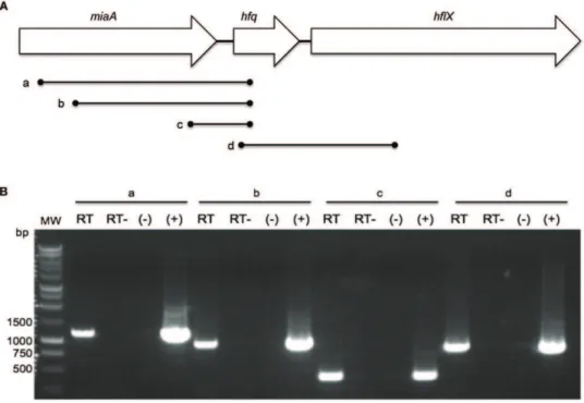
While theF. tularensisLVShfqgene locus, promoter region, and corresponding protein sequence have previously been described [13], it was unknown if theF. novicidahomolog possessed the same characteristics. TheF. novicida hfqgene (FTN_1051) is located on the negative strand at bps 1110271–1110600 of the genome. The gene encodes a 109 amino acid protein and is 99.39% identical to itsF. tularensisLVS homolog at the nucleotide level and 100% at the amino acid level. In both species, thehfqgene is flanked by the miaAgene upstream [encoding tRNA delta(2)-isopentenylpyropho-sphate transferase] and hflX downstream [encoding a GTP-binding protein] (Fig. 1A) [13], a configuration found in a number of bacteria such asE. colithat may form part of an operon [35].
To test ifF. novicida hfqwas part of an operon we performed RT-PCR experiments targeting the intergenic regions directly up- and downstream ofhfq. Transcription ofhfqinE. colihas been shown to initiate from multiple upstream promoter sequences, including two located within themiaAgene [36]. To test ifhfqtranscription inF. novicida originated from other promoters besides the single one identified 65 bp upstream of the F. holarctica hfqgene [13],
RT-PCR was performed using reverse primer FN-hfq-151R and forward primers 25F, 274F, and FN_miaA-833F (designated lines a, b, and c respectively) internal tomiaA (Fig. 1A). The resulting PCR products (1164 bp, 915 bp, and 357 bp) indicated co-transcription ofhfqandmiaAthroughout the length of the miaA ORF, suggesting at least one additional promoter region is responsible forhfqtranscription (Fig. 1B). 59
RACE experiments placed the start site ofhfqtranscription265 bases relative to its ATG start codon, which matches similar work performed inF. tularensisLVS [13]. However, 59RACE did not indicate additional transcription start sites further upstream. In order to ascertain ifhfqis co-transcribed with the downstream gene hflX, RT-PCR using primers FN_hfq-22F and FN_hflX-501R (line d) was performed (Fig. 1A). An 853 bp product resulted, suggesting co-transcription ofhfqwith the downstream genehflX (Fig. 1B).
F. novicidaHfq is unable to complement anE. coli hfq
mutation
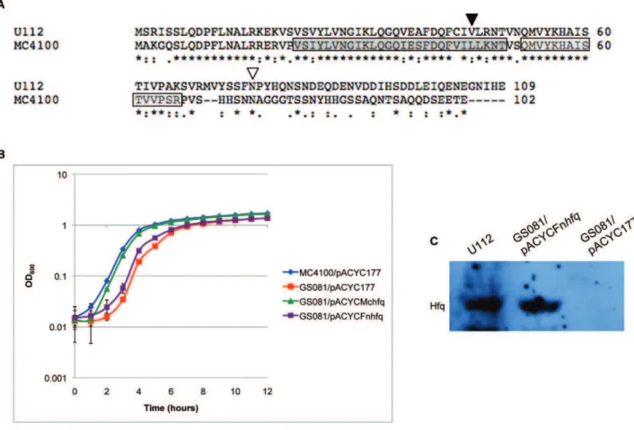
TheF. novicidaHfq protein is slightly larger than the Hfq protein ofE. colistrain MC4100 (109 a.a. versus 102 a.a.) and is 39% and 49% identical at the nucleotide and amino acid level respectively (Fig. 2A). To investigate the functionality ofF. novicidaHfq in the E. coli hfqmutant strain GS081, a region spanning thehfqgene was ligated to the E. coli hfq promoter region, cloned into plasmid pACYC177 and transformed into GS081 [24] yielding strain GS081/pACYCFnhfq. A region containing the nativeE. colistrain MC4100 hfq gene and promoter region was also cloned into pACYC177 as a control yielding pACYCMchfq, which was transformed into GS081. These two transformants, along with the E. coli parent strain MC4100 andE. coli hfq mutant strain GS081, were each grown in LB broth plus appropriate antibiotics at 37uC to allow comparison of their growth rates. Additionally,
MC4100 and GS081 were each grown harboring empty pACYC177 as a control, both of which displayed the same growth rate as the parent strains (data not shown). TheE. coli hfq mutant GS081 showed a modest growth defect relative to the parent strain. Expression of E. coliHfq, under the control of its native promoter, almost fully restored normal growth. However, the strain containingF. novicida hfqexhibited a slower growth rate than the strain containing native MC4100hfqand did not appear to complement the hfq mutant phenotype (Fig. 2B). Non-quantitative immunoblot analysis verified expression ofF. novicida Hfq protein in theE. colimutant strain GS081 (Fig. 2C).
Phenotypic analysis ofhfqtransposon insertion mutants To determine what effect interruption of thehfqgene has on theF. novicidaphenotype, two separate transposon insertion mutants were obtained from a F. novicida library created by Gallagher and co-workers [37]. Locations of the EZ-Tn5,KAN-2.insertions mapped to positions 229 and 133 bp relative to the 327 bphfqORF for strains Tnhfq1 and Tnhfq2, respectively (Fig. 2A). Phenotypic analysis of these twohfqinsertion mutants compared to U112 at 37uC indicated no growth difference for Tnhfq1 or Tnhfq2 (Fig. 3A). Further analysis using 42uC heat stress showed a slower rate of growth for Tnhfq1 compared to U112 at 42uC and its own growth phenotype at 37uC. A more severe defect in growth rate was observed for Tnhfq2, which took
over fifteen hours to exit lag phase (Fig. 3B). The different phenotypes observed between Tnhfq1 and Tnhfq2, suggests that the N-terminal half of theF. novicidaHfq is more important for growth during growth in certain stress conditions than the C-terminal half.
Construction ofF. novicida hfq deletion mutant and growthin vitro
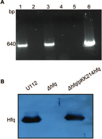
Homologous recombination was used to create a deletion mutant in which theF. novicida hfqORF was replaced by anermC ORF. After verifying that marker exchange had occurred by PCR and Southern blotting (data not shown), RT-PCR using primers FN_hflX-373F and FN_hflX-1013R internal tohflXverified that deletion ofhfqdid not have a polar effect on transcription of the downstream gene hflX (Fig. 4A). We then used the low copy-number vector pKK214gfpto generate a plasmid-based comple-menting clone of hfqunder the control of its native promoter, creating strain Dhfq/pKK214hfq. Subsequent non-quantitative immunoblot analysis verified the production of Hfq protein in both the wild-type U112 and the complemented strain, and the loss of Hfq in the mutant (Fig. 4B). Immunoblot analysis of thehfq mutant strain harboring empty pKK214 as a control also exhibited a loss of Hfq (data not shown).
To examine if the entire Hfq protein is important forF. novicida growth, we compared the growth of U112, Dhfq, and Dhfq/ Figure 2. Complementation ofE. coli hfqmutant withF. novicidaHfq.(A) Alignment of the Hfq proteins fromF. novicidastrain U112 andE. coli
strain MC4100 using the ClustalW program. Periods designate semiconserved substitutions, colons designate conserved substitutions, and asterisks designate residues found in both strains. A dark gray and light gray box indicates the location ofE. coliSm1 and Sm2 sequence motifs respectively. Inverted arrows designate the location of transposon insertion withinF. novicida hfq. The arrow at position 45 a.a. (.) corresponds to strain Tnhfq2 insertion, and the arrow at position 76 a.a. (,) corresponds to strain Tnhfq1 insertion. (B) Growth ofE. colistrains MC4100/pACYC177, GS081/ pACYC177, GS081/pACYCMchfq, and GS081/pACYCFnhfqin LB broth at 37uC with appropriate antibiotics. (C) Detection ofF. novicidaHfq assessed by immunoblot analysis inF. novicidastrain U112, GS081/pACYCFnhfq, and GS081/pACYC177 using anti-Hfq antisera.
pKK214hfqstrains in liquid medium at 37uC. Both the wild-type and hfqmutant were grown with empty pKK214 vector to account for any plasmid maintenance effects in the complemented strain. The deletion strain, Dhfq/pKK214, exhibited a longer lag phase compared to wild-type U112/pKK214. The complemented strain,
Dhfq/pKK214hfq, exhibited a similar growth compared to U112/ pKK214 but eventually reached a higher final optical density (Fig. 5).
Hfq is important for certainF. novicida stress responses Francisellapossesses a diverse lifestyle and like most pathogens is exposed to various stressors. We therefore examined if Hfq is important in the stress resistance ofF. novicidaby subjecting U112/ pKK214,Dhfq/pKK214, andDhfq/pKK214hfqstrains to certain stress conditions during growth in liquid medium. When adding
1% NaCl to the medium, the U112/pKK214 strain exhibited an enhanced growth rate and reached a higher final optical density than in medium lacking this supplement, however a rapid decrease in cell density quickly followed. Growth of the Dhfq/pKK214 strain was also slightly enhanced with additional 1% NaCl compared to growth in medium without this supplement (Figs. 5 and 6A). In medium containing 2% NaCl, Dhfq/pKK214 exhibited a prolonged lag phase relative to medium containing 1% NaCl (Fig. 6B). Upon entry into stationary phase, U112/ pKK214 cell density decreased while the hfq mutant remained unchanged compared to growth in unadjusted medium (data not shown). The growth of the strains was also compared in medium adjusted to pH 5. Strain U112 exhibited a slightly slower rate of growth and reached a lower final optical density compared to Figure 3. Growth characterization ofhfqtransposon strains.(A)F. novicidastrains U112, Tnhfq1, and Tnhfq2 were cultured in TS broth supplemented with cysteine and appropriate antibiotics at 37uC and the OD600 nmof each culture was measured at select time points. (B) Growth
unadjusted medium. TheDhfqstrain also showed a slower growth rate and prolonged lag phase in the acidic medium compared to unadjusted medium but still reached a higher density than U112 (Fig. 6C). Growth of Dhfq/pKK214hfqand pKK214-containing control strains exhibited little or no growth during the initial 24 hours of incubation at acidic pH (data not shown). At 42uC, the U112/pKK214 rate of growth was unchanged but the final density was lower compared to growth at 37uC. The Dhfq/ pKK214 strain exhibited a slightly slower rate of growth at 42uC. TheDhfq/pKK214 strain also reached a lower final optical density and never surpassed the density of U112/pKK214 (Fig. 6D). Addition of 0.0015% hydrogen peroxide led to a prolonged lag phase for both U112/pKK214 and Dhfq/pKK214, after which both strains exhibited a rate of growth similar to incubation in unadjusted medium. However, the complemented strain Dhfq/ pKK214hfqwas able to exit lag phase sooner than even U112/ pKK214 (Fig. 6E).
To further examine the role of Hfq in the stress response ofF. novicidawe monitoredhfqexpression using qRT-PCR experiments. Cultures of U112 were grown to exponential phase, then separated into two portions as described in the methods. One portion was allowed to continue growth at 37uC while the other was incubated with additional 1% NaCl, in medium adjusted to pH 5, or in unadjusted medium at 42uC. Expression ofhfqwas quantified (using primers Q_Fn_hfq-84F/Q_Fn_hfq-195R) from RNA extracted at 20 minutes, one hour, and four hours post-separation and compared tohfqlevels from time 0 (pre-separation). Measurements of culture optical density at these time points indicate an approximate correlation to exponential, early stationary, and late stationary phases respectively. Expression of hfqexhibited a 5.21-fold decrease after prolonged growth (4 hr) in medium containing additional 1% NaCl. A similar decrease (3.55-fold) in hfqexpression was observed shortly after incubation at 42uC. Conversely, a 2.02-fold increase in expression was observed after one hour of growth in acidic medium. We did not observe significant changes inhfqexpression for the other measured time points (Fig. 7).
Figure 4. Affect of hfq deletion onhflX expression and Hfq protein expression inF. novicidastrains.(A) RT-PCR using primers internal tohflXwas performed with RNA isolated from U112 with RT (lane 1), RNA from U112 without RT (lane 2), RNA isolated from thehfq
mutant with RT (lane 3), RNA from thehfqmutant without RT (lane 4), PCR with no DNA template as a negative control (lane 5), and PCR with DNA isolated from U112 as a positive control (lane 6). (B) Immunoblot analysis of U112, the hfqmutant, andDhfq/pKK214hfq strains using anti-Hfq antiserum.
doi:10.1371/journal.pone.0019797.g004
Figure 5. Growth characteristics of the U112hfqmutant.F. novicidastrains U112/pKK214,Dhfq/pKK214, andDhfq/pKK214hfqwere cultured in TS broth supplemented with cysteine and appropriate antibiotics at 37uC and the OD600 nmof each culture was measured over the course of time.
Hfq promotes formation of biofilms inF. novicida
Biofilm growth has been shown to confer both enhanced survival during stress conditions and environmental persistence. BecauseFrancisellahas been shown to produce biofilms [10,33,38], we decided to test biofilm formation by U112 and theDhfqstrain. Cultures were grown statically in microtiter plates at 37uC for 48 hours followed by staining with 0.1% crystal violet and measurement of the OD590 nm. BothF. novicidastrain U112 and
thehfqmutant showed similar crystal violet staining when grown in TS broth supplemented with cysteine (data not shown). However, thehfqmutant exhibited increased crystal violet staining
compared to wild-type when grown in MH broth indicating increased accumulation of adherent biomass (Fig. 8).
Loss of Hfq results in expression changes for a variety of genes
From microarray analysis of a F. tularensis LVS hfq mutant, numerous genes were identified as being regulated by Hfq [13]. We selected genes from that study which exhibited at least a 2-fold change in expression to profile inF. novicidastrains U112 andDhfq. Quantitative real-time PCR was performed on RNA extracted from cultures of U112 and Dhfq grown to exponential and Figure 6. Stress resistance of the U112 hfqmutant. Growth characteristics of F. novicidastrain U112/pKK214, Dhfq/pKK214, andDhfq/ pKK214hfqstrains in TS broth supplemented with cysteine and appropriate antibiotics under the following conditions: (A) additional 1% NaCl at 37uC, (B) additional 2% NaCl at 37uC, (C) adjusted to pH 5 at 37uC, (D) at 42uC, and (E) addition of 0.0015% H2O2at 37uC.
doi:10.1371/journal.pone.0019797.g006
Figure 7. Quantification ofhfqexpression during stress exposure using quantitative real-time PCR.Normalized fold expression ofhfq
transcripts in U112 grown in TS broth supplemented with cysteine at 37uC, at 37uC with the addition of 1% NaCl, at 37uC in medium adjusted to pH 5, and in unadjusted medium at 42uC. Transcript levels were normalized to the level of DNA helicase gene expression (FTN_1594).
stationary phase. During exponential phase, the expression of two genes decreased significantly (bioD [FTN_0812] and pdpB [FTN_1310]). One of these genes,pdpB, is part of theFrancisella pathogenicity island [7]. Four genes displayed significantly altered expression between U112 and Dhfqduring growth in stationary phase. Two of these genes were upregulated in the hfq mutant (dnaG [FTN_0914] and pyrF [FTN_0035]), while the other two genes were downregulated (katG [FTN_0633] and pdpA [FTN_1309]). Additionally, we examined expression of the mglA gene (FTN_1290) which encodes a global transcriptional regulator previously shown to regulatehfq[39]. While no significant change was found during exponential phase, expression of mglA was upregulated during stationary phase in thehfqmutant.
Discussion
The ability to monitor environmental changes within the host organism and adjust the expression of stress- and virulence-associated genes accordingly is critical for the growth and survival of pathogenic bacteria. The importance of the small RNA chaperone Hfq in modulating such genes has been demonstrated in a variety of both intra- and extracellular bacterial pathogens [19,20,40]. This study demonstrates the role Hfq plays in stress tolerance, gene expression, and biofilm formation in the intracellular pathogenFrancisella novicida.
Previously, it was shown thathfq transcription in F. tularensis LVS is controlled by a single promoter located in between the miaAandhfqORFs [13]. Interestingly, we found that whilehfqis transcribed from an identical promoter region inF. novicida, it is also co-transcribed with the upstream genemiaA. Thus expression of the locus inF. novicidaappears to differ from that ofF. tularensis LVS. Alternatively, this conflicting data could be explained by differences in experimental design. In E. coli, multiple upstream promoters, including two withinmiaA, drive transcription of hfq, which is located within the miaA-hfq-hflX operon [36]. This genomic locus is conserved inF. novicida, making it tempting to speculate thathfqexpression is also under the control of multiple promoters both within and upstream ofmiaA. However, because our results only indicate a minimum of co-transcription with the
majority of the miaA transcript (Fig. 1), the exact location and number of additional promoter regions remain to be determined. Despite the shared operon structure, Hfq inF. novicidais unable to complement an E. coli hfq mutant (Fig. 2B). This inability to complement anE. coli hfqmutant is likely due to basic structural differences between the two Hfq proteins as reflected in the amino acid sequence, especially in the two Sm-domains (Fig. 2A).
Complete deletion ofF. novicida hfq only resulted in a modest growth defect compared to wild-type U112 (Fig. 5). The transposon mutant analysis illustrates the importance of the F. novicidaHfq N-terminal domain during 42uC growth (Fig. 3B). The N-terminal insertion of strain Tnhfq2 would disrupt a region similar to the Sm1 domain ofE. coli Hfq, while the C-terminal disruption of strain Tnhfq1 would leave both Sm domains intact (Fig. 2A) [41]. The Sm motifs found in Hfq proteins are vital for hexameric formation and riboregulation, while the exact function of the C-terminal half remains unclear [22,42]. However, stability of the two transposon mutant variants is unknown since a Western blot signal could not be observed using theF. novicidaHfq specific antisera (data not shown). While neither insertion strain exhibited a more dramatic phenotype than the completehfqdeletion strain during growth at 37uC, the lag phase of Tnhfq2 during heat shock was more pronounced than thehfqknockout strain (Fig. 3B and Fig. 6D). The reason for this discrepancy is unclear, but further studies are currently underway to elucidate this phenotype.
The importance of Hfq for stress tolerance has been reported in a variety of other bacteria, including pathogens such asPseudomonas aeruginosaandNeisseria gonorrhoeae[43]. As such, the importance of F. novicidaHfq in growth and survival during stress conditions was examined by comparing phenotypic differences between the hfq mutant strain and wild-type U112, as well as monitoring changes inhfqtranscription. The slightly faster growth rate for both U112/ pKK214 and Dhfq/pKK214 when exposed to increased salt concentration suggests that additional NaCl provides a growth advantage to both strains (Figs. 5, 6A, and 6B), while the increased lag phase ofDhfq/pKK214 when exposed to 2% additional NaCl and the subsequent reduced transcription ofhfqduring growth in 1% NaCl (Fig. 6B and 7) indicate a potential contribution for Hfq Figure 8. Biofilm formation byF. novicidaU112 andDhfqstrains.Both strains were assayed for biofilm formation using crystal violet staining after 48 hours of static growth in MH broth at 37uC. Culture OD590 nmwas determined for multiple replicates. U112 demonstrated decreased crystal
during osmotic stress. This finding is also supported by previous work inF. tularensissubspecies holarctica[13]. Similar phenotypes have been observed in Gram-negative pathogens such as Burkholderia cepacia,Moraxella catarrhalis, andNeisseria gonorrhoeae[43]. Tolerance to low pH is important inF. tularensisdue to its ability to survive and escape the phagosome during intracellular infection of certain host cell types such as macrophages. Phagosomes have previously been shown to become acidic in response toF. tularensis infection [44]. While both U112 andDhfqexhibited a reduced rate during growth in acidic media, the prolonged lag phase ofDhfq (Fig. 6C) as well as increased expression ofhfqafter one hour in acidic conditions (Fig. 7) suggest a role for Hfq in the initial response to low pH. Interestingly, growth of the complemented strain and strains harboring empty pKK214 was severely inhibited by the decreased pH suggesting difficulties in plasmid maintenance during growth at pH 5, and was therefore not included in this analysis. Additionally, we examined the growth rate of U112/pKK214 and Dhfq/pKK214 during heat shock. Compared to the salt and low pH exposure, heat shock had a more negative effect on the growth rate ofDhfq/pKK214 and resulted in entry into stationary phase at a lower optical density than all other conditions tested (Fig. 6). This phenotype suggests a role for Hfq in the heat shock response even though transcription ofhfqwas initially repressed upon introduction of the stressor before returning to normal levels (Fig. 7).
Reactive oxygen species are one defense mechanism used by macrophages, monocytes, and neutrophils to combat intracellular pathogens. F. tularensis has been reported to inhibit particular neutrophil killing mechanisms such as the respiratory burst as part of its infection lifecycle [45,46]. Oxidative stress induced by addition of hydrogen peroxide had a severe effect on both U112/pKK214 and Dhfq/pKK214 as indicated by their pronounced lag phases (Fig. 6E). However, the additional six-hour lag phase exhibited by Dhfq/pKK214 indicates that Hfq is necessary for combating initial exposure to H2O2. Similar growth responses to oxidative stress have
been reported forhfqmutants in an array of pathogens [43]. While expression ofhfqwas not assayed under this condition, expression of mglAwas upregulated in the mutant (Fig. 9). Previous studies using a
F. novicida mglAmutant have shown that lack of MglA results in increased resistance to H2O2 [39]. It is therefore tempting to
speculate that the increased expression ofmglAinDhfqis directly linked to the increased lag phase of the strain during H2O2
exposure. However, further studies are necessary to determine the role Hfq plays in regulating mglA expression. As a whole, these experiments demonstrate a role forF. novicidaHfq during growth under a variety of environmental conditions.
The highly pleiotropic phenotype of an E. coli hfq mutant is largely due to the importance of Hfq in sRNA-mediated regulation. A number of sRNA targets are global regulators such as the alternative sigma factors rpoH, encoding the heat-shock sigma factors32, andrpoS, which encodes the general stress signal
factorsS[47,48].F. novicidalacks an annotatedrpoSgene and to date no known master regulator controlling the transition from exponential to stationary phase has been characterized. Interest-ingly, we observed thehfqmutant reaching a higher cell density before entry into stationary phase compared to wild-type under most conditions tested (Figs. 5 and 6). The complemented strain (Dhfq/pKK214hfq) also surpassed the cell density of U112/ pKK214 before entering stationary phase. Because the native promoter region directly upstream ofhfqis solely responsible for expression of Hfq in the complemented strain, the presence of multiple promoter regions controlling transcription ofhfqin U112 suggests that the partial-complementation observed in Dhfq/ pKK214hfq is due to insufficient cellular concentration of Hfq, especially as cellular density increases. The importance of controlling intracellular Hfq concentration has been shown in several bacteria, includingAcinetobacter baylyi, where overexpression led to an inability of the cells to assemble into chains [49]. Additionally, while investigating the role of Hfq in Yersinia pseudotuberculosis, Bai and colleagues found thathfqmRNA levels in a partially complemented strain were lower than those of the wild-type due to differential transcription from multiple promot-ers. A multicopy construct was required to express hfqto levels necessary for full complementation to be observed [50]. Guina and coworkers have also shown that MglA is important forF. novicida
Figure 9. Quantification of selected gene transcription using quantitative real-time PCR.Changes in gene expression in thehfqmutant compared to wild-type U112 were examined during both exponential (exp) and stationary phase (stat) using RNA derived from growth in TS broth supplemented with cysteine at 37uC. Each gene was analyzed using RNA derived from three separate cultures and each reaction was performed in triplicate. Transcript levels were normalized to the level of DNA helicase (FTN_1594).
survival during stationary-phase growth [39]. Thus the increased expression of mglA in Dhfqreported here could also be directly related to the increased cell density observed during stationary-phase. Additionally, global analysis of gene expression in a F. tularensisLVShfqmutant strain revealed upregulation ofrpoD, the gene encoding the exponential phase s70 [13]. Farewell et al. demonstrated that overexpression of s70
in E. coli dramatically alters gene expression and inhibits development ofsS
-dependent phenotypes, mimicking the effect of anrpoSmutation [51]. Despite the lack of an annotatedsS
inF. novicida, we speculate that Hfq plays a role in the transition from exponential to stationary phase and that this role is dose-dependent.
Biofilm formation plays an important part in the transmission and environmental persistence of many bacterial pathogens. BecauseF. novicidahas recently been shown to form biofilms on natural and chitin surfaces [33], we wanted to examine if Hfq contributes to biofilm formation inF. novicida. While no significant differences in crystal violet staining were observed between the two strains after growth in TS broth, the higher crystal violet staining exhibited by the hfq mutant strain when grown in the more restrictive MH broth is indicative of increased biofilm formation (Fig. 8). This level of biofilm formation was similar to that reported for F. novicida on chitin surfaces [33]. Biofilm formation in Campylobacter jejuniis significantly increased during growth in MH broth compared to more nutrient-rich media, so it is possible that F. novicidagrown in rich TS broth is unable to form a substantial enough biofilm to observe a phenotypic difference between wild-type and thehfqmutant strain [52]. However, a ready explanation for increased biofilm formation in thehfqmutant during growth in MH broth is not apparent. Attia et al. demonstrated a growth advantage for aMoraxella catarrhalis hfqmutant over the wild-type in a continuous flow biofilm system. The authors suggest this phenotype results from altered gene expression in the Dhfq background leading to changes in the overall outer membrane architecture [53]. In accordance with this hypothesis, Meibom et al. found expression of an outer membrane protein (FTL_0535) and several Type IV pili-associated proteins (FTL_0797, FTL_0798, and FTL_0827) upregulated in a F. tularensis LVS hfq mutant strain [13]. While further expression analyses are needed to confirm this hypothesis, it seems possible that changes in outer membrane protein architecture could account for the increased biofilm formation observed in theF. novicida hfqmutant. Hfq is an important regulator for a variety of genes in numerous bacteria, includingF. tularensisLVS [13,20,40]. In order to evaluate its role in F. novicida, we monitored expression of nine genes identified by microarray analysis of aF. tularensisLVShfqmutant [13]. Seven of these genes showed significant (greater than 2-fold) changes in expression. Two genes,pdpAandpdpB, encoded within theFrancisellapathogenicity island were downregulated at different times in the hfq mutant. Previous studies have shown that inactivation of these two genes inF. novicidaleads to growth defects within host cells and attenuation of virulence [6,8]. Another downregulated gene, katG, encodes a protein annotated as possessing catalase/peroxidase activity, potentially a factor in the
severeDhfqgrowth defect observed during oxidative stress (Fig. 6E), as inactivation of katG increased susceptibility of F. novicida to hydrogen peroxide [39]. Three of the genes examined in this study, katG,dnaG, andpyrF, exhibited similar expression changes to those previously reported by Meibom and colleagues while three other genes, pdpA, pdpB, and bioD, exhibited different changes in expression [13]. While the reason behind these differences is unclear, it is interesting to note that two of the genes,pdpAandpdpB, are virulence factors encoded in the FPI, of which there is only one copy in theF. novicidagenome compared to more virulent strains of Francisella which contain two copies [54]. Additionally, the transcriptional regulator MglA was upregulated during stationary phase growth in the hfq mutant. MglA has been implicated in coordinating both virulence gene expression and the stress response ofF. tularensisby interacting with SspA (Stringent starvation protein A) and binding RNA polymerase [12,39]. Absence of MglA has also been shown to result in increasedkatGexpression inF. novicida[39], thus the downregulation ofkatGobserved in this study could be a consequence of increasedmglAexpression in theDhfqstrain. While we are unable to determine if the expression changes observed for the profiled genes are directly or indirectly affected by Hfq, the protein still appears to play a role in their expression.
The ability of Hfq to affect such wide-ranging cellular functions is likely due to its role as an RNA chaperone for sRNAs. Hfq facilitates pairing interactions between small RNAs and their mRNA targets in several bacteria leading to post-transcriptional regulation of the target gene [55]. To date, no small RNAs have been identified inF. novicida so the exact regulatory function of Hfq remains to be elucidated, but it is likely that many of the effects observed here are due to interactions with as yet unidentified sRNAs. Our work demonstrates the importance of Hfq in cell growth, stress resistance, gene expression, and biofilm formation inF. novicida. Information from these studies is critical to understanding how this pathogen responds to and survives in the diverse environments it occupies.
Supporting Information
Figure S1 Expression of qRT-PCR reporter geneuvrD.
Relative change in uvrD expression across multiple time points using RNA derived from U112 growth in TS broth supplemented with cysteine at 37uC.
(TIF)
Acknowledgments
We thank Tendai Mutangadura, University of Missouri, for creating theF. novicida hfq deletion cassette, Brian Beal, AgReliant Genetics LLC, for expression of theF. novicidaHfq for antisera generation, and Gisela Storz, National Institute of Health, for providing theE. coliGS081 (hfqmutant) and MC4100 strains.
Author Contributions
Conceived and designed the experiments: JRC KSB. Performed the experiments: JRC. Analyzed the data: JRC KSB. Contributed reagents/ materials/analysis tools: KSB. Wrote the paper: JRC KSB.
References
1. McLendon MK, Apicella MA, Allen L-AH (2006) Francisella tularensis: Taxonomy, genetics, and immunopathogenesis of a potential agent of biowarfare. Annual Reviews in Microbiology 60: 167–185.
2. Ellis J, Oyston PCF, Green M, Titball RW (2002) Tularemia. Clinical Microbiology Reviews 15: 631–646.
3. Mo¨rner T (1992) The ecology of tularemia. Revue scientifique et technique 11: 1123–1130.
4. Parker RR, Steinhaus EA, Kohls GM, Jellison WL (1951) Contamination of natural waters and mud withPastuerella tularensis. Bulletin National Institute of Health 193: 7–33.
5. Sjo¨stedt A (2006) Intracellular survival mechanisms ofFrancisella tularensis, a stealth pathogen. Microbes and Infection 8: 561–567.
6. Nano FE, Zhang N, Cowley SC, Klose KE, Cheung KKM, et al. (2004) A
Francisella tularensispathogenicity island required for intramacrophage growth. Journal of Bacteriology 186: 6430–6436.
7. Schmerk CL, Duplantis BN, Wang D, Burke RD, Chou AY, et al. (2009) Characterization of the pathogenicity island protein PdpA and its role in the virulence ofFrancisella novicida. Microbiology 155: 1489–1497.
8. Weiss DS, Brotcke A, Henry T, Margolis JJ, Chan K, et al. (2007)In vivo
Proceedings of the National Academy of Sciences USA 104: 6037– 6042.
9. Su J, Yang J, Zhao D, Kawula TH, Banas JA, et al. (2007) Genome-wide identification of Francisella tularensis virulence determinants. Infection and Immunity 75: 3089–3101.
10. Dean RE, Ireland PM, Jordan JE, Titball RW, Oyston PC (2009) RelA regulates virulence and intracellular survival ofFrancisella novicida. Microbiology 155: 4104–4113.
11. Brotcke A, Monack DM (2008) Identification of fevR, a novel regulator of virulence gene expression inFrancisella novicida. Infection and Immunity 76: 3473–3480.
12. Charity JC, Costante-Hamm MM, Balon EL, Boyd DH, Rubin EJ, et al. (2007) Twin RNA Polymerase-associated proteins control virulence gene expression in
Francisella tularensis. PLoS Pathogens 3: e84.
13. Meibom KL, Forslund A-L, Kuoppa K, Alkhuder K, Dubail I, et al. (2009) Hfq, a novel pleiotropic regulator of virulence-associated genes inFrancisella tularensis. Infection and Immunity 77: 1866–1880.
14. Sittka A, Pfeiffer V, Tedin K, Vogel J (2007) The RNA chaperone Hfq is essential for the virulence ofSalmonella typhimurium. Molecular Microbiology 63: 193–217.
15. Schiano CA, Bellows LE, Lathem WW (2010) The small RNA chaperone Hfq is required for the virulence ofYersinia pseudotuberculosis. Infection and Immunity 78: 2034–2044.
16. Mellin JR, McClure R, Lopez D, Green O, Reinhard B, et al. (2010) Role of Hfq in iron-dependent and -independent gene regulation inNeisseria meningitidis. Microbiology 156: 2316–2326.
17. Franze de Fernandez MT, Eoyang L, August JT (1968) Factor fraction required for the synthesis of bacteriophage Qbeta-RNA. Nature 219: 588–590. 18. Møller T, Franch T, Højrup P, Keene DR, Ba¨chinger HP, et al. (2002) Hfq: a
bacterial Sm-like protein that mediates RNA-RNA interaction. Molecular Cell 9: 23–30.
19. Christiansen JK, Larsen MH, Ingmer H, Søgaard-Anderson L, Kallipolitis BH (2004) The RNA-binding protein Hfq ofListeria monocytogenes: Role in stress tolerance and virulence. Journal of Bacteriology 186: 3355–3362.
20. Dietrich M, Munke R, Gottschald M, Ziska E, Boettcher JP, et al. (2009) The effect ofhfqon global gene expression and virulence inNeisseria gonorrhoeae. FEBS Journal 276: 5507–5520.
21. Sonnleitner E, Schuster M, Sorger-Domenigg T, Greenberg EP, Bla¨si U (2006) Hfq-dependent alterations of the trancriptome profile and effects on quorum sensing inPseudomonas aeruginosa. Molecular Microbiology 59: 1542–1558. 22. Tsui H-CT, Leung H-CE, Winkler ME (1994) Characterization of broadly
pleiotropic phenotypes caused by anhfqinsertion mutation inEscherichia coli K-12. Molecular Microbiology 13: 35–49.
23. Geng J, Song Y, Yang L, Feng Y, Qiu Y, et al. (2009) Involvement of the post-transcriptional regulator Hfq inYersinia pestisvirulence. PLoS ONE 4: e6213. 24. Zhang A, Wassarman KM, Ortega J, Steven AC, Storz G (2002) The Sm-like
Hfq protein increases OxyS RNA interaction with target mRNAs. Molecular Cell 9: 11–22.
25. Gottesman S, Storz G (2010) Bacterial small RNA regulators: versatile roles and rapidly evolving variations. Cold Spring Harb Perspect Biol: doi:10.1101/ cshperspect.a003798.
26. Meibom KL, Dubail I, Dupuis M, Barel M, Lenco J, et al. (2008) The heat-shock protein ClpB ofFrancisella tularensisis invoved in stress tolerance and is required for multiplication in target organs of infected mice. Molecular Microbiology 67: 1384–1401.
27. Bell BL, Mohapatra NP, Gunn JS (2010) Regulation of virulence gene transcripts by theFrancisella novicidaorphan response regulator PmrA: role of phosphorylation and evidence of MglA/SspA interaction. Infection and Immunity 78: 2189–2198.
28. Shevchuk NA, Bryksin AV, Nusinovich YA, Cabello FC, Sutherland M, et al. (2004) Construction of long DNA molecules using long PCR-based fusion of several fragments simultaneously. Nucleic Acids Research 32: e19.
29. Hamilton HL, Schwartz KJ, Dillard JP (2001) Insertion-duplication mutagenesis ofNeisseria: Use in characterization of DNA transfer genes in the Gonococcal genetic island. Journal of Bacteriology 183: 4718–4726.
30. Abd H, Johansson T, Golovliov I, Sandstro¨m G, Forsman M (2003) Survival and growth of Francisella tularensis in Acanthamoeba castellanii. Applied and Environmental Microbiology 69: 600–606.
31. LoVullo ED, Sherrill LA, Perez LL, Pavelka MS, Jr. (2006) Genetic tools for highly pathogenic Francisella tularensis subsp. tularensis. Microbiology 152: 3425–3435.
32. Djordjevic D, Wiedmann M, McLandsborough LA (2002) Microtiter plate assay for assesment ofListeria monocytogenesbiofilm formation. Applied and Environ-mental Microbiology 68: 2950–2958.
33. Margolis JJ, El-Etr S, Joubert L-M, Moore E, Robinson R, et al. (2010) Contributions ofFrancisella tularensissubsp.novicidachitinases and Sec secretion system to biofilm formation on chitin. Applied and Environmental Microbiology 76: 596–608.
34. Pfaffl MW (2001) A new mathematical model for relative quantification in real-time RT-PCR. Nucleic Acids Research 29: e45.
35. Tsui H-CT, Winkler ME (1994) Transcriptional patterns of themutL-miaA
superoperon of Escherichia coli K-12 suggest a model for posttranscriptional regulation. Biochimie 76: 1168–1177.
36. Tsui H-CT, Feng G, Winkler ME (1996) Transcription of themutLrepair,miaA
tRNA modification,hfqpleiotropic regulator, andhflAregion protease genes of
Escherichia coliK-12 from clustered Es32
-Specific promoters during heat shock. Journal of Bacteriology 178: 5719–5731.
37. Gallagher LA, Ramage E, Jacobs MA, Kaul R, Brittnacher M, et al. (2007) A comprehensive transposon mutant library ofFrancisella novicida, a bioweapon surrogate. Proceedings of the National Academy of Sciences USA 104: 1009–1014.
38. Durham-Colleran MW, Verhoeven AB, van Hoek ML (2009)Francisella novicida
forms in vitro biofilms mediated by an orphan response regulator. Environ-mental Microbiology 59: 457–465.
39. Guina T, Radulovic D, Bahrami AJ, Bolton DL, Rohmer L, et al. (2007) MglA regulates Francisella tularensis subsp. novicida (Francisella novicida) response to starvation and oxidative stress. Journal of Bacteriology 189: 6580–6586. 40. Ding Y, Davis BM, Waldor MK (2004) Hfq is essential forVibrio cholerae
virulence and downregulates sE
expression. Molecular Microbiology 53: 345–354.
41. Olsen AS, Møller-Jensen J, Brennan RG, Valentin-Hansen P (2010) C-Terminally truncated derivatives of Escherichia coli Hfq are proficient in riboregulation. Journal of Molecular Biology 404: 173–182.
42. Vecˇerek B, Rajkowitsh L, Sonnleitner E, Schroeder R, Bla¨si U (2008) The C-terminal domain ofEscherichia coliHfq is required for regulation. Nucleic Acids Research 36: 133–143.
43. Chao Y, Vogel J (2010) The role of Hfq in bacterial pathogens. Current Opinion in Microbiology 13: 24–33.
44. Chong A, Wehrly TD, Nair V, Fischer ER, Barker JR, et al. (2008) The early phagosomal stage ofFrancisella tularensisdetermines optimal phagosomal escape andFrancisellapathogenicity island protein expression. Infection and Immunity 76: 5488–5499.
45. McCaffrey RL, Allen L-AH (2006)Francisella tularensisLVS evades killing by human neutrophils via inhibition of the respiratory burst phagosome escape. Journal of Leukocyte Biology 80: 1222–1223.
46. Schulert GS, McCaffrey RL, Buchan BW, Lindemann SR, Hollenback C, et al. (2009) Francisella tularensis genes required for inhibition of the neutrophil respiratory burst and intramacrophage growth identified by random transposon mutagensis of strain LVS. Infection and Immunity 77: 1324–1336.
47. Hengge-Aronis R (2002) Signal transduction and regulatory mechanisms involved in control of thesS
(RpoS) subunit of RNA Polymerase. Microbiology and Molecular Biology Reviews 66: 373–395.
48. Guisbert E, Rhodius VA, Ahuja N, Witkin E, Gross CA (2007) Hfq modulates thesE
-mediated envelope stress response and thes32
-mediated cytoplasmic stress response inEscherichia coli. Journal of Bacteriology 189: 1963–1973. 49. Schilling D, Gerischer U (2009) TheAcinetobacter baylyi hfqgene encodes a large
protein with an unusual C terminus. Journal of Bacteriology 191: 5553–5562. 50. Bai G, Golubov A, Smith EA, McDonough KA (2010) The importance of the
small RNA chaperone Hfq for growth of epidemicYersinia pestis, but notYersinia pseudotuberculosis, with implications for plague biology. Journal of Bacteriology 192: 4239–4245.
51. Farewell A, Kvint K, Nystro¨m T (1998) Negative regulation by RpoS: a case of sigma factor competition. Molecular Microbiology 29: 1039–1051.
52. Reeser RJ, Medler RT, Billington SJ, Jost BH, Joens LA (2007) Characterization ofCampylobacter jejunibiofilms under defined growth conditions. Applied and Environmental Microbiology 73: 1908–1913.
53. Attia AS, Sedillo JL, Wang W, Liu W, Brautigam CA, et al. (2008)Moraxella catarrhalis expresses an unusual Hfq protein. Infection and Immunity 76: 2520–2530.
54. Nano FE, Schmerk C (2007) TheFrancisellapathogenicity island. Annals of the New York Academy of Sciences 1105: 122–137.
55. Brennan RG, Link TM (2007) Hfq structure, function, and ligand binding. Current Opinion in Microbiology 10: 125–133.
56. Larson CL, Wicht W, Jellison WL (1955) A new organism resemblingP. tularensis