Immunolocalization of FGF-2 and VEGF in rat periodontal
ligament during experimental tooth movement
Milene Freitas Lima Salomão1, Sílvia Regina de Almeida Reis2, Vera Lúcia Costa Vale3, Cintia de Vasconcellos Machado4, Roberto Meyer5, Ivana Lucia Oliveira Nascimento5
How to cite this article: Salomão MFL, Reis SRA, Vale VLC, Machado CV, Meyer R, Nascimento ILO. Immunolocalization of FGF-2 and VEGF in rat periodontal ligament during experimental tooth movement. Dental Press J Orthod. 2014 May-June;19(3):67-74. DOI: http://dx.doi.org/10.1590/2176-9451.19.3.067-074.oar
» Patients displayed in this article previously approved the use of their facial and in-traoral photographs.
Contact address: Cíntia de Vasconcellos Machado
Rua Marechal Floriano, 354/701 – Canela – Salvador/BA — Brazil CEP: 40110-010 – E-mail: [email protected]
1 Assistant professor, School of Medicine and Public Health of Bahia (EBMSP). 2 Adjunct professor, EBMSP.
3 Full professor, University of the State of Bahia (UNEB). 4 Visiting professor, Brazilian Dental Association/Bahia (ABO-BA). 5 Associate Professor, UFBA.
» The authors report no commercial, proprietary or financial interest in the products or companies described in this article.
Objective:This article aimed at identifying the expression of fibroblast growth factor-2 (FGF-2) and vascular endothelial growth factor (VEGF) in the tension and pressure areas of rat periodontal ligament, in different periods of experimental orthodontic tooth movement. Methods: An orthodontic force of 0.5 N was applied to the upper right first molar of 18 male Wistar rats for periods of 3 (group I), 7 (group II) and 14 days (group III). The counter-side first molar was used as a control. The animals were euthanized at the aforementioned time periods, and their maxillary bone was removed and fixed. After demineralization, the specimens were histologically processed and embedded in paraffin. FGF-2 and VEGF expressions were studied through immunohistochemistry and morphological analysis. Results: The experimental side showed a higher expression of both FGF-2 and VEGF in all groups, when compared with the control side (P < 0.05). Statistically significant differences were also found between the tension and pressure areas in the experimental side. Conclusion: Both FGF-2 and VEGF are expressed in rat periodontal tissue. Additionally, these growth factors are upregulated when orthodontic forces are applied, thereby suggesting that they play an important role in changes that occur in periodontal tissue during orthodontic movement.
Keywords:Periodontal ligament. Orthodontics. Vascular endothelial growth factor A. Fibroblast growth factor 1.
DOI: http://dx.doi.org/10.1590/2176-9451.19.3.067-074.oar
Submitted: October 01, 2012
Revised and accepted: January 20, 2013
Objetivo:o objetivo desse estudo foi identificar a expressão do fator de crescimento de fibroblastos 2 (FGF-2) e do fator de crescimento vascular endotelial (VEGF) nos lados de tensão e pressão do ligamento periodontal de ratos, durante movimento ortodôntico experi-mental, em diferentes períodos de tempo. Métodos: uma força ortodôntica de 0,5N foi aplicada no primeiro molar superior direito de 18 ratos Wistar machos, por períodos de 3 (grupo I), 7 (grupo II) e 14 dias (grupo III). O primeiro molar do lado oposto foi utilizado como controle. Os animais foram sacrificados nos períodos de tempo mencionados, sendo a arcada superior removida e fixada. Após a desmineralização, os espécimes foram processados histologicamente e embebidos em parafina. A expressão do FGF-2 e do VEGF foram estudadas por meio de análise imuno-histoquímica. Resultados: o ligamento periodontal dos dentes submetidos à movimentação orto-dôntica mostraram maior expressão tanto de FGF-2 quanto de VEGF, em todos os grupos experimentais, quando comparados com os dentes do lado controle (p < 0,05). Diferenças estatisticamente significativas entre os lados de tensão e pressão também foram encontradas nos dentes submetidos à movimentação ortodôntica. Conclusões: tanto o FGF-2 quanto o VEGF são expressos no tecido periodontal de ratos, e esses fatores de crescimento são aumentados quando forças ortodônticas são aplicadas, sugerindo que esses desempenham um papel importante na reorganização do periodonto durante o movimento ortodôntico.
INTRODUCTION
Orthodontic tooth movement is achieved by re-modeling the alveolar bone and periodontal ligament
(PDL) in response to mechanical loading.17 It is a
highly sophisticated biological process that leads to local inflammation, with vascular, cellular and extra-cellular matrix (ECM) alterations that allow
remod-eling events and, ultimately, tooth displacement.17,37
Orthodontic tooth movement is characterized by the abrupt creation of compression and tension sides in the PDL, with a repeated process of alveolar bone resorption on the pressure side and new bone
forma-tion on the tension side.3,6,35
Although the exact mechanism of periodontal tis-sue remodeling is not clearly understood, a milieu of cytokines, growth factors, neurotransmitters, ECM components, colony-stimulating factors and inflam-matory mediators have been reported to be synthe-sized and released in PDL during orthodontic
move-ment.2,7,17,25,28,36 These molecules interact with
vari-ous dental and paradental cells and stimulate them to initiate and sustain tissue remodeling, inducing bone
deposition and resorption.6,42
Continuous orthodontic forces can exert pres-sure that compromises the integrity of the vascu-lar compartment in PDL. Over-compression re-sults in ischemia, gradual reduction of capillaries, presence of thrombi, interruption of nutrition and
cell death21,29 with almost unavoidable formation of
a necrotic or hyaline zone, mainly on the pressure
side.27,38 In contrast, dilated blood vessels were found
in the tension side.33,38
These vascular alterations can be mediated by dif-ferent growth factors, such as fibroblast growth fac-tor-2 (FGF-2) and vascular endothelial growth factor (VEGF). FGF-2, also known as basic FGF, is a potent angiogenic factor that shows increased expression in
hypoxic conditions and during wound healing.5,15
This growth factor enhances endothelial cell
prolif-eration and induces endothelial cell sprouting.3
Like-wise, FGF-2 is a component of bone matrix and plays
an important role in regulating bone remodeling.13,19
VEGF is considered the most important regulator of vasculogenesis and angiogenesis in physiological
as well as in pathological conditions.4,9In vivo, VEGF
enhances vascular permeability and induces potent
angiogenic responses.8,10 There is solid evidence for
a functional link between vasculogenesis and bone
development.41 Furthermore, VEGF may participate
in the regulation of bone metabolism and wound
healing during orthodontic tooth movement.16,23
This growth factor has the ability to induce func-tional osteoclasts when injected in PDL, thereby
in-creasing the rate of tooth movement in mice.16
Thus, this study was designed to assess the ex-pression levels of FGF-2 and VEGF in rat periodon-tal tissue submitted to mechanical forces in an ex-perimental model of orthodontic tooth movement.
MATERIAL AND METHODS
Animal model and experimental orthodontic tooth movement
All experiments were conducted according to the guidelines of the Ethics Committee on Animal Use from the Federal University of Bahia (Brazil) where this study was submitted and approved.
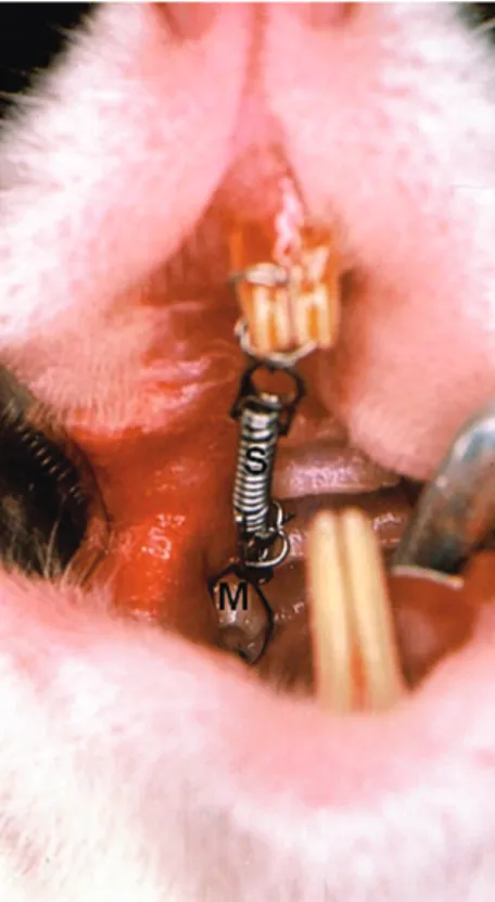
The study sample comprised 18 male Wistar rats aged between 60 ± 5 days (mean ± SD), with a mean weight of 170 g. The upper right first mo-lar in each animal was mesially moved by means of a closed coil spring (3M Brasil, Sumaré, Brazil) which was fixed to the upper incisor from the same side (Fig 1), as previously described by Heller and
Nanda.11 Grooves were made on the incisors to
sup-port the appliance. Forces of 0.5 N were applied for periods of 3 (group I, n = 6), 7 (group II, n = 6) and 14 days (group III, n = 6). The intensity of force was assessed using a dynamometer (Dentaurum Brasil, São Paulo, Brazil) while the spring was being fixed and then every day during the three different experi-mental periods. The upper left first molar, which was not subjected to any orthodontic movement, served as control. The orthodontic appliance was fixed and activated under anesthesia induced by intraperitoneal injection of ketamine (0.12 ml/100 g) and xylazine (0.06 ml/100 g). The animals had access to food and
water ad libitum, and were kept on a reversed 12-h
light/12-h dark cycle (dark period 08.00–20.00 h).
Tissue processing
The specimens were decalciied in 10% EDTA at room temperature (pH 7.2) for 12 weeks, processed histo-logically and parain embedded. Sections of 5 μm, parallel to the long axis of the irst upper molar, were obtained and mounted on glass slides.
Morphological and
immunohistochemical analysis
For morphological analysis, one section of each sample was stained with hematoxy-lin and eosin and analyzed by light microscopy.
For immunohistochemical assay, a streptavidin-bio-tin complex (LSAB, Dako Cytomation, Carpinteria, USA) was used. For detection of FGF-2 and VEGF, polyclonal anti-FGF-2 (dilution 1:1000; clone 147; Santa Cruz Biotechnology, Santa Cruz, CA) and monoclonal anti-VEGF (dilution 1:50; clone C-1; Santa Cruz Biotechnology, Santa Cruz, CA) anti-bodies were respectively used. The sections were dewaxed, rehydrated and washed in distilled water. The antigen retrieval was performed by enzymat-ic digestion with 1% trypsin (Sigma, Saint Louis, USA) for 20 min at 37°C. Endogenous peroxidase was blocked by treatment with 3% hydrogen perox-ide for 10 min at 25°C. The slperox-ides were then incu-bated with the primary antibody in a humid cham-ber overnight at 4°C. Subsequently, the slides were washed with 1% PBS/BSA and incubated with bio-tinylated secondary antibodies (link reagent, Dako Cytomation, Carpinteria, USA) for 60 min at room temperature, followed by washing and incubation with the streptavidin-biotin-peroxidase complex. Diaminobenzidine (Dako Cytomation, Carpinteria, USA) was used as chromogen and the slides were counterstained with Harris hematoxylin (Sigma, Saint Louis, USA) for 15 seconds. Negative controls included replacement of primary antibodies with non-immune bovine serum albumin.
Specific areas of the PDL were selected for mor-phological and immunohistochemical assessment. They corresponded to pressure and tension sides of the upper first molar submitted to orthodontic movement, as shown in Figure 2. The same areas in control teeth were chosen for analysis.
Quantitative analysis of immunohistochemis-try was performed by means of a microscope (Axi-olab, Zeiss, Germany) with a coupled camera (Ax-iocam HRP, Zeiss, Germany) linked to the Image J Software. Calibrations for each objective were per-formed using an image captured from calibration slides provided by the manufacturer. The slides were
examined by random selection of two 0.1 mm2 areas.
An experienced observer examined these images and identified DAB cells and excluded unstained tissue. Immunohistochemical staining was quantified by de-termining the percentage of the stained area. There-after, each area was captured under a final magnifica-tion of 400 x and saved in TIFF format.
Figure 1 - Occlusal view of orthodontic appliance placed on rat upper right first molar. The closed-coil spring (S) is attached to the molar (M) and inci-sor.
Figure 2 - Diagrammatic representation of the areas chosen for morpho-logical and immunohistochemical analyses. A and C correspond to pres-sure areas; B and D correspond to tension areas; arrow indicates direction of experimental orthodontic tooth movement.
A
B C
Immunohistochemistry
FGF-2 and VEGF immunoreactivity was detected in fibroblasts, osteoblasts, osteoclasts and endothelial cells in PDL of both the control and experimental sides (Figs 4 and 5). For FGF-2 expression, statisti-cally significant differences were found between ex-perimental and control groups at 3, 7 and 14 days (P < 0.05), as shown in Figure 6. When the pres-sure and tension sides were compared in the teeth that had undergone orthodontic movement, FGF-2 expression was significantly higher after 3 days of orthodontic movement on the pressure side, but not after 7 or 14 days (P < 0.05; Table 1). On the pressure side, all three experimental groups were statistically different for this growth factor, with group I show-ing the strongest expression (P < 0.05). On the ten-sion side, FGF-2 expresten-sion was higher after 14 days of treatment, when compared with groups I and II (P < 0.05) (Table 1).
There were also significant differences between the experimental and control groups after 3, 7 and 14 days of orthodontic movement for VEGF expression (P < 0.05; Fig 6). On the tension side, the expres-sion of VEGF was statistically less in group I, when compared with groups II and III (P < 0.05). Con-versely, on the pressure side, it was statistically high-er on group I, when compared with the othhigh-er groups (P < 0.05). When pressure and tension sides were compared, the expression of VEGF was higher on the pressure side after 3 days of orthodontic move-ment, but not after 7 and 14 days (P < 0.05) (Table 1).
DISCUSSION
Orthodontic tooth movement is produced by mechanical forces that evoke biological responses. Mechanics and biology act together to produce desir-able and predictdesir-able alterations in the form and
func-tion of the dentoalveolar complex.18 In this study,
histological assessment revealed that experimental tooth movement induced periodontal remodeling. The main characteristic of the tension side was al-veolar bone formation, whereas on the pressure side it was bone resorption, which is in accordance with
other researches.22 Some hyalinized areas were
ob-served, mainly on the pressure side. Previous studies show the presence of hyalinized areas in the peri-odontium after orthodontic tooth movement, even
Statistical analysis
Wilcoxon Signed Ranks test was used to com-pare differences between the experimental and the control side as well as to compare the tension and the pressure areas within each experimental group. Kruskal-Wallis test and Dunn’s post hoc test were used to compare the three experimental groups. Sig-nificance level was set at P < 0.05. Statistical analysis was performed with SPSS 17.0 for Windows.
RESULTS
Histology
All specimens comprising the control group ex-hibited a PDL without signs of alteration, as shown in Figure 3. In the groups submitted to orthodontic movement, marked alterations were observed on the PDL, especially in the interradicular space. Hyalin-ization areas were observed, mainly on the pressure side. Bone resorption was also observed with the presence of numerous osteoclasts. Most blood vessels collapsed and periodontal ligament fibers were ren-dered disorganized. On the tension side, the fibers were distended and sometimes disrupted. Hyperemic and dilated blood vessels were observed throughout the PDL extension on the tension side. Some areas of bone formation were found.
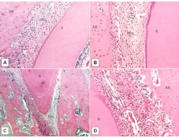
Figure 3 - Histological findings in the control (A) and experimental groups (B-D) after 3 days of orthodontic tooth movement, stained with H&E.
(A) PDL without signs of alteration (100x). (B) Disrupted fibers (arrow) ob-served on the tension side (100x). (C) Hyalinized areas (h) seen on the pres-sure side (40x). (D) Resorption lacunae with osteoclasts (arrows) observed on the pressure side (200x). AB indicates alveolar bone; PDL, periodontal ligament; R, root; T, tension side; P, pressure side.
AB
AB
AB
AB PDL
PDL R
R
T
P h
R
R
PDL
Figure 4 - FGF-2 immunohistochemistry staining of the control (A,B) and ex-perimental groups (C-H) after 3, 7 and 14 days of orthodontic tooth move-ment. (A) magnification of 40x; (B, C, H) 200x; (D, E, F) 100x; (G) 400x. AB indicates alveolar bone; PDL, periodontal ligament; R, root; Ob, osteoblasts; E, endothelial cells; h, hialinized area; F, fibroblasts.
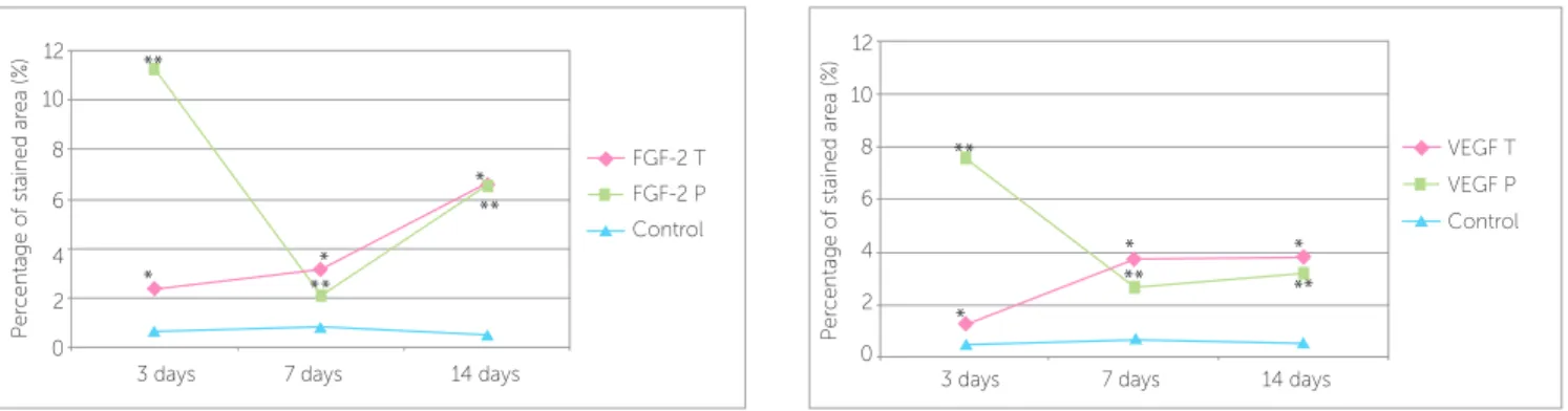
Figure 6 - Percentage of FGF-2 and VEGF stained area (%) in the experimental and control groups, after 3, 7 and 14 days of orthodontic tooth movement; * and ** indicate statistically significant differences between experimental and control groups (P < 0.05). T indicates tension side; P, pressure side. FGF-2 control val-ues: group I (0.68 ± 0.26); group II (0.80 ± 0.20); group III (0.50 ± 0.19). VEGF control valval-ues: group I (0.50 ± 0.21); group II (0.72 ± 0.10); group III (0.56 ± 0.22).
Figure 5 - VEGF immunohistochemistry staining of the control (A,B) and ex-perimental groups (C-H) after 3, 7 and 14 days of orthodontic tooth move-ment. (A, E, F) magnification of 100x; (B, C, D, G, H) 200x. AB indicates alveolar bone; PDL, periodontal ligament; R, root; E, endothelial cells; Oc, osteoclasts; h, hialinized area.
Table 1 - Percentage of FGF-2 and VEGF stained areas (%) on tension and pressure sides of the three experimental groups.
Values account for mean and standard deviation (±). Different letters on the column indicate statistically significant difference between groups as well as between tension and pressure sides (P < 0.05).
FGF-2 VEGF
Tension Pression Tension Pressure
Group 1 2.40 ± 0.69a 11.25 ± 3.30a* 1.28 ± 0.52a 7.56 ± 2.34a*
Group 2 3.16 ± 0.96a 2.06 ± 0.94b 3.78 ± 1.04b 2.70 ± 0.54b
Group 3 6.63 ± 1.07b 6.56 ± 0.77c 3.83 ± 1.25b 3.20 ± 0.66b
FGF-2 T VEGF T
FGF-2 P VEGF P
Control Control
14 days 14 days
7 days 7 days
3 days 3 days
0 0
P
er
c
entage o
f stained ar
ea (%)
P
er
c
entage o
f stained ar
ea (%)
2 2
4 4
6 6
8 8
10 10
12 12
AB
PDL
AB
Ob
PDL
PDL
F PDL
PDL
PDL PDL AB
PDL
h
AB
AB AB
R R
R
E AB
R R
Gr
oup III
Gr
oup II
Gr
oup I
Contr
ol
Tension Pressure
Oc
R R
R
R
PDL
PDL PDL
h
E
E E
E E
E
PDL PDL
PDL
AB
AB AB
AB AB AB
AB PDL
Gr
oup III
Gr
oup II
Gr
oup I
Tension Pressure
Contr
when light forces are used. Likewise, accumulation of osteoclasts near the hyalinization areas, which led to bone resorption on the pressure side, has been
de-scribed.39,40 In our study, the presence of resorption
lacunae containing osteoclasts on alveolar bone sur-faces close to the hyalinized areas on the pressure side of PDL was observed. Cells such as macrophages, foreign body giant cells and osteoclasts remove this hyalinized necrotic tissue after a few days of force application, allowing tooth movement through the
alveolar bone.17
We demonstrated that FGF-2 and VEGF are ex-pressed in rat periodontal ligament cells, even at bas-al levels in control areas of PDL, thereby suggesting a constitutive production of these molecules by PDL cells. The expression of both FGF-2 and VEGF was assessed during experimental orthodontic tooth move-ment, indicating that upregulation of these cytokines could be associated with PDL remodeling. It is pos-sible that this process sufers inluence of these impor-tant angiogenic growth factors, since orthodontic forc-es alter blood low in the periodontal region, initiating a cascade of biochemical and cellular processes that are
responsible for these biological events.12,23,24
On the pressure side, PDL cells showed intense expression of FGF-2 three days after experimental tooth movement, which was concomitant with the observation of a higher number of osteoclasts and bone resorption in this group, thus indicating that this growth factor plays an important role during orthodontic movement. On day 7, a significant de-crease of FGF-2 expression, as well as a lower num-ber of osteoclasts were noted. One possible explana-tion is that there is dissipaexplana-tion of the applied orth-odontic force due to tooth movement in the arch. A new increase in FGF-2 expression recorded on day 14 could be associated with PDL remodeling in this phase of orthodontic movement. FGF-2 has the abil-ity to accelerate periodontal tissue regeneration at the final phase of tissue repair in alveolar bone defects by promoting angiogenesis and inducing growth of
immature PDL cells.24
On the tension side, a gradual increase in FGF-2 was observed from day 3 to 14 of the induced orth-odontic tooth movement, which is in agreement with the neoformation events observed in this
re-gion of PDL.16,32 After 14 days of orthodontic force
application, a regeneration of periodontal tissue was observed, as well as a significant expression of FGF-2. It seems that FGF-2 is capable of inducing chemotaxis and mitogenesis of various PDL cells,
thus, inducing tissue regeneration processes.24,34
There was a higher expression of VEGF on day 3 on the pressure side, probably due to the elevated number of osteoclasts observed in this area on the first days of experimental tooth movement. This could be explained by the ability of VEGF in inducing
os-teoclast differentiation.1,14 Continuous compressive
forces enhance VEGF production and angiogenic ac-tivity in PDL cells, which may contribute to peri-odontal remodeling during orthodontic tooth
move-ment.23 These reports suggest that VEGF expression
in compressed periodontal tissue may play an impor-tant role in bone resorption, as well as in the promo-tion of angiogenesis in hyalinized tissues and adjacent areas on the pressure side. Moreover, through bio-logical properties such as vascular permeability and chemotaxis, VEGF may provide the degenerated tis-sues with many cell types, for instance, fibroblasts,
macrophages and multinucleated giant cells.23
On the tension side, there was a moderate expres-sion of VEGF by the PDL cells, although an increase in this cytokine was observed along the three experi-mental periods. This is consistent with the demon-stration of VEGF expression in osteoblasts on the tension side of mouse incisors and the predominance of alveolar bone formation that is characteristic of
this region.16,22 Constitutive VEGF expression may
contribute to PDL homeostasis by regulating blood
circulation and bone metabolism.23
The higher expression of FGF-2 observed in this study during the first days of experimental tooth movement, when compared to VEGF, could be re-lated to cellular events observed in the initial phase of inflammatory response resulting from the orth-odontic force applied to the tooth. The generation of an acute inflammatory process, characteristic of orthodontic movement, may be responsible for the
secretion of FGF-2.20,26,30 This growth factor is
con-sidered the most potent mitogen for periodontal cells and it may be important in wound healing, since it promotes angiogenesis and induces the development of immature PDL cells, thus, accelerating
is an optimal compressive force for VEGF produc-tion in PDL cells, and an excessive force results in
decreased VEGF production.23
CONCLUSION
The present study demonstrates that important alterations occur in PDL during experimental orth-odontic tooth movement, in which bone formation and apposition on tension side and bone resorption on pressure side are the main events. The expression
1. Aldridge SE, Lennard TW, Williams JR, Birch MA. Vascular endothelial growth factor receptors in osteoclast diferentiation and function. Biochem Biophys Res Commun. 2005;335(3):793-8.
2. Anastasi G, Cordasco G, Matarese G, Rizzo G, Nucera R, Mazza M, et al. An immunohistochemical, histological, and electron-microscopic study of the human periodontal ligament during orthodontic treatment. Int J Molec Med. 2008;21(5):545-54.
3. Bafour R, Berman J, Garb JL, Rhee SW, Kaufman J, Friedmann P. Enhanced angiogenesis and growth of collaterals by in vivo administration of recombinant basic ibroblast growth factor in a rabbit model of acute lower limb ischemia: dose-response efect of basic ibroblast growth factor. J Vasc Surg. 1992;16(2):181-91.
4. Carmeliet P, Collen D. Molecular analysis of blood vessel formation and disease. Am J Physiol. 1997;273(5 Pt 2):H2091-104.
5. Clarke MSF, Caldewell RW, Chiao H, Miyake K, McNeil PL. Contraction-induced cell wounding and release of ibroblast growth factor in heart. Circ Res. 1995;76(6):927-34.
6. Davidovitch Z, Nicolay OF, Ngan PW, Shanfeld JL. Neurotransmitters, cytokines, and the control of alveolar bone remodeling in orthodontics. Dent Clin North Am. 1988;32(3):411-35.
7. Davidovitch Z. Tooth movement. Crit Rev Oral Biol Med. 1991;2(4):411-50.
8. Ferrara N. Molecular and biological properties of vascular endothelial growth factor. J Mol Med. 1999;77(7):527-43.
9. Ferrara N. Vascular endothelial growth factor. Arterioscler Thromb Vasc Biol. 2009;29(6):789-91.
10. Ferrara N, Gerber HP, Lecouter J. The biology of VEGF and its receptors. Nat Med. 2003;9(6):669-76.
11. Heller IJ, Nanda R. Efect of metabolic alteration of periodontal ibers on orthodontic tooth movement. An experimental study. Am J Orthod. 1979;75(3):239-58.
12. Henneman S, Von Den Hof JW, Maltha JC. Mechanobiology of tooth movement. Eur J Orthod. 2008;30(3):299-306.
13. Iwaniec UTL, Mosekilde L, Mitova-Caneva NG, Thomsen JS, Wronski TJ. Sequential treatment with basic ibroblast growth factor and PTH is more eicacious than treatment with PTH alone for increasing vertebral bone mass and strength in osteopenic ovariectomized rats. Endocrinology. 2002;143(7):2515-26.
14. Kaku M, Kohno S, Kawata T, Fujita T, Tokimasa C, Tsutsui K, et al. Efects of vascular endothelial growth factor on osteoclast induction during tooth movement on mice. J Dent Res. 2001;80(10):1880-83.
15. Karami E. Fibroblast growth factor-2 and cardioprotection. Heart Fail Rev. 2007;12(3-4):267-77.
16. Kohno S, Kaku M, Tsutsui K, Motokawa M, Ohtani J, Tenjo K, et al. Expression of vascular endothelial growth factor and the efects on bone remodeling during experimental tooth movement. J Dent Res. 2003;82(3):177-82.
17. Krishnan V, Davidovitch Z. Cellular, molecular, and tissue-level reactions to orthodontic force. Am J Orthod Dentofacial Orthop. 2006;129(4):469.e1-32.
18. Krishnan V, Davidovitch Z. On a path to unfolding the biological mechanisms of orthodontic tooth movement. J Dent Res. 2009;88(7):597-608.
19. Lane NE, Kumer J, Yao W, Breunig T, Wronski T, Modin G, et al. Basic ibroblast growth factor forms new trabeculae that physically connect with pre-existing trabeculae, and this new bone is maintained with an anti-resorptive agent and enhanced with an anabolic agent in an osteopenic rat model. Osteoporos Int. 2003;14(5):374-82. 20. Lara VS, Figueiredo F, Silva TA, Cunha FQ. Dentin-induced in vivo
inlammatory response and in vitro activation of murine macrophages. J Dent Res. 2003;82(6):460-5.
21. Lew K, Sims MR, Leppard PI. Tooth extrusion efects on microvessel volumes, endothelial areas, and fenestrae in molar apical periodontal ligament. Am J Orthod Dentofacial Orthop. 1989;96(3):221-31. REFERENCES
22. Liang Y, Zhou Y, Jiang T, Zhang Z, Wang S, Wang Y. Expression of LIF and LIFR in periodontal tissue during orthodontic tooth movement. Angle Orthod. 2011;81(4):600-8.
23. Miyagawa A, Chiba M, Hayashi H, Igarashi K. Compressive forces induces VEGF production in periodontal tissues. J Dent Res. 2009;88(8):752-6. 24. Murakami S, Takayama S, Ikesawa K, Shimabukuro Y, Kitamura M, Nozaki
T, et al. Regeneration of periodontal tissues by basic ibroblast growth factor. J Periodontal Res. 1999;34(7):425-30.
25. Nakanishi H, Seki Y, Kohno T, Muramoto T, Toda K, Soma K. Changes in response properties of periodontal mechanoreceptors after experimental orthodontic tooth movement in rats. Angle Orthod. 2004;74(1):93-9. 26. Perinetti G, Paolantonio M, D’Attilio M, D’Archivio D, Tripodi D,
Femminella B, et al. Alkaline phosphatase activity in gingival crevicular luid during human orthodontic tooth movement. Am J Orthod Dentofacial Ortop. 2002;122(5):548-56.
27. Reitan K, Kvam E. Comparative behavior of human and animal tissue during experimental tooth movement. Angle Orthod. 1971;41(1):1-14. 28. Ren Y, Vissink A. Cytokines in crevicular luid and orthodontic tooth
movement. Eur J Oral Sci. 2008;116(2):89-97.
29. Rygh P. Elimination of hyalinized periodontal tissues associated with orthodontic tooth movement. Scand J Dent Res. 1974;82(1):57-73. 30. Schulze-Osthof K, Risau W, Vollmer E, Sorg C. In situ detection of ibroblast growth factor by highly speciic antibodies. Am J Pathol. 1990;137(1):85-92.
31. Shimazu A, Morishita M. Basic ibroblast growth factor induces the expression of matrix metalloproteinase-3 in human periodontal ligament cells through the MEK2 mitogen-activated protein kinase pathway. J Periodontal Res. 2003;38(2):122-9.
32. Shirazi M, Nilforoushan D, Alghasi H, Dehpour AR. The role of nitric oxide in orthodontic tooth movement in rats. Angle Orthod. 2002;72(3):211-5. 33. Tang MP, Sims MR, Sampson WJ, Dreyer CW. Evidence for endothelial
junctions acting as a luid lux pathway in tensioned periodontal ligament. Archs Oral Biol. 1993;38(3):273-6.
34. Terranova VP, Odziemiec C, Tweden KS, Spadone DP. Repopulation of dentin surfaces by periodontal ligament cells and endothelial cells. Efect of basic ibroblast growth factor. J Periodontol. 1989;60(6):293-301. 35. Toms SR, Lemons JE, Bartolucci AA, Eberhardt AW. Nonlinear
stress-strain behavior of periodontal ligament under orthodontic loading. Am J Orthod Dentofacial Orthop. 2002;122(2):174-9.
36. Vandevska-Radunovic V. Neural modulation of inlammatory reactions in dental tissues incident to orthodontic tooth movement. A review of the literature. Eur J Orthod. 1999;21(3):231-47.
37. Vandevska-Radunovic V, Kristiansen AB, Heyeraas KJ, Kvinnsland S. Changes in blood circulation in teeth and supporting tissues incident to experimental tooth movement. Eur J Orthod. 1994;16(5):361-9. 38. Von Böhl M, Kuijpers-Jagtman AM. Hyalinization during orthodontic
tooth movement: a systematic review on tissue directions. Eur J Orthod. 2009;31(1):30-6.
39. Von Böhl M, Maltha J, Von Den Hoff H, Kuijpers-Jagtman AM. Changes in the periodontal ligament after experimental tooth movement using high and low continuous forces in beagle dogs. Angle Orthod. 2004;74(1):16-25.
40. Von Böhl M, Maltha JC, Von Den Hof JW, Kuijpers-Jagtman AM. Focal hyalinization during experimental tooth movement in beagle dogs. Am J Orthod Dentofacial Orthop. 2004;125(5):615-23.
41. Wang Y, Wan C, Deng L, Liu W, Cao X, Gilbert SR, et al. The hypoxia-inducible factor alpha pathway couples angiogenesis to osteogenesis during skeletal development. J Clin Invest. 2007;117(6):1616-26. 42. Yamaguchi M, Kojima T, Kanekawa M, Aihara N, Nogimura A, Kasai K.