CLINICAL SCIENCE
Clinical predictors of prosthesis-patient mismatch
after aortic valve replacement for aortic stenosis
Luis M. Astudillo,IOrlando Santana,IPablo A. Urbandt,IAlexandre M. Benjo,ILior U. Elkayam,IFrancisco O. Nascimento,IGervasio A. Lamas,IJoseph LamelasII
IColumbia University Division of Cardiology, Mount Sinai Heart Institute, Miami Beach, Florida, USA.IIColumbia University Division of Cardiac Surgery, Mount Sinai Heart Institute, Miami Beach, Florida, USA.
OBJECTIVE: We sought to ascertain predictors of Patient Prosthesis Mismatch, an independent predictor of mortality, in patients with aortic stenosis using bioprosthetic valves.
METHOD:We analyzed 2,107 sequential surgeries. Patient Prosthesis Mismatch was calculated using the effective orifice area of the prosthesis divided by the patient’s body surface area. We defined nonsignificant, moderate, and severe Patient Prosthesis Mismatch as effective orifice area indexes of .0.85 cm2/m, 0.85-0.66 cm2/m2, and
#0.65 cm2/m2, respectively.
RESULTS: A total of 311 bioprosthetic patients were identified. The incidence of nonsignificant, moderate, and severe Patient Prosthesis Mismatch was 41%, 42, and 16%, respectively. Severe Patient Prosthesis Mismatch was significantly more prevalent in females (82%). In severe Patient Prosthesis Mismatch, the perfusion and the cross-clamp times were considerably lower when compared with nonsignificant Patient Prosthesis Mismatch and moderate Patient Prosthesis Mismatch. Patients with severe Patient Prosthesis Mismatch had a significantly higher likelihood of spending time in the intensive care unit and a significantly longer length of stay in the hospital. Body surface area was not different in severe Patient Prosthesis Mismatch when compared with nonsignificant Patient Prosthesis Mismatch. In-hospital mortality in patients with nonsignificant, moderate, and severe Patient Prosthesis Mismatch was 2.3%, 6.1%, and 8%, respectively. Minimally invasive surgery was significantly associated with moderate Patient Prosthesis Mismatch in 49% of the patients, but not with severe Patient Prosthesis Mismatch.
CONCLUSION: Severe Patient Prosthesis Mismatch is more common in females, but not in those with minimal available body surface area. Though operative times were shorter in these patients, intensive care unit and hospital lengths of stay were longer. Surgeons and cardiologists should be cognizant of these clinical predictors and complications prior to valve surgery.
KEYWORDS: Aortic Stenosis; Patient Prosthesis Mismatch; Bioprosthesis Female; Gender Clinical Predictors.
Astudillo LM, Santana O, Urbandt PA, Benjo AM, Elkayam LU, Nascimento FO, et al. Clinical predictors of prosthesis-patient mismatch after aortic valve replacement for aortic stenosis. Clinics. 2012;67(1):55-60.
Received for publication onOctober 14, 2011;First review completed onOctober 26, 2011;Accepted for publication onNovember 3, 2011
E-mail: [email protected] Tel.: 305 674-2168
INTRODUCTION
Aortic valve stenosis affects 2% to 4% of older adults and is the most common reason for valve surgery. An estimated 50,000 aortic valve procedures are performed each year in the United States (1). However, once the aortic valve is replaced, issues related to prosthesis-patient mismatch (PPM) may emerge.
PPM was first described by Rahimtoola in 1978 (2). PPM is considered to be present when the prosthetic valve used in the patient is smaller than the normal native valve. As a
consequence of the smaller prosthetic valve, the left ventricle has to produce higher pressures to overcome the mechanical resistance produced by the new device. This is transmitted in higher transvalvular pressure gradients as measured by Doppler (2). It has been demonstrated that the high-pressure gradients through the prosthesis are associated with higher morbidity and impact both short- and long-term mortality, when com-pared with prostheses of adequate size, based on the patient’s body surface area (4-5).
It has been shown that PPM is a common problem in patients undergoing AVR, occurring in up to 70% of aortic valve replacements (3). At the same time, this problem can be prevented through the use of a systematic approach prior to surgery (3). This may result in enhanced recovery with lower morbidity and mortality. The aim of this study was to determine the prevalence, clinical predictors, and short-term impact of severe PPM.
Copyrightß2012CLINICS– This is an Open Access article distributed under
the terms of the Creative Commons Attribution Non-Commercial License (http:// creativecommons.org/licenses/by-nc/3.0/) which permits unrestricted non-commercial use, distribution, and reproduction in any medium, provided the original work is properly cited.
METHODS
Patient population: After obtaining approval from the independent review board, we retrospectively reviewed 2,107 consecutive heart surgeries performed at our institution between January 2005 and June 2009. Of these, 964 were coronary artery bypass surgeries, 683 involved single-valve surgeries, 411 were concomitant valve and bypass surgeries, and 144 were multivalve surgeries. From this group, patients with a diagnosis of severe aortic stenosis (AVA#1 cm2) were included in the study, along with those in whom a bioprosthetic valve was used for the aortic valve replace-ment. Patients with concomitant valve or coronary artery bypass surgery were also included in the study, but patients with a diagnosis of aortic insufficiency as the reason for AVR were excluded. Baseline preoperative and operative variables were used based on the information provided by the Society of Thoracic Surgeons database in Tables 1 and 2.
Measurements: The effective orifice area (EOA) of each valve was obtained from a list that the American Society of Echocardiography published in their 2009 Recommenda-tions for the Evaluation of Prosthetic Valves with Echocardiography and Doppler Ultrasound (6), which was based on a published report of normally functioning aortic valve prostheses (7). The effective orifice area values obtained from in vivo measurements for the different bioprosthetic aortic valves used during this analysis are given in Table 3. PPM was calculated using the EOA of the aortic valve prosthesis, which was divided by the patient’s body surface area to obtain the indexed EOA. We defined nonsignificant, moderate, and severe PPM as an indexed EOA of.0.85 cm2/m2, 0.66 cm2/m2to
#0.85 cm2/m2, and
#0.65 cm2/m2, respectively (12). We defined the nonsigni-ficant PPM group as our control because they received the optimal therapy. Mortality was analyzed at the time of discharge from the hospital.
Statistical Analysis: Continuous variables are expressed as the mean ¡1 SD; comparisons were conducted by t-tests/ANOVAs or nonparametric Mann-Whitney/Kruskal-Wallis tests if the normality assumption was violated when comparing severe PPM with no PPM or severe PPM with moderate PPM. Discrete variables are presented as percen-tages and relatives frequencies; comparisons were con-ducted by chi-squared statistics or Fisher’s exact test, as appropriate. A p-value ,0.05 was considered statistically significant. A stepwise logistic regression analysis (back-ward Wald) was used for the variables with ap-value,0.05 to assess the independent predictors of PPM and length of stay in the intensive care unit. We used a software system (Statistical Package for the Social Sciences, SPSS version 17 Chicago, Illinois) for all analyses.
RESULTS
A total of 311 patients (128 females and 183 males) with a diagnosis of severe aortic stenosis who underwent AVR with a bioprosthetic valve were identified. Based on the severity of the PPM, the cohort was divided in three groups: non-significant PPM (n = 129), 41%; moderate PPM (n = 132), 42%; and severe PPM, (n = 50) 16%. PPM was present in 58% of the total population, in 42% as moderate PPM and in 16% as severe PPM.
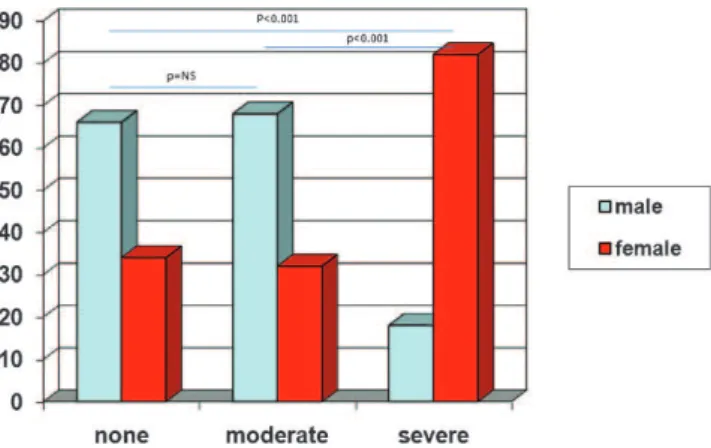
Patient characteristics: The baseline characteristics are presented in Table 1. The mean age was 75¡11, 73¡9, and 77¡7 years for nonsignificant, moderate, and severe PPM, respectively (p= 0.6). Females represented 82% of the severe PPM group compared with 34% and 32% of the nonsigni-ficant and moderate PPM groups, respectively (p,0.001) (Figure 1). There was no discrepancy in age between the different groups of PPM, (p= 0.6). The body surface area was lowest in the severe PPM group (1.75¡0.5 m2) compared with 1.97¡0.2 m2 of the moderate PPM
Table 1 -Preoperative Data.
Patients
Non PPM n = 129 (41%)
Moderate PPM n = 132 (42%)
Severe
PPM n = 50(16%) p-value
Age (years) 75¡11 73¡9 77¡7 0.6
Female 45 (34.9%) 42 (32.1%) 41 (82%) ,0.001
Body surface area (m2) 1.80¡0.2 1.97¡0.2 1.75¡0.25 0.155*
Body mass index (Kg/m2) 26.6
¡4.27 29.4¡5.4 27.8¡6.2 0.120*
NYHA class$III 49 (38%) 49 (37%) 28 (56%) 0.05
Diabetes mellitus 34 (26%) 59 (44%) 17 (34%) 0.008
Renal failure 3 (2.3%) 4 (3%) 6 (12%) 0.01
Hypertension 111 (86%) 125 (95%) 47 (94%) 0.04
Cerebrovascular disease 12 (9.3%) 10 (7.6%) 6 (12%) 0.64
Smoker 33 (25%) 37 (28%) 17 (34%) 0.53
Chronic lung disease 79 (61%) 86 (65%) 34 (68%) 0.65
Peripheral vascular disease 14 (11%) 7 (5.3%) 4 (8%) 0.25
Prior coronary artery bypass grafting 11 (8.5%) 16 (12%) 6 (12%) 0.60
Myocardial infarction 11 (8.5%) 16 (12%) 10 (20%) 0.10
Left ventricular ejection fraction#55% 45 (35%) 47 (36%) 14 (28%) 0.60
Mean ejection fraction 52¡12 52¡13 54¡12 0.62
Preoperative aortic valve gradient (mmHg) 53¡19 53¡22 51¡18 0.38
Severe mitral insufficiency 13 (10%) 18 (14%) 14 (28%) 0.009
Preoperative Medical Therapy Beta-blockers
74 (57%) 85 (64%) 30 (60%) 0.50
Angiotensin converting enzyme inhibitors 43 (33%) 59 (45%) 15 (30%) 0.08
Aspirin 46 (36%) 55 (42%) 19 (38%) 0.60
Lipid lowering medications 66 (51%) 75 (57%) 32 (64%) 0.30
*represents the
(p,0.001), but this was not significantly different when compared with the nonsignificant PPM group value of 1.80¡0.2 m2(p= 0.155).
Body mass index did not differ between the severe (27.8¡6.2 Kg/m2) and moderate groups (29.4¡5.4 Kg/m2), (p= 0.10), or when comparing nonsignificant (26.6¡4.27 Kg/ m2) and severe mismatch groups, (p= 0.12). Advanced heart failure (based on the New York Heart Association criteria) was found in 56% of patients in the severe group (p= 0.05). Diabetes mellitus was more prevalent in patients with moderate PPM (p= 0.008). Hypertension was prevalent in all of the groups, in particular, in those with moderate and severe mismatch (p= 0.04). Renal failure (serum creatinine.2 mg/ dl) was higher in the severe PPM group, (p= 0.01). Coronary artery disease, with previous coronary artery bypass grafting (CABG) was similar in all groups as well, (p= 0.6).
The echocardiographic basal characteristics of the study population presented no differences in the mean ejection fraction: no PPM, 52¡12%; moderate PPM, 52¡13%; and severe PPM, 54¡12%, (p= 0.62). Likewise, the aortic valve gradient was not significantly different: 53¡19 mmHg, 53¡22 mmHg, and 51¡18 mmHg, in that order, (p= 0.38). Surgical data: Most of the procedures were elective. Coronary artery bypass graft surgery was performed in 41%
(nonsignificant PPM), 36% (moderate PPM), and 42% (severe PPM), respectively, (p= 0.67). Concomitant mitral valve surgery was performed in 34% of the severe PPM group, 17% of the moderate PPM group, and 12% in the no-PPM group (p= 0.003). Tricuspid valve surgery was com-pleted in 8% of the severe PPM cases. A total of six (6) aortic annular enlargement procedures was performed; two in the no PPM, one in the moderate PPM, and three in the severe PPM groups.
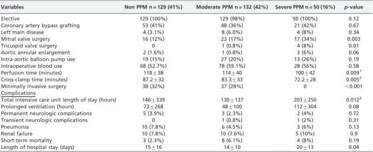
Interestingly, the operative data in Table 2 show that patients in the severe PPM group had the shortest perfusion (p= 0.009) and cross-clamp times (p= 0.005) (Figure 2). However, the same group had the highest total intensive care unit length of stay with a mean value of 203 hours compared with 146 and 130 hours in the nonsignificant and moderate PPM groups, respectively (Figure 3). This was statistically significant (p= 0.012) when comparing severe and moderate PPM. No other factors were associated with the amount of intensive care unit hours except for the PPM difference. However, there was a statistical trend (p= 0.08) for prolonged mechanical ventilation in hours in the severe mismatch group (Figure 4). Along with these findings, patients with severe mismatch had the longest hospital stay with a mean value of 20 days compared with 15 and 14 days Table 2 -Operative Data.
Variables Non PPM n = 129 (41%) Moderate PPM n = 132 (42%) Severe PPM n = 50 (16%) p-value
Elective 129 (100%) 129 (98%) 50 (100%) 0.12
Coronary artery bypass grafting 53 (41%) 48 (36%) 21 (42%) 0.67
Left main disease 4 (3.1%) 8 (6.0%) 4 (8%) 0.34
Mitral valve surgery 16 (12%) 23 (17%) 17 (34%) 0.003
Tricuspid valve surgery 0 1 (0.8%) 4 (8%) 0.01
Aortic annular enlargement 2 (1.6%) 1 (0.8%) 3 (6%) 0.06
Intra-aortic balloon pump use 19 (15%) 27 (20%) 13 (26%) 0.19
Intraoperative blood use 68 (52.7%) 78 (59.1%) 28 (56%) 0.58
Perfusion time (minutes) 118¡38 114¡40 100¡42 0.0091
Cross-clamp time (minutes) 87.2¡32 83.3¡33 72.2¡28 0.0052
Minimally invasive surgery 38 (32%) 37 (28%) 0 ,0.001
Complications
Total intensive care unit length of stay (hours) 146¡339 130¡137 203¡250 0.0123
Prolonged ventilation (hours) 73¡268 48¡100 112¡304 0.08
Permanent neurologic complications 5 (3.9%) 3 (2.3%) 2 (4%) 0.72
Transient neurologic complications 0 1 (0.8%) 1 (2%) 0.31
Pneumonia 10 (7.8%) 6 (4.5%) 3 (6%) 0.13
Renal failure 10 (7.8%) 10 (7.6%) 5 (10%) 0.9
Short-term mortality 3 (2.3%) 8 (6.1%) 4 (8%) 0.19
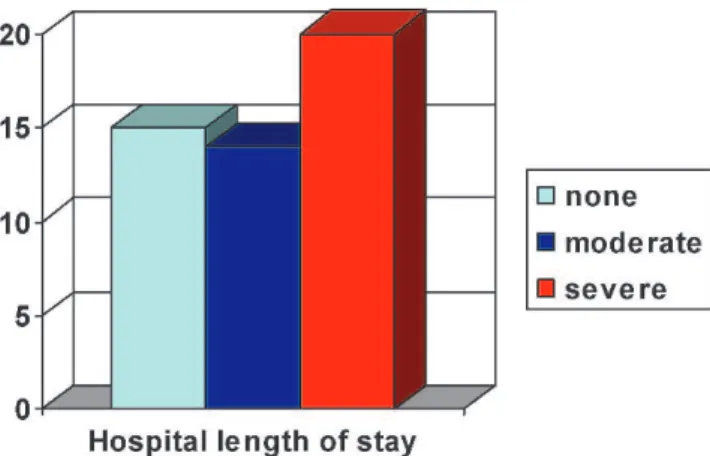
Length of hospital stay (days) 15¡16 14¡10 20¡13 0.04
1
p-value between severe PPM versus no PPM.p= 0.05 between severe PPM versus moderate PPM. 2
p-value between severe PPM versus no PPM.p= 0.04 between severe PPM vs. moderate PPM. 3
p-value between severe PPM versus moderate PPM.p= 0.27 between severe PPM and no PPM.
Table 3 -Reference values of effective orifice area for bioprosthetic aortic valves.
Size, mm Prosthetic Valve
19 21 23 25 27 29
No. of Patients 54 98 90 51 17 1
Mosaic Porcine 121 1.4 1.5 1.8 1.9 2.1
Freestyle 4 1.4 1.7 2.1 2.5
Mitroflow 1 1.1 1.3 1.5 1.8
Carpentier Edwards Standard 166 0.9 1.5 1.7 1.9 2.3 2.8
stented porcine
Carpentier Edwards Pericardial 19 1.2 1.5 1.8
in the nonsignificant and moderate PPM groups, respec-tively (p= 0.04) (Figure 5). Fifteen cases of in-hospital mortality were identified, three in nonsignificant PPM, eight in moderate PPM, and four in the severe PPM groups (p= 0.19). Seventy-five minimally invasive surgeries were performed, 38 (50%) had nonsignificant PPM and 37 (50%) had moderate PPM. No case of severe PPM was found to be associated with minimally invasive surgery.
Table 3 shows the different types of bioprosthetic valves used. The Carpentier Edwards Standard (Edwards Life-sciences, Irvine, CA), and the Mosaic Porcine (Medtronic, Minneapolis, MN) were the most commonly used, 53% and 39% of the time, respectively. A valve size of 21 mm was implanted in 31% of the surgeries, making it the most frequently used, while a valve size of 23 mm was the second most common, being used 29% of the time.
Independent Risk Factors: Univariate analysis from preoperative data showed that female gender and renal failure were statistically associated with severe PPM. Diabetes mellitus and ACE inhibitors use were associated with the moderate PPM group but not with the severe PPM group. Further multivariable analysis demonstrated that female gender was associated with severe mismatch (p,0.001), even when compared with the nonsignificant as well as the moderate PPM groups.
DISCUSSION
In patients undergoing AVR, PPM is a commonly encountered problem that leads to worsened hemodynamic function, less regression of left ventricular hypertrophy, and more cardiac events with lower survival rates (4,11,12). Other studies have demonstrated an association between female gender and higher operative mortality after valvular heart surgery (15).
Our results indicate that female gender is an indepen-dent risk factor for severe prosthesis-patient mismatch. This association was observed after the body surface area of the severe mismatch group proved not to be significantly different when compared with the group without any mismatch. When compared with men, women in general have a smaller body surface area and a smaller aortic annulus. Accordingly, PPM has proven to be more common in older females with concomitant coronary artery disease (16-18).
Our study demonstrated that 82 percent of severe mismatches corresponded to females without differences in age or any other baseline characteristics. Interestingly, this particular group had the shortest operative times. It is our impression that the use of smaller prosthetic valves for small and severely calcified annuli may have shortened the Figure 1 -Percentage of prosthesis-patient mismatch by gender.
Figure 2 -Total amount of intensive care unit hours distributed according to patient-prosthesis mismatch.
Figure 3 - Hours of mechanical ventilation according to the severity of patient-prosthesis mismatch.
surgical times. It is possible that the reason is associated with the fact that surgeons did not have to enlarge the aortic root. PPM is therefore more frequently encountered in females, and the discrepancy between the inadequate size of both the prosthesis and the patient have proven to be risk factors for mortality (4,12,13). At the same time, the female heart responds differently than the male heart does for the same degree of left ventricular outflow obstruction, result-ing in smaller end-systolic chamber size and higher pressures in the female heart response (18). This is particularly common with elderly patients with an excessive or inappropriate degree of hypertrophy; wall thickness is greater than necessary to counterbalance the high intraca-vitary pressures. As a result, the systolic wall stress is lower, and the ejection fraction is higher; such inappropriate LV hypertrophy has been associated with higher perioperative morbidity and mortality (17-19).
We found a significant impact of severe PPM on the intensive care unit length of stay and on the total hospital length of stay. Because of the nature of the study, we cannot establish a causal relationship between the degree of PPM and the findings. However, it has been demonstrated that abnormal intracavitary flow accelerations (defined as the presence of a dagger-shaped intracavitary flow signal on continuous-wave Doppler) after aortic valve replacement for severe aortic stenosis are associated with concentric left ventricular hypertrophy and supernormal systolic function. The abnormal intracavitary flow acceleration and concentric left ventricular hypertrophy were associated with higher in-hospital mortality, morbidity and prolonged intubation (17-20). Although not directly investigated in our study, this may have played a role in our patients.
We demonstrated that minimally invasive surgery may be related to a higher frequency of moderate PPM, but also to a lower rate of severe PPM; we believe that if this is confirmed, it is likely to have clinical implications with respect to the selection of a particular surgical technique. For this reason, we are currently prospectively exploring this possibility in our institution.
This study highlights the importance of avoiding PPM, particularly in females who react differently from a pathophysiologic standpoint. Aortic PPM can be avoided through optimal prosthesis selection in the individual patient by calculating the necessary indexed EOA prior to surgery (3). This is particularly important in patients with
left ventricular dysfunction and/or severe left ventricular hypertrophy (3).
Study Limitations: The current study was based on data obtained from a patient registry with retrospective analysis and was not able to establish a causal relationship. The post-operative intracavitary flow accelerations were not available but may have helped to confirm our hypothesis. The assess-ment of these parameters along with the patient’s outcome may have an impact on selecting the appropriate therapy.
AUTHOR CONTRIBUTIONS
Lamelas GA and Lamas J coordinated the study. Urbandt PA and Santana O wrote and reviewed the article. Astudillo LM and Nascimento FO collected the data. Benjo AM performed the statistical analysis. Elkayam LU performed the statistical analysis and reviewed the manuscript.
REFERENCES
1. Freeman RV, Otto CM. Spectrum of calcific aortic valve disease. Pathogenesis, disease progression, and treatment strategies. Circulation 2005;111:3316-26.
2. Rahimtoola SH. The problem of valve prosthesis-patient mismatch. Circulation 1978;58:20-4.
3. Pibarot P, Dumesnil JG. Prosthetic Heart Valves. Selection of the optimal prosthesis and long-term management. Circulation 2009;119:1034-48. 4. Blais C, Dumesnil JG, Baillot R, Simard S, Doyle D, Pibarot P. Impact of
valve prosthesis-patient mismatch on short-term mortality after aortic valve replacement. Circulation. 2003;108:983-8.
5. Mohty D, Malouf JF, Girard SE, Schaff HV, Grill DE, Enriquez-Sarano ME, et al. Impact of prosthesis-patient mismatch on long-term survival in patients with small St. Jude medical mechanical prosthesis in the aortic position. Circulation. 2006;113:420-6.
6. Zoghbi WA, Chambers JB, Dumesnil JG, Foster E, Gottdiener JS, Grayburn PA, et al. Recommendations for evaluation of prosthetic valves with echocardiography and doppler ultrasound: a report From the American Society of Echocardiography’s Guidelines and Standards Committee and the Task Force on Prosthetic Valves, developed in conjunction with the American College of Cardiology Cardiovascular Imaging Committee, Cardiac Imaging Committee of the American Heart Association, the European Association of Echocardiography, a registered branch of the European Society of Cardiology, the Japanese Society of Echocardiography and the Canadian Society of Echocardiography, endorsed by the American College of Cardiology Foundation, American Heart Association, European Assocation of Echocardiography, a registered branch of the European Society of Cardiology, the Japanese Society of Echocardiography, and Canadian Society of Echocardiography. J Am Soc Echo. 2009;22:975-1014. 7. Rajani R, Mukherjee D, Chambers JB. Doppler echocardiography in
normally functioning replacement aortic valves: a review of 129 studies. J Heart Valve Dis. 2007;16:521-35.
8. Oakley RE, Kleine P, Bach DS. Choice of prosthetic heart valve in today’s practice. Circulation. 2008;117:253-6.
9. Pibarot P, Dumesnil JG. Hemodynamic and clinical impact of prosthesis patient mismatch in the aortic valve position and its prevention. J Am Coll Cardiol. 2000;36:1131-41.
10. Rahimtoola SH. Choice of prosthetic heart valve for adult patients. J Am Coll Cardiol. 2003;41:893-904.
11. Rubio Alvarez J, Sierra Quiroga J, Vega Fernandez M, Adrio Nazar B, Gude Sampedro F, Martinez Comendador JM, et al. Up to twenty-five-year survival after aortic valve replacement with size 19 mm valves. Inter Cardiovasc and Thorac Surg. 2010;10:32-6.
12. Pibarot P, Dumesnil JG. Prosthesis-patient mismatch: definition, clinical impact, and prevention. Heart. 2006;92:1022-9.
13. Walther T, Rastan A, Falk V, Lehmann S, Garbade J, Funkat AK, et al. Patient prosthesis mismatch affects short and long term outcomes after aortic valve replacement. Eur J Cardiothorac Surg. 2006;30:15-9. 14. Pai RG, Kapoor N, Bansal RC, Varadarajan P. Malignant natural history
of asymptomatic severe aortic stenosis: benefit of aortic valve replace-ment. Ann Thorac Surg. 2006;82:2116-22.
15. Edwards FH, Peterson ED, Coombs LP, DeLong ER, Jamieson WR, Shroyer ALW, et al. Prediction of operative mortality after valve replacement surgery. J Am Coll Cardiol. 2001;37:885-92.
16. Taggart DP. Prosthesis patient mismatch in aortic valve replacement: possible but pertinent? Eur Heart J. 2006;27:644-6.
17. Aurigemma G, Battista S, Orsinelli D, Sweeney A, Pape L, Cuenoud H. Abnormal left ventricular intracavitary flow acceleration in patients undergoing aortic valve replacement for aortic stenosis: a marker for high postoperative morbidity and mortality. Circulation. 1992;86:926-36. Figure 5 -Total length of in-hospital stay in days distributed
18. Carroll JD, Carroll EP, Feldman T, Ward DM, Lang RM, McGaughey D, et al. Sex-associated differences in left ventricular function in aortic stenosis of the elderly. Circulation. 1992;86:1099-107.
19. Orsinelli DA, Aurigemma GP, Battista S, Krendel S, Gaasch WH. Left ventricular hypertrophy and mortality after aortic valve replacement for
aortic stenosis: a high risk subgroup identified by preoperative relative wall thickness. J Am Coll Cardiol. 1993;22:1679-83.